Congenital emphysema in children: Segmental lungresection as an alternative to lobectomyDaniil U. Krivchenya, Eugene O. Rudenko⁎, Alexander G. Dubrovin
Department of Paediatric Surgery, Bogomolets National Medical University, National Children Specialized Hospital“OHMATDYT,” Kyiv 01135, Ukraine
Received 4 November 2012; accepted 12 November 2012
Congenital emphysema is a rare malformation that is causing an increase in parenchymal lung volume and a
characterized by hypertrophy, hyperinflation and over-distention of the lung parenchyma. Although usuallydescribed as affecting lobes it may affect individual segments
decrease in blood flow and potentially compromisingventilation. It may present early with respiratory distress ininfants and hence require urgent surgery. Although “con-genital lobar emphysema” is the commonest phrase used inthe literature [1–5], it does not necessarily correspond withthe anatomic location of the affected zone, especially if thesuperior division (S1, S2, S3) bronchopulmonary segments ofthe left lung are affected.
The goals of the study were to clarify the pathogenesis,particularly of the contribution of its vascular elements, andto report the outcome of lung-sparing segmental resections.
Fig. 1 Chest radiographs showing congenital emphysema ofsuperior (S1–S3) segments of left upper lobe (A) and congenitalemphysema of right upper lobe (B). There is increased translucencyof the lung field on the affected side, and a diminished vascularpattern with shift of mediastinum to the opposite side—pseudodex-trocardia (A). There is also ipsilateral flattening of the diaphragmcupola and widening of the intercostal spaces. The contralaterallung field is reduced and has diminished translucency.
1. Materials and methods
All patients were identified from retrospective review ofclinical records from the Department of Pediatric Surgery ofBogomolets National Medical University from 1982 to2012. Thirteen cases from this series were first published in1987 [6].
The diagnosis of congenital lobar or segmental emphy-sema was based on clinical findings, chest radiography,digital subtraction pulmonary angiography (DSA) (from1986) and computed tomography (CT) with intravenouscontrast enhancement (from 1998). The differential diag-nosis for such cases included pneumonia; respiratory tractforeign bodies; bronchial obstruction by aberrant bloodvessels; pulmonary agenesis, aplasia, hypoplasia anddysplasia; and agenesis of pulmonary artery and itsbranches. Bronchoscopy was used in some cases to excludelung aplasia or foreign bodies. We did not use broncho-graphy and consider its use dangerous particularly in thosewith an acute presentation.
The main radiographic signs consistent with congenitalemphysema were as follows: a) increase of the lungtransparency on the affected side with decreased vascularmarkings; b) shift of the mediastinum to the contralateral side(mediastinal herniation); c) pseudodextrocardia in left-sidedcases; d) ipsilateral flattening of the diaphragm cupola; e)ipsilateral expansion of intercostal spaces and g) reduction ofcontralateral lung field with a reduction in transparency(Figs. 1 and 2).
Three types of clinical presentations were defined:decompensated, sub-compensated, and compensated. Thustypical symptoms in the decompensated form were acuterespiratory insufficiency with shortness of breath at rest, skinpallor, and in critical cases, cyanosis, asphyxia andconvulsions. In the sub-compensated form children hadsymptoms of shortness of breath, coughing, and sweating onparticipation in minor physical activity.
Segmental lung anatomy was reported as follows: leftupper lobe—superior division (S1, S2 and S3), lingual (S4
and S5) and lower lobe (S6–10) segments [7].All surgeries were open and performed through a lateral
fourth space thoracotomy. The lung was examined thor-oughly with regard to the color, size and consistency ofoverinflated and collapsed areas. Emphysematous segmentsor lobes were pale pink, doughy consistency compared withunaffected ones that were bright pink in color, reduced involume and sometimes atelectatic. Segmental arteries andveins were exposed, ligated and divided and segmentalresection was performed using stapling devices or by suturesapplied to the parenchyma. Suture lines were sealed with the
peeled off visceral pleura of the resected segments. Lung-sparing surgery removing only abnormal lung parenchymawas the aim and implied segmental resection in most left-sided cases. Division of a patent ductus arteriosus or shortligamentum arteriosum was performed where necessary.
Follow-up examinations and chest radiography (byquestionnaire in those who could not be reached for face-to-face interview) were performed from 6 months to10 years after surgery and defined clinical symptoms andtolerance to physical activity.
2. Results
In the 30-year period, 1982–2012, 43 (30 male) childrenwere diagnosed with congenital emphysema and underwent
Table 1 Age at presentation (n=43).
Age b1 y (months) N1 year Total
b1 1–3 4–6 7–12 1–3 4–7 8–10
Number 3 15 9 6 4 1 5 43 (100%)33 (77%) 10 (23%)
311Congenital emphysema in children
surgical lung resection. Their median age was 4 months(range 10 days to 10 years). The majority of children wereb1 year of age (n=33; 77%) (Table 1).
Twenty-four (56%) children had involvement of the leftside, and 19 (44%) of the right side. Table 2 illustrates thepattern of segmental involvement in left-sided lesions. Thecommonest pattern was emphysema of all superior division(S1, S2 and S3) segments (n=19, 79%). Spared lingualsegments appeared to be decreased in volume but with acolor at surgery identical to that of left lower lobe. The entireleft upper lobe (S1–5) was involved in a single child. Bycontrast, the pattern of right lung involvement was rightupper lobe (RUL) (n=12, 63%), right middle lobe (RML)(n=4, 21%), and right lower lobe (RLL) (n=1, 5%), and intwo cases the emphysema was bilobar (RML and RLL).
The absence of blood perfusion in the affected segmentswas well shown by DSA and particularly the characteristicsparing of the lingual (S4 and S5) segments in left-sideddisease. Fig. 2 illustrates the typical vascular pattern ofaffected lung parenchyma. Angiography was also able toexclude other cardiac and major blood vessel anomalies.Fig. 3 illustrates typical CT scan findings includingtracheal deviation.
The degree of symptoms was assessed as decompensated(n=10, 23%), sub-compensated (n=29, 67%) and compen-sated (n=4, 9%). A common characteristic of older affectedchildren was a delay in their physical development caused bymalnutrition due to chronic respiratory disease.
Surgical resection was performed in 42 children. Aninfant aged 2 months presented with decompensated left-sided emphysema (involving S1–S3 segments) was admit-ted to our clinic in a terminal condition after a prolongedjourney from distant region and died before surgery couldbe carried out.
Fig. 2 Digital subtraction angiography (DSA) in a 2-year-oldchild with congenital emphysema of S1–S3 of LUL. The catheter isin the right ventricle. (A) Pulmonary arterial phase showing virtualabsence of vascular pattern in the affected left-sided segments, witharteries of the left lower lobe and lingual (S4 and S5) segmentsshifted downward. (B) Delayed contrast within the right pulmonaryveins, heart and aorta. The heart is shifted to the right with a rotatedaortic arch, visualization of right-side pulmonary veins but absenceof left-sided perfusion. (C) DSA volume blood flow measuringmode (videodensitometry). Perfusion can be seen mainly in theright lung (“black picture”) with much reduction on the affected leftside—only seen in the collapsed left lower lobe and lingual (S4andS5) segments.
Table 3 Characteristic of lung resections in congenitalemphysema (n=42).
Segmental resections (left lung) Lobectomy
Resected area n % Resected area n %
S1–S3 a 18 43 RUL 12 29S1 and S3 a 2 5 LUL 1 2S4 1 2 RML 4 10S1, S2, S3 and S6 1 2 RLL 1 2
RUL+RML 2 5Total 22 52% Total 20 48%
a In 5 cases segmental resections of S1–S3(n=3) and S1, S3 (n=2)were supplemented with division of patent ductus arteriosus or ductalligament.
312 D.U. Krivchenya et al.
Lung-sparing segmental resection (with preservation oflingual segments) was performed in 22 children with left-sided emphysema (S1-3 n=19; S1 and S3 n=2; S1-3 andsuperior (S6) segment of left lower lobe (LLL), n=1). Onlyin one child was the entire left upper lobe was removed(Table 3).
Fig. 3 Computed tomograms showing congenital emphysema ofS1–S3 of the left upper lobe (A) and right upper lobe (B). Affectedlung tissue is overinflated, with a reduced vascular pattern, cardiacshift to the contralateral side and a large mediastinal hernia. Thetrachea is narrowed and displaced above its bifurcation.
Single lobectomy was performed in 17 children withright-sided emphysema (RUL, n=12; RML, n=4; RLL, n=1) and bilobectomy (RUL and RML) in 2 children.
Vascular abnormalities were considered to be the reasonfor emphysema at surgery in 10 children. These includedthe following: short veins and arteries of the RUL,duplicated RML arteries and early branching of segmentalarteries. Left-sided emphysema cases showed short seg-mental arteries or veins, early branching of segmentalarteries, patent ductus arteriosus and short ligamentumarteriosum all seeming to cause bronchial compression orkinking of affected segments. Thus in these left-sided casesadditional ligation and division of patent ductus arteriosus(n=4) or short ligamentum arteriosum (n=1) was carriedout. Pulmonary artery and aortic arch also appeared closerthan usual and may have also contributed to bronchialcompression. Certainly, ventilation of the affected areaimproved after division of duct or ligament.
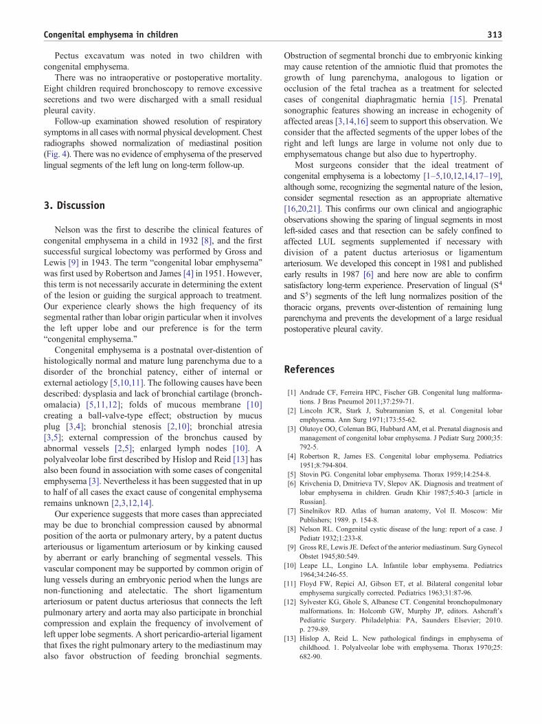
Fig. 4 Postoperative chest radiograph after segmental (S1–S3)resection of the left upper lobe. The mediastinum is in normalposition (thick arrow); the line of metal stapler sutures aftersegmental resection can be seen (thin arrow). There are no signsof recurrence of emphysema despite preserved lingual (S4 andS5) segments.
Pectus excavatum was noted in two children withcongenital emphysema.
There was no intraoperative or postoperative mortality.Eight children required bronchoscopy to remove excessivesecretions and two were discharged with a small residualpleural cavity.
Follow-up examination showed resolution of respiratorysymptoms in all cases with normal physical development. Chestradiographs showed normalization of mediastinal position(Fig. 4). There was no evidence of emphysema of the preservedlingual segments of the left lung on long-term follow-up.
3. Discussion
Nelson was the first to describe the clinical features ofcongenital emphysema in a child in 1932 [8], and the firstsuccessful surgical lobectomy was performed by Gross andLewis [9] in 1943. The term “congenital lobar emphysema”was first used by Robertson and James [4] in 1951. However,this term is not necessarily accurate in determining the extentof the lesion or guiding the surgical approach to treatment.Our experience clearly shows the high frequency of itssegmental rather than lobar origin particular when it involvesthe left upper lobe and our preference is for the term“congenital emphysema.”
Congenital emphysema is a postnatal over-distention ofhistologically normal and mature lung parenchyma due to adisorder of the bronchial patency, either of internal orexternal aetiology [5,10,11]. The following causes have beendescribed: dysplasia and lack of bronchial cartilage (bronch-omalacia) [5,11,12]; folds of mucous membrane [10]creating a ball-valve-type effect; obstruction by mucusplug [3,4]; bronchial stenosis [2,10]; bronchial atresia[3,5]; external compression of the bronchus caused byabnormal vessels [2,5]; enlarged lymph nodes [10]. Apolyalveolar lobe first described by Hislop and Reid [13] hasalso been found in association with some cases of congenitalemphysema [3]. Nevertheless it has been suggested that in upto half of all cases the exact cause of congenital emphysemaremains unknown [2,3,12,14].
Our experience suggests that more cases than appreciatedmay be due to bronchial compression caused by abnormalposition of the aorta or pulmonary artery, by a patent ductusarteriousus or ligamentum arteriosum or by kinking causedby aberrant or early branching of segmental vessels. Thisvascular component may be supported by common origin oflung vessels during an embryonic period when the lungs arenon-functioning and atelectatic. The short ligamentumarteriosum or patent ductus arteriosus that connects the leftpulmonary artery and aorta may also participate in bronchialcompression and explain the frequency of involvement ofleft upper lobe segments. A short pericardio-arterial ligamentthat fixes the right pulmonary artery to the mediastinum mayalso favor obstruction of feeding bronchial segments.
Obstruction of segmental bronchi due to embryonic kinkingmay cause retention of the amniotic fluid that promotes thegrowth of lung parenchyma, analogous to ligation orocclusion of the fetal trachea as a treatment for selectedcases of congenital diaphragmatic hernia [15]. Prenatalsonographic features showing an increase in echogenity ofaffected areas [3,14,16] seem to support this observation. Weconsider that the affected segments of the upper lobes of theright and left lungs are large in volume not only due toemphysematous change but also due to hypertrophy.
Most surgeons consider that the ideal treatment ofcongenital emphysema is a lobectomy [1–5,10,12,14,17–19],although some, recognizing the segmental nature of the lesion,consider segmental resection as an appropriate alternative[16,20,21]. This confirms our own clinical and angiographicobservations showing the sparing of lingual segments in mostleft-sided cases and that resection can be safely confined toaffected LUL segments supplemented if necessary withdivision of a patent ductus arteriosus or ligamentumarteriosum. We developed this concept in 1981 and publishedearly results in 1987 [6] and here now are able to confirmsatisfactory long-term experience. Preservation of lingual (S4
and S5) segments of the left lung normalizes position of thethoracic organs, prevents over-distention of remaining lungparenchyma and prevents the development of a large residualpostoperative pleural cavity.
[20] Costa AS, Perfeito JAJ, Forte V. Surgical treatment of 60 patients withpulmonary malformations: what have we learned? J Bras Pneumonol2008;34:661-6.
[21] Lilly JR, Wesenberg RL, Shikes RH. Segmental lung resection in thefirst year of life. Ann Thorac Surg 1976;22:16-22.