32
CONGENITAL GLAUCOMA MADE BY : SWATI PANARA FROM : BHARTIMAIYA COLLEGE OF OPTOMETRY 3 rd YEAR 6 th SEMESTER
| Date post: | 16-Jul-2015 |
| Category: |
Health & Medicine |
| Upload: | student |
| View: | 44 times |
| Download: | 0 times |

CONGENITAL GLAUCOMA
MADE BY : SWATI PANARAFROM : BHARTIMAIYA COLLEGE OF
OPTOMETRY3rd YEAR 6th SEMESTER
GLAUCOMA
• Glaucoma is not a single disease process but a group of disorders characterized by a progressive optic neuropathy resulting in a characterstic appearance of the optic disc and a specific pattern of irreversible visual field defects that are associated frequently but not invariably with raised intraocular pressure (IOP).

TYPES
CONGENITAL GLAUCOMA
PRIMARY ADULT
GLAUCOMA
SECONDARY GLAUCOMA

CONGENITAL GLAUCOMA
• The congenital glaucoma are a group of diverse disorders in which abnormal high intraocular pressure results due to developmental abnormalities of the angle of anterior chamber obstructing the drainage of aqueous humour.

TYPES
• PRIMARY DEVELOPMENTAL / CONGENITAL GLAUCOMA
• DEVELOPMENTAL GLAUCOMA WITH ASSOCIATED SPECIFIC OCULAR OR SYSTEMIC CONGENITAL ANOMALIES

PRIMARY DEVELOPMENTAL / CONGENITAL GLAUCOMA
• It refers to abnormally high IOP which results due to developmental anomaly of the angle of the anterior chamber, not associated with any other ocular or systemic anomaly.
• There are three types of primary congenital glaucoma.

NEW BORN GLAUCOMA
INFANTILE GLAUCOMA
JUVENILE GLAUCOMA

NEW BORN GLAUCOMA
• when IOP is raised during intrauterine life and child is born with ocular enlargement.
• It occurs in about 40 percent of cases.

INFANTILE GLAUCOMA
• when the disease manifests prior to the child's third birthday.
• It occurs in about 50 percent of cases.
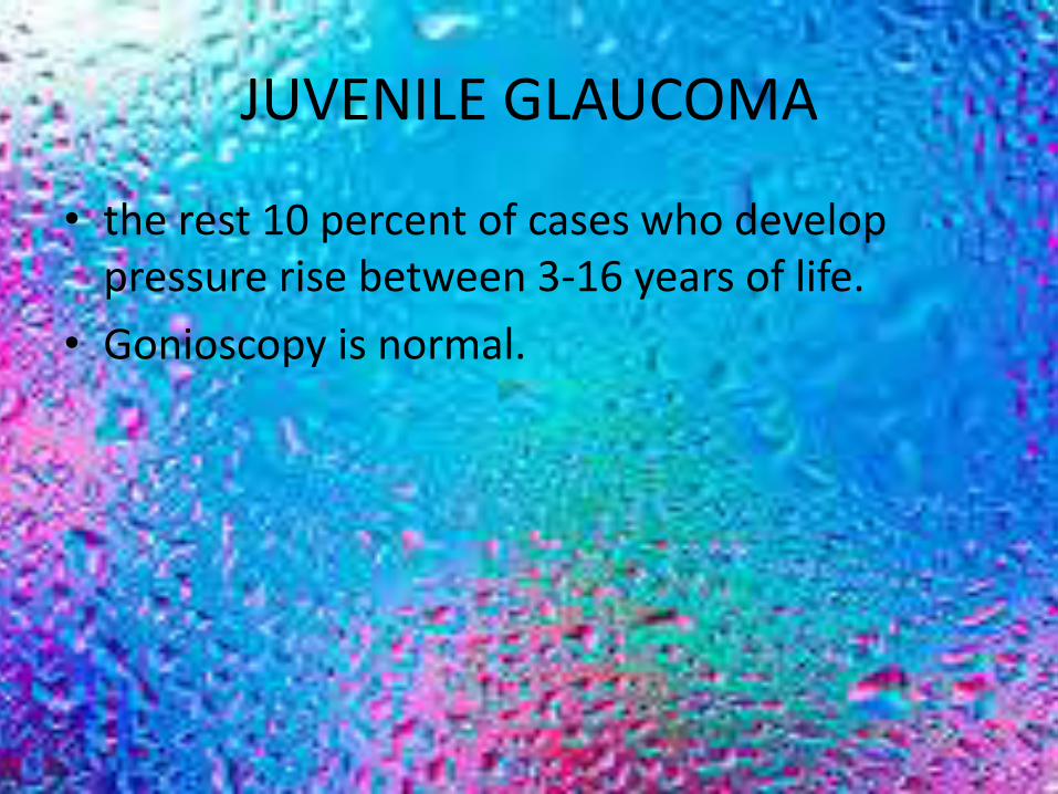
JUVENILE GLAUCOMA
• the rest 10 percent of cases who develop pressure rise between 3-16 years of life.
• Gonioscopy is normal.

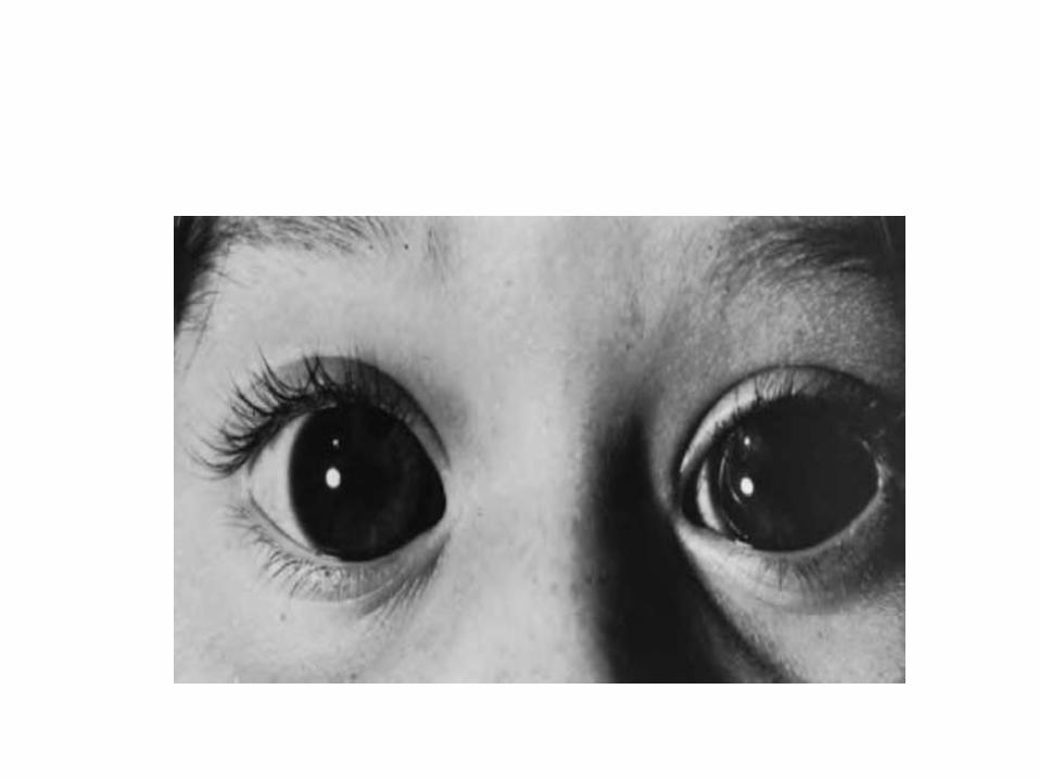
• Most infant corneas measure less than 10.5mm in horizontal diameter.
• A measurement over 12mm is considered diagnostic of congenital glaucoma.
• These eyes, hazy and enlarged, appear so grotesque that the term buphthalmos.
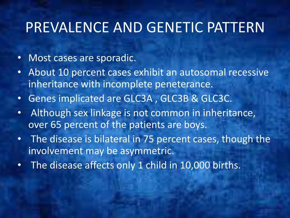
PREVALENCE AND GENETIC PATTERN
• Most cases are sporadic.
• About 10 percent cases exhibit an autosomal recessive inheritance with incomplete peneterance.
• Genes implicated are GLC3A , GLC3B & GLC3C.
• Although sex linkage is not common in inheritance, over 65 percent of the patients are boys.
• The disease is bilateral in 75 percent cases, though the involvement may be asymmetric.
• The disease affects only 1 child in 10,000 births.

PATHOGENESIS
• Maldevelopment of trabeculum including the iridotrabecular junction is responsible for impaired aqueous outflow resulting in raised IOP.
• In primary congenital glaucoma the trabeculodysgenesis is not associated with any other major ocular anomalies.

CLINICAL FEATURES
• Photophobia, blepharospasm, lacrimation and eye rubbing often occur together.
• Corneal signs. Corneal signs include its oedema, enlargement and Descemet’s breaks.
• IOP is raised which is neither marked nor acute.• Axial myopia may occur because of increase in axial length which
may give rise to anisometropic amblyopia


• Sclera becomes thin and appears blue due to underlying uveal tissue.
• Anterior chamber becomes deep.• Iris may show iridodonesis and atrophic patches in late stage.• Lens becomes flat due to stretching of zonules and may even
subluxate.• Optic disc may show variable cupping and atrophy especially
after third year.

EXAMINATION
• A complete examination under general anaesthesia should be performed on each child suspected of having congenital glaucoma:
(1) MEASUREMENT OF IOP : Schiotz or preferably hand held Perkin’s applanation tonometer since scleral rigidity is very low in children.
(2) MEASUREMENT OF CORNEAL DIAMETER.

(3) OPHTHALMOSCOPY TO EVALUATE OPTIC DISC
(4) GONIOSCOPIC EXAMINATION
(5) CENTRAL CORNEAL THICKNESS
(6) ULTRA SOUND


DIFFERENTIAL DIAGNOSIS
• Cloudy cornea. In unilateral cases the commonest cause is trauma with rupture of Descemet’s membrane. In bilateral cases causes may be trauma, interstitial keratitis and corneal endothelial dystrophy.
• Large cornea due to buphthalmos should be differentiated from megalocornea.
• Lacrimation in an infant is usually considered to be due to congenital nasolacrimal duct blockage and thus early diagnosis of congenital glaucoma may be missed.
• Photophobia may be due to keratitis or uveitis.• Raised IOP in infants may also be associated with retinoblastoma,
retinopathy of prematurity, persistent primary hyperplastic vitreous, traumatic glaucoma and secondary congenital glaucoma seen in rubella, aniridia.
TREATMENT
• Treatment of congenital glaucoma is primarily surgical :
(1) Incisional angle surgery
(2) Filteration surgery

DEVELOPMENTAL GLAUCOMAS WITHASSOCIATED ANOMALIES
• A wide variety of systemic and/or ocular anomalies have an associated raised IOP, usually due to developmental defects of the anterior chamber angle.
(1) Glaucoma associated with iridocorneal dysgenesis. These include: posterior embryotoxon characterized by a prominent Schwalbe’s ring Rieger anomaly, Rieger syndrome, Peter’s anomaly and combined Rieger syndrome and Peter’s anomaly.
(2) Glaucoma associated with aniridia (50% cases).

SCHWALBE’S RING
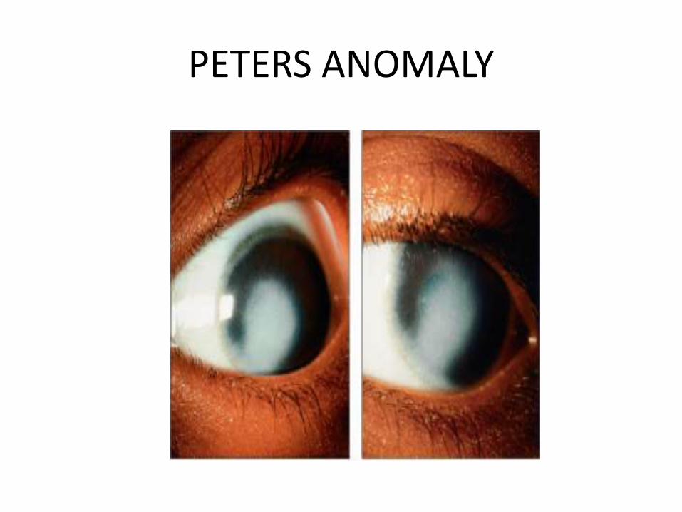
PETERS ANOMALY

ANIRIDIA

RIEGER SYNDROME
• (3) Glaucoma associated with ectopia lentis syndromes, which include Marfan’s syndrome, Weil - Marchesani syndrome and homocystinuria.
• (4) Glaucoma associated with phakomatosis is seen in Sturge-Weber syndrome ( 50% cases) and Von Recklinghausen’s neurofibromatosis (25% cases).


• (5) Miscellaneous conditions. Lowe’s syndrome naevus of Ota, nanophthalmos, congenital ectropion uveae, congenital microcornea and rubella syndrome.


