Page 1
ORIGINAL COMMUNICATION
Congenital myasthenic syndrome with tubular aggregates causedby GFPT1 mutations
Velina Guergueltcheva • Juliane S. Muller • Marina Dusl • Jan Senderek • Anders Oldfors •
Christopher Lindbergh • Susan Maxwell • Jaume Colomer • Cecilia Jimenez Mallebrera • Andres Nascimento •
Juan J. Vilchez • Nuria Muelas • Janbernd Kirschner • Shahriar Nafissi • Ariana Kariminejad •
Yalda Nilipour • Bita Bozorgmehr • Hossein Najmabadi • Carmelo Rodolico • Jorn P. Sieb •
Beate Schlotter • Benedikt Schoser • Ralf Herrmann • Thomas Voit • Ortrud K. Steinlein • Abdolhamid Najafi •
Andoni Urtizberea • Doriette M. Soler • Francesco Muntoni • Michael G. Hanna • Amina Chaouch •
Volker Straub • Kate Bushby • Jacqueline Palace • David Beeson • Angela Abicht • Hanns Lochmuller
Received: 20 July 2011 / Revised: 13 September 2011 / Accepted: 15 September 2011 / Published online: 6 October 2011
� Springer-Verlag 2011
Abstract Congenital myasthenic syndrome (CMS) is a
clinically and genetically heterogeneous group of inherited
disorders of the neuromuscular junction. A difficult to
diagnose subgroup of CMS is characterised by proximal
muscle weakness and fatigue while ocular and facial
involvement is only minimal. DOK7 mutations have been
identified as causing the disorder in about half of the cases.
More recently, using classical positional cloning, we have
identified mutations in a previously unrecognised CMS
gene, GFPT1, in a series of DOK7-negative cases. How-
ever, detailed description of clinical features of GFPT1
patients has not been reported yet. Here we describe the
clinical picture of 24 limb-girdle CMS (LG-CMS) patients
and pathological findings of 18 of them, all carrying
GFPT1 mutations. Additional patients with CMS, but
without tubular aggregates, and patients with non-fatigable
weakness with tubular aggregates were also screened. In
most patients with GFPT1 mutations, onset of the disease
occurs in the first decade of life with characteristicV. Guergueltcheva and J. S. Muller contributed equally to the study.
V. Guergueltcheva � M. Dusl � B. Schlotter � B. Schoser �A. Abicht
Department of Neurology, Friedrich-Baur-Institut,
Ludwig Maximilians University, Munich, Germany
V. Guergueltcheva
Clinic of Neurology, University Hospital Alexandrovska,
Sofia, Bulgaria
J. S. Muller � A. Chaouch � V. Straub � K. Bushby �H. Lochmuller (&)
Institute of Genetic Medicine, Newcastle University,
International Centre for Life, Central Parkway,
Newcastle upon Tyne NE1 3BZ, UK
e-mail: [email protected]
J. Senderek
Institute of Neuropathology, RWTH Aachen University,
Aachen, Germany
J. Senderek
Institute of Human Genetics, RWTH Aachen University,
Aachen, Germany
A. Oldfors
Department of Pathology, Institute of Biomedicine,
University of Gothenburg, Gothenburg, Sweden
C. Lindbergh
Neuromuscular Center, Sahlgrenska University Hospital,
Gothenburg, Sweden
S. Maxwell � D. Beeson
Neurosciences Group, Department of Clinical Neurology,
Weatherall Institute of Molecular Medicine,
University of Oxford, Oxford, UK
J. Colomer � C. J. Mallebrera � A. Nascimento
Unitat de Patologia Neuromuscular, Servei de Neurologia,
Hospital Sant Joan de Deu, Esplugues, Barcelona, Spain
J. J. Vilchez � N. Muelas
Servicio de Neurologıa, Hospital Universitario y Politecnico
La Fe and CIBER de Enfermedades Neurodegenerativas
(CIBERNED), Valencia, Spain
J. Kirschner
Division of Neuropaediatrics and Muscle Disorders,
University Medical Center, Freiburg, Germany
S. Nafissi
Department of Neurology, Tehran University of Medical
Sciences, Tehran, Iran
123
J Neurol (2012) 259:838–850
DOI 10.1007/s00415-011-6262-z
Page 2
limb-girdle weakness and fatigue. A common feature was
beneficial and sustained response to acetylcholinesterase
inhibitor treatment. Most of the patients who had a muscle
biopsy showed tubular aggregates in myofibers. Analysis of
endplate morphology in one of the patients revealed
unspecific abnormalities. Our study delineates the pheno-
type of CMS associated with GFPT1 mutations and
expands the understanding of neuromuscular junction dis-
orders. As tubular aggregates in context of a neuromuscular
transmission defect appear to be highly indicative, we
suggest calling this condition congenital myasthenic syn-
drome with tubular aggregates (CMS-TA).
Keywords Congenital myasthenic syndromes �Limb-girdle myasthenia � Tubular aggregates �GFPT1 � Dok-7
Abbreviations
AChE Acetylcholinesterase
AChR Acetylcholine receptor
CK Creatine kinase
CMAP Compound muscle action potential
CMS Congenital myasthenic syndrome
3,4-DAP 3,4-Diaminopyridine
DOK7 Downstream of kinase 7 gene
EM Electron microscopy
EMG Electromyography
LG-CMS Limb-girdle congenital myasthenic
syndrome
NMJ Neuromuscular junction
RNS Repetitive nerve stimulation
SFEMG Single-fiber EMG
TA Tubular aggregates
GFPT1/GFAT1 Glutamine-fructose-6-phosphate
transaminase 1
Introduction
Congenital myasthenic syndrome (CMS) is a rare and het-
erogeneous group of inherited muscle disorders caused by
genetic defects that affect signal transmission at the neu-
romuscular junction (NMJ) [1, 2]. The clinical phenotype of
CMS is fatigable weakness presenting usually from birth
but later onset is also possible. To date, 14 different genes
are known to cause CMS if mutated (http://neuromuscular.
wustl.edu/synmg.html).
Limb-girdle congenital myasthenic syndrome (LG-CMS)
is a previously recognised clinical entity [3] with prominent
shoulder and pelvic girdle weakness and fatigue and minimal
ocular and facial involvement. In 2006, mutations in the
DOK7 gene were identified to cause a form of CMS with
limb-girdle weakness where patients do not benefit from
pyridostigmine treatment [4]. Subsequently, detailed clini-
cal analysis of DOK7 patients revealed that many of them
show external eye muscle involvement (often ptosis, less
frequent ophthalmoplegia) contrary to the original concept
of pure limb girdle weakness [4–9].
A. Kariminejad � B. Bozorgmehr � H. Najmabadi
Kariminejad-Najmabadi Pathology and Genetics Center,
Tehran, Iran
Y. Nilipour
Neuropathology Lab, Toos Hospital, Tehran, Iran
C. Rodolico
Departments of Neurosciences, Psychiatry and Anaesthesiology,
A.O.U. ‘‘G. Martino’’, Messina, Italy
J. P. Sieb
Department of Neurology, Geriatric Medicine and Palliative
Care, Hanse-Klinikum, Stralsund, Germany
R. Herrmann
Department of Paediatrics I, University Hospital Essen,
Essen, Germany
T. Voit
Institut de Myologie, Unite Mixte de Recherche
UPMC-INSERM-CNRS-AIM UM 76, U974, UMR 7215,
Groupe Hospitalier Pitie-Salpetriere, Paris, France
O. K. Steinlein
Institute of Human Genetics, Ludwig Maximilians University,
Munich, Germany
A. Najafi
Azad University Medical Branch, Tehran, Iran
A. Urtizberea
Hopital Marin, Hendaye, France
D. M. Soler
Department of Paediatrics, Mater Dei Hospital,
Tal-Qroqq, Msida, Malta
F. Muntoni
The Dubowitz Neuromuscular Centre,
UCL Institute of Child Health, London, UK
M. G. Hanna
MRC Centre for Neuromuscular Diseases,
UCL Institute of Neurology, London, UK
J. Palace
CMS NCG Group, Department of Clinical Neurology,
John Radcliffe Hospital, Oxford, UK
J Neurol (2012) 259:838–850 839
123
Page 3
Recently, we have identified the underlying gene
mutations in a second subset of CMS patients with prom-
inent limb-girdle weakness responding well to esterase
inhibitor therapy. We mapped the gene defect to the
GFPT1 (glutamine-fructose-6-phosphate transaminase 1)
gene on chromosome 2p13.3 [10]. GFPT1 is the key
enzyme of the hexosamine pathway yielding the amino
sugar UDP-N-acetylglucosamine, an essential substrate for
protein glycosylation [11].
In this report, we describe the clinical features of 24
patients with GFPT1 mutations; muscle histopathology
was available in 18 of these patients. This is the first
detailed description of the phenotypic presentation of
GFPT1 patients which should expedite diagnosis and
treatment of LG-CMS in the future.
Patients and methods
All studies were carried out with informed consent of the
patients or patients’ parents and approved by the institu-
tional ethics review boards. Consent has been obtained for
publishing any recognizable persons in photographs, vid-
eos, or other information. Nine families are derived from
the CMS patient cohort referred to the Friedrich-Baur-
Institute in Munich, Germany, for genetic testing over the
last 15 years. Two families (LGM7 and 8) were recruited
through the CMS service in Oxford, UK and reported
previously in [7] (patients 1 and 3 in Table 3). Two LGM
families with tubular aggregates were previously reported
in [12] (family LGM13) and [13] (family LGM3). GFPT1
mutations of all patients except family LGM17 have been
reported in [10]. Family pedigrees are shown in [10],
supplementary Fig. S2. Family LGM17 has not been
reported previously.
Three further patients described with CMS in a limb-
girdle distribution, tubular aggregates and benefit from
esterase inhibitors (LGM4, 15 and 16 in [10] and [12]) and
two patients reported in [7] did not carry GFPT1 mutations.
GFPT1 mutations were also absent from a cohort of 52
unsolved cases with a wide range of different CMS phe-
notypes, but without tubular aggregates. We also screened
a cohort of four patients with unexplained muscle weak-
ness and tubular aggregates on biopsy, but without clear
fatigability and without evidence of a neuromuscular
transmission defect, for mutations in GFPT1, but did not
detect any.
All pedigrees are compatible with autosomal recessive
inheritance; all parents of the probands are reported to be
healthy. Families LGM1-LGM4, LGM10 and LGM11 are
consanguineous. The patients from families LGM1,
LGM3, LGM5, LGM12, LGM13 and LGM17 are siblings.
The age at examination varied between 7 and 63 years.
Tubular aggregates in muscle biopsies were present in 13
of 18 biopsies from the 14 families. Formal clinical assess-
ment was performed in all patients as well as measurement
of serum creatine kinase levels (CK) and titres of anti-ace-
tylcholine receptor (anti-AChR) antibodies. Electromyog-
raphy (EMG), nerve conduction studies (NCS), repetitive
nerve stimulation (RNS) and single fibre EMG (SFEMG)
were performed using standard techniques. Muscle biopsies
were performed by open or needle technique. Electron
microscopical analysis of one muscle biopsy specimen was
performed after fixation in 2.5% glutaraldehyde, postfix-
ation in OsO4 and embedding in resin. Ultrathin sections
were contrasted with uranyl acetate and lead citrate.
Results
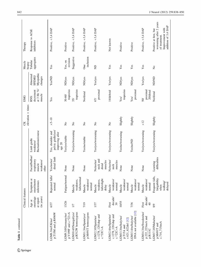
The main clinical, electrophysiological and muscle biopsy
features as well as the response to treatment of each patient
and their GFPT1 genotypes are presented in Table 1.
Clinical features
The first symptoms were noted in the first decade of life in
21 out of 24 patients. (range: first year of life–40s, median
6 years). Symptoms included difficulty in rising from a
squatting position, climbing stairs, lifting the arms above
the head, holding heavy objects and falls. All patients had
normal motor milestones except patients LGM7.3,
LGM12.3 and LGM17.3 (delayed achievement of inde-
pendent walking at age 18–24 months). Three patients
manifested after the first decade of life: patient LGM9.3
experienced weakness on physical activity at age 13 years,
patient LGM5.4 complained of shoulder girdle weakness in
her 40s, her brother (LGM5.3) had similar problems and
presented at age 14 years.
On examination, weakness was more pronounced in the
pelvic girdle muscles for most patients (Fig. 1f), but with
the following exceptions. The shoulder girdle was initially
involved in patient LGM5.3, LGM5.4 and in LGM3 family
[13]. The pelvic and shoulder girdles were equally affected
in patients LGM7.3 and LGM8.3, although weakness was
first noted in the pelvic girdle in patient LGM8.3 that
progressed to involve the shoulder girdle muscles 6 years
later. Scapular winging and waddling gait were evident in
half of the patients (Fig. 1e).
Only one patient had slight ptosis (patient LGM7.3) with
all other patients exhibiting no significant ocular muscle
involvement (Fig. 1a–c). Five patients exhibited mild
facial weakness (LGM1.4, LGM1.5, LGM5.4, LGM7.3
and LGM8.3). Additionally, distal muscle weakness was
noted in family LGM1 (long finger flexors and extensors as
well as foot extensors), patients LGM5.3, LGM5.5,
840 J Neurol (2012) 259:838–850
123
Page 4
Ta
ble
1C
lin
ical
feat
ure
so
fo
ur
pat
ien
tsw
ith
GF
PT
1m
uta
tio
ns
Cli
nic
alfe
atu
res
CK
elev
atio
n9
tim
es
EM
GM
usc
le
bio
psy
Th
erap
y
Ag
eat
on
set/
age
atre
po
rt
(in
yea
rs)
Sy
mp
tom
at
on
set/
mo
tor
mil
esto
nes
Fac
ial/
bu
lbar
/
resp
irat
ory
mu
scle
wea
kn
ess/
oth
er
Lim
bg
ird
le
wea
kn
ess/
flu
ctu
atio
ns/
cou
rse
RN
S:
dec
rem
ent
at3
Hz
%/
mu
scle
Ab
no
rmal
SF
EM
G/
My
op
ath
ic
chan
ges
Tu
bu
lar
agg
reg
ates
Res
po
nse
toA
Ch
E
inh
ibit
ors
LG
M1
.4/m
/Ira
n/y
es/
p.D
34
8Y
ho
mo
zyg
ou
s
6/3
1M
usc
le
wea
kn
ess,
fati
gu
e,w
ors
e
insu
mm
er/
no
rmal
Yes
/no
/no
/no
/
dis
tal
lim
b
Yes
/yes
/im
pro
vin
g
afte
rag
e2
0
91
.5Y
es/
pro
xim
al
ND
/ND
Yes
Po
siti
ve,
sid
eef
fect
s
LG
M1
.5/m
/Ira
n/y
es/
p.D
34
8Y
ho
mo
zyg
ou
s
6/2
6M
usc
le
wea
kn
ess,
fati
gu
e,w
ors
e
insu
mm
er/
no
rmal
Yes
/no
/no
/no
/
dis
tal
lim
b
Yes
/yes
/im
pro
vin
g
afte
rag
e2
0
91
.56
7/d
elto
idN
D/y
esN
DP
osi
tiv
e,si
de
effe
cts
LG
M2
.4/f
/Tu
rkey
/yes
/
p.W
24
0X
ho
mo
zyg
ou
s
6/2
6M
usc
le
wea
kn
ess,
fati
gu
ean
d
pai
n/n
orm
al
No
ne
Yes
/yes
/wo
rsen
ing
No
14
/dis
tal
Yes
/yes
Yes
Po
siti
ve
LG
M3
.3,
LG
M3
.5,
LG
M3
.6,
LG
M3
.8,
LG
M3
.9/2
m,3
f/L
iby
a/
yes
/p.R
11
1C
ho
mo
zyg
ou
s[1
3]
6/2
3–
35
Mu
scle
wea
kn
ess,
fati
gu
e/
no
rmal
No
ne
Yes
,
sho
uld
er[
pel
vic
gir
dle
/ND
/ND
No
65
-35
/
trap
eziu
s
ND
/yes
Yes
,b
iop
sy
per
form
ed
ino
ne
of
the
sib
lin
gs
Par
tial
lyp
osi
tiv
e
LG
M5
.3/m
/Sp
ain
/no
/
p.M
49
2T
and
c.*
22
C[
A
14
/55
Wea
kn
ess
in
the
up
per
lim
bs/
no
rmal
No
/no
/no
/
dis
tal
lim
b
Yes
/no
/sli
gh
t
wo
rsen
ing
91
.52
1/d
elto
idY
es/y
esY
esP
osi
tiv
e
LG
M5
.4/f
/Sp
ain
/no
/
p.M
49
2T
and
c.*
22
C[
A
40
s/5
4W
eak
nes
sin
sho
uld
er
gir
dle
/no
rmal
No
/no
/no
/
nec
k
mu
scle
s
Yes
/no
/sli
gh
t
wo
rsen
ing
No
12
/del
toid
Yes
/ND
ND
LG
M5
.5/m
/Sp
ain
/no
/
p.M
49
2T
and
c.*
22
C[
A
10
/50
Mu
scle
wea
kn
ess,
fall
s/n
orm
al
No
/no
/no
/
dis
tal
lim
b
Yes
/yes
/sli
gh
t
wo
rsen
ing
91
.52
6/d
elto
idY
es/y
esY
esP
osi
tiv
e,?
3,4
DA
P
LG
M6
.4/m
/Ger
man
y/n
o/
p.D
43
Van
dp
.I1
21
T
5/1
6M
usc
le
wea
kn
ess/
no
rmal
No
ne
Yes
/yes
/sli
gh
t
wo
rsen
ing
92
–8
Yes
ND
/ND
Yes
,o
n
re-r
evis
ion
Po
siti
ve
LG
M7
.3/m
/UK
/no
/
p.R
38
5H
and
p.R
43
4H
8/2
3F
atig
ue
on
wal
k/s
lig
ht
del
ay
Yes
/no
/sli
gh
t/
slig
ht
nec
k
and
dis
tal
lim
b
wea
kn
ess
Yes
,sh
ou
lder
and
pel
vic
gir
dle
/no
/
wo
rsen
ing
92
Yes
/
anco
neu
s
Yes
/ND
Yes
Po
siti
ve,
?3
,4D
AP
J Neurol (2012) 259:838–850 841
123
Page 5
Ta
ble
1co
nti
nu
ed
Cli
nic
alfe
atu
res
CK
elev
atio
n9
tim
es
EM
GM
usc
le
bio
psy
Th
erap
y
Ag
eat
on
set/
age
atre
po
rt
(in
yea
rs)
Sy
mp
tom
at
on
set/
mo
tor
mil
esto
nes
Fac
ial/
bu
lbar
/
resp
irat
ory
mu
scle
wea
kn
ess/
oth
er
Lim
bg
ird
le
wea
kn
ess/
flu
ctu
atio
ns/
cou
rse
RN
S:
dec
rem
ent
at3
Hz
%/
mu
scle
Ab
no
rmal
SF
EM
G/
My
op
ath
ic
chan
ges
Tu
bu
lar
agg
reg
ates
Res
po
nse
toA
Ch
E
inh
ibit
ors
LG
M8
.3/m
/UK
/no
/
p.T
15
Man
dp
.R4
96
W
6/3
7R
epea
ted
fall
s/
no
rmal
Yes
/no
/no
/
dis
tal
lim
b
Yes
,sh
ou
lder
and
pel
vic
gir
dle
/yes
/
imp
rov
ing
afte
r
age
20
95
–1
0Y
esY
es/N
DY
esP
osi
tiv
e,?
3,4
DA
P
LG
M9
.3/f
/Ger
man
y/n
o/
p.V
19
9F
and
c.*
22[
A
13
/26
Fat
igu
e/n
orm
alN
on
eY
es/y
es/s
tab
leN
o3
0-6
0/
trap
eziu
s
ND
/yes
Yes
,o
n
rev
isio
n
Po
siti
ve
LG
M1
0.4
/f/S
eneg
al/y
es/
p.R
51
2W
ho
mo
zyg
ou
s
1/7
Mu
scle
wea
kn
ess,
fall
s/n
orm
al
No
ne
Yes
/yes
/wo
rsen
ing
No
35
/ trap
eziu
s
ND
/yes
Su
gg
esti
ve
Po
siti
ve,
?3
,4D
AP
LG
M1
1.3
/m/S
pai
n/y
es/
p.M
49
1T
ho
mo
zyg
ou
s
7/1
9M
usc
le
wea
kn
ess/
no
rmal
No
ne
Yes
/no
/sta
ble
No
No
/dis
tal
ND
/yes
On
e
incl
usi
on
Po
siti
ve,
?3
,4D
AP
LG
M1
2.3
/f/S
pai
n/n
o/
c.1
27
8_
12
81
du
pan
d
c.*
22
C[
A
1/3
7M
usc
le
wea
kn
ess,
fall
s/sl
igh
t
del
ay
No
/no
/no
/
nec
kan
d
dis
tal
mu
scle
s
Yes
/yes
/wo
rsen
ing
No
67
/ pro
xim
al
Yes
/yes
ND
Po
siti
ve,
?3
,4D
AP
LG
M1
2.4
/m/S
pai
n/n
o/
c.1
27
8_
12
81
du
pan
d
c.*
22
C[
A
Fir
st dec
ade/
39
Mu
scle
wea
kn
ess/
no
rmal
No
/no
/no
/
nec
k
mu
scle
s
Yes
/yes
/wo
rsen
ing
No
14
/del
toid
Yes
/yes
Yes
No
tk
no
wn
LG
M1
3.3
/m/I
taly
/no
/
p.T
15
Aan
d
c.6
21
_6
22
del
[12]
10
/55
Mu
scle
wea
kn
ess/
no
rmal
No
ne
Yes
/no
/wo
rsen
ing
Sli
gh
tly
Yes
/
trap
eziu
s
ND
/yes
Yes
Po
siti
ve
LG
M1
3.4
/m/I
taly
/no
/
DN
An
ot
avai
lab
le[1
2]
7/3
6M
usc
le
wea
kn
ess/
no
rmal
No
ne
Yes
/no
/ND
Sli
gh
tly
Yes
/
pro
xim
al
ND
/yes
Yes
Po
siti
ve
LG
M1
4.3
/m/S
wed
en/n
o/
p.2
22
_2
23
insA
and
p.R
11
1C
Fir
st dec
ade/
40
Mu
scle
wea
kn
ess/
no
rmal
No
ne
Yes
/yes
/wo
rsen
ing
91
25
0/ pro
xim
al
20
/dis
tal
Yes
/yes
Yes
Po
siti
ve,
?3
,4D
AP
LG
M1
7.3
/f/M
alta
/no
/
p.M
49
1T
and
c.7
14
_7
15
insA
8/9
Fre
qu
ent
fall
s,
fati
gab
ilit
y
wh
en
wal
kin
g/
del
ayed
Lea
rnin
g
dif
ficu
ltie
s
Yes
/yes
/wo
rsen
ing
Sli
gh
tly
No
/dis
tal
ND
/ND
No
Po
siti
ve
atfi
rst,
bu
t
wo
rsen
ing
afte
r2
yea
rs
of
trea
tmen
t.
Imp
rov
emen
tw
ith
add
itio
no
f3
,4D
AP
842 J Neurol (2012) 259:838–850
123
Page 6
LGM7.3, LGM8.3 and LGM12.3. Neck muscles were
weak in patients LGM7.3, LGM12.3, LGM12.4 and
LGM5.4. One patient (LGM7.3) showed more generalised
and severe muscle involvement (ptosis, facial, and neck
weakness along with proximal and distal limb weakness).
He also had subclinical involvement of the respiratory
muscles and became non-ambulant within 4 years from
presentation. Muscle atrophy was rarely observed, the two
brothers from the Iranian family LGM1 were reported to
have slight generalized muscle atrophy and the affectd
members of family LGM5––scapular winging. Mild prox-
imal wasting was reported in family LGM3 [13].
The majority of patients reported prominent fluctuation
of symptoms, both improvement and worsening over short
periods of time (Table 1). Fixed muscle weakness was
reported in six patients (patients LGM5.3, LGM5.4,
LGM7.3, LGM11.3, LGM13.3 and LGM13.4). Diurnal
fluctuations were reported in patients LGM2.4 and
LGM6.4 while patient LGM14.3 experienced significant
day-to-day fluctuations. Both heat and infections were
noted to exacerbate neuromuscular weakness (LGM1,
LGM2.4, LGM3 family and LGM8.2). Disease progres-
sion, e.g. reduced walking distance, was noted in the first
two decades of life in most patients. Some patients expe-
rienced gradual worsening over decades. All patients
retained independent walking abilities during the periods of
observation except LGM7.3, LGM8.3 and LGM17.4. It
was lost permanently or for walking outdoors in all three
patients around age 12 years.
Laboratory tests and electrophysiology (Table 1)
CK levels were normal or slightly elevated in most patients
except in three individuals in whom the CK levels were
moderately elevated (up to 8–12 times) (LGM6.4, LGM8.3
and LGM14.3). Anti-AChR antibodies were not detected in
any of the patients.
When recording from distal muscles, RNS did not yield
a decremental response in some patients (family LGM1,
patients LGM5.4, LGM10.4, LGM11.3 and LGM17.4), but
clear decrement was obtained from proximal muscles in all
tested patients (Table 1). There was a single CMAP
response to single nerve stimuli except in patient LGM10.4
with a double CMAP response tested twice on and off
AChE inhibitors treatment. SFEMG showed abnormal jit-
ter in all nine patients. Needle EMG performed in 13
patients showed mild myopathic changes in proximal
muscles with no spontaneous activity.
Muscle pathological studies
Most muscle biopsies showed unspecific or mild myopathic
changes (summarised in Table 2). In addition, tubularTa
ble
1co
nti
nu
ed
Cli
nic
alfe
atu
res
CK
elev
atio
n9
tim
es
EM
GM
usc
le
bio
psy
Th
erap
y
Ag
eat
on
set/
age
atre
po
rt
(in
yea
rs)
Sy
mp
tom
at
on
set/
mo
tor
mil
esto
nes
Fac
ial/
bu
lbar
/
resp
irat
ory
mu
scle
wea
kn
ess/
oth
er
Lim
bg
ird
le
wea
kn
ess/
flu
ctu
atio
ns/
cou
rse
RN
S:
dec
rem
ent
at3
Hz
%/
mu
scle
Ab
no
rmal
SF
EM
G/
My
op
ath
ic
chan
ges
Tu
bu
lar
agg
reg
ates
Res
po
nse
toA
Ch
E
inh
ibit
ors
LG
M1
7.4
/m/M
alta
/no
/
p.M
49
1T
and
c.7
14
_7
15
insA
7/1
3D
iffi
cult
ies
in
run
nin
g,
fati
gab
ilit
y
wh
en
wal
kin
g/
no
rmal
Lea
rnin
g
dif
ficu
ltie
s
Yes
/yes
/wo
rsen
ing
Sli
gh
tly
ND
ND
/ND
No
Po
siti
ve
atfi
rst,
bu
tef
fect
lost
afte
r3
yea
rso
f
trea
tmen
td
esp
ite
add
ing
3,4
DA
P
Th
ecl
inic
alfe
atu
res
wer
en
ot
rep
ort
edto
be
asy
mm
etri
cal
Yes
,o
nre
visi
on
TA
-neg
ativ
eb
iop
sies
wer
eex
amin
edag
ain
afte
rth
eg
enet
icd
iag
no
sis
was
mad
ean
dG
FP
T1
mu
tati
on
sw
ere
fou
nd
inth
ose
pat
ien
ts
ND
no
td
on
e,C
Kcr
eati
ne
kin
ase,
EM
Gel
ectr
om
yo
gra
ph
y,
3,4
-DA
P3
,4-D
iam
ino
py
rid
ine,
RN
Sre
pet
itiv
en
erv
est
imu
lati
on
,S
FE
MG
sin
gle
-fib
erE
MG
,A
Ch
Eac
ety
lch
oli
nes
tera
se
J Neurol (2012) 259:838–850 843
123
Page 7
aggregates (TAs) were identified in 11 families, best seen
on NADH staining (Fig. 2). NADH staining and EM
images of the TAs of affected individuals from families
LGM3 and four have been published previously [12, 13].
The muscle pathology studies of patients LGM5.3,
LGM5.5 and LGM12.4 revealed TAs but so small that they
could be overlooked or misinterpreted as mitochondrial
proliferation. In two patients (LGM10.4 and LGM11.3) we
detected inconclusive histological findings that did not
fully match the criteria for TAs and were not further ana-
lysed under electron microscopy (Fig. 2d). Electron
microscopy (EM) images of the tubular aggregates
observed in patient LGM14.3 are shown in Fig. 2e, f. End
plate morphology in patient 14.3 (Fig. 3) revealed
pronounced simplification of the postsynaptic membrane
compared to the normal NMJ. There were numerous
apparently normal synaptic vesicles in the axon terminals.
Response to therapy
Twenty of 22 treated patients responded well to AChE
inhibitor treatment at a daily dosage of 80–540 mg (median
217 mg/day for patients aged 7–55 years), the remaining
two patients (LGM5.4 and LGM12.4) did not receive
treatment (Table 1). However, the benefit was not sus-
tained in family LGM1 and LGM17. In family LGM1,
although AChE inhibitors treatment led to significant
improvement of the muscle weakness at a dosage of
Fig. 1 Photographs of GFPT1-
related CMS patients reported in
this study. a, b Patients LGM1.4
and LGM1.5. Note the absence
of ptosis in the two brothers.
c Patient LGM10.4. Eye
movements are not restricted.
d Patient LGM6.4. Note the
absence of facial muscle
weakness. e, f Patient LGM11.3
shows weakness in the shoulder
and pelvic girdle, and has
difficulties rising from the floor
and lifting the arms sidewards
844 J Neurol (2012) 259:838–850
123
Page 8
2.8 mg/kg/day in the older brother and 2.4 mg/kg/day in
the younger brother, treatment was later discontinued due
to possible side effects (muscle twitching, depression and
anxiety). The addition of 3,4-Diaminopyridine (3,4-DAP)
in some patients was effective in improving or stabilizing
the disease. In LGM 17, both siblings benefited from AChE
in the first 18 months but seemed to relapse after that.
Following an initial improvement with pyridostigmine by
12 years of age patient LGM17.3, started complaining of
increased general fatigue, weakness in her hands and
without medication she could barely walk for 2–3 min.
Despite optimisation of pyridostigmine dose at 7.7 mg/kg/
day, she continued to deteriorate and by 13 years of age,
the 28 feet walking time was 9 s. By 14 years of age, she
was started on 3,4-DAP at a dose of 50 mg/day. On a
current optimised doses of combined pyridostigmine and
3,4-DAP her condition appeared to have stabilized. She
resumed walk for 10–15 min, her writing endurance
improved and she could now reach for heavy objects.
Additional features
Retinal involvement was reported in two of the families.
Both affected brothers from family LGM1 were diagnosed
as having juvenile macular degeneration causing signifi-
cant visual loss, more severe in the younger brother
LGM1.5. No other family members showed clinical signs
of retinal disease. Patient LGM7.3 exhibited an early squint
and was subsequently diagnosed as having retinitis pig-
mentosa at the age of 5 years. None of the other families
shows clinical signs of retinal disease, although ophthal-
mological studies were not performed in all patients. We
were interested to note learning difficulties in patient LGM
17.3 and 4.
Table 2 Muscle biopsy findings
Patient Analysed muscle Tubular aggregates (TAs) Additional findings
LGM1.4 Biceps brachii Yes (NADH staining) Fiber size variation, type 1 fibre predominance,
round or angular fibres
LGM2.4 Vastus lateralis Yes, TAs exclusively in type 2 fibres Chronic myopathic changes
LGM3 [13] Biceps brachii Yes, small subsarcolemmal aggregates
(NADH staining), TAs in EM
–
LGM5.3 Deltoid Yes, subsarcolemmal enhancement
(NADH staining), TAs in EM
Mild myopathic changes, type 1 fibres
predominance and ragged red-like fibres
LGM5.5 Deltoid Yes, subsarcolemmal enhancement
(NADH staining), TAs in EM
Unspecific myopathic changes, type 1 fibre
predominance, ragged red-like fibres
LGM6.4 Unknown Yes (NADH staining) Unspecific myopathic changes
LGM7.3 Unknown Yes Muscle atrophy, multiple internal nuclei,
vacuoles, features consistent with denervation
LGM8.3 Unknown Yes –
LGM9.3 Biceps brachii Yes –
LGM10.4 Deltoid Not clear, suggestive of TAs (NADH staining) Uneven oxidative staining, accumulation of
mitochondria
LGM11.3 Deltoid No, only one fibre with a possible inclusion
(NADH staining)
–
LGM12.3 Deltoid NADH staining and EM not done Unspecific myopathic changes
LGM12.4 Biceps brachii Yes, enhancement and subsarcolemmal
aggregates (NADH staining), TAs in EM
Unspecific myopathic changes
LGM13.3 [12] Vastus lateralis Yes, TAs in EM –
LGM13.4 [12] Vastus lateralis Yes –
LGM14.3 Deltoid Yes, frequent fibres with TAs, TAs in EM Increased fiber size variability
Frequent fibres with internalized nuclei and
autophagic vacuoles
LGM17.3 Quadriceps No Fibre size variability, oxidative enzyme staining
showed type 2 predominance with occasional
core-like areas and subtle uneven staining
LGM17.4 Quadriceps No Fibre size variability, oxidative enzyme staining
was pale and inconclusive
EM electron microscopy
J Neurol (2012) 259:838–850 845
123
Page 9
Discussion
We report on 14 families with CMS due to mutations in the
GFPT1 gene. The clinical phenotype associated with
GFPT1 mutations seems to be distinct, and includes fati-
gable weakness of the shoulder and hip girdle muscles,
normal eye movements, good response to esterase inhibi-
tors, and evidence of tubular aggregates on muscle biopsy.
Screening for GFPT1 mutations in 52 unsolved CMS cases
with a wide range of different clinical phenotypes, but
without tubular aggregates, was negative confirming that
GFPT1 mutations are associated with a distinct and rec-
ognisable CMS phenotype. Tubular aggregates in muscle
biopsies of patients with a proven neuromuscular trans-
mission defect seem to be highly indicative of GFPT1
defects. Therefore, we would like to suggest naming this
condition congenital myasthenic syndrome with tubular
aggregates (CMS-TA). This follows the example of
another clinically distinct form of CMS, congenital myas-
thenic syndrome with episodic apnea (CMS-EA), which is
caused by mutations in CHAT [14].
Notably, five patients with CMS and tubular aggregates
(three described in [12], 2 in [7]) did not have GFPT1
mutations. This may indicate that a small proportion of
patients may carry cryptic mutations in GFPT1 not
detectable by standard exon sequencing of genomic DNA,
or may carry mutations in other, yet unknown genes.
Moreover, patients with unexplained muscle weakness and
tubular aggregates on muscle biopsy, but without evidence
of a neuromuscular transmission defect, seem to be less
likely to carry mutations in GFPT1, and other causes of
tubular aggregates, such as periodic paralysis or chronic
alcohol consumption, need to be considered.
Previous reports on so-called limb-girdle congenital
myasthenia with and without tubular aggregates [7, 12, 15–
22] described patients with a phenotype that may be
Fig. 2 NADH staining and range of tubular aggregates present in
muscle biopsy sections of CMS patients with GFPT1 mutations. a,
b Patient LGM1.4. Biopsy from the biceps brachii muscle showing
variation in fiber size and increased internal nuclei. Some fibres show
abundant TAs, other show tiny cytoplasmic vacuoles containing
granular material. b A magnified view of the highlighted area in
a. c Patient LGM9.3. Biopsy from the biceps brachii muscle, NADH
reaction reveals TAs in a few fibres (arrows). d Patient LGM10.4.
Significant subsarcolemmal accumulations of mitochondria and
unevenness of staining, but unequivocal TAs were not seen in this
patient’s biopsy. e, f Patient 14.3. Electron microscopy images of
tubular aggregates beneath the sarcolemma. The tubules vary in
diameter
846 J Neurol (2012) 259:838–850
123
Page 10
compatible with the clinical phenotype observed in our
GFPT1 patients. However, it was difficult to ascertain
whether these patients belong to a single disease entity
distinguishable from other forms of inherited neuromus-
cular junction defects. Genetic testing of these patients for
GFPT1 can now be undertaken.
CMS with tubular aggregates caused by GFPT1 defects
has clinical and pathological features that may help dis-
tinguishing it from other forms of CMS. This is particularly
important as patients with an inherited neuromuscular
transmission defect and predominant limb-girdle weakness
seem to fall into two major categories. Half of the patients
with CMS and prominent limb-girdle weakness carry
mutations in the DOK7 gene [4, 6–8]. CMS caused by
DOK7 mutations shows clear clinical and pathological
differences from CMS with GFPT1 mutations: DOK7
patients may have ptosis, facial, bulbar and respiratory
involvement and do not show sustained benefit from
esterase inhibitor treatment, while muscle weakness may
improve under ephedrine treatment [23, 24]. Muscle
biopsies do not show tubular aggregates. Table 3 compares
the main features of DOK7 and GFPT1 caused forms of
CMS. Rarely, CMS patients with mutations in RAPSN or
COLQ show a limb-girdle pattern of weakness. However,
they were never found to have tubular aggregates on
muscle biopsies [25–27].
The moderately elevated CK levels in three patients
(LGM 6.4, LGM 8.3 and LGM 14.3) correspond to severe
Fig. 3 Ultrastructural findings
at the neuromuscular junction
(NMJ) in patient LGM14.3.
a Normal NMJ from a control
demonstrating normal nerve
terminal (N) and highly
complex postsynaptic
membrane folding with well
formed secondary synaptic
clefts (arrow head). Schwann
cell processes (S) cover the
nerve terminal, without
extending into the normal
primary synaptic cleft (arrow).
The postsynaptic membrane
(arrow heads) is well
developed. b–f NMJ of patient
14.3, deltoid muscle, three
endplates were studied. The
NMJ which is illustrated in
(b) is shown at higher
magnification (c) and (d).
Additional NMJs are illustrated
in (e) and (f). Note pronounced
simplification of the
postsynaptic membrane
compared to the normal NMJ.
There are numerous apparently
normal synaptic vesicles in the
axon terminals
J Neurol (2012) 259:838–850 847
123
Page 11
and fixed muscle weakness in just one of them (LGM 8.3)
and clear dystrophic features are not found in their muscle
biopsies. Myopathic changes in needle EMG are found in
all patients examined and do not correspond to CK levels.
Given the lack of clear fluctuations in eight of the patients,
one has to consider a wider differential diagnosis of pri-
mary muscle disorders like congenital myopathies and
muscular dystrophies. Indeed, many of our GFPT1 patients
were assigned a clinical diagnosis of a myopathy or mus-
cular dystrophy prior to the elucidation of a neuromuscular
transmission defect. Evidence of decrement on repeat nerve
stimulation and the positive response to AChE inhibitors
may be invaluable clinical clues to help distinguish CMS
with tubular aggregates from other muscle disorders.
We were interested to note retinal involvement in two of
our families (macular degeneration in family LGM1 and
retinitis pigmentosa in patient LGM7.3). It is presently
unclear whether retinal disease is associated with the
GFPT1 mutations in these families or whether the patients
are affected by two genetically distinct conditions.
The GFPT1 enzyme is expressed in both nerve and
muscle tissue [28, 29]. Endplate morphology analysis in
patient LGM14.3 showed unspecific abnormalities and the
ultrastructural data we have available so far do not clarify
Table 3 Comparison of main typical clinical features of CMS patients with DOK7 and GFPT1 mutations
CMS with GFPT1 mutations CMS with DOK7 mutations
Disease onset
Average age at onset (range) First decade (birth–40s) Second year of life, sometimes late onset (birth–late 20s)
First symptoms Walking difficulties, weakness
of shoulder or pelvic muscles
Walking difficulties; ptosis, floppy tone and bulbar
problems if onset at birth
Delayed motor milestones Rare, 3/24 patients in our study Walking onset usually not delayed
Pattern of muscle weakness
Ocular muscles
Bilateral ptosis Not present Yes
Ophthalmoparesis Not present Not present
Facial muscles Rarely affected (4/24 patients) Affected
Bulbar muscles Not affected Affected
Extremities, shoulder and pelvic
muscles
Limb-girdle weakness pattern Limb-girdle weakness pattern, waddling or sinuous gait
Respiratory problems No Deterioration of respiratory function during the course of
the disease, respiratory crises in some patients
Fluctuation of weakness Daytime-dependent, day–day
fluctuations, fluctuations over
longer periods
day–day fluctuations, fluctuations over longer periods
Progression Worsening in 11 patients, but
independent walking ability
retained in most
Progressive long-term deterioration leading to intermittent
or permanent wheelchair use
Other features Fixed weakness in some (8/24
patients)
Myopathic-like phenotype with permanent weakness,
thinness of muscles, spinal deformities
Muscle pathology
CK Elevated in 8/23 patients Elevated in some
Tubular aggregates Present in 13/18 biopsies Not present
Endplate pathology Unspecific changes (findings from
only one patient)
Small simplified endplates, degenerating or highly
simplified junctional folds, reinnervated, denervated and
ectopic junctions
Therapeutic options
Response to esterase inhibitors Clearly positive in most No effect or only short-term improvement, sometimes
worsening
Successful long-term therapy with Esterase inhibitors Ephedrine, salbutamol
Mutation analysis No obvious common mutation or
mutation hotspot. No patient
with complete loss of GFPT1
identified
Common mutation 1124_1127dupTGCC present in the
majority of patients, majority of patients have mutations
in exon 7. No CMS patient with complete loss of Dok-7
identified
848 J Neurol (2012) 259:838–850
123
Page 12
if the origin of the neurotransmission defect is primarily
presynaptic or postsynaptic, or indeed a combination of
both pre- and postsynaptic abnormalities. Analysis of
additional patients, as well as deciphering the molecular
consequences of impaired GFPT1 function or reduced
GFPT1 levels in patients will be required to fully under-
stand the molecular pathogenesis of this disorder.
The origin and functional consequences of TAs have
been investigated over the last 40 years. It is still unknown
whether TAs are pathological structures or represent
compensatory reactions to diverse pathogenic events such
as periodic paralysis, dyskalaemia, intoxication, inflam-
matory myopathies, cramps and myalgias, myotonia con-
genita, familial myopathies, and several other myopathies
of uncertain etiology [30, 31]. TAs are characterised as
more or less densely packed aggregates of vesicular or
tubular membranes of variable forms and sizes thought to
derive from the sarcoplasmic reticulum (review in [15,
30]). TAs can be seen by light microscopy as dark inclu-
sions in the NADH stain of muscle biopsies. A more spe-
cific way to identify TAs is by electron microscopy. This
however is not done on a routine basis for all muscle
biopsies. Caveolin-2(-/-)-deficient mice represent one
animal model with TAs in muscle [32]. Currently, a
molecular pathway linking TAs and the NMJ has not been
established. Some families in our study (LGM10.4 and
LGM11.3) share the same clinical features and harbour
GFPT1 mutations but TAs were not conclusively detected.
These are also some of the youngest patients in our study.
This may hint to variable expression of TAs in different
muscles or during life time of patients. Alternatively they
may be an unspecific feature not directly related to the
underlying pathomechanism. It will be interesting to see
whether some of the patients described as unspecific
myopathy with TAs carry mutations in GFPT1.
Our study confirms the phenotypic and genetic hetero-
geneity of CMS. In addition to DOK7–related CMS,
GFPT1 associated CMS seems to be an important and
distinct clinical and genetic entity associated with limb-
girdle fatigable weakness, clear response to pyridostig-
mine, and frequent TAs on muscle biopsies. The identifi-
cation of GFPT1 as the predominant gene involved CMS-
TA will allow better diagnosis, treatment and counselling
of patients and their families.
Acknowledgments We wish to thank the patients and their families
for participating in this study. The Institute of Genetic Medicine in
Newcastle is part of the MRC centre for Neuromuscular Diseases.
AA, JK, BS, RH, TV and HL are members of the German Muscular
Dystrophy Network (MD-NET 01GM0601) funded by the German
Ministry of Education and Research (BMBF, Bonn, Germany;
http://www.md-net.org). Newcastle University and MD-NET are
partners of TREAT-NMD (EC, 6th FP, proposal #036825; http://
www.treat-nmd.eu). VG is a research fellow of the Alexander von
Humboldt Foundation. JS is a Heisenberg fellow of the Deutsche
Forschungsgemeinschaft. AA is supported by a grant from the
Deutsche Forschungsgemeinschaft (Ab 130/2-1), DB by grants from
the Medical Research Council, the Myasthenia Gravis Association
and the Muscular Dystrophy Campaign. JSM receives a research
fellowship by the Faculty of Medical Sciences, Newcastle University.
NM received a fellowship from the Instituto de Salud Carlos III and
Fundacion para la Investigacion del Hospital Universitario La Fe
(CM06/00154). JJV and NM are members of CIBER de Enfermed-
ades Neurodegenerativas (CIBERNED), Valencia, Spain.
Conflict of interest None.
References
1. Engel AG, Sine SM (2005) Current understanding of congenital
myasthenic syndromes. Curr Opin Pharmacol 5:308–321
2. Muller JS, Mihaylova V, Abicht A, Lochmuller H (2007) Con-
genital myasthenic syndromes: spotlight on genetic defects of
neuromuscular transmission. Expert Rev Mol Med 9:1–20
3. McQuillen MP (1966) Familial limb-girdle myasthenia. Brain
89:121–132
4. Beeson D, Higuchi O, Palace J, Cossins J, Spearman H, Maxwell
S, Newsom-Davis J, Burke G, Fawcett P, Motomura M et al
(2006) Dok-7 mutations underlie a neuromuscular junction syn-
aptopathy. Science (New York, NY) 313:1975–1978
5. Ben Ammar A, Petit F, Alexandri N, Gaudon K, Bauche S,
Rouche A, Gras D, Fournier E, Koenig J, Stojkovic T et al (2010)
Phenotype genotype analysis in 15 patients presenting a con-
genital myasthenic syndrome due to mutations in DOK7. J Neurol
257:754–766
6. Muller JS, Herczegfalvi A, Vilchez JJ, Colomer J, Bachinski LL,
Mihaylova V, Santos M, Schara U, Deschauer M, Shevell M et al
(2007) Phenotypical spectrum of DOK7 mutations in congenital
myasthenic syndromes. Brain 130:1497–1506
7. Palace J, Lashley D, Newsom-Davis J, Cossins J, Maxwell S,
Kennett R, Jayawant S, Yamanashi Y, Beeson D (2007) Clinical
features of the DOK7 neuromuscular junction synaptopathy.
Brain 130:1507–1515
8. Selcen D, Milone M, Shen XM, Harper CM, Stans AA, Wieben
ED, Engel AG (2008) Dok-7 myasthenia: phenotypic and
molecular genetic studies in 16 patients. Ann Neurol 64:71–87
9. Anderson JA, Ng JJ, Bowe C, McDonald C, Richman DP,
Wollmann RL, Maselli RA (2008) Variable phenotypes associ-
ated with mutations in DOK7. Muscle Nerve 37:448–456
10. Senderek J, Muller JS, Dusl M, Strom TM, Guergueltcheva V,
Diepolder I, Laval SH, Maxwell S, Cossins J, Krause S et al
(2011) Hexosamine biosynthetic pathway mutations cause neu-
romuscular transmission defect. Am J Hum Genet 88:162–172
11. Haltiwanger RS, Lowe JB (2004) Role of glycosylation in
development. Annu Rev Biochem 73:491–537
12. Rodolico C, Toscano A, Autunno M, Messina S, Nicolosi C,
Aguennouz M, Laura M, Girlanda P, Messina C, Vita G (2002)
Limb-girdle myasthenia: clinical, electrophysiological and mor-
phological features in familial and autoimmune cases. Neurom-
uscul Disord 12:964–969
13. Sieb JP, Tolksdorf K, Dengler R, Jerusalem F (1996) An auto-
somal-recessive congenital myasthenic syndrome with tubular
aggregates in a Libyan family. Neuromuscul Disord 6:115–119
14. Ohno K, Tsujino A, Brengman JM, Harper CM, Bajzer Z, Udd B,
Beyring R, Robb S, Kirkham FJ, Engel AG (2001) Choline
acetyltransferase mutations cause myasthenic syndrome associ-
ated with episodic apnea in humans. Proc Natl Acad Sci USA
98:2017–2022
J Neurol (2012) 259:838–850 849
123
Page 13
15. Chevessier F, Bauche-Godard S, Leroy JP, Koenig J, Paturneau-
Jouas M, Eymard B, Hantai D, Verdiere-Sahuque M (2005) The
origin of tubular aggregates in human myopathies. J Pathol
207:313–323
16. Slater CR, Fawcett PR, Walls TJ, Lyons PR, Bailey SJ, Beeson
D, Young C, Gardner-Medwin D (2006) Pre- and post-synaptic
abnormalities associated with impaired neuromuscular transmis-
sion in a group of patients with ‘limb-girdle myasthenia’. Brain
129:2061–2076
17. Azulay JP, Pouget J, Figarella-Branger D, Colamarino R, Pel-
lissier JF, Serratrice G (1994) Isolated proximal muscular
weakness disclosing myasthenic syndrome. Rev Neurol (Paris)
150:377–381
18. Dobkin BH, Verity MA (1978) Familial neuromuscular disease
with type 1 fiber hypoplasia, tubular aggregates, cardiomyopathy,
and myasthenic features. Neurology 28:1135–1140
19. Furui E, Fukushima K, Sakashita T, Sakato S, Matsubara S,
Takamori M (1997) Familial limb-girdle myasthenia with tubular
aggregates. Muscle Nerve 20:599–603
20. Johns TR, Campa JF, Adelman LS (1973) Familial myasthenia
with ‘tubular aggregates’ treated with prednisone. Neurology
73:426
21. Johns TR, Campa JF, Crowley WJ, Miller JQ (1971) Familial
myasthenic myopathy. Neurology 71:449
22. Zephir H, Stojkovic T, Maurage CA, Hurtevent JF, Vermersch P
(2001) Tubular aggregate congenital myopathy associated with
neuromuscular block. Rev Neurol (Paris) 157:1293–1296
23. Schara U, Barisic N, Deschauer M, Lindberg C, Straub V, Strigl-
Pill N, Wendt M, Abicht A, Muller JS, Lochmuller H (2009)
Ephedrine therapy in eight patients with congenital myasthenic
syndrome due to DOK7 mutations. Neuromuscul Disord
19:828–832
24. Lashley D, Palace J, Jayawant S, Robb S, Beeson D (2010)
Ephedrine treatment in congenital myasthenic syndrome due to
mutations in DOK7. Neurology 74:1517–1523
25. Mihaylova V, Muller JS, Vilchez JJ, Salih MA, Kabiraj MM,
D’Amico A, Bertini E, Wolfle J, Schreiner F, Kurlemann G et al
(2008) Clinical and molecular genetic findings in COLQ-mutant
congenital myasthenic syndromes. Brain 131:747–759
26. Muller JS, Mildner G, Muller-Felber W, Schara U, Krampfl K,
Petersen B, Petrova S, Stucka R, Mortier W, Bufler J et al (2003)
Rapsyn N88 K is a frequent cause of congenital myasthenic
syndromes in European patients. Neurology 60:1805–1810
27. Cossins J, Burke G, Maxwell S, Spearman H, Man S, Kuks J,
Vincent A, Palace J, Fuhrer C, Beeson D (2006) Diverse
molecular mechanisms involved in AChR deficiency due to
rapsyn mutations. Brain 129:2773–2783
28. Oki T, Yamazaki K, Kuromitsu J, Okada M, Tanaka I (1999)
cDNA cloning and mapping of a novel subtype of gluta-
mine:fructose-6-phosphate amidotransferase (GFAT2) in human
and mouse. Genomics 57:227–234
29. Niimi M, Ogawara T, Yamashita T, Yamamoto Y, Ueyama A,
Kambe T, Okamoto T, Ban T, Tamanoi H, Ozaki K et al (2001)
Identification of GFAT1-L, a novel splice variant of human
glutamine: fructose-6-phosphate amidotransferase (GFAT1) that
is expressed abundantly in skeletal muscle. J Hum Genet
46:566–571
30. Pavlovicova M, Novotova M, Zahradnik I (2003) Structure and
composition of tubular aggregates of skeletal muscle fibres. Gen
Physiol Biophys 22:425–440
31. Engel WK, Bishop DW, Cunningham GG (1970) Tubular
aggregates in type II muscle fibers: ultrastructural and histo-
chemical correlation. J Ultrastruct Res 31:507–525
32. Schubert W, Sotgia F, Cohen AW, Capozza F, Bonuccelli G,
Bruno C, Minetti C, Bonilla E, Dimauro S, Lisanti MP (2007)
Caveolin-1(-/-)- and caveolin-2(-/-)-deficient mice both dis-
play numerous skeletal muscle abnormalities, with tubular
aggregate formation. Am J Pathol 170:316–333
850 J Neurol (2012) 259:838–850
123