the bmj | BMJ 2016;354:i3899 | doi: 10.1136/bmj.i3899 RESEARCH 1 OPEN ACCESS 1 Association for Assistance of Disabled Children, AACD, Recife, Brazil 2 Barão de Lucena Hospital, HBL, Recife, Brazil 3 Federal University of Pernambuco, UFPE, Recife, Brazil 4 Prof Fernando Figueira Integral Medicine Institute, IMIP, Recife, Brazil 5 Centro Diagnóstico Multimagem, Recife, Brazil 6 Mauricio de Nassau University, Recife, Brazil 7 University of Pernambuco, UPE, Recife, Brazil 8 Oswaldo Cruz University Hospital, HUOC, Recife, Brazil 9 Hospital Infantil Jorge de Medeiros Recife, Brazil 10 Altino Ventura Foundation, FAV, Recife, Brazil 11 Pernambuco’s Eye Hospital, Recife, Brazil Correspondence to: V van der Linden vanessavdlinden@ hotmail.com Cite this as: BMJ 2016;354:i3899 http://dx.doi.org/10.1136/bmj.i3899 Accepted: 05 July 2016 Congenital Zika syndrome with arthrogryposis: retrospective case series study Vanessa van der Linden, 1,2 Epitacio Leite Rolim Filho, 1,3 Otavio Gomes Lins, 3 Ana van der Linden, 4 Maria de Fátima Viana Vasco Aragão, 5,6 Alessandra Mertens Brainer-Lima, 6,7 Danielle Di Cavalcanti Sousa Cruz, 4 Maria Angela Wanderley Rocha, 8 Paula Fabiana Sobral da Silva, 8 Maria Durce Costa Gomes Carvalho, 8 Fernando José do Amaral, 2 Joelma Arruda Gomes, 2 Igor Colaço Ribeiro de Medeiros, 9 Camila V Ventura, 10,11 Regina Coeli Ramos 8 ABSTRACT OBJECTIVE To describe the clinical, radiological, and electromyographic features in a series of children with joint contractures (arthrogryposis) associated with congenital infection presumably caused by Zika virus. DESIGN Retrospective case series study. SETTING Association for Assistance of Disabled Children, Pernambuco state, Brazil. PARTICIPANTS Seven children with arthrogryposis and a diagnosis of congenital infection presumably caused by Zika virus during the Brazilian microcephaly epidemic. MAIN OUTCOME MEASURES Main clinical, radiological, and electromyographic findings, and likely correlation between clinical and primary neurological abnormalities. RESULTS The brain images of all seven children were characteristic of congenital infection and arthrogryposis. Two children tested positive for IgM to Zika virus in the cerebrospinal fluid. Arthrogryposis was present in the arms and legs of six children (86%) and the legs of one child (14%). Hip radiographs showed bilateral dislocation in seven children, subluxation of the knee associated with genu valgus in three children (43%), which was bilateral in two (29%). All the children underwent high definition ultrasonography of the joints, and there was no evidence of abnormalities. Moderate signs of remodeling of the motor units and a reduced recruitment pattern were found on needle electromyography (monopolar). Five of the children underwent brain computed tomography (CT) and magnetic resonance imaging (MRI) and the remaining two CT only. All presented malformations of cortical development, calcifications predominantly in the cortex and subcortical white matter (especially in the junction between the cortex and white matter), reduction in brain volume, ventriculomegaly, and hypoplasia of the brainstem and cerebellum. MRI of the spine in four children showed apparent thinning of the cord and reduced ventral roots. CONCLUSIONS Congenital Zika syndrome should be added to the differential diagnosis of congenital infections and arthrogryposis. The arthrogryposis was unrelated to the abnormalities of the joints themselves, but was possibly of neurogenic origin, with chronic involvement of central and peripheral motor neurones leading to deformities as a result of fixed postures in utero. Based on the neurophysiological observations, we suggest two possible mechanisms: tropism of neurones, with involvement of peripheral and central motor neurones, or a relation with vascular disorders. Introduction Epidemiological data suggested that cases of micro- cephaly in Brazil might be associated with the introduc- tion of the Zika virus. 1 One study detected Zika virus genome and anti-Zika virus IgM in the amniotic fluid of pregnant women with microcephalic fetuses. 2 An autopsy study described the complete recovery of the Zika virus genome from a fetus’s brain. 3 In April 2016, the US Centers for Disease Control and Prevention con- cluded that there is a causal relation between prenatal Zika virus infection and microcephaly and other seri- ous brain anomalies. 4 Brain impairment in the presence of microcephaly is the main characteristic of a congenital Zika virus syn- drome. However, little is still known about this condi- tion and its clinical spectrum, which also concerns newborns with a normal head circumference. Two stud- ies have described the association between arthrogryp- osis and microcephaly in newborns presumed to have congenital Zika virus infection. 5 6 Arthrogryposis multiplex congenita is characterised by joint contractures at birth, 7 which can be divided WHAT IS ALREADY KNOWN ON THIS TOPIC Until recently there were no reports of an association between congenital viral infection and arthrogryposis Aſter the outbreak of microcephaly in Brazil associated with Zika virus, two reports appeared suggesting an association, but they did not describe the deformities in detail WHAT THIS STUDY ADDS This case series provides detailed information about the clinical, imaging, and electromyographic findings in babies with arthrogryposis associated with congenital Zika virus infection Tests to evaluate arthrogryposis were consistent with a neurogenic pattern, with electromyelographic findings and spinal magnetic resonance imaging suggesting involvement of the lower motor neurones The pathophysiology of this condition might be related to the tropism of the virus by the upper and lower motor neurons, or to embryonic vascular change affecting these two segments
Transcript
thethinspbmj | BMJ 2016354i3899 | doi 101136bmji3899
RESEARCH
1
open access
1Association for Assistance of Disabled Children AACD Recife Brazil2Baratildeo de Lucena Hospital HBL Recife Brazil3Federal University of Pernambuco UFPE Recife Brazil4Prof Fernando Figueira Integral Medicine Institute IMIP Recife Brazil5Centro Diagnoacutestico Multimagem Recife Brazil6Mauricio de Nassau University Recife Brazil7University of Pernambuco UPE Recife Brazil8Oswaldo Cruz University Hospital HUOC Recife Brazil9Hospital Infantil Jorge de Medeiros Recife Brazil10Altino Ventura Foundation FAV Recife Brazil11Pernambucorsquos Eye Hospital Recife BrazilCorrespondence to V van der Linden vanessavdlindenhotmailcomCite this as BMJ 2016354i3899httpdxdoiorg101136bmji3899
Accepted 05 July 2016
Congenital Zika syndrome with arthrogryposis retrospective case series studyVanessa van der Linden12 Epitacio Leite Rolim Filho13 Otavio Gomes Lins3 Ana van der Linden4 Maria de Faacutetima Viana Vasco Aragatildeo56 Alessandra Mertens Brainer-Lima67 Danielle Di Cavalcanti Sousa Cruz4 Maria Angela Wanderley Rocha8 Paula Fabiana Sobral da Silva8 Maria Durce Costa Gomes Carvalho8 Fernando Joseacute do Amaral2 Joelma Arruda Gomes2 Igor Colaccedilo Ribeiro de Medeiros9 Camila V Ventura1011 Regina Coeli Ramos8
ABSTRACTObjeCtiveTo describe the clinical radiological and electromyographic features in a series of children with joint contractures (arthrogryposis) associated with congenital infection presumably caused by Zika virusDesignRetrospective case series studysettingAssociation for Assistance of Disabled Children Pernambuco state BrazilPartiCiPantsSeven children with arthrogryposis and a diagnosis of congenital infection presumably caused by Zika virus during the Brazilian microcephaly epidemicMain OutCOMe MeasuresMain clinical radiological and electromyographic findings and likely correlation between clinical and primary neurological abnormalitiesresultsThe brain images of all seven children were characteristic of congenital infection and arthrogryposis Two children tested positive for IgM to Zika virus in the cerebrospinal fluid Arthrogryposis was present in the arms and legs of six children (86) and the legs of one child (14) Hip radiographs showed bilateral dislocation in seven children subluxation of the knee associated with genu valgus in three children (43) which was bilateral in two (29) All the children underwent high definition ultrasonography of the joints and there was no
evidence of abnormalities Moderate signs of remodeling of the motor units and a reduced recruitment pattern were found on needle electromyography (monopolar) Five of the children underwent brain computed tomography (CT) and magnetic resonance imaging (MRI) and the remaining two CT only All presented malformations of cortical development calcifications predominantly in the cortex and subcortical white matter (especially in the junction between the cortex and white matter) reduction in brain volume ventriculomegaly and hypoplasia of the brainstem and cerebellum MRI of the spine in four children showed apparent thinning of the cord and reduced ventral rootsCOnClusiOnsCongenital Zika syndrome should be added to the differential diagnosis of congenital infections and arthrogryposis The arthrogryposis was unrelated to the abnormalities of the joints themselves but was possibly of neurogenic origin with chronic involvement of central and peripheral motor neurones leading to deformities as a result of fixed postures in utero Based on the neurophysiological observations we suggest two possible mechanisms tropism of neurones with involvement of peripheral and central motor neurones or a relation with vascular disorders
IntroductionEpidemiological data suggested that cases of micro-cephaly in Brazil might be associated with the introduc-tion of the Zika virus1 One study detected Zika virus genome and anti-Zika virus IgM in the amniotic fluid of pregnant women with microcephalic fetuses2 An autopsy study described the complete recovery of the Zika virus genome from a fetusrsquos brain3 In April 2016 the US Centers for Disease Control and Prevention con-cluded that there is a causal relation between prenatal Zika virus infection and microcephaly and other seri-ous brain anomalies4
Brain impairment in the presence of microcephaly is the main characteristic of a congenital Zika virus syn-drome However little is still known about this condi-tion and its clinical spectrum which also concerns newborns with a normal head circumference Two stud-ies have described the association between arthrogryp-osis and microcephaly in newborns presumed to have congenital Zika virus infection5 6
Arthrogryposis multiplex congenita is characterised by joint contractures at birth7 which can be divided
WhAT IS AlReAdy knoWn on ThIS TopICUntil recently there were no reports of an association between congenital viral infection and arthrogryposisAfter the outbreak of microcephaly in Brazil associated with Zika virus two reports appeared suggesting an association but they did not describe the deformities in detail
WhAT ThIS STudy AddSThis case series provides detailed information about the clinical imaging and electromyographic findings in babies with arthrogryposis associated with congenital Zika virus infectionTests to evaluate arthrogryposis were consistent with a neurogenic pattern with electromyelographic findings and spinal magnetic resonance imaging suggesting involvement of the lower motor neuronesThe pathophysiology of this condition might be related to the tropism of the virus by the upper and lower motor neurons or to embryonic vascular change affecting these two segments
doi 101136bmji3899 | BMJ 2016354i3899 | thethinspbmj
RESEARCH
2
into isolated and multiple contractures Isolated con-tractures affect only one area of the body most com-monly the foot The term arthrogryposis is often used as shorthand to describe multiple congenital contractures affecting two or more areas of the body8 Thus arthro-gryposis might be considered more a sign than a spe-cific disease and it might be associated with several disorders However there are no reports in the literature about other congenital infections in humans associated with arthrogryposis9 10
We describe the clinical radiological and electro-myographic findings in a series of seven children with arthrogryposis associated with congenital infection presumably caused by Zika virus and try to establish a likely correlation between the clinical and primary neu-rological abnormalities found
MethodsWe conducted a descriptive retrospective study by reviewing the medical records of children with arthro-gryposis associated with congenital infection presum-ably caused by Zika virus during the Brazilian microcephaly epidemic The children were seen at the rehabilitation centre of the Association for Assistance of Disabled Children (AACD) in Pernambuco Brazil which follows up patients with congenital Zika virus infection Two children were treated by the AACD team in the intensive care unit of other hospitals
According to the Brazilian Department of Healthrsquos protocol all children with suspected microcephaly are referred to one of two paediatric infectious disease departments and to one of four rehabilitation centres All investigations described were conducted as part of the clinical protocol or clinical indication none was conducted for research reasons and therefore neither ethical approval nor informed consent was necessary (other than for the use of photographs in this paper) The first criterion for referral was a head circumference of less than 33 cm from 2 December the criterion was changed to 32 cm for gestational age 37 weeks or more and two standard deviations below the mean for age and sex in the Fenton curve for preterm babies
In this series we describe seven patients with a diag-nosis of congenital infection presumably caused by Zika virus who had arthrogryposis and met the inclu-sion criteria of brain imaging suggestive of congenital infection a negative test result for the five other main infectious causes of microcephalymdashtoxoplasmosis cytomegalovirus rubella syphilis and HIV and pres-ence of arthrogryposis defined as congenital contrac-tures affecting two or more areas of the body
A standard form was used to collect personal and clinical data including maternal reports of a rash during pregnancy
Cytomegalovirus toxoplasmosis rubella syphilis and HIV are the main causes of congenital infections that result in brain calcifications and microcephaly We tested for these viruses using paired serology (IgM and IgG) of both the mother and the newborn If cytomega-lovirus IgG was present in both samples we carried out polymerase chain reaction on urine specimens
We excluded patients with known causes of microceph-aly other than the Zika virus
IgM antibody capture enzyme-linked immunosor-bent assay was used to test for Zika virus in the cerebro-spinal fluid samples of two children following the CDC protocol11
Microcephaly is an important sign however it is not present in all cases of congenital Zika virus infection The Fetal International and Newborn Growth Consor-tium for the 21st Century (Intergrowth-21st) defines microcephaly as a head circumference two standard deviations below the mean for gestational age and sex and severe microcephaly as three standard deviations below this mean12 13 We also used the Intergrowth-21st curve to evaluate birth weight classified as appropri-ate small or large for gestational age and sex13
All the children underwent neurological and ortho-paedic examinations along with several other investiga-tions radiography brain computed tomography (CT) or brain magnetic resonance imaging (MRI) without con-trast high definition ultrasonography of the joints (with specific attention to cartilage synovia pericapsular structures and muscular tissue around joints) nerve conduction studies and needle electromyography If cal-cifications were present on brain imaging (CT or MRI) we considered the possibility of congenital infections
Four children underwent MRI of the spine MRI was not possible in two children as they were receiving mechanical ventilation on an intensive unit care
According to Pernambuco statersquos protocol for microcephaly all seven children underwent fundo-scopic assessment and six also underwent hearing screening by otoacoustic emissions or brainstem evoked potentials
One of the children underwent orthopaedic surgery for correction of foot and hip deformities Assessment of range of motion was carried out under anaesthesia and the muscles evaluated macroscopically
Patient involvementNo patients were involved in setting the research ques-tion or the outcome measures nor were they involved in developing plans for design or implementation of the study No patients were asked to advise on interpreta-tion or writing up of results There are no plans to dis-seminate the results of the research to study participants or the relevant patient community
ResultsAt the time of writing March 2016 104 children were under evaluation at AACD for congenital infection pre-sumably caused by Zika virus Seven (7) met the inclusion criteria (brain imaging suggestive of congeni-tal infection a negative test result for congenital infec-tions and presence of arthrogryposis) two of whom were girls (29) Two of the seven children tested posi-tive for IgM for Zika virus in the cerebrospinal fluid All seven children met the protocol criteria for congenital infection presumably caused by Zika virus even with-out being tested for IgM for Zika virus which is not yet available on a routine basis
thethinspbmj | BMJ 2016354i3899 | doi 101136bmji3899
RESEARCH
3
Table 1 summarises the characteristics of the chil-dren All were born at term in Pernambuco state Brazil during October to November 2015 Four mothers (57) described having a rash between the second and fourth gestational months The head circumference was in the normal range in one child (14) two standard devia-tions below the mean for gestational age and sex in two (29) and three standard deviations below this mean for gestational age and sex in four (57) Three of the children (43) were of appropriate birth weight for ges-tational age and four (571) were small for gestational age Six of the seven (86) children showed evidence of craniofacial disproportion three (43) had redundant skin on the scalp at birth Dysphagia was present in six children (86) two underwent gastrostomy and tra-cheostomy All five boys had cryptorchidism which was unilateral in one child
Arthrogryposis was present in both the arms and the legs of six children (86) and in the legs of one child (14) Several leg deformities were observed congen-ital clubfoot in six children (86) which was bilateral in three (43) knee flexion contracture in five chil-dren (71) which was bilateral in three (43) and unilateral in two (29) hyperextension associated with subluxation of the knee in three children (43) which was bilateral in two (29) and contractures of hip flexion adduction and external rotation associ-ated with irreducible bilateral dislocation that is not reducible to manoeuvre of Ortolani in all seven chil-dren In all seven children spinal deformities were not identified in either the sagittal or the coronal plane on plain radiography The chest was barrel-like in four children (57) Deformities identified in the arms were camptodactyly in six children (86) which was bilateral in five (714) and deformations of flexion in the second to fifth chirodactylus in all seven children Adduction of the thumb was present in five children (71) abduction of the thumb in two (29) a bilat-eral simian crease in one (14) deformities in hyper-extension of the elbow in four (57) flexion contracture in two (29) which was bilateral and decreased range of motion of the shoulder with con-tracture in adduction and internal rotation in two (29) Figure 1 shows the clinical pictures of children with arthrogryposis
None of the children had deformities or limitation of motion of the cervical spine
Other findings were ligamentous laxity in one child (14) and skin haemangioma in four children (57)mdashone frontal three occipital and one on the left paratho-racic region
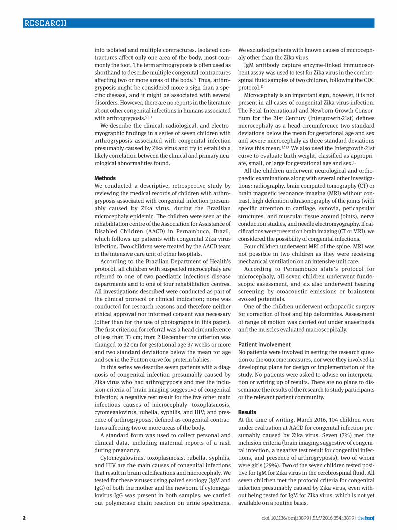
Hip radiographs showed bilateral dislocation in seven children subluxation of the knee associated with genu valgus in three children (43) bilateral in two (29) In all the children no dysplastic changes were identified on simple radiographs of the appen-dicular skeleton and spine and none were identified in either the sagittal or coronal plane dysplastic changes were identified in the dislocated hips related to dysplastic acetabular (acetabular index gt30 degrees) Table 2 shows the main deformities most often found in the children and figure 2 shows some radiological features
All seven children underwent high definition ultraso-nography of the joints with specific attention to carti-lage synovia pericapsular structures and muscular tissue around the joints There was no evidence of joint abnormalities
Nerve conduction studies and needle electromyogra-phy was performed in all seven children Nerves stud-ied were median and ulnar (sensory and motor conduction studies) tibial and fibular (motor conduc-tion studies) and medial plantar (sensory conduction
table 1 | Characteristics of the childrenPatient no sex
Zika igM status
birth weight for ga
Maternal rash during pregnancy
Head circumference at birth Microcephalus
Craniofacial disproportion
redundant scalp skin
1 Boy Positive Appropriate 2nd month 33 cm No No No2 Girl Positive Small 2nd month 30 cm 2 SD Yes No3 Boy Not done Small 3rd month 27 cm 3 SD Yes Yes4 Girl Not done Appropriate No 29 cm 2 SD Yes No5 Boy Not done Appropriate No 30 cm 3 SD Yes No6 Boy Not done Small No 27 cm 3 SD Yes Yes7 Boy Not done Small 4th month 26 cm 3 SD Yes YesGA=gestational ageStandard deviations below mean for age and sex
Fig 1 | (a) Contracture in flexion of knee (b) hyperextension of knee (knee dislocation) (C) clubfeet (D) deformities in 2nd 3rd and 4th fingers (e) joint contractures in legs and arms without involvement of trunk
doi 101136bmji3899 | BMJ 2016354i3899 | thethinspbmj
RESEARCH
4
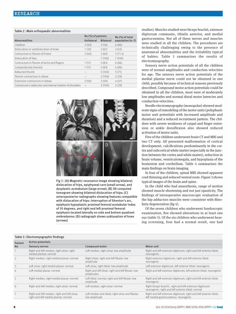
studies) Muscles studied were biceps brachii extensor digitorum communis tibialis anterior and medial gastrocnemius Not all of these nerves and muscles were studied in all the children The procedures are technically challenging owing to the presence of anatomical abnormalities and the irritability typical of babies Table 3 summarises the results of electromyography
Sensory nerve action potentials of all the children were of normal amplitudes and conduction velocities for age The sensory nerve action potentials of the medial plantar nerve could not be obtained in one child possibly because of technical reasons previously described Compound motor action potentials could be obtained in all the children most were of moderately low amplitudes and normal distal motor latencies and conduction velocities
Needle electromyography (monopolar) showed mod-erate signs of remodeling of the motor units (polyphasic motor unit potentials with increased amplitude and duration) and a reduced recruitment pattern The chil-dren with severe weakness of carpal and finger exten-sion or ankle dorsiflexion also showed reduced activation of motor units
Five of the children underwent brain CT and MRI and two CT only All presented malformation of cortical development calcifications predominantly in the cor-tex and subcortical white matter (especially in the junc-tion between the cortex and white matter) reduction in brain volume ventriculomegaly and hypoplasia of the brainstem and cerebellum Table 4 summarises the main findings on brain imaging
In four of the children spinal MRI showed apparent cord thinning and reduced ventral roots Figure 3 shows typical images of the brain and spine
In the child who had anaesthesia range of motion showed muscle shortening and not just spasticity The findings of intraoperative macroscopic evaluation of the hip adductors muscles were consistent with fibro-fatty degeneration (fig 4)
Of the seven children who underwent fundoscopic examination five showed alterations in at least one eye (table 5) Of the six children who underwent hear-ing screening four had a normal result one had
Fig 2 | (a) Magnetic resonance image showing bilateral dislocation of hips epiphyseal core (small arrow) and dysplastic acetabulum (large arrow) (b) 3D computed tomogram showing bilateral dislocation of hips (C) anteroposterior radiographs showing features compatible with dislocation of hips interruption of shentonrsquos arc epiphysis hypoplastic proximal femoral acetabular index of 35 degrees and right and left proximal femoral epiphysis located laterally on side and bottom quadrant ombredanne (D) radiograph shows subluxation of knee (arrows)
table 3 | electromyographic findings
Patient no
action potentialssensory nerves Compound motor Motor unit
1 Right and left median right ulnar right medial plantar normal
Left median right ulnar low amplitude Right and left extensor digitorum right and left anterior tibial neurogenic
2 Right median right medial planar normal Right tibial right and left fibular low amplitude
Right extensor digitorum right and left anterior tibial neurogenic
3 Left ulnar right medial planar normal Left ulnar right tibial low amplitude Left extensor digitorum left anterior tibial neurogenic4 Left medial planar normal Right and left tibial right and left fibular low
amplitudesRight and left extensor digitorum left anterior tibial neurogenic
5 Right median right medial planar normal Left tibial normal right and left fibular low amplitude
Right and left extensor digitorum right and left anterior tibial neurogenic
6 Right and left median right ulnar normal Left median right ulnar normal Right biceps brachii right and left extensor digitorum neurogenic right and left anterior tibial normal
7 Right and left median right and left ulnar right and left medial plantar normal
Left median and tibial right ulnar and fibular low amplitude
Right and left extensor digitorum right and left anterior tibial left medial gastrocnemius neurogenic
table 2 | Main orthopaedic abnormalities
abnormalitiesno () of patients no () of total
population (n=7)unilateral bilateralClubfoot 3 (50) 3 (50) 6 (86)Dislocation or subdislocation of knee 1 (33) 2 (67) 3 (43)Contracture in flexion of knees 2 (40) 3 (60) 5 (714)Dislocation of hips mdash 7 (100) 7 (100)Contracture in flexion of wrist and fingers 1 (17) 5 (83) 6 (86)Camptodactyly (hands) 1 (17) 5 (83) 6 (86)Adducted thumb mdash 5 (100) 5 (71)Flexion contracture in elbow mdash 2 (100) 2 (29)Extension contracture in elbow 2 (50) 2 (50) 4 (57)Contracture in adduction and internal rotation of shoulders mdash 2 (100) 2 (29)
thethinspbmj | BMJ 2016354i3899 | doi 101136bmji3899
RESEARCH
5
abnormalities in one ear and one had abnormalities in both ears
discussionIn this series of children with joint contractures ( arthrogryposis) associated with congenital infection presumably caused by Zika virus all seven showed changes on brain imaging with calcifications pre-dominantly in the cortex and subcortical white mat-ter (especially in the junction between the cortex and white matter) with abnormalities of cortical develop-ment and brainstem and cerebellar atrophy Tests to evaluate arthrogryposis were consistent with a neu-rogenic pattern with electromyelographic findings and spinal MRI suggesting involvement of the lower motor neurones Microcephaly and craniofacial dis-proportion have been common but were not present in all cases
Comparison with other studiesIn microcephaly a babyrsquos head is smaller than that of a baby of the same sex and age Microcephaly is a clini-cal sign and not a disease Increased rates of congeni-tal microcephaly have been reported during the Zika virus outbreak in Brazil beginning in late 201514 15 Genetic or environmental brain damage in utero can result in congenital microcephaly at birth and infec-tious causes are well known eg rubella cytomegalo-virus and toxoplasmosis12 Before 2015 evidence for a congenital infection presumed to be caused by Zika virus was lacking
This disease goes beyond microcephaly with other symptoms such as visual and hearing impairment and unusual signs and symptoms different from other congenital infections such as arthrogryposis and no microcephaly suggesting that the term congenital Zika syndrome is more appropriate The visual changes in this syndrome have been described previously16
The presence of disorders of cortical development suggest that the insult occurred within the first five months of pregnancy17 An earlier study reported three infants with fetal brain disruption sequence a recog-nisable pattern of defects consisting of severe micro-cephaly overlapping sutures prominence of the occipital bone and scalp rugae18 This condition seems to be produced by partial brain destruction during the second or third trimester diminution in intracranial hydrostatic pressure and subsequent col-lapse of the fetal skull18 Several causes for this condi-tion have been suggested including partial disruption of the blood supply to the brain and prenatal infection with viruses18 This finding is similar to that in our patients Dysphagia was a common symptom proba-bly related to the severity of changes apparent on brain imaging including hypoplasia of the brainstem and cerebellum
Arthrogryposis is derived from the Greek words arthro (joint) and gryposis (crooked)19 The term arthro-gryposis is often used as shorthand to describe multiple congenital contractures that affect two or more areas of ta
ble
4 | b
rain
imag
ing
findi
ngs
Patie
nt
noCa
licifi
catio
nsDe
crea
sed
brai
n vo
lum
e
Sym
met
ry
of fi
ndin
gsCe
rebe
llum
or
bra
inst
em
hypo
plas
ia
Vent
ricul
omeg
aly
Larg
e ex
traxi
al
CSF s
pace
Mal
form
atio
ns
of c
ortic
al
deve
lopm
ent
Sim
plifi
ed
gyra
l pa
ttern
Corp
us
callo
sum
Corti
cal a
nd
subc
ortic
al
white
mat
ter
Basa
l ga
nglia
Cere
bellu
mPe
riven
tricu
lar
Brai
nste
mPr
edom
inan
ce
(gt20
)En
larg
ed
cist
erna
m
agna
1M
ildYe
sYe
sYe
sNo
Yes
Yes
Norm
alYe
sYe
sNo
NoYe
sBa
sal g
angl
iaYe
s2
Mod
erat
eNo
Yes
Yes
Mild
Yes
Yes
Pron
ounc
ed
hypo
gene
ticYe
sNo
NoYe
sYe
sPe
riven
tricu
lar
Pron
ounc
ed
3Ye
sYe
sYe
sYe
sYe
sYe
sYe
sHy
poge
ntic
Yes
Yes
NoYe
sNo
Cort
ical
su
bcor
tical
w
hite
mat
ter
Yes
4M
oder
ate
Yes
Yes
Yes
Yes
Yes
Yes
Hypo
plas
icYe
sYe
sNo
Yes
Yes
Cort
ical
su
bcor
tical
w
hite
mat
ter
Yes
5Ye
sYe
sYe
sYe
sM
ildYe
sYe
sAg
enet
ic o
r pr
onou
nced
hy
poge
netic
Yes
Yes
NoNo
NoCo
rtic
al
subc
ortic
al
whi
te m
atte
r
Pron
ounc
ed
6Se
vere
Yes
Yes
Yes
Yes
Yes
Yes
Hypo
gene
ticYe
sYe
sYe
sYe
sNo
Cort
ical
su
bcor
tical
w
hite
mat
ter
Pron
ounc
ed
7Se
vere
Yes
Yes
Yes
Yes
Yes
Yes
Pron
ounc
ed
hypo
gene
ticYe
sYe
sYe
sYe
sYe
sCo
rtic
al
subc
ortic
al
whi
te m
atte
r
Yes
doi 101136bmji3899 | BMJ 2016354i3899 | thethinspbmj
RESEARCH
6
the body Arthrogryposis is not a specific diagnosis but rather a clinical finding and it is a characteristic of more than 300 disorders19 Arthrogryposis can be divided into subgroups as a way of generating a differential diagnosis which includes neurological dis-eases (brain spine or peripheral nerve) connective tissue defects (diastrophic dysplasia) muscle abnor-malities (muscular dystrophies or mitochondrial abnor-malities) space limitations within the uterus (oligohydramnios fibroids uterine malformations or multiple pregnancy) intrauterine or fetal vascular com-promise (impaired normal development of nerves or
anterior horn cell death) and maternal diseases (diabe-tes mellitus multiple sclerosis myasthenia gravis infection drugs or trauma)9
Neurological abnormalities seem to be one of the most common causes of arthrogryposis (about 70-80 of cases)9 Developmental abnormalities affecting the forebrain (eg hydranencephaly microcephaly fore-brain neuronal migration disorders) whether primarily due to genetic factors or a consequence of infection in the fetal central nervous system are sometimes associated with arthrogryposis In most such cases joint contractures are probably caused by diminished corticospinal tract activation of spinal cord motor neu-rones or sometimes the underlying disease also directly injures spinal cord motor neurones contributing to fetal hypomotility9 10
By 2015 there were no reports of congenital infec-tions associated with arthrogryposis in humans Two groups described the association between arthrogry-posis and microcephaly in newborns with congenital infection presumably due to Zika virus5 6 but with-out an indepth investigation of the possible causes and characterisation of deformities The Arkabane virus an arbovirus of the Simbu group of the family Bunyaviridae might cause abortions stillbirths premature births and deformed or anomalous bovine caprine and ovine fetuses or neonates including brain malformations and arthrogryposis Evidence for humans being infected by Akabane virus is lacking20
In our case series the arthrogryposis did not result from abnormalities of the joints themselves but was likely to be of neurogenic origin with chronic involve-ment of central and peripheral motor neurones lead-ing to fixed postures in utero and consequently deformities Electromyographic findings suggest chronic involvement of peripheral motor neurones
Fig 3 | spine and brain magnetic resonance imaging of baby with arthrogryposis sagittal t2 weighted fast imaging employing steady state acquisition (Fiesta) (a) showing apparently reduced spinal cord thickness (short arrows) and mega cisterna magna (long arrow) axial reconstruction of t2 weighted Fiesta (b) showing reduction of medullary cone ventral roots (long arrows) compared with dorsal roots (short arrows) sagittal t2 weighted image (C) showing hypogenesis of corpus callosum (long white arrow) enlarged cisterna magna (long black arrow) enlarged fourth ventricle (short black arrow) and pons hypoplasia (short white arrow) axial t2 weighted imaging (D) showing pachygyria in frontal lobes (black arrows) and severe ventriculomegaly mainly at posterior part of lateral ventricles axial susceptibility weighted image (e and F) showing some hypointense small dystrophic calcifications (white arrows) in junction between cortical and subcortical white matter (e) and in midbrain (F)
Fig 4 | adductor longus muscle of child with irreducible dislocation of hips before surgery Colour is characteristic of fibrofatty infiltration typical of initial phase of neuropathies
thethinspbmj | BMJ 2016354i3899 | doi 101136bmji3899
RESEARCH
7
In severely weak muscles the activation of the motor units was severely reduced suggesting reduced central drive and involvement of central motor neurones The pattern of peripheral denervation seems to corre-spond to the pattern of central involvement which could suggest a component of trans-synaptic degener-ation Spinal MRI showed apparent thinning of the spi-nal cord and a reduction in the ventral roots of medullary conus corroborate the findings on electro-myography An intraoperative macroscopic evaluation under anaesthesia is also consistent with findings on electromyography and spinal MRI
Interestingly developmental abnormalities of the cortex and arthrogryposis are found together in syn-dromes that result from exposure to misoprostrol in utero and in perisylvian polymicrogyria21 22 One study described neuropathological findings at autopsy and suggested a possible location of the virus in neurones3 Another study found that Zika virus infection leads to cell cycle arrest apoptosis and inhibition of the differ-entiation of neural progenitor cells resulting in cortical thinning and microcephaly23
Multiple hypotheses have been proposed to explain the presence of congenital joint contractures in some patients with abnormalities of brain development these include an in utero vascular insult affecting both central and peripheral nervous systems a common developmental mechanism of altered migration in both the brain and the spinal cord and a direct central effect of the brain malformation on joint mobility in the fetus20 On the basis of our neurophysiological observations and the literature we suggest two possi-ble mechanisms tropism for the neurones or neural progenitor cells with involvement of peripheral motor neurons and central motor neurones or a relation with vascular disorders
strengths and limitations of this studyIn this case series we evaluated all seven children and carried out extensive imaging along with neurological and orthopaedic investigations including fundo-scopic examination and auditory screening Further research is needed with a larger number of cases to study the neurological abnormalities behind arthro-gryposis including histopathology of autopsy sam-ples or tissues from stillborn babies As we do not know the potential implications of congenital Zika virus infection as it evolves children must receive orthopaedic follow-up even those with a standard first orthopaedic evaluation because they could
develop musculoskeletal deformities secondary to neurological impairment central or peripheral or both as these occur in patients with cerebral palsy and other chronic encephalopathies
ConclusionCongenital Zika syndrome should be added to the dif-ferential diagnosis of congenital infections and arthro-gryposisContributors VvdL and ELRF coordinated all work and did most of the writing VvdL AvdL PFSdS and MDCGdC were responsible for the neurological data ELRF was responsible for the orthopaedic data OGL was responsible for the electromyographic data RCR MAWR and DDCSC were responsible for laboratory data to exclude other congenital infections FJdA was responsible for the ultrasonographic data MdFVA and AMB-L helped to analyse brain and spinal cord imaging CVV assisted with the ophthalmologic data FJdA and ICRdM assisted the patients admitted to an intensive care unit All authors reviewed and commented on drafts and approved the final manuscript and the decision to submit for publication VvdL is the guarantorCompeting interests All authors have completed the ICMJE uniform disclosure form at wwwicmjeorgcoi_disclosurepdf and declare no support from any organisation for the submitted work no financial relationships with any organisations that might have an interest in the submitted work in the previous three years no other relationships or activities that could appear to have influenced the submitted workEthical approval All investigations described were conducted as part of a clinical protocol approved by the Brazilian government and analysed retrospectively no investigations were conducted for research reasons and therefore neither ethical approval nor informed consent was necessaryPatient consent The authors obtained written consent from parents for publication of the images in figures 1 to 4 All mothers gave consent for neuroimaging studies to be performed as part of the Brazilian microcephaly outbreak protocol or clinical indication All seven cases have been deidentified and we decided to proceed with publication in the interests of public healthData sharing No additional data availableTransparency The lead author (VvdL) affirms that the manuscript is an honestaccurate and transparent account of the study being reported that no important aspects of the study have been omittedThis is an Open Access article distributed in accordance with the Creative Commons Attribution Non Commercial (CC BY-NC 30) license which permits others to distribute remix adapt build upon this work non-commercially and license their derivative works on different terms provided the original work is properly cited and the use is non-commercial See httpcreativecommonsorglicensesby-nc30
1 Heukelbach J Alencar CH Kelvin AA de Oliveira WK Pamplona de Goacutees Cavalcanti L Zika virus outbreak in Brazil J Infect Dev Ctries 201610116-20
2 Calvet G Aguiar RS Melo ASO et al Detection and sequencing of Zika virus from amniotic fluid of fetuses with microcephaly in Brazil a case study Lancet Infect Dis 201616653-60
3 Mlakar J Korva M Tul N et al Zika virus associated with microcephaly N Engl J Med 2016374951-8
4 Rasmussen SA Jamieson DJ Honein MA Petersen LR Zika Virus and Birth DefectsndashReviewing the Evidence for Causality N Engl J Med 20163741981-7
table 5 | Ocular findingsPatient no
Findings 1 2 3 4 5 6 7Macular pigment mottling No Both eyes Both eyes Left eye No Right eye NoOptic disc pallor No Both eyes No No No Both eyes NoOptic disc hypoplasia No Both eyes No No No No NoMacular chorioretinal atrophic lesion Right eye Right eye No Left eye No Right eye NoChorioretinal atrophic macular lesion colobomatous No No No No No Left eye No
RESEARCH
No commercial reuse See rights and reprints httpwwwbmjcompermissions Subscribe httpwwwbmjcomsubscribe
5 Schuler-Faccini L Ribeiro EM Feitosa IML et al Brazilian Medical Genetics SocietyndashZika Embryopathy Task Force Possible Association Between Zika Virus Infection and MicrocephalyndashBrazil 2015 MMWR Morb Mortal Wkly Rep 20166559-62
6 Oliveira Melo AS Malinger G Ximenes R Szejnfeld PO Alves Sampaio S Bispo de Filippis AM Zika virus intrauterine infection causes fetal brain abnormality and microcephaly tip of the iceberg Ultrasound Obstet Gynecol 2016476-7
7 Fedrizzi E Botteon G Inverno M Ciceri E DrsquoIncerti L Dworzak F Neurogenic arthrogryposis multiplex congenita clinical and MRI findings Pediatr Neurol 19939343-8
8 Bamshad M Van Heest AE Pleasure D Arthrogryposis a review and update J Bone Joint Surg Am 200991(Suppl 4)40-6
9 Kalampokas E Kalampokas T Sofoudis C Deligeoroglou E Botsis D Diagnosing arthrogryposis multiplex congenita a review ISRN Obstet Gynecol 20122012264918 doi1054022012264918
10 Bamshad M Van Heest AE Pleasure D Arthrogryposis a review and update J Bone Joint Surg Am 200991(Suppl 4)40-6
11 Martin DA Muth DA Brown T Johnson AJ Karabatsos N Roehrig JT Standardization of immunoglobulin M capture enzyme-linked immunosorbent assays for routine diagnosis of arboviral infections J Clin Microbiol 2000381823-6
12 World Health Organization Assessment of infants with microcephaly in the context of Zika Interim guidance 25 February 2016 WHOZIKVMOC163 WHO
13 Villar J Cheikh Ismail L Victora CG et al International Fetal and Newborn Growth Consortium for the 21st Century (INTERGROWTH-21st) International standards for newborn weight length and head circumference by gestational age and sex the Newborn Cross-Sectional Study of the INTERGROWTH-21st Project Lancet 2014384857-68
14 Brasil-Ministeacuterio da Sauacutede Protocolo de vigilacircncia e resposta agrave ocorrecircncia de microcefalia relacionada agrave infecccedilatildeo pelo viacuterus Zika Available at httpportalsaudesaudegovbrimagespdf2015dezembro09Microcefalia---Protocolo-de-vigil--ncia-e-resposta---vers--o-1----09dez2015-8hpdf [Acessed January 26 2016]
15 Brasil-Ministeacuterio da Sauacutede Ministeacuterio da sauacutede atualiza casos suspeitos de microcefalia Available at httpportalsaudesaudegovbrindexphpcidadaoprincipalagencia-saude21890-ministerio-da-saude-investiga-3-448-casos-suspeitos-de-microcefalia [Accessed February 1 2016]
16 Ventura CV Maia M Ventura BV et al Ophthalmological findings in infants with microcephaly and presumable intra-uterus Zika virus infection Arq Bras Oftalmol 2016791-3
17 Volpe JJ Neurology of the Newborn4th ed WB Saunders 2008
18 Russell LJ Weaver DD Bull MJ Weinbaum M In utero brain destruction resulting in collapse of the fetal skull microcephaly scalp rugae and neurologic impairment the fetal brain disruption sequence Am J Med Genet 198417509-21
21 Marques-Dias MJ Physiopathogeny of moebius syndrome and arthrogriposis due to in utero misoprostrol exposure Thesis Satildeo Paulo 1999
22 Poduri A Chitsazzadeh V DrsquoArrigo S et al The syndrome of perisylvian polymicrogyria with congenital arthrogryposis Brain Dev 201032550-5
23 Li C Xu D Ye Q et al Zika Virus Disrupts Neural Progenitor Development and Leads to Microcephaly in Mice Cell Stem Cell 201619120-6
24 Dick GW Kitchen SF Haddow AJ Zika virus I Isolations and serological specificity Trans R Soc Trop Med Hyg 195246509-20
25 Dick GW Zika virus II Pathogenicity and physical properties Trans R Soc Trop Med Hyg 195246521-34
26 Duffy MR Chen TH Hancock WT et al Zika virus outbreak on Yap Island Federated States of Micronesia N Engl J Med 2009360 2536-43
27 Besnard M Lastere S Teissier A Cao-Lormeau V Musso D Evidence of perinatal transmission of Zika virus French Polynesia December 2013 and February 2014 Euro surveill 2014319(13)
28 Tognarelli J Ulloa S Villagra E et al A report on the outbreak of zika virus on Easter Island South Pacific 2014 Arch Virol 2016161 665-8
29 Zanluca C Melo VC Mosimann AL Santos GI Santos CN Luz K First report of autochthonous transmission of Zika virus in Brazil Mem Inst Oswaldo Cruz 2015110569-72
30 Tavernise S McNeil DG Jr Zika virus a global health emergence WHO says 2016 Feb 1 [cited 2016 Feb 20] In New York Times httpwwwnytimescom20160202healthzika-virus-world-health-organizationhtml_r=0
31 Mussi-Pinhata MM Yamamoto AY [Congenital and perinatal infections] J Pediatr (Rio J) 199975(Suppl 1)S15-30
doi 101136bmji3899 | BMJ 2016354i3899 | thethinspbmj
RESEARCH
2
into isolated and multiple contractures Isolated con-tractures affect only one area of the body most com-monly the foot The term arthrogryposis is often used as shorthand to describe multiple congenital contractures affecting two or more areas of the body8 Thus arthro-gryposis might be considered more a sign than a spe-cific disease and it might be associated with several disorders However there are no reports in the literature about other congenital infections in humans associated with arthrogryposis9 10
We describe the clinical radiological and electro-myographic findings in a series of seven children with arthrogryposis associated with congenital infection presumably caused by Zika virus and try to establish a likely correlation between the clinical and primary neu-rological abnormalities found
MethodsWe conducted a descriptive retrospective study by reviewing the medical records of children with arthro-gryposis associated with congenital infection presum-ably caused by Zika virus during the Brazilian microcephaly epidemic The children were seen at the rehabilitation centre of the Association for Assistance of Disabled Children (AACD) in Pernambuco Brazil which follows up patients with congenital Zika virus infection Two children were treated by the AACD team in the intensive care unit of other hospitals
According to the Brazilian Department of Healthrsquos protocol all children with suspected microcephaly are referred to one of two paediatric infectious disease departments and to one of four rehabilitation centres All investigations described were conducted as part of the clinical protocol or clinical indication none was conducted for research reasons and therefore neither ethical approval nor informed consent was necessary (other than for the use of photographs in this paper) The first criterion for referral was a head circumference of less than 33 cm from 2 December the criterion was changed to 32 cm for gestational age 37 weeks or more and two standard deviations below the mean for age and sex in the Fenton curve for preterm babies
In this series we describe seven patients with a diag-nosis of congenital infection presumably caused by Zika virus who had arthrogryposis and met the inclu-sion criteria of brain imaging suggestive of congenital infection a negative test result for the five other main infectious causes of microcephalymdashtoxoplasmosis cytomegalovirus rubella syphilis and HIV and pres-ence of arthrogryposis defined as congenital contrac-tures affecting two or more areas of the body
A standard form was used to collect personal and clinical data including maternal reports of a rash during pregnancy
Cytomegalovirus toxoplasmosis rubella syphilis and HIV are the main causes of congenital infections that result in brain calcifications and microcephaly We tested for these viruses using paired serology (IgM and IgG) of both the mother and the newborn If cytomega-lovirus IgG was present in both samples we carried out polymerase chain reaction on urine specimens
We excluded patients with known causes of microceph-aly other than the Zika virus
IgM antibody capture enzyme-linked immunosor-bent assay was used to test for Zika virus in the cerebro-spinal fluid samples of two children following the CDC protocol11
Microcephaly is an important sign however it is not present in all cases of congenital Zika virus infection The Fetal International and Newborn Growth Consor-tium for the 21st Century (Intergrowth-21st) defines microcephaly as a head circumference two standard deviations below the mean for gestational age and sex and severe microcephaly as three standard deviations below this mean12 13 We also used the Intergrowth-21st curve to evaluate birth weight classified as appropri-ate small or large for gestational age and sex13
All the children underwent neurological and ortho-paedic examinations along with several other investiga-tions radiography brain computed tomography (CT) or brain magnetic resonance imaging (MRI) without con-trast high definition ultrasonography of the joints (with specific attention to cartilage synovia pericapsular structures and muscular tissue around joints) nerve conduction studies and needle electromyography If cal-cifications were present on brain imaging (CT or MRI) we considered the possibility of congenital infections
Four children underwent MRI of the spine MRI was not possible in two children as they were receiving mechanical ventilation on an intensive unit care
According to Pernambuco statersquos protocol for microcephaly all seven children underwent fundo-scopic assessment and six also underwent hearing screening by otoacoustic emissions or brainstem evoked potentials
One of the children underwent orthopaedic surgery for correction of foot and hip deformities Assessment of range of motion was carried out under anaesthesia and the muscles evaluated macroscopically
Patient involvementNo patients were involved in setting the research ques-tion or the outcome measures nor were they involved in developing plans for design or implementation of the study No patients were asked to advise on interpreta-tion or writing up of results There are no plans to dis-seminate the results of the research to study participants or the relevant patient community
ResultsAt the time of writing March 2016 104 children were under evaluation at AACD for congenital infection pre-sumably caused by Zika virus Seven (7) met the inclusion criteria (brain imaging suggestive of congeni-tal infection a negative test result for congenital infec-tions and presence of arthrogryposis) two of whom were girls (29) Two of the seven children tested posi-tive for IgM for Zika virus in the cerebrospinal fluid All seven children met the protocol criteria for congenital infection presumably caused by Zika virus even with-out being tested for IgM for Zika virus which is not yet available on a routine basis
thethinspbmj | BMJ 2016354i3899 | doi 101136bmji3899
RESEARCH
3
Table 1 summarises the characteristics of the chil-dren All were born at term in Pernambuco state Brazil during October to November 2015 Four mothers (57) described having a rash between the second and fourth gestational months The head circumference was in the normal range in one child (14) two standard devia-tions below the mean for gestational age and sex in two (29) and three standard deviations below this mean for gestational age and sex in four (57) Three of the children (43) were of appropriate birth weight for ges-tational age and four (571) were small for gestational age Six of the seven (86) children showed evidence of craniofacial disproportion three (43) had redundant skin on the scalp at birth Dysphagia was present in six children (86) two underwent gastrostomy and tra-cheostomy All five boys had cryptorchidism which was unilateral in one child
Arthrogryposis was present in both the arms and the legs of six children (86) and in the legs of one child (14) Several leg deformities were observed congen-ital clubfoot in six children (86) which was bilateral in three (43) knee flexion contracture in five chil-dren (71) which was bilateral in three (43) and unilateral in two (29) hyperextension associated with subluxation of the knee in three children (43) which was bilateral in two (29) and contractures of hip flexion adduction and external rotation associ-ated with irreducible bilateral dislocation that is not reducible to manoeuvre of Ortolani in all seven chil-dren In all seven children spinal deformities were not identified in either the sagittal or the coronal plane on plain radiography The chest was barrel-like in four children (57) Deformities identified in the arms were camptodactyly in six children (86) which was bilateral in five (714) and deformations of flexion in the second to fifth chirodactylus in all seven children Adduction of the thumb was present in five children (71) abduction of the thumb in two (29) a bilat-eral simian crease in one (14) deformities in hyper-extension of the elbow in four (57) flexion contracture in two (29) which was bilateral and decreased range of motion of the shoulder with con-tracture in adduction and internal rotation in two (29) Figure 1 shows the clinical pictures of children with arthrogryposis
None of the children had deformities or limitation of motion of the cervical spine
Other findings were ligamentous laxity in one child (14) and skin haemangioma in four children (57)mdashone frontal three occipital and one on the left paratho-racic region
Hip radiographs showed bilateral dislocation in seven children subluxation of the knee associated with genu valgus in three children (43) bilateral in two (29) In all the children no dysplastic changes were identified on simple radiographs of the appen-dicular skeleton and spine and none were identified in either the sagittal or coronal plane dysplastic changes were identified in the dislocated hips related to dysplastic acetabular (acetabular index gt30 degrees) Table 2 shows the main deformities most often found in the children and figure 2 shows some radiological features
All seven children underwent high definition ultraso-nography of the joints with specific attention to carti-lage synovia pericapsular structures and muscular tissue around the joints There was no evidence of joint abnormalities
Nerve conduction studies and needle electromyogra-phy was performed in all seven children Nerves stud-ied were median and ulnar (sensory and motor conduction studies) tibial and fibular (motor conduc-tion studies) and medial plantar (sensory conduction
table 1 | Characteristics of the childrenPatient no sex
Zika igM status
birth weight for ga
Maternal rash during pregnancy
Head circumference at birth Microcephalus
Craniofacial disproportion
redundant scalp skin
1 Boy Positive Appropriate 2nd month 33 cm No No No2 Girl Positive Small 2nd month 30 cm 2 SD Yes No3 Boy Not done Small 3rd month 27 cm 3 SD Yes Yes4 Girl Not done Appropriate No 29 cm 2 SD Yes No5 Boy Not done Appropriate No 30 cm 3 SD Yes No6 Boy Not done Small No 27 cm 3 SD Yes Yes7 Boy Not done Small 4th month 26 cm 3 SD Yes YesGA=gestational ageStandard deviations below mean for age and sex
Fig 1 | (a) Contracture in flexion of knee (b) hyperextension of knee (knee dislocation) (C) clubfeet (D) deformities in 2nd 3rd and 4th fingers (e) joint contractures in legs and arms without involvement of trunk
doi 101136bmji3899 | BMJ 2016354i3899 | thethinspbmj
RESEARCH
4
studies) Muscles studied were biceps brachii extensor digitorum communis tibialis anterior and medial gastrocnemius Not all of these nerves and muscles were studied in all the children The procedures are technically challenging owing to the presence of anatomical abnormalities and the irritability typical of babies Table 3 summarises the results of electromyography
Sensory nerve action potentials of all the children were of normal amplitudes and conduction velocities for age The sensory nerve action potentials of the medial plantar nerve could not be obtained in one child possibly because of technical reasons previously described Compound motor action potentials could be obtained in all the children most were of moderately low amplitudes and normal distal motor latencies and conduction velocities
Needle electromyography (monopolar) showed mod-erate signs of remodeling of the motor units (polyphasic motor unit potentials with increased amplitude and duration) and a reduced recruitment pattern The chil-dren with severe weakness of carpal and finger exten-sion or ankle dorsiflexion also showed reduced activation of motor units
Five of the children underwent brain CT and MRI and two CT only All presented malformation of cortical development calcifications predominantly in the cor-tex and subcortical white matter (especially in the junc-tion between the cortex and white matter) reduction in brain volume ventriculomegaly and hypoplasia of the brainstem and cerebellum Table 4 summarises the main findings on brain imaging
In four of the children spinal MRI showed apparent cord thinning and reduced ventral roots Figure 3 shows typical images of the brain and spine
In the child who had anaesthesia range of motion showed muscle shortening and not just spasticity The findings of intraoperative macroscopic evaluation of the hip adductors muscles were consistent with fibro-fatty degeneration (fig 4)
Of the seven children who underwent fundoscopic examination five showed alterations in at least one eye (table 5) Of the six children who underwent hear-ing screening four had a normal result one had
Fig 2 | (a) Magnetic resonance image showing bilateral dislocation of hips epiphyseal core (small arrow) and dysplastic acetabulum (large arrow) (b) 3D computed tomogram showing bilateral dislocation of hips (C) anteroposterior radiographs showing features compatible with dislocation of hips interruption of shentonrsquos arc epiphysis hypoplastic proximal femoral acetabular index of 35 degrees and right and left proximal femoral epiphysis located laterally on side and bottom quadrant ombredanne (D) radiograph shows subluxation of knee (arrows)
table 3 | electromyographic findings
Patient no
action potentialssensory nerves Compound motor Motor unit
1 Right and left median right ulnar right medial plantar normal
Left median right ulnar low amplitude Right and left extensor digitorum right and left anterior tibial neurogenic
2 Right median right medial planar normal Right tibial right and left fibular low amplitude
Right extensor digitorum right and left anterior tibial neurogenic
3 Left ulnar right medial planar normal Left ulnar right tibial low amplitude Left extensor digitorum left anterior tibial neurogenic4 Left medial planar normal Right and left tibial right and left fibular low
amplitudesRight and left extensor digitorum left anterior tibial neurogenic
5 Right median right medial planar normal Left tibial normal right and left fibular low amplitude
Right and left extensor digitorum right and left anterior tibial neurogenic
6 Right and left median right ulnar normal Left median right ulnar normal Right biceps brachii right and left extensor digitorum neurogenic right and left anterior tibial normal
7 Right and left median right and left ulnar right and left medial plantar normal
Left median and tibial right ulnar and fibular low amplitude
Right and left extensor digitorum right and left anterior tibial left medial gastrocnemius neurogenic
table 2 | Main orthopaedic abnormalities
abnormalitiesno () of patients no () of total
population (n=7)unilateral bilateralClubfoot 3 (50) 3 (50) 6 (86)Dislocation or subdislocation of knee 1 (33) 2 (67) 3 (43)Contracture in flexion of knees 2 (40) 3 (60) 5 (714)Dislocation of hips mdash 7 (100) 7 (100)Contracture in flexion of wrist and fingers 1 (17) 5 (83) 6 (86)Camptodactyly (hands) 1 (17) 5 (83) 6 (86)Adducted thumb mdash 5 (100) 5 (71)Flexion contracture in elbow mdash 2 (100) 2 (29)Extension contracture in elbow 2 (50) 2 (50) 4 (57)Contracture in adduction and internal rotation of shoulders mdash 2 (100) 2 (29)
thethinspbmj | BMJ 2016354i3899 | doi 101136bmji3899
RESEARCH
5
abnormalities in one ear and one had abnormalities in both ears
discussionIn this series of children with joint contractures ( arthrogryposis) associated with congenital infection presumably caused by Zika virus all seven showed changes on brain imaging with calcifications pre-dominantly in the cortex and subcortical white mat-ter (especially in the junction between the cortex and white matter) with abnormalities of cortical develop-ment and brainstem and cerebellar atrophy Tests to evaluate arthrogryposis were consistent with a neu-rogenic pattern with electromyelographic findings and spinal MRI suggesting involvement of the lower motor neurones Microcephaly and craniofacial dis-proportion have been common but were not present in all cases
Comparison with other studiesIn microcephaly a babyrsquos head is smaller than that of a baby of the same sex and age Microcephaly is a clini-cal sign and not a disease Increased rates of congeni-tal microcephaly have been reported during the Zika virus outbreak in Brazil beginning in late 201514 15 Genetic or environmental brain damage in utero can result in congenital microcephaly at birth and infec-tious causes are well known eg rubella cytomegalo-virus and toxoplasmosis12 Before 2015 evidence for a congenital infection presumed to be caused by Zika virus was lacking
This disease goes beyond microcephaly with other symptoms such as visual and hearing impairment and unusual signs and symptoms different from other congenital infections such as arthrogryposis and no microcephaly suggesting that the term congenital Zika syndrome is more appropriate The visual changes in this syndrome have been described previously16
The presence of disorders of cortical development suggest that the insult occurred within the first five months of pregnancy17 An earlier study reported three infants with fetal brain disruption sequence a recog-nisable pattern of defects consisting of severe micro-cephaly overlapping sutures prominence of the occipital bone and scalp rugae18 This condition seems to be produced by partial brain destruction during the second or third trimester diminution in intracranial hydrostatic pressure and subsequent col-lapse of the fetal skull18 Several causes for this condi-tion have been suggested including partial disruption of the blood supply to the brain and prenatal infection with viruses18 This finding is similar to that in our patients Dysphagia was a common symptom proba-bly related to the severity of changes apparent on brain imaging including hypoplasia of the brainstem and cerebellum
Arthrogryposis is derived from the Greek words arthro (joint) and gryposis (crooked)19 The term arthro-gryposis is often used as shorthand to describe multiple congenital contractures that affect two or more areas of ta
ble
4 | b
rain
imag
ing
findi
ngs
Patie
nt
noCa
licifi
catio
nsDe
crea
sed
brai
n vo
lum
e
Sym
met
ry
of fi
ndin
gsCe
rebe
llum
or
bra
inst
em
hypo
plas
ia
Vent
ricul
omeg
aly
Larg
e ex
traxi
al
CSF s
pace
Mal
form
atio
ns
of c
ortic
al
deve
lopm
ent
Sim
plifi
ed
gyra
l pa
ttern
Corp
us
callo
sum
Corti
cal a
nd
subc
ortic
al
white
mat
ter
Basa
l ga
nglia
Cere
bellu
mPe
riven
tricu
lar
Brai
nste
mPr
edom
inan
ce
(gt20
)En
larg
ed
cist
erna
m
agna
1M
ildYe
sYe
sYe
sNo
Yes
Yes
Norm
alYe
sYe
sNo
NoYe
sBa
sal g
angl
iaYe
s2
Mod
erat
eNo
Yes
Yes
Mild
Yes
Yes
Pron
ounc
ed
hypo
gene
ticYe
sNo
NoYe
sYe
sPe
riven
tricu
lar
Pron
ounc
ed
3Ye
sYe
sYe
sYe
sYe
sYe
sYe
sHy
poge
ntic
Yes
Yes
NoYe
sNo
Cort
ical
su
bcor
tical
w
hite
mat
ter
Yes
4M
oder
ate
Yes
Yes
Yes
Yes
Yes
Yes
Hypo
plas
icYe
sYe
sNo
Yes
Yes
Cort
ical
su
bcor
tical
w
hite
mat
ter
Yes
5Ye
sYe
sYe
sYe
sM
ildYe
sYe
sAg
enet
ic o
r pr
onou
nced
hy
poge
netic
Yes
Yes
NoNo
NoCo
rtic
al
subc
ortic
al
whi
te m
atte
r
Pron
ounc
ed
6Se
vere
Yes
Yes
Yes
Yes
Yes
Yes
Hypo
gene
ticYe
sYe
sYe
sYe
sNo
Cort
ical
su
bcor
tical
w
hite
mat
ter
Pron
ounc
ed
7Se
vere
Yes
Yes
Yes
Yes
Yes
Yes
Pron
ounc
ed
hypo
gene
ticYe
sYe
sYe
sYe
sYe
sCo
rtic
al
subc
ortic
al
whi
te m
atte
r
Yes
doi 101136bmji3899 | BMJ 2016354i3899 | thethinspbmj
RESEARCH
6
the body Arthrogryposis is not a specific diagnosis but rather a clinical finding and it is a characteristic of more than 300 disorders19 Arthrogryposis can be divided into subgroups as a way of generating a differential diagnosis which includes neurological dis-eases (brain spine or peripheral nerve) connective tissue defects (diastrophic dysplasia) muscle abnor-malities (muscular dystrophies or mitochondrial abnor-malities) space limitations within the uterus (oligohydramnios fibroids uterine malformations or multiple pregnancy) intrauterine or fetal vascular com-promise (impaired normal development of nerves or
anterior horn cell death) and maternal diseases (diabe-tes mellitus multiple sclerosis myasthenia gravis infection drugs or trauma)9
Neurological abnormalities seem to be one of the most common causes of arthrogryposis (about 70-80 of cases)9 Developmental abnormalities affecting the forebrain (eg hydranencephaly microcephaly fore-brain neuronal migration disorders) whether primarily due to genetic factors or a consequence of infection in the fetal central nervous system are sometimes associated with arthrogryposis In most such cases joint contractures are probably caused by diminished corticospinal tract activation of spinal cord motor neu-rones or sometimes the underlying disease also directly injures spinal cord motor neurones contributing to fetal hypomotility9 10
By 2015 there were no reports of congenital infec-tions associated with arthrogryposis in humans Two groups described the association between arthrogry-posis and microcephaly in newborns with congenital infection presumably due to Zika virus5 6 but with-out an indepth investigation of the possible causes and characterisation of deformities The Arkabane virus an arbovirus of the Simbu group of the family Bunyaviridae might cause abortions stillbirths premature births and deformed or anomalous bovine caprine and ovine fetuses or neonates including brain malformations and arthrogryposis Evidence for humans being infected by Akabane virus is lacking20
In our case series the arthrogryposis did not result from abnormalities of the joints themselves but was likely to be of neurogenic origin with chronic involve-ment of central and peripheral motor neurones lead-ing to fixed postures in utero and consequently deformities Electromyographic findings suggest chronic involvement of peripheral motor neurones
Fig 3 | spine and brain magnetic resonance imaging of baby with arthrogryposis sagittal t2 weighted fast imaging employing steady state acquisition (Fiesta) (a) showing apparently reduced spinal cord thickness (short arrows) and mega cisterna magna (long arrow) axial reconstruction of t2 weighted Fiesta (b) showing reduction of medullary cone ventral roots (long arrows) compared with dorsal roots (short arrows) sagittal t2 weighted image (C) showing hypogenesis of corpus callosum (long white arrow) enlarged cisterna magna (long black arrow) enlarged fourth ventricle (short black arrow) and pons hypoplasia (short white arrow) axial t2 weighted imaging (D) showing pachygyria in frontal lobes (black arrows) and severe ventriculomegaly mainly at posterior part of lateral ventricles axial susceptibility weighted image (e and F) showing some hypointense small dystrophic calcifications (white arrows) in junction between cortical and subcortical white matter (e) and in midbrain (F)
Fig 4 | adductor longus muscle of child with irreducible dislocation of hips before surgery Colour is characteristic of fibrofatty infiltration typical of initial phase of neuropathies
thethinspbmj | BMJ 2016354i3899 | doi 101136bmji3899
RESEARCH
7
In severely weak muscles the activation of the motor units was severely reduced suggesting reduced central drive and involvement of central motor neurones The pattern of peripheral denervation seems to corre-spond to the pattern of central involvement which could suggest a component of trans-synaptic degener-ation Spinal MRI showed apparent thinning of the spi-nal cord and a reduction in the ventral roots of medullary conus corroborate the findings on electro-myography An intraoperative macroscopic evaluation under anaesthesia is also consistent with findings on electromyography and spinal MRI
Interestingly developmental abnormalities of the cortex and arthrogryposis are found together in syn-dromes that result from exposure to misoprostrol in utero and in perisylvian polymicrogyria21 22 One study described neuropathological findings at autopsy and suggested a possible location of the virus in neurones3 Another study found that Zika virus infection leads to cell cycle arrest apoptosis and inhibition of the differ-entiation of neural progenitor cells resulting in cortical thinning and microcephaly23
Multiple hypotheses have been proposed to explain the presence of congenital joint contractures in some patients with abnormalities of brain development these include an in utero vascular insult affecting both central and peripheral nervous systems a common developmental mechanism of altered migration in both the brain and the spinal cord and a direct central effect of the brain malformation on joint mobility in the fetus20 On the basis of our neurophysiological observations and the literature we suggest two possi-ble mechanisms tropism for the neurones or neural progenitor cells with involvement of peripheral motor neurons and central motor neurones or a relation with vascular disorders
strengths and limitations of this studyIn this case series we evaluated all seven children and carried out extensive imaging along with neurological and orthopaedic investigations including fundo-scopic examination and auditory screening Further research is needed with a larger number of cases to study the neurological abnormalities behind arthro-gryposis including histopathology of autopsy sam-ples or tissues from stillborn babies As we do not know the potential implications of congenital Zika virus infection as it evolves children must receive orthopaedic follow-up even those with a standard first orthopaedic evaluation because they could
develop musculoskeletal deformities secondary to neurological impairment central or peripheral or both as these occur in patients with cerebral palsy and other chronic encephalopathies
ConclusionCongenital Zika syndrome should be added to the dif-ferential diagnosis of congenital infections and arthro-gryposisContributors VvdL and ELRF coordinated all work and did most of the writing VvdL AvdL PFSdS and MDCGdC were responsible for the neurological data ELRF was responsible for the orthopaedic data OGL was responsible for the electromyographic data RCR MAWR and DDCSC were responsible for laboratory data to exclude other congenital infections FJdA was responsible for the ultrasonographic data MdFVA and AMB-L helped to analyse brain and spinal cord imaging CVV assisted with the ophthalmologic data FJdA and ICRdM assisted the patients admitted to an intensive care unit All authors reviewed and commented on drafts and approved the final manuscript and the decision to submit for publication VvdL is the guarantorCompeting interests All authors have completed the ICMJE uniform disclosure form at wwwicmjeorgcoi_disclosurepdf and declare no support from any organisation for the submitted work no financial relationships with any organisations that might have an interest in the submitted work in the previous three years no other relationships or activities that could appear to have influenced the submitted workEthical approval All investigations described were conducted as part of a clinical protocol approved by the Brazilian government and analysed retrospectively no investigations were conducted for research reasons and therefore neither ethical approval nor informed consent was necessaryPatient consent The authors obtained written consent from parents for publication of the images in figures 1 to 4 All mothers gave consent for neuroimaging studies to be performed as part of the Brazilian microcephaly outbreak protocol or clinical indication All seven cases have been deidentified and we decided to proceed with publication in the interests of public healthData sharing No additional data availableTransparency The lead author (VvdL) affirms that the manuscript is an honestaccurate and transparent account of the study being reported that no important aspects of the study have been omittedThis is an Open Access article distributed in accordance with the Creative Commons Attribution Non Commercial (CC BY-NC 30) license which permits others to distribute remix adapt build upon this work non-commercially and license their derivative works on different terms provided the original work is properly cited and the use is non-commercial See httpcreativecommonsorglicensesby-nc30
1 Heukelbach J Alencar CH Kelvin AA de Oliveira WK Pamplona de Goacutees Cavalcanti L Zika virus outbreak in Brazil J Infect Dev Ctries 201610116-20
2 Calvet G Aguiar RS Melo ASO et al Detection and sequencing of Zika virus from amniotic fluid of fetuses with microcephaly in Brazil a case study Lancet Infect Dis 201616653-60
3 Mlakar J Korva M Tul N et al Zika virus associated with microcephaly N Engl J Med 2016374951-8
4 Rasmussen SA Jamieson DJ Honein MA Petersen LR Zika Virus and Birth DefectsndashReviewing the Evidence for Causality N Engl J Med 20163741981-7
table 5 | Ocular findingsPatient no
Findings 1 2 3 4 5 6 7Macular pigment mottling No Both eyes Both eyes Left eye No Right eye NoOptic disc pallor No Both eyes No No No Both eyes NoOptic disc hypoplasia No Both eyes No No No No NoMacular chorioretinal atrophic lesion Right eye Right eye No Left eye No Right eye NoChorioretinal atrophic macular lesion colobomatous No No No No No Left eye No
RESEARCH
No commercial reuse See rights and reprints httpwwwbmjcompermissions Subscribe httpwwwbmjcomsubscribe
5 Schuler-Faccini L Ribeiro EM Feitosa IML et al Brazilian Medical Genetics SocietyndashZika Embryopathy Task Force Possible Association Between Zika Virus Infection and MicrocephalyndashBrazil 2015 MMWR Morb Mortal Wkly Rep 20166559-62
6 Oliveira Melo AS Malinger G Ximenes R Szejnfeld PO Alves Sampaio S Bispo de Filippis AM Zika virus intrauterine infection causes fetal brain abnormality and microcephaly tip of the iceberg Ultrasound Obstet Gynecol 2016476-7
7 Fedrizzi E Botteon G Inverno M Ciceri E DrsquoIncerti L Dworzak F Neurogenic arthrogryposis multiplex congenita clinical and MRI findings Pediatr Neurol 19939343-8
8 Bamshad M Van Heest AE Pleasure D Arthrogryposis a review and update J Bone Joint Surg Am 200991(Suppl 4)40-6
9 Kalampokas E Kalampokas T Sofoudis C Deligeoroglou E Botsis D Diagnosing arthrogryposis multiplex congenita a review ISRN Obstet Gynecol 20122012264918 doi1054022012264918
10 Bamshad M Van Heest AE Pleasure D Arthrogryposis a review and update J Bone Joint Surg Am 200991(Suppl 4)40-6
11 Martin DA Muth DA Brown T Johnson AJ Karabatsos N Roehrig JT Standardization of immunoglobulin M capture enzyme-linked immunosorbent assays for routine diagnosis of arboviral infections J Clin Microbiol 2000381823-6
12 World Health Organization Assessment of infants with microcephaly in the context of Zika Interim guidance 25 February 2016 WHOZIKVMOC163 WHO
13 Villar J Cheikh Ismail L Victora CG et al International Fetal and Newborn Growth Consortium for the 21st Century (INTERGROWTH-21st) International standards for newborn weight length and head circumference by gestational age and sex the Newborn Cross-Sectional Study of the INTERGROWTH-21st Project Lancet 2014384857-68
14 Brasil-Ministeacuterio da Sauacutede Protocolo de vigilacircncia e resposta agrave ocorrecircncia de microcefalia relacionada agrave infecccedilatildeo pelo viacuterus Zika Available at httpportalsaudesaudegovbrimagespdf2015dezembro09Microcefalia---Protocolo-de-vigil--ncia-e-resposta---vers--o-1----09dez2015-8hpdf [Acessed January 26 2016]
15 Brasil-Ministeacuterio da Sauacutede Ministeacuterio da sauacutede atualiza casos suspeitos de microcefalia Available at httpportalsaudesaudegovbrindexphpcidadaoprincipalagencia-saude21890-ministerio-da-saude-investiga-3-448-casos-suspeitos-de-microcefalia [Accessed February 1 2016]
16 Ventura CV Maia M Ventura BV et al Ophthalmological findings in infants with microcephaly and presumable intra-uterus Zika virus infection Arq Bras Oftalmol 2016791-3
17 Volpe JJ Neurology of the Newborn4th ed WB Saunders 2008
18 Russell LJ Weaver DD Bull MJ Weinbaum M In utero brain destruction resulting in collapse of the fetal skull microcephaly scalp rugae and neurologic impairment the fetal brain disruption sequence Am J Med Genet 198417509-21
21 Marques-Dias MJ Physiopathogeny of moebius syndrome and arthrogriposis due to in utero misoprostrol exposure Thesis Satildeo Paulo 1999
22 Poduri A Chitsazzadeh V DrsquoArrigo S et al The syndrome of perisylvian polymicrogyria with congenital arthrogryposis Brain Dev 201032550-5
23 Li C Xu D Ye Q et al Zika Virus Disrupts Neural Progenitor Development and Leads to Microcephaly in Mice Cell Stem Cell 201619120-6
24 Dick GW Kitchen SF Haddow AJ Zika virus I Isolations and serological specificity Trans R Soc Trop Med Hyg 195246509-20
25 Dick GW Zika virus II Pathogenicity and physical properties Trans R Soc Trop Med Hyg 195246521-34
26 Duffy MR Chen TH Hancock WT et al Zika virus outbreak on Yap Island Federated States of Micronesia N Engl J Med 2009360 2536-43
27 Besnard M Lastere S Teissier A Cao-Lormeau V Musso D Evidence of perinatal transmission of Zika virus French Polynesia December 2013 and February 2014 Euro surveill 2014319(13)
28 Tognarelli J Ulloa S Villagra E et al A report on the outbreak of zika virus on Easter Island South Pacific 2014 Arch Virol 2016161 665-8
29 Zanluca C Melo VC Mosimann AL Santos GI Santos CN Luz K First report of autochthonous transmission of Zika virus in Brazil Mem Inst Oswaldo Cruz 2015110569-72
30 Tavernise S McNeil DG Jr Zika virus a global health emergence WHO says 2016 Feb 1 [cited 2016 Feb 20] In New York Times httpwwwnytimescom20160202healthzika-virus-world-health-organizationhtml_r=0
31 Mussi-Pinhata MM Yamamoto AY [Congenital and perinatal infections] J Pediatr (Rio J) 199975(Suppl 1)S15-30
thethinspbmj | BMJ 2016354i3899 | doi 101136bmji3899
RESEARCH
3
Table 1 summarises the characteristics of the chil-dren All were born at term in Pernambuco state Brazil during October to November 2015 Four mothers (57) described having a rash between the second and fourth gestational months The head circumference was in the normal range in one child (14) two standard devia-tions below the mean for gestational age and sex in two (29) and three standard deviations below this mean for gestational age and sex in four (57) Three of the children (43) were of appropriate birth weight for ges-tational age and four (571) were small for gestational age Six of the seven (86) children showed evidence of craniofacial disproportion three (43) had redundant skin on the scalp at birth Dysphagia was present in six children (86) two underwent gastrostomy and tra-cheostomy All five boys had cryptorchidism which was unilateral in one child
Arthrogryposis was present in both the arms and the legs of six children (86) and in the legs of one child (14) Several leg deformities were observed congen-ital clubfoot in six children (86) which was bilateral in three (43) knee flexion contracture in five chil-dren (71) which was bilateral in three (43) and unilateral in two (29) hyperextension associated with subluxation of the knee in three children (43) which was bilateral in two (29) and contractures of hip flexion adduction and external rotation associ-ated with irreducible bilateral dislocation that is not reducible to manoeuvre of Ortolani in all seven chil-dren In all seven children spinal deformities were not identified in either the sagittal or the coronal plane on plain radiography The chest was barrel-like in four children (57) Deformities identified in the arms were camptodactyly in six children (86) which was bilateral in five (714) and deformations of flexion in the second to fifth chirodactylus in all seven children Adduction of the thumb was present in five children (71) abduction of the thumb in two (29) a bilat-eral simian crease in one (14) deformities in hyper-extension of the elbow in four (57) flexion contracture in two (29) which was bilateral and decreased range of motion of the shoulder with con-tracture in adduction and internal rotation in two (29) Figure 1 shows the clinical pictures of children with arthrogryposis
None of the children had deformities or limitation of motion of the cervical spine
Other findings were ligamentous laxity in one child (14) and skin haemangioma in four children (57)mdashone frontal three occipital and one on the left paratho-racic region
Hip radiographs showed bilateral dislocation in seven children subluxation of the knee associated with genu valgus in three children (43) bilateral in two (29) In all the children no dysplastic changes were identified on simple radiographs of the appen-dicular skeleton and spine and none were identified in either the sagittal or coronal plane dysplastic changes were identified in the dislocated hips related to dysplastic acetabular (acetabular index gt30 degrees) Table 2 shows the main deformities most often found in the children and figure 2 shows some radiological features
All seven children underwent high definition ultraso-nography of the joints with specific attention to carti-lage synovia pericapsular structures and muscular tissue around the joints There was no evidence of joint abnormalities
Nerve conduction studies and needle electromyogra-phy was performed in all seven children Nerves stud-ied were median and ulnar (sensory and motor conduction studies) tibial and fibular (motor conduc-tion studies) and medial plantar (sensory conduction
table 1 | Characteristics of the childrenPatient no sex
Zika igM status
birth weight for ga
Maternal rash during pregnancy
Head circumference at birth Microcephalus
Craniofacial disproportion
redundant scalp skin
1 Boy Positive Appropriate 2nd month 33 cm No No No2 Girl Positive Small 2nd month 30 cm 2 SD Yes No3 Boy Not done Small 3rd month 27 cm 3 SD Yes Yes4 Girl Not done Appropriate No 29 cm 2 SD Yes No5 Boy Not done Appropriate No 30 cm 3 SD Yes No6 Boy Not done Small No 27 cm 3 SD Yes Yes7 Boy Not done Small 4th month 26 cm 3 SD Yes YesGA=gestational ageStandard deviations below mean for age and sex
Fig 1 | (a) Contracture in flexion of knee (b) hyperextension of knee (knee dislocation) (C) clubfeet (D) deformities in 2nd 3rd and 4th fingers (e) joint contractures in legs and arms without involvement of trunk
doi 101136bmji3899 | BMJ 2016354i3899 | thethinspbmj
RESEARCH
4
studies) Muscles studied were biceps brachii extensor digitorum communis tibialis anterior and medial gastrocnemius Not all of these nerves and muscles were studied in all the children The procedures are technically challenging owing to the presence of anatomical abnormalities and the irritability typical of babies Table 3 summarises the results of electromyography
Sensory nerve action potentials of all the children were of normal amplitudes and conduction velocities for age The sensory nerve action potentials of the medial plantar nerve could not be obtained in one child possibly because of technical reasons previously described Compound motor action potentials could be obtained in all the children most were of moderately low amplitudes and normal distal motor latencies and conduction velocities
Needle electromyography (monopolar) showed mod-erate signs of remodeling of the motor units (polyphasic motor unit potentials with increased amplitude and duration) and a reduced recruitment pattern The chil-dren with severe weakness of carpal and finger exten-sion or ankle dorsiflexion also showed reduced activation of motor units
Five of the children underwent brain CT and MRI and two CT only All presented malformation of cortical development calcifications predominantly in the cor-tex and subcortical white matter (especially in the junc-tion between the cortex and white matter) reduction in brain volume ventriculomegaly and hypoplasia of the brainstem and cerebellum Table 4 summarises the main findings on brain imaging
In four of the children spinal MRI showed apparent cord thinning and reduced ventral roots Figure 3 shows typical images of the brain and spine
In the child who had anaesthesia range of motion showed muscle shortening and not just spasticity The findings of intraoperative macroscopic evaluation of the hip adductors muscles were consistent with fibro-fatty degeneration (fig 4)
Of the seven children who underwent fundoscopic examination five showed alterations in at least one eye (table 5) Of the six children who underwent hear-ing screening four had a normal result one had
Fig 2 | (a) Magnetic resonance image showing bilateral dislocation of hips epiphyseal core (small arrow) and dysplastic acetabulum (large arrow) (b) 3D computed tomogram showing bilateral dislocation of hips (C) anteroposterior radiographs showing features compatible with dislocation of hips interruption of shentonrsquos arc epiphysis hypoplastic proximal femoral acetabular index of 35 degrees and right and left proximal femoral epiphysis located laterally on side and bottom quadrant ombredanne (D) radiograph shows subluxation of knee (arrows)
table 3 | electromyographic findings
Patient no
action potentialssensory nerves Compound motor Motor unit
1 Right and left median right ulnar right medial plantar normal
Left median right ulnar low amplitude Right and left extensor digitorum right and left anterior tibial neurogenic
2 Right median right medial planar normal Right tibial right and left fibular low amplitude
Right extensor digitorum right and left anterior tibial neurogenic
3 Left ulnar right medial planar normal Left ulnar right tibial low amplitude Left extensor digitorum left anterior tibial neurogenic4 Left medial planar normal Right and left tibial right and left fibular low
amplitudesRight and left extensor digitorum left anterior tibial neurogenic
5 Right median right medial planar normal Left tibial normal right and left fibular low amplitude
Right and left extensor digitorum right and left anterior tibial neurogenic
6 Right and left median right ulnar normal Left median right ulnar normal Right biceps brachii right and left extensor digitorum neurogenic right and left anterior tibial normal
7 Right and left median right and left ulnar right and left medial plantar normal
Left median and tibial right ulnar and fibular low amplitude
Right and left extensor digitorum right and left anterior tibial left medial gastrocnemius neurogenic
table 2 | Main orthopaedic abnormalities
abnormalitiesno () of patients no () of total
population (n=7)unilateral bilateralClubfoot 3 (50) 3 (50) 6 (86)Dislocation or subdislocation of knee 1 (33) 2 (67) 3 (43)Contracture in flexion of knees 2 (40) 3 (60) 5 (714)Dislocation of hips mdash 7 (100) 7 (100)Contracture in flexion of wrist and fingers 1 (17) 5 (83) 6 (86)Camptodactyly (hands) 1 (17) 5 (83) 6 (86)Adducted thumb mdash 5 (100) 5 (71)Flexion contracture in elbow mdash 2 (100) 2 (29)Extension contracture in elbow 2 (50) 2 (50) 4 (57)Contracture in adduction and internal rotation of shoulders mdash 2 (100) 2 (29)
thethinspbmj | BMJ 2016354i3899 | doi 101136bmji3899
RESEARCH
5
abnormalities in one ear and one had abnormalities in both ears
discussionIn this series of children with joint contractures ( arthrogryposis) associated with congenital infection presumably caused by Zika virus all seven showed changes on brain imaging with calcifications pre-dominantly in the cortex and subcortical white mat-ter (especially in the junction between the cortex and white matter) with abnormalities of cortical develop-ment and brainstem and cerebellar atrophy Tests to evaluate arthrogryposis were consistent with a neu-rogenic pattern with electromyelographic findings and spinal MRI suggesting involvement of the lower motor neurones Microcephaly and craniofacial dis-proportion have been common but were not present in all cases
Comparison with other studiesIn microcephaly a babyrsquos head is smaller than that of a baby of the same sex and age Microcephaly is a clini-cal sign and not a disease Increased rates of congeni-tal microcephaly have been reported during the Zika virus outbreak in Brazil beginning in late 201514 15 Genetic or environmental brain damage in utero can result in congenital microcephaly at birth and infec-tious causes are well known eg rubella cytomegalo-virus and toxoplasmosis12 Before 2015 evidence for a congenital infection presumed to be caused by Zika virus was lacking
This disease goes beyond microcephaly with other symptoms such as visual and hearing impairment and unusual signs and symptoms different from other congenital infections such as arthrogryposis and no microcephaly suggesting that the term congenital Zika syndrome is more appropriate The visual changes in this syndrome have been described previously16
The presence of disorders of cortical development suggest that the insult occurred within the first five months of pregnancy17 An earlier study reported three infants with fetal brain disruption sequence a recog-nisable pattern of defects consisting of severe micro-cephaly overlapping sutures prominence of the occipital bone and scalp rugae18 This condition seems to be produced by partial brain destruction during the second or third trimester diminution in intracranial hydrostatic pressure and subsequent col-lapse of the fetal skull18 Several causes for this condi-tion have been suggested including partial disruption of the blood supply to the brain and prenatal infection with viruses18 This finding is similar to that in our patients Dysphagia was a common symptom proba-bly related to the severity of changes apparent on brain imaging including hypoplasia of the brainstem and cerebellum
Arthrogryposis is derived from the Greek words arthro (joint) and gryposis (crooked)19 The term arthro-gryposis is often used as shorthand to describe multiple congenital contractures that affect two or more areas of ta
ble
4 | b
rain
imag
ing
findi
ngs
Patie
nt
noCa
licifi
catio
nsDe
crea
sed
brai
n vo
lum
e
Sym
met
ry
of fi
ndin
gsCe
rebe
llum
or
bra
inst
em
hypo
plas
ia
Vent
ricul
omeg
aly
Larg
e ex
traxi
al
CSF s
pace
Mal
form
atio
ns
of c
ortic
al
deve
lopm
ent
Sim
plifi
ed
gyra
l pa
ttern
Corp
us
callo
sum
Corti
cal a
nd
subc
ortic
al
white
mat
ter
Basa
l ga
nglia
Cere
bellu
mPe
riven
tricu
lar
Brai
nste
mPr
edom
inan
ce
(gt20
)En
larg
ed
cist
erna
m
agna
1M
ildYe
sYe
sYe
sNo
Yes
Yes
Norm
alYe
sYe
sNo
NoYe
sBa
sal g
angl
iaYe
s2
Mod
erat
eNo
Yes
Yes
Mild
Yes
Yes
Pron
ounc
ed
hypo
gene
ticYe
sNo
NoYe
sYe
sPe
riven
tricu
lar
Pron
ounc
ed
3Ye
sYe
sYe
sYe
sYe
sYe
sYe
sHy
poge
ntic
Yes
Yes
NoYe
sNo
Cort
ical
su
bcor
tical
w
hite
mat
ter
Yes
4M
oder
ate
Yes
Yes
Yes
Yes
Yes
Yes
Hypo
plas
icYe
sYe
sNo
Yes
Yes
Cort
ical
su
bcor
tical
w
hite
mat
ter
Yes
5Ye
sYe
sYe
sYe
sM
ildYe
sYe
sAg
enet
ic o
r pr
onou
nced
hy
poge
netic
Yes
Yes
NoNo
NoCo
rtic
al
subc
ortic
al
whi
te m
atte
r
Pron
ounc
ed
6Se
vere
Yes
Yes
Yes
Yes
Yes
Yes
Hypo
gene
ticYe
sYe
sYe
sYe
sNo
Cort
ical
su
bcor
tical
w
hite
mat
ter
Pron
ounc
ed
7Se
vere
Yes
Yes
Yes
Yes
Yes
Yes
Pron
ounc
ed
hypo
gene
ticYe
sYe
sYe
sYe
sYe
sCo
rtic
al
subc
ortic
al
whi
te m
atte
r
Yes
doi 101136bmji3899 | BMJ 2016354i3899 | thethinspbmj
RESEARCH
6
the body Arthrogryposis is not a specific diagnosis but rather a clinical finding and it is a characteristic of more than 300 disorders19 Arthrogryposis can be divided into subgroups as a way of generating a differential diagnosis which includes neurological dis-eases (brain spine or peripheral nerve) connective tissue defects (diastrophic dysplasia) muscle abnor-malities (muscular dystrophies or mitochondrial abnor-malities) space limitations within the uterus (oligohydramnios fibroids uterine malformations or multiple pregnancy) intrauterine or fetal vascular com-promise (impaired normal development of nerves or
anterior horn cell death) and maternal diseases (diabe-tes mellitus multiple sclerosis myasthenia gravis infection drugs or trauma)9
Neurological abnormalities seem to be one of the most common causes of arthrogryposis (about 70-80 of cases)9 Developmental abnormalities affecting the forebrain (eg hydranencephaly microcephaly fore-brain neuronal migration disorders) whether primarily due to genetic factors or a consequence of infection in the fetal central nervous system are sometimes associated with arthrogryposis In most such cases joint contractures are probably caused by diminished corticospinal tract activation of spinal cord motor neu-rones or sometimes the underlying disease also directly injures spinal cord motor neurones contributing to fetal hypomotility9 10
By 2015 there were no reports of congenital infec-tions associated with arthrogryposis in humans Two groups described the association between arthrogry-posis and microcephaly in newborns with congenital infection presumably due to Zika virus5 6 but with-out an indepth investigation of the possible causes and characterisation of deformities The Arkabane virus an arbovirus of the Simbu group of the family Bunyaviridae might cause abortions stillbirths premature births and deformed or anomalous bovine caprine and ovine fetuses or neonates including brain malformations and arthrogryposis Evidence for humans being infected by Akabane virus is lacking20
In our case series the arthrogryposis did not result from abnormalities of the joints themselves but was likely to be of neurogenic origin with chronic involve-ment of central and peripheral motor neurones lead-ing to fixed postures in utero and consequently deformities Electromyographic findings suggest chronic involvement of peripheral motor neurones
Fig 3 | spine and brain magnetic resonance imaging of baby with arthrogryposis sagittal t2 weighted fast imaging employing steady state acquisition (Fiesta) (a) showing apparently reduced spinal cord thickness (short arrows) and mega cisterna magna (long arrow) axial reconstruction of t2 weighted Fiesta (b) showing reduction of medullary cone ventral roots (long arrows) compared with dorsal roots (short arrows) sagittal t2 weighted image (C) showing hypogenesis of corpus callosum (long white arrow) enlarged cisterna magna (long black arrow) enlarged fourth ventricle (short black arrow) and pons hypoplasia (short white arrow) axial t2 weighted imaging (D) showing pachygyria in frontal lobes (black arrows) and severe ventriculomegaly mainly at posterior part of lateral ventricles axial susceptibility weighted image (e and F) showing some hypointense small dystrophic calcifications (white arrows) in junction between cortical and subcortical white matter (e) and in midbrain (F)
Fig 4 | adductor longus muscle of child with irreducible dislocation of hips before surgery Colour is characteristic of fibrofatty infiltration typical of initial phase of neuropathies
thethinspbmj | BMJ 2016354i3899 | doi 101136bmji3899
RESEARCH
7
In severely weak muscles the activation of the motor units was severely reduced suggesting reduced central drive and involvement of central motor neurones The pattern of peripheral denervation seems to corre-spond to the pattern of central involvement which could suggest a component of trans-synaptic degener-ation Spinal MRI showed apparent thinning of the spi-nal cord and a reduction in the ventral roots of medullary conus corroborate the findings on electro-myography An intraoperative macroscopic evaluation under anaesthesia is also consistent with findings on electromyography and spinal MRI
Interestingly developmental abnormalities of the cortex and arthrogryposis are found together in syn-dromes that result from exposure to misoprostrol in utero and in perisylvian polymicrogyria21 22 One study described neuropathological findings at autopsy and suggested a possible location of the virus in neurones3 Another study found that Zika virus infection leads to cell cycle arrest apoptosis and inhibition of the differ-entiation of neural progenitor cells resulting in cortical thinning and microcephaly23
Multiple hypotheses have been proposed to explain the presence of congenital joint contractures in some patients with abnormalities of brain development these include an in utero vascular insult affecting both central and peripheral nervous systems a common developmental mechanism of altered migration in both the brain and the spinal cord and a direct central effect of the brain malformation on joint mobility in the fetus20 On the basis of our neurophysiological observations and the literature we suggest two possi-ble mechanisms tropism for the neurones or neural progenitor cells with involvement of peripheral motor neurons and central motor neurones or a relation with vascular disorders
strengths and limitations of this studyIn this case series we evaluated all seven children and carried out extensive imaging along with neurological and orthopaedic investigations including fundo-scopic examination and auditory screening Further research is needed with a larger number of cases to study the neurological abnormalities behind arthro-gryposis including histopathology of autopsy sam-ples or tissues from stillborn babies As we do not know the potential implications of congenital Zika virus infection as it evolves children must receive orthopaedic follow-up even those with a standard first orthopaedic evaluation because they could
develop musculoskeletal deformities secondary to neurological impairment central or peripheral or both as these occur in patients with cerebral palsy and other chronic encephalopathies
ConclusionCongenital Zika syndrome should be added to the dif-ferential diagnosis of congenital infections and arthro-gryposisContributors VvdL and ELRF coordinated all work and did most of the writing VvdL AvdL PFSdS and MDCGdC were responsible for the neurological data ELRF was responsible for the orthopaedic data OGL was responsible for the electromyographic data RCR MAWR and DDCSC were responsible for laboratory data to exclude other congenital infections FJdA was responsible for the ultrasonographic data MdFVA and AMB-L helped to analyse brain and spinal cord imaging CVV assisted with the ophthalmologic data FJdA and ICRdM assisted the patients admitted to an intensive care unit All authors reviewed and commented on drafts and approved the final manuscript and the decision to submit for publication VvdL is the guarantorCompeting interests All authors have completed the ICMJE uniform disclosure form at wwwicmjeorgcoi_disclosurepdf and declare no support from any organisation for the submitted work no financial relationships with any organisations that might have an interest in the submitted work in the previous three years no other relationships or activities that could appear to have influenced the submitted workEthical approval All investigations described were conducted as part of a clinical protocol approved by the Brazilian government and analysed retrospectively no investigations were conducted for research reasons and therefore neither ethical approval nor informed consent was necessaryPatient consent The authors obtained written consent from parents for publication of the images in figures 1 to 4 All mothers gave consent for neuroimaging studies to be performed as part of the Brazilian microcephaly outbreak protocol or clinical indication All seven cases have been deidentified and we decided to proceed with publication in the interests of public healthData sharing No additional data availableTransparency The lead author (VvdL) affirms that the manuscript is an honestaccurate and transparent account of the study being reported that no important aspects of the study have been omittedThis is an Open Access article distributed in accordance with the Creative Commons Attribution Non Commercial (CC BY-NC 30) license which permits others to distribute remix adapt build upon this work non-commercially and license their derivative works on different terms provided the original work is properly cited and the use is non-commercial See httpcreativecommonsorglicensesby-nc30
1 Heukelbach J Alencar CH Kelvin AA de Oliveira WK Pamplona de Goacutees Cavalcanti L Zika virus outbreak in Brazil J Infect Dev Ctries 201610116-20
2 Calvet G Aguiar RS Melo ASO et al Detection and sequencing of Zika virus from amniotic fluid of fetuses with microcephaly in Brazil a case study Lancet Infect Dis 201616653-60
3 Mlakar J Korva M Tul N et al Zika virus associated with microcephaly N Engl J Med 2016374951-8
4 Rasmussen SA Jamieson DJ Honein MA Petersen LR Zika Virus and Birth DefectsndashReviewing the Evidence for Causality N Engl J Med 20163741981-7
table 5 | Ocular findingsPatient no
Findings 1 2 3 4 5 6 7Macular pigment mottling No Both eyes Both eyes Left eye No Right eye NoOptic disc pallor No Both eyes No No No Both eyes NoOptic disc hypoplasia No Both eyes No No No No NoMacular chorioretinal atrophic lesion Right eye Right eye No Left eye No Right eye NoChorioretinal atrophic macular lesion colobomatous No No No No No Left eye No
RESEARCH
No commercial reuse See rights and reprints httpwwwbmjcompermissions Subscribe httpwwwbmjcomsubscribe
5 Schuler-Faccini L Ribeiro EM Feitosa IML et al Brazilian Medical Genetics SocietyndashZika Embryopathy Task Force Possible Association Between Zika Virus Infection and MicrocephalyndashBrazil 2015 MMWR Morb Mortal Wkly Rep 20166559-62
6 Oliveira Melo AS Malinger G Ximenes R Szejnfeld PO Alves Sampaio S Bispo de Filippis AM Zika virus intrauterine infection causes fetal brain abnormality and microcephaly tip of the iceberg Ultrasound Obstet Gynecol 2016476-7
7 Fedrizzi E Botteon G Inverno M Ciceri E DrsquoIncerti L Dworzak F Neurogenic arthrogryposis multiplex congenita clinical and MRI findings Pediatr Neurol 19939343-8
8 Bamshad M Van Heest AE Pleasure D Arthrogryposis a review and update J Bone Joint Surg Am 200991(Suppl 4)40-6
9 Kalampokas E Kalampokas T Sofoudis C Deligeoroglou E Botsis D Diagnosing arthrogryposis multiplex congenita a review ISRN Obstet Gynecol 20122012264918 doi1054022012264918
10 Bamshad M Van Heest AE Pleasure D Arthrogryposis a review and update J Bone Joint Surg Am 200991(Suppl 4)40-6
11 Martin DA Muth DA Brown T Johnson AJ Karabatsos N Roehrig JT Standardization of immunoglobulin M capture enzyme-linked immunosorbent assays for routine diagnosis of arboviral infections J Clin Microbiol 2000381823-6
12 World Health Organization Assessment of infants with microcephaly in the context of Zika Interim guidance 25 February 2016 WHOZIKVMOC163 WHO
13 Villar J Cheikh Ismail L Victora CG et al International Fetal and Newborn Growth Consortium for the 21st Century (INTERGROWTH-21st) International standards for newborn weight length and head circumference by gestational age and sex the Newborn Cross-Sectional Study of the INTERGROWTH-21st Project Lancet 2014384857-68
14 Brasil-Ministeacuterio da Sauacutede Protocolo de vigilacircncia e resposta agrave ocorrecircncia de microcefalia relacionada agrave infecccedilatildeo pelo viacuterus Zika Available at httpportalsaudesaudegovbrimagespdf2015dezembro09Microcefalia---Protocolo-de-vigil--ncia-e-resposta---vers--o-1----09dez2015-8hpdf [Acessed January 26 2016]
15 Brasil-Ministeacuterio da Sauacutede Ministeacuterio da sauacutede atualiza casos suspeitos de microcefalia Available at httpportalsaudesaudegovbrindexphpcidadaoprincipalagencia-saude21890-ministerio-da-saude-investiga-3-448-casos-suspeitos-de-microcefalia [Accessed February 1 2016]
16 Ventura CV Maia M Ventura BV et al Ophthalmological findings in infants with microcephaly and presumable intra-uterus Zika virus infection Arq Bras Oftalmol 2016791-3
17 Volpe JJ Neurology of the Newborn4th ed WB Saunders 2008
18 Russell LJ Weaver DD Bull MJ Weinbaum M In utero brain destruction resulting in collapse of the fetal skull microcephaly scalp rugae and neurologic impairment the fetal brain disruption sequence Am J Med Genet 198417509-21
21 Marques-Dias MJ Physiopathogeny of moebius syndrome and arthrogriposis due to in utero misoprostrol exposure Thesis Satildeo Paulo 1999
22 Poduri A Chitsazzadeh V DrsquoArrigo S et al The syndrome of perisylvian polymicrogyria with congenital arthrogryposis Brain Dev 201032550-5
23 Li C Xu D Ye Q et al Zika Virus Disrupts Neural Progenitor Development and Leads to Microcephaly in Mice Cell Stem Cell 201619120-6
24 Dick GW Kitchen SF Haddow AJ Zika virus I Isolations and serological specificity Trans R Soc Trop Med Hyg 195246509-20
25 Dick GW Zika virus II Pathogenicity and physical properties Trans R Soc Trop Med Hyg 195246521-34
26 Duffy MR Chen TH Hancock WT et al Zika virus outbreak on Yap Island Federated States of Micronesia N Engl J Med 2009360 2536-43
27 Besnard M Lastere S Teissier A Cao-Lormeau V Musso D Evidence of perinatal transmission of Zika virus French Polynesia December 2013 and February 2014 Euro surveill 2014319(13)
28 Tognarelli J Ulloa S Villagra E et al A report on the outbreak of zika virus on Easter Island South Pacific 2014 Arch Virol 2016161 665-8
29 Zanluca C Melo VC Mosimann AL Santos GI Santos CN Luz K First report of autochthonous transmission of Zika virus in Brazil Mem Inst Oswaldo Cruz 2015110569-72
30 Tavernise S McNeil DG Jr Zika virus a global health emergence WHO says 2016 Feb 1 [cited 2016 Feb 20] In New York Times httpwwwnytimescom20160202healthzika-virus-world-health-organizationhtml_r=0
31 Mussi-Pinhata MM Yamamoto AY [Congenital and perinatal infections] J Pediatr (Rio J) 199975(Suppl 1)S15-30
doi 101136bmji3899 | BMJ 2016354i3899 | thethinspbmj
RESEARCH
4
studies) Muscles studied were biceps brachii extensor digitorum communis tibialis anterior and medial gastrocnemius Not all of these nerves and muscles were studied in all the children The procedures are technically challenging owing to the presence of anatomical abnormalities and the irritability typical of babies Table 3 summarises the results of electromyography
Sensory nerve action potentials of all the children were of normal amplitudes and conduction velocities for age The sensory nerve action potentials of the medial plantar nerve could not be obtained in one child possibly because of technical reasons previously described Compound motor action potentials could be obtained in all the children most were of moderately low amplitudes and normal distal motor latencies and conduction velocities
Needle electromyography (monopolar) showed mod-erate signs of remodeling of the motor units (polyphasic motor unit potentials with increased amplitude and duration) and a reduced recruitment pattern The chil-dren with severe weakness of carpal and finger exten-sion or ankle dorsiflexion also showed reduced activation of motor units
Five of the children underwent brain CT and MRI and two CT only All presented malformation of cortical development calcifications predominantly in the cor-tex and subcortical white matter (especially in the junc-tion between the cortex and white matter) reduction in brain volume ventriculomegaly and hypoplasia of the brainstem and cerebellum Table 4 summarises the main findings on brain imaging
In four of the children spinal MRI showed apparent cord thinning and reduced ventral roots Figure 3 shows typical images of the brain and spine
In the child who had anaesthesia range of motion showed muscle shortening and not just spasticity The findings of intraoperative macroscopic evaluation of the hip adductors muscles were consistent with fibro-fatty degeneration (fig 4)
Of the seven children who underwent fundoscopic examination five showed alterations in at least one eye (table 5) Of the six children who underwent hear-ing screening four had a normal result one had
Fig 2 | (a) Magnetic resonance image showing bilateral dislocation of hips epiphyseal core (small arrow) and dysplastic acetabulum (large arrow) (b) 3D computed tomogram showing bilateral dislocation of hips (C) anteroposterior radiographs showing features compatible with dislocation of hips interruption of shentonrsquos arc epiphysis hypoplastic proximal femoral acetabular index of 35 degrees and right and left proximal femoral epiphysis located laterally on side and bottom quadrant ombredanne (D) radiograph shows subluxation of knee (arrows)
table 3 | electromyographic findings
Patient no
action potentialssensory nerves Compound motor Motor unit
1 Right and left median right ulnar right medial plantar normal
Left median right ulnar low amplitude Right and left extensor digitorum right and left anterior tibial neurogenic
2 Right median right medial planar normal Right tibial right and left fibular low amplitude
Right extensor digitorum right and left anterior tibial neurogenic
3 Left ulnar right medial planar normal Left ulnar right tibial low amplitude Left extensor digitorum left anterior tibial neurogenic4 Left medial planar normal Right and left tibial right and left fibular low
amplitudesRight and left extensor digitorum left anterior tibial neurogenic
5 Right median right medial planar normal Left tibial normal right and left fibular low amplitude
Right and left extensor digitorum right and left anterior tibial neurogenic
6 Right and left median right ulnar normal Left median right ulnar normal Right biceps brachii right and left extensor digitorum neurogenic right and left anterior tibial normal
7 Right and left median right and left ulnar right and left medial plantar normal
Left median and tibial right ulnar and fibular low amplitude
Right and left extensor digitorum right and left anterior tibial left medial gastrocnemius neurogenic
table 2 | Main orthopaedic abnormalities
abnormalitiesno () of patients no () of total
population (n=7)unilateral bilateralClubfoot 3 (50) 3 (50) 6 (86)Dislocation or subdislocation of knee 1 (33) 2 (67) 3 (43)Contracture in flexion of knees 2 (40) 3 (60) 5 (714)Dislocation of hips mdash 7 (100) 7 (100)Contracture in flexion of wrist and fingers 1 (17) 5 (83) 6 (86)Camptodactyly (hands) 1 (17) 5 (83) 6 (86)Adducted thumb mdash 5 (100) 5 (71)Flexion contracture in elbow mdash 2 (100) 2 (29)Extension contracture in elbow 2 (50) 2 (50) 4 (57)Contracture in adduction and internal rotation of shoulders mdash 2 (100) 2 (29)
thethinspbmj | BMJ 2016354i3899 | doi 101136bmji3899
RESEARCH
5
abnormalities in one ear and one had abnormalities in both ears
discussionIn this series of children with joint contractures ( arthrogryposis) associated with congenital infection presumably caused by Zika virus all seven showed changes on brain imaging with calcifications pre-dominantly in the cortex and subcortical white mat-ter (especially in the junction between the cortex and white matter) with abnormalities of cortical develop-ment and brainstem and cerebellar atrophy Tests to evaluate arthrogryposis were consistent with a neu-rogenic pattern with electromyelographic findings and spinal MRI suggesting involvement of the lower motor neurones Microcephaly and craniofacial dis-proportion have been common but were not present in all cases
Comparison with other studiesIn microcephaly a babyrsquos head is smaller than that of a baby of the same sex and age Microcephaly is a clini-cal sign and not a disease Increased rates of congeni-tal microcephaly have been reported during the Zika virus outbreak in Brazil beginning in late 201514 15 Genetic or environmental brain damage in utero can result in congenital microcephaly at birth and infec-tious causes are well known eg rubella cytomegalo-virus and toxoplasmosis12 Before 2015 evidence for a congenital infection presumed to be caused by Zika virus was lacking
This disease goes beyond microcephaly with other symptoms such as visual and hearing impairment and unusual signs and symptoms different from other congenital infections such as arthrogryposis and no microcephaly suggesting that the term congenital Zika syndrome is more appropriate The visual changes in this syndrome have been described previously16
The presence of disorders of cortical development suggest that the insult occurred within the first five months of pregnancy17 An earlier study reported three infants with fetal brain disruption sequence a recog-nisable pattern of defects consisting of severe micro-cephaly overlapping sutures prominence of the occipital bone and scalp rugae18 This condition seems to be produced by partial brain destruction during the second or third trimester diminution in intracranial hydrostatic pressure and subsequent col-lapse of the fetal skull18 Several causes for this condi-tion have been suggested including partial disruption of the blood supply to the brain and prenatal infection with viruses18 This finding is similar to that in our patients Dysphagia was a common symptom proba-bly related to the severity of changes apparent on brain imaging including hypoplasia of the brainstem and cerebellum
Arthrogryposis is derived from the Greek words arthro (joint) and gryposis (crooked)19 The term arthro-gryposis is often used as shorthand to describe multiple congenital contractures that affect two or more areas of ta
ble
4 | b
rain
imag
ing
findi
ngs
Patie
nt
noCa
licifi
catio
nsDe
crea
sed
brai
n vo
lum
e
Sym
met
ry
of fi
ndin
gsCe
rebe
llum
or
bra
inst
em
hypo
plas
ia
Vent
ricul
omeg
aly
Larg
e ex
traxi
al
CSF s
pace
Mal
form
atio
ns
of c
ortic
al
deve
lopm
ent
Sim
plifi
ed
gyra
l pa
ttern
Corp
us
callo
sum
Corti
cal a
nd
subc
ortic
al
white
mat
ter
Basa
l ga
nglia
Cere
bellu
mPe
riven
tricu
lar
Brai
nste
mPr
edom
inan
ce
(gt20
)En
larg
ed
cist
erna
m
agna
1M
ildYe
sYe
sYe
sNo
Yes
Yes
Norm
alYe
sYe
sNo
NoYe
sBa
sal g
angl
iaYe
s2
Mod
erat
eNo
Yes
Yes
Mild
Yes
Yes
Pron
ounc
ed
hypo
gene
ticYe
sNo
NoYe
sYe
sPe
riven
tricu
lar
Pron
ounc
ed
3Ye
sYe
sYe
sYe
sYe
sYe
sYe
sHy
poge
ntic
Yes
Yes
NoYe
sNo
Cort
ical
su
bcor
tical
w
hite
mat
ter
Yes
4M
oder
ate
Yes
Yes
Yes
Yes
Yes
Yes
Hypo
plas
icYe
sYe
sNo
Yes
Yes
Cort
ical
su
bcor
tical
w
hite
mat
ter
Yes
5Ye
sYe
sYe
sYe
sM
ildYe
sYe
sAg
enet
ic o
r pr
onou
nced
hy
poge
netic
Yes
Yes
NoNo
NoCo
rtic
al
subc
ortic
al
whi
te m
atte
r
Pron
ounc
ed
6Se
vere
Yes
Yes
Yes
Yes
Yes
Yes
Hypo
gene
ticYe
sYe
sYe
sYe
sNo
Cort
ical
su
bcor
tical
w
hite
mat
ter
Pron
ounc
ed
7Se
vere
Yes
Yes
Yes
Yes
Yes
Yes
Pron
ounc
ed
hypo
gene
ticYe
sYe
sYe
sYe
sYe
sCo
rtic
al
subc
ortic
al
whi
te m
atte
r
Yes
doi 101136bmji3899 | BMJ 2016354i3899 | thethinspbmj
RESEARCH
6
the body Arthrogryposis is not a specific diagnosis but rather a clinical finding and it is a characteristic of more than 300 disorders19 Arthrogryposis can be divided into subgroups as a way of generating a differential diagnosis which includes neurological dis-eases (brain spine or peripheral nerve) connective tissue defects (diastrophic dysplasia) muscle abnor-malities (muscular dystrophies or mitochondrial abnor-malities) space limitations within the uterus (oligohydramnios fibroids uterine malformations or multiple pregnancy) intrauterine or fetal vascular com-promise (impaired normal development of nerves or
anterior horn cell death) and maternal diseases (diabe-tes mellitus multiple sclerosis myasthenia gravis infection drugs or trauma)9
Neurological abnormalities seem to be one of the most common causes of arthrogryposis (about 70-80 of cases)9 Developmental abnormalities affecting the forebrain (eg hydranencephaly microcephaly fore-brain neuronal migration disorders) whether primarily due to genetic factors or a consequence of infection in the fetal central nervous system are sometimes associated with arthrogryposis In most such cases joint contractures are probably caused by diminished corticospinal tract activation of spinal cord motor neu-rones or sometimes the underlying disease also directly injures spinal cord motor neurones contributing to fetal hypomotility9 10
By 2015 there were no reports of congenital infec-tions associated with arthrogryposis in humans Two groups described the association between arthrogry-posis and microcephaly in newborns with congenital infection presumably due to Zika virus5 6 but with-out an indepth investigation of the possible causes and characterisation of deformities The Arkabane virus an arbovirus of the Simbu group of the family Bunyaviridae might cause abortions stillbirths premature births and deformed or anomalous bovine caprine and ovine fetuses or neonates including brain malformations and arthrogryposis Evidence for humans being infected by Akabane virus is lacking20
In our case series the arthrogryposis did not result from abnormalities of the joints themselves but was likely to be of neurogenic origin with chronic involve-ment of central and peripheral motor neurones lead-ing to fixed postures in utero and consequently deformities Electromyographic findings suggest chronic involvement of peripheral motor neurones
Fig 3 | spine and brain magnetic resonance imaging of baby with arthrogryposis sagittal t2 weighted fast imaging employing steady state acquisition (Fiesta) (a) showing apparently reduced spinal cord thickness (short arrows) and mega cisterna magna (long arrow) axial reconstruction of t2 weighted Fiesta (b) showing reduction of medullary cone ventral roots (long arrows) compared with dorsal roots (short arrows) sagittal t2 weighted image (C) showing hypogenesis of corpus callosum (long white arrow) enlarged cisterna magna (long black arrow) enlarged fourth ventricle (short black arrow) and pons hypoplasia (short white arrow) axial t2 weighted imaging (D) showing pachygyria in frontal lobes (black arrows) and severe ventriculomegaly mainly at posterior part of lateral ventricles axial susceptibility weighted image (e and F) showing some hypointense small dystrophic calcifications (white arrows) in junction between cortical and subcortical white matter (e) and in midbrain (F)
Fig 4 | adductor longus muscle of child with irreducible dislocation of hips before surgery Colour is characteristic of fibrofatty infiltration typical of initial phase of neuropathies
thethinspbmj | BMJ 2016354i3899 | doi 101136bmji3899
RESEARCH
7
In severely weak muscles the activation of the motor units was severely reduced suggesting reduced central drive and involvement of central motor neurones The pattern of peripheral denervation seems to corre-spond to the pattern of central involvement which could suggest a component of trans-synaptic degener-ation Spinal MRI showed apparent thinning of the spi-nal cord and a reduction in the ventral roots of medullary conus corroborate the findings on electro-myography An intraoperative macroscopic evaluation under anaesthesia is also consistent with findings on electromyography and spinal MRI
Interestingly developmental abnormalities of the cortex and arthrogryposis are found together in syn-dromes that result from exposure to misoprostrol in utero and in perisylvian polymicrogyria21 22 One study described neuropathological findings at autopsy and suggested a possible location of the virus in neurones3 Another study found that Zika virus infection leads to cell cycle arrest apoptosis and inhibition of the differ-entiation of neural progenitor cells resulting in cortical thinning and microcephaly23
Multiple hypotheses have been proposed to explain the presence of congenital joint contractures in some patients with abnormalities of brain development these include an in utero vascular insult affecting both central and peripheral nervous systems a common developmental mechanism of altered migration in both the brain and the spinal cord and a direct central effect of the brain malformation on joint mobility in the fetus20 On the basis of our neurophysiological observations and the literature we suggest two possi-ble mechanisms tropism for the neurones or neural progenitor cells with involvement of peripheral motor neurons and central motor neurones or a relation with vascular disorders
strengths and limitations of this studyIn this case series we evaluated all seven children and carried out extensive imaging along with neurological and orthopaedic investigations including fundo-scopic examination and auditory screening Further research is needed with a larger number of cases to study the neurological abnormalities behind arthro-gryposis including histopathology of autopsy sam-ples or tissues from stillborn babies As we do not know the potential implications of congenital Zika virus infection as it evolves children must receive orthopaedic follow-up even those with a standard first orthopaedic evaluation because they could
develop musculoskeletal deformities secondary to neurological impairment central or peripheral or both as these occur in patients with cerebral palsy and other chronic encephalopathies
ConclusionCongenital Zika syndrome should be added to the dif-ferential diagnosis of congenital infections and arthro-gryposisContributors VvdL and ELRF coordinated all work and did most of the writing VvdL AvdL PFSdS and MDCGdC were responsible for the neurological data ELRF was responsible for the orthopaedic data OGL was responsible for the electromyographic data RCR MAWR and DDCSC were responsible for laboratory data to exclude other congenital infections FJdA was responsible for the ultrasonographic data MdFVA and AMB-L helped to analyse brain and spinal cord imaging CVV assisted with the ophthalmologic data FJdA and ICRdM assisted the patients admitted to an intensive care unit All authors reviewed and commented on drafts and approved the final manuscript and the decision to submit for publication VvdL is the guarantorCompeting interests All authors have completed the ICMJE uniform disclosure form at wwwicmjeorgcoi_disclosurepdf and declare no support from any organisation for the submitted work no financial relationships with any organisations that might have an interest in the submitted work in the previous three years no other relationships or activities that could appear to have influenced the submitted workEthical approval All investigations described were conducted as part of a clinical protocol approved by the Brazilian government and analysed retrospectively no investigations were conducted for research reasons and therefore neither ethical approval nor informed consent was necessaryPatient consent The authors obtained written consent from parents for publication of the images in figures 1 to 4 All mothers gave consent for neuroimaging studies to be performed as part of the Brazilian microcephaly outbreak protocol or clinical indication All seven cases have been deidentified and we decided to proceed with publication in the interests of public healthData sharing No additional data availableTransparency The lead author (VvdL) affirms that the manuscript is an honestaccurate and transparent account of the study being reported that no important aspects of the study have been omittedThis is an Open Access article distributed in accordance with the Creative Commons Attribution Non Commercial (CC BY-NC 30) license which permits others to distribute remix adapt build upon this work non-commercially and license their derivative works on different terms provided the original work is properly cited and the use is non-commercial See httpcreativecommonsorglicensesby-nc30
1 Heukelbach J Alencar CH Kelvin AA de Oliveira WK Pamplona de Goacutees Cavalcanti L Zika virus outbreak in Brazil J Infect Dev Ctries 201610116-20
2 Calvet G Aguiar RS Melo ASO et al Detection and sequencing of Zika virus from amniotic fluid of fetuses with microcephaly in Brazil a case study Lancet Infect Dis 201616653-60
3 Mlakar J Korva M Tul N et al Zika virus associated with microcephaly N Engl J Med 2016374951-8
4 Rasmussen SA Jamieson DJ Honein MA Petersen LR Zika Virus and Birth DefectsndashReviewing the Evidence for Causality N Engl J Med 20163741981-7
table 5 | Ocular findingsPatient no
Findings 1 2 3 4 5 6 7Macular pigment mottling No Both eyes Both eyes Left eye No Right eye NoOptic disc pallor No Both eyes No No No Both eyes NoOptic disc hypoplasia No Both eyes No No No No NoMacular chorioretinal atrophic lesion Right eye Right eye No Left eye No Right eye NoChorioretinal atrophic macular lesion colobomatous No No No No No Left eye No
RESEARCH
No commercial reuse See rights and reprints httpwwwbmjcompermissions Subscribe httpwwwbmjcomsubscribe
5 Schuler-Faccini L Ribeiro EM Feitosa IML et al Brazilian Medical Genetics SocietyndashZika Embryopathy Task Force Possible Association Between Zika Virus Infection and MicrocephalyndashBrazil 2015 MMWR Morb Mortal Wkly Rep 20166559-62
6 Oliveira Melo AS Malinger G Ximenes R Szejnfeld PO Alves Sampaio S Bispo de Filippis AM Zika virus intrauterine infection causes fetal brain abnormality and microcephaly tip of the iceberg Ultrasound Obstet Gynecol 2016476-7
7 Fedrizzi E Botteon G Inverno M Ciceri E DrsquoIncerti L Dworzak F Neurogenic arthrogryposis multiplex congenita clinical and MRI findings Pediatr Neurol 19939343-8
8 Bamshad M Van Heest AE Pleasure D Arthrogryposis a review and update J Bone Joint Surg Am 200991(Suppl 4)40-6
9 Kalampokas E Kalampokas T Sofoudis C Deligeoroglou E Botsis D Diagnosing arthrogryposis multiplex congenita a review ISRN Obstet Gynecol 20122012264918 doi1054022012264918
10 Bamshad M Van Heest AE Pleasure D Arthrogryposis a review and update J Bone Joint Surg Am 200991(Suppl 4)40-6
11 Martin DA Muth DA Brown T Johnson AJ Karabatsos N Roehrig JT Standardization of immunoglobulin M capture enzyme-linked immunosorbent assays for routine diagnosis of arboviral infections J Clin Microbiol 2000381823-6
12 World Health Organization Assessment of infants with microcephaly in the context of Zika Interim guidance 25 February 2016 WHOZIKVMOC163 WHO
13 Villar J Cheikh Ismail L Victora CG et al International Fetal and Newborn Growth Consortium for the 21st Century (INTERGROWTH-21st) International standards for newborn weight length and head circumference by gestational age and sex the Newborn Cross-Sectional Study of the INTERGROWTH-21st Project Lancet 2014384857-68
14 Brasil-Ministeacuterio da Sauacutede Protocolo de vigilacircncia e resposta agrave ocorrecircncia de microcefalia relacionada agrave infecccedilatildeo pelo viacuterus Zika Available at httpportalsaudesaudegovbrimagespdf2015dezembro09Microcefalia---Protocolo-de-vigil--ncia-e-resposta---vers--o-1----09dez2015-8hpdf [Acessed January 26 2016]
15 Brasil-Ministeacuterio da Sauacutede Ministeacuterio da sauacutede atualiza casos suspeitos de microcefalia Available at httpportalsaudesaudegovbrindexphpcidadaoprincipalagencia-saude21890-ministerio-da-saude-investiga-3-448-casos-suspeitos-de-microcefalia [Accessed February 1 2016]
16 Ventura CV Maia M Ventura BV et al Ophthalmological findings in infants with microcephaly and presumable intra-uterus Zika virus infection Arq Bras Oftalmol 2016791-3
17 Volpe JJ Neurology of the Newborn4th ed WB Saunders 2008
18 Russell LJ Weaver DD Bull MJ Weinbaum M In utero brain destruction resulting in collapse of the fetal skull microcephaly scalp rugae and neurologic impairment the fetal brain disruption sequence Am J Med Genet 198417509-21
21 Marques-Dias MJ Physiopathogeny of moebius syndrome and arthrogriposis due to in utero misoprostrol exposure Thesis Satildeo Paulo 1999
22 Poduri A Chitsazzadeh V DrsquoArrigo S et al The syndrome of perisylvian polymicrogyria with congenital arthrogryposis Brain Dev 201032550-5
23 Li C Xu D Ye Q et al Zika Virus Disrupts Neural Progenitor Development and Leads to Microcephaly in Mice Cell Stem Cell 201619120-6
24 Dick GW Kitchen SF Haddow AJ Zika virus I Isolations and serological specificity Trans R Soc Trop Med Hyg 195246509-20
25 Dick GW Zika virus II Pathogenicity and physical properties Trans R Soc Trop Med Hyg 195246521-34
26 Duffy MR Chen TH Hancock WT et al Zika virus outbreak on Yap Island Federated States of Micronesia N Engl J Med 2009360 2536-43
27 Besnard M Lastere S Teissier A Cao-Lormeau V Musso D Evidence of perinatal transmission of Zika virus French Polynesia December 2013 and February 2014 Euro surveill 2014319(13)
28 Tognarelli J Ulloa S Villagra E et al A report on the outbreak of zika virus on Easter Island South Pacific 2014 Arch Virol 2016161 665-8
29 Zanluca C Melo VC Mosimann AL Santos GI Santos CN Luz K First report of autochthonous transmission of Zika virus in Brazil Mem Inst Oswaldo Cruz 2015110569-72
30 Tavernise S McNeil DG Jr Zika virus a global health emergence WHO says 2016 Feb 1 [cited 2016 Feb 20] In New York Times httpwwwnytimescom20160202healthzika-virus-world-health-organizationhtml_r=0
31 Mussi-Pinhata MM Yamamoto AY [Congenital and perinatal infections] J Pediatr (Rio J) 199975(Suppl 1)S15-30
thethinspbmj | BMJ 2016354i3899 | doi 101136bmji3899
RESEARCH
5
abnormalities in one ear and one had abnormalities in both ears
discussionIn this series of children with joint contractures ( arthrogryposis) associated with congenital infection presumably caused by Zika virus all seven showed changes on brain imaging with calcifications pre-dominantly in the cortex and subcortical white mat-ter (especially in the junction between the cortex and white matter) with abnormalities of cortical develop-ment and brainstem and cerebellar atrophy Tests to evaluate arthrogryposis were consistent with a neu-rogenic pattern with electromyelographic findings and spinal MRI suggesting involvement of the lower motor neurones Microcephaly and craniofacial dis-proportion have been common but were not present in all cases
Comparison with other studiesIn microcephaly a babyrsquos head is smaller than that of a baby of the same sex and age Microcephaly is a clini-cal sign and not a disease Increased rates of congeni-tal microcephaly have been reported during the Zika virus outbreak in Brazil beginning in late 201514 15 Genetic or environmental brain damage in utero can result in congenital microcephaly at birth and infec-tious causes are well known eg rubella cytomegalo-virus and toxoplasmosis12 Before 2015 evidence for a congenital infection presumed to be caused by Zika virus was lacking
This disease goes beyond microcephaly with other symptoms such as visual and hearing impairment and unusual signs and symptoms different from other congenital infections such as arthrogryposis and no microcephaly suggesting that the term congenital Zika syndrome is more appropriate The visual changes in this syndrome have been described previously16
The presence of disorders of cortical development suggest that the insult occurred within the first five months of pregnancy17 An earlier study reported three infants with fetal brain disruption sequence a recog-nisable pattern of defects consisting of severe micro-cephaly overlapping sutures prominence of the occipital bone and scalp rugae18 This condition seems to be produced by partial brain destruction during the second or third trimester diminution in intracranial hydrostatic pressure and subsequent col-lapse of the fetal skull18 Several causes for this condi-tion have been suggested including partial disruption of the blood supply to the brain and prenatal infection with viruses18 This finding is similar to that in our patients Dysphagia was a common symptom proba-bly related to the severity of changes apparent on brain imaging including hypoplasia of the brainstem and cerebellum
Arthrogryposis is derived from the Greek words arthro (joint) and gryposis (crooked)19 The term arthro-gryposis is often used as shorthand to describe multiple congenital contractures that affect two or more areas of ta
ble
4 | b
rain
imag
ing
findi
ngs
Patie
nt
noCa
licifi
catio
nsDe
crea
sed
brai
n vo
lum
e
Sym
met
ry
of fi
ndin
gsCe
rebe
llum
or
bra
inst
em
hypo
plas
ia
Vent
ricul
omeg
aly
Larg
e ex
traxi
al
CSF s
pace
Mal
form
atio
ns
of c
ortic
al
deve
lopm
ent
Sim
plifi
ed
gyra
l pa
ttern
Corp
us
callo
sum
Corti
cal a
nd
subc
ortic
al
white
mat
ter
Basa
l ga
nglia
Cere
bellu
mPe
riven
tricu
lar
Brai
nste
mPr
edom
inan
ce
(gt20
)En
larg
ed
cist
erna
m
agna
1M
ildYe
sYe
sYe
sNo
Yes
Yes
Norm
alYe
sYe
sNo
NoYe
sBa
sal g
angl
iaYe
s2
Mod
erat
eNo
Yes
Yes
Mild
Yes
Yes
Pron
ounc
ed
hypo
gene
ticYe
sNo
NoYe
sYe
sPe
riven
tricu
lar
Pron
ounc
ed
3Ye
sYe
sYe
sYe
sYe
sYe
sYe
sHy
poge
ntic
Yes
Yes
NoYe
sNo
Cort
ical
su
bcor
tical
w
hite
mat
ter
Yes
4M
oder
ate
Yes
Yes
Yes
Yes
Yes
Yes
Hypo
plas
icYe
sYe
sNo
Yes
Yes
Cort
ical
su
bcor
tical
w
hite
mat
ter
Yes
5Ye
sYe
sYe
sYe
sM
ildYe
sYe
sAg
enet
ic o
r pr
onou
nced
hy
poge
netic
Yes
Yes
NoNo
NoCo
rtic
al
subc
ortic
al
whi
te m
atte
r
Pron
ounc
ed
6Se
vere
Yes
Yes
Yes
Yes
Yes
Yes
Hypo
gene
ticYe
sYe
sYe
sYe
sNo
Cort
ical
su
bcor
tical
w
hite
mat
ter
Pron
ounc
ed
7Se
vere
Yes
Yes
Yes
Yes
Yes
Yes
Pron
ounc
ed
hypo
gene
ticYe
sYe
sYe
sYe
sYe
sCo
rtic
al
subc
ortic
al
whi
te m
atte
r
Yes
doi 101136bmji3899 | BMJ 2016354i3899 | thethinspbmj
RESEARCH
6
the body Arthrogryposis is not a specific diagnosis but rather a clinical finding and it is a characteristic of more than 300 disorders19 Arthrogryposis can be divided into subgroups as a way of generating a differential diagnosis which includes neurological dis-eases (brain spine or peripheral nerve) connective tissue defects (diastrophic dysplasia) muscle abnor-malities (muscular dystrophies or mitochondrial abnor-malities) space limitations within the uterus (oligohydramnios fibroids uterine malformations or multiple pregnancy) intrauterine or fetal vascular com-promise (impaired normal development of nerves or
anterior horn cell death) and maternal diseases (diabe-tes mellitus multiple sclerosis myasthenia gravis infection drugs or trauma)9
Neurological abnormalities seem to be one of the most common causes of arthrogryposis (about 70-80 of cases)9 Developmental abnormalities affecting the forebrain (eg hydranencephaly microcephaly fore-brain neuronal migration disorders) whether primarily due to genetic factors or a consequence of infection in the fetal central nervous system are sometimes associated with arthrogryposis In most such cases joint contractures are probably caused by diminished corticospinal tract activation of spinal cord motor neu-rones or sometimes the underlying disease also directly injures spinal cord motor neurones contributing to fetal hypomotility9 10
By 2015 there were no reports of congenital infec-tions associated with arthrogryposis in humans Two groups described the association between arthrogry-posis and microcephaly in newborns with congenital infection presumably due to Zika virus5 6 but with-out an indepth investigation of the possible causes and characterisation of deformities The Arkabane virus an arbovirus of the Simbu group of the family Bunyaviridae might cause abortions stillbirths premature births and deformed or anomalous bovine caprine and ovine fetuses or neonates including brain malformations and arthrogryposis Evidence for humans being infected by Akabane virus is lacking20
In our case series the arthrogryposis did not result from abnormalities of the joints themselves but was likely to be of neurogenic origin with chronic involve-ment of central and peripheral motor neurones lead-ing to fixed postures in utero and consequently deformities Electromyographic findings suggest chronic involvement of peripheral motor neurones
Fig 3 | spine and brain magnetic resonance imaging of baby with arthrogryposis sagittal t2 weighted fast imaging employing steady state acquisition (Fiesta) (a) showing apparently reduced spinal cord thickness (short arrows) and mega cisterna magna (long arrow) axial reconstruction of t2 weighted Fiesta (b) showing reduction of medullary cone ventral roots (long arrows) compared with dorsal roots (short arrows) sagittal t2 weighted image (C) showing hypogenesis of corpus callosum (long white arrow) enlarged cisterna magna (long black arrow) enlarged fourth ventricle (short black arrow) and pons hypoplasia (short white arrow) axial t2 weighted imaging (D) showing pachygyria in frontal lobes (black arrows) and severe ventriculomegaly mainly at posterior part of lateral ventricles axial susceptibility weighted image (e and F) showing some hypointense small dystrophic calcifications (white arrows) in junction between cortical and subcortical white matter (e) and in midbrain (F)
Fig 4 | adductor longus muscle of child with irreducible dislocation of hips before surgery Colour is characteristic of fibrofatty infiltration typical of initial phase of neuropathies
thethinspbmj | BMJ 2016354i3899 | doi 101136bmji3899
RESEARCH
7
In severely weak muscles the activation of the motor units was severely reduced suggesting reduced central drive and involvement of central motor neurones The pattern of peripheral denervation seems to corre-spond to the pattern of central involvement which could suggest a component of trans-synaptic degener-ation Spinal MRI showed apparent thinning of the spi-nal cord and a reduction in the ventral roots of medullary conus corroborate the findings on electro-myography An intraoperative macroscopic evaluation under anaesthesia is also consistent with findings on electromyography and spinal MRI
Interestingly developmental abnormalities of the cortex and arthrogryposis are found together in syn-dromes that result from exposure to misoprostrol in utero and in perisylvian polymicrogyria21 22 One study described neuropathological findings at autopsy and suggested a possible location of the virus in neurones3 Another study found that Zika virus infection leads to cell cycle arrest apoptosis and inhibition of the differ-entiation of neural progenitor cells resulting in cortical thinning and microcephaly23
Multiple hypotheses have been proposed to explain the presence of congenital joint contractures in some patients with abnormalities of brain development these include an in utero vascular insult affecting both central and peripheral nervous systems a common developmental mechanism of altered migration in both the brain and the spinal cord and a direct central effect of the brain malformation on joint mobility in the fetus20 On the basis of our neurophysiological observations and the literature we suggest two possi-ble mechanisms tropism for the neurones or neural progenitor cells with involvement of peripheral motor neurons and central motor neurones or a relation with vascular disorders
strengths and limitations of this studyIn this case series we evaluated all seven children and carried out extensive imaging along with neurological and orthopaedic investigations including fundo-scopic examination and auditory screening Further research is needed with a larger number of cases to study the neurological abnormalities behind arthro-gryposis including histopathology of autopsy sam-ples or tissues from stillborn babies As we do not know the potential implications of congenital Zika virus infection as it evolves children must receive orthopaedic follow-up even those with a standard first orthopaedic evaluation because they could
develop musculoskeletal deformities secondary to neurological impairment central or peripheral or both as these occur in patients with cerebral palsy and other chronic encephalopathies
ConclusionCongenital Zika syndrome should be added to the dif-ferential diagnosis of congenital infections and arthro-gryposisContributors VvdL and ELRF coordinated all work and did most of the writing VvdL AvdL PFSdS and MDCGdC were responsible for the neurological data ELRF was responsible for the orthopaedic data OGL was responsible for the electromyographic data RCR MAWR and DDCSC were responsible for laboratory data to exclude other congenital infections FJdA was responsible for the ultrasonographic data MdFVA and AMB-L helped to analyse brain and spinal cord imaging CVV assisted with the ophthalmologic data FJdA and ICRdM assisted the patients admitted to an intensive care unit All authors reviewed and commented on drafts and approved the final manuscript and the decision to submit for publication VvdL is the guarantorCompeting interests All authors have completed the ICMJE uniform disclosure form at wwwicmjeorgcoi_disclosurepdf and declare no support from any organisation for the submitted work no financial relationships with any organisations that might have an interest in the submitted work in the previous three years no other relationships or activities that could appear to have influenced the submitted workEthical approval All investigations described were conducted as part of a clinical protocol approved by the Brazilian government and analysed retrospectively no investigations were conducted for research reasons and therefore neither ethical approval nor informed consent was necessaryPatient consent The authors obtained written consent from parents for publication of the images in figures 1 to 4 All mothers gave consent for neuroimaging studies to be performed as part of the Brazilian microcephaly outbreak protocol or clinical indication All seven cases have been deidentified and we decided to proceed with publication in the interests of public healthData sharing No additional data availableTransparency The lead author (VvdL) affirms that the manuscript is an honestaccurate and transparent account of the study being reported that no important aspects of the study have been omittedThis is an Open Access article distributed in accordance with the Creative Commons Attribution Non Commercial (CC BY-NC 30) license which permits others to distribute remix adapt build upon this work non-commercially and license their derivative works on different terms provided the original work is properly cited and the use is non-commercial See httpcreativecommonsorglicensesby-nc30
1 Heukelbach J Alencar CH Kelvin AA de Oliveira WK Pamplona de Goacutees Cavalcanti L Zika virus outbreak in Brazil J Infect Dev Ctries 201610116-20
2 Calvet G Aguiar RS Melo ASO et al Detection and sequencing of Zika virus from amniotic fluid of fetuses with microcephaly in Brazil a case study Lancet Infect Dis 201616653-60
3 Mlakar J Korva M Tul N et al Zika virus associated with microcephaly N Engl J Med 2016374951-8
4 Rasmussen SA Jamieson DJ Honein MA Petersen LR Zika Virus and Birth DefectsndashReviewing the Evidence for Causality N Engl J Med 20163741981-7
table 5 | Ocular findingsPatient no
Findings 1 2 3 4 5 6 7Macular pigment mottling No Both eyes Both eyes Left eye No Right eye NoOptic disc pallor No Both eyes No No No Both eyes NoOptic disc hypoplasia No Both eyes No No No No NoMacular chorioretinal atrophic lesion Right eye Right eye No Left eye No Right eye NoChorioretinal atrophic macular lesion colobomatous No No No No No Left eye No
RESEARCH
No commercial reuse See rights and reprints httpwwwbmjcompermissions Subscribe httpwwwbmjcomsubscribe
5 Schuler-Faccini L Ribeiro EM Feitosa IML et al Brazilian Medical Genetics SocietyndashZika Embryopathy Task Force Possible Association Between Zika Virus Infection and MicrocephalyndashBrazil 2015 MMWR Morb Mortal Wkly Rep 20166559-62
6 Oliveira Melo AS Malinger G Ximenes R Szejnfeld PO Alves Sampaio S Bispo de Filippis AM Zika virus intrauterine infection causes fetal brain abnormality and microcephaly tip of the iceberg Ultrasound Obstet Gynecol 2016476-7
7 Fedrizzi E Botteon G Inverno M Ciceri E DrsquoIncerti L Dworzak F Neurogenic arthrogryposis multiplex congenita clinical and MRI findings Pediatr Neurol 19939343-8
8 Bamshad M Van Heest AE Pleasure D Arthrogryposis a review and update J Bone Joint Surg Am 200991(Suppl 4)40-6
9 Kalampokas E Kalampokas T Sofoudis C Deligeoroglou E Botsis D Diagnosing arthrogryposis multiplex congenita a review ISRN Obstet Gynecol 20122012264918 doi1054022012264918
10 Bamshad M Van Heest AE Pleasure D Arthrogryposis a review and update J Bone Joint Surg Am 200991(Suppl 4)40-6
11 Martin DA Muth DA Brown T Johnson AJ Karabatsos N Roehrig JT Standardization of immunoglobulin M capture enzyme-linked immunosorbent assays for routine diagnosis of arboviral infections J Clin Microbiol 2000381823-6
12 World Health Organization Assessment of infants with microcephaly in the context of Zika Interim guidance 25 February 2016 WHOZIKVMOC163 WHO
13 Villar J Cheikh Ismail L Victora CG et al International Fetal and Newborn Growth Consortium for the 21st Century (INTERGROWTH-21st) International standards for newborn weight length and head circumference by gestational age and sex the Newborn Cross-Sectional Study of the INTERGROWTH-21st Project Lancet 2014384857-68
14 Brasil-Ministeacuterio da Sauacutede Protocolo de vigilacircncia e resposta agrave ocorrecircncia de microcefalia relacionada agrave infecccedilatildeo pelo viacuterus Zika Available at httpportalsaudesaudegovbrimagespdf2015dezembro09Microcefalia---Protocolo-de-vigil--ncia-e-resposta---vers--o-1----09dez2015-8hpdf [Acessed January 26 2016]
15 Brasil-Ministeacuterio da Sauacutede Ministeacuterio da sauacutede atualiza casos suspeitos de microcefalia Available at httpportalsaudesaudegovbrindexphpcidadaoprincipalagencia-saude21890-ministerio-da-saude-investiga-3-448-casos-suspeitos-de-microcefalia [Accessed February 1 2016]
16 Ventura CV Maia M Ventura BV et al Ophthalmological findings in infants with microcephaly and presumable intra-uterus Zika virus infection Arq Bras Oftalmol 2016791-3
17 Volpe JJ Neurology of the Newborn4th ed WB Saunders 2008
18 Russell LJ Weaver DD Bull MJ Weinbaum M In utero brain destruction resulting in collapse of the fetal skull microcephaly scalp rugae and neurologic impairment the fetal brain disruption sequence Am J Med Genet 198417509-21
21 Marques-Dias MJ Physiopathogeny of moebius syndrome and arthrogriposis due to in utero misoprostrol exposure Thesis Satildeo Paulo 1999
22 Poduri A Chitsazzadeh V DrsquoArrigo S et al The syndrome of perisylvian polymicrogyria with congenital arthrogryposis Brain Dev 201032550-5
23 Li C Xu D Ye Q et al Zika Virus Disrupts Neural Progenitor Development and Leads to Microcephaly in Mice Cell Stem Cell 201619120-6
24 Dick GW Kitchen SF Haddow AJ Zika virus I Isolations and serological specificity Trans R Soc Trop Med Hyg 195246509-20
25 Dick GW Zika virus II Pathogenicity and physical properties Trans R Soc Trop Med Hyg 195246521-34
26 Duffy MR Chen TH Hancock WT et al Zika virus outbreak on Yap Island Federated States of Micronesia N Engl J Med 2009360 2536-43
27 Besnard M Lastere S Teissier A Cao-Lormeau V Musso D Evidence of perinatal transmission of Zika virus French Polynesia December 2013 and February 2014 Euro surveill 2014319(13)
28 Tognarelli J Ulloa S Villagra E et al A report on the outbreak of zika virus on Easter Island South Pacific 2014 Arch Virol 2016161 665-8
29 Zanluca C Melo VC Mosimann AL Santos GI Santos CN Luz K First report of autochthonous transmission of Zika virus in Brazil Mem Inst Oswaldo Cruz 2015110569-72
30 Tavernise S McNeil DG Jr Zika virus a global health emergence WHO says 2016 Feb 1 [cited 2016 Feb 20] In New York Times httpwwwnytimescom20160202healthzika-virus-world-health-organizationhtml_r=0
31 Mussi-Pinhata MM Yamamoto AY [Congenital and perinatal infections] J Pediatr (Rio J) 199975(Suppl 1)S15-30