Page 1
7/21/2019 Connective tissue images.ppt
http://slidepdf.com/reader/full/connective-tissue-imagesppt 1/54
CONNECTIVE TISSUE
Connective tissue is composed of cells
surrounded by an extensive extracellularmatrix. There are a number of different
types of connective tissues that areclassified by the orani!ation and
composition of the cellular componentsand the extracellular matrix. The follo"inslides demonstrate and describe these
connective tissues and their components.
Page 2
7/21/2019 Connective tissue images.ppt
http://slidepdf.com/reader/full/connective-tissue-imagesppt 2/54
#asic Connective Tissue Types
$ The basic connective tissue types include
loose connective tissue% dense irreular
connective tissue and dense reular
connective tissue. These are classifiedbased on the relative density and
orani!ation of the extracellular matrix
components. There are a number of celltypes that are resident to connective tissue.
These are described in subse&uent slides.
Page 3
7/21/2019 Connective tissue images.ppt
http://slidepdf.com/reader/full/connective-tissue-imagesppt 3/54
$ This sho"s a thin%
plastic section of looseconnective tissue fromthe oviduct stained"ith methylene blue'a!ure II. This type of
connective tissue isvery cellular and hasfe" collaen fibers.The lare elonatedcell (arro") is afibroblast "ith an ovalnucleus and mucheuchromatin.
Page 4
7/21/2019 Connective tissue images.ppt
http://slidepdf.com/reader/full/connective-tissue-imagesppt 4/54
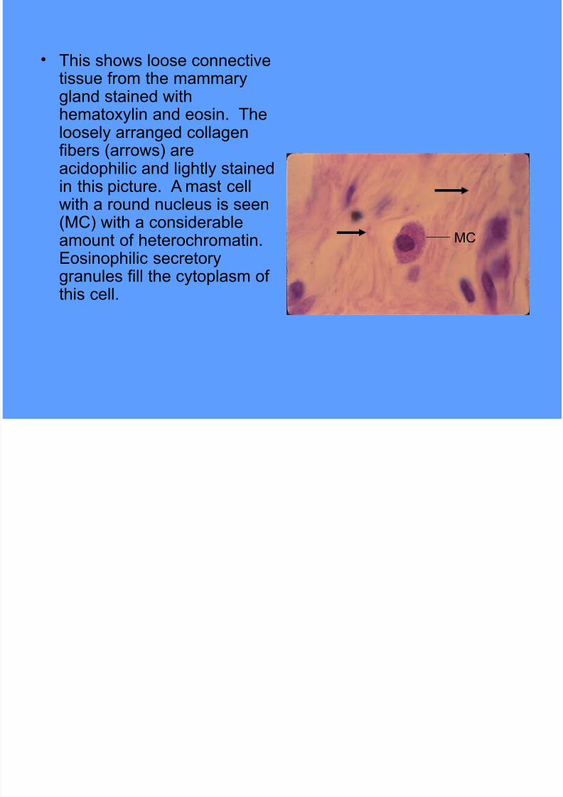
$ This sho"s loose connectivetissue from the mammary
land stained "ithhematoxylin and eosin. Theloosely arraned collaenfibers (arro"s) areacidophilic and lihtly stainedin this picture. * mast cell"ith a round nucleus is seen(+C) "ith a considerableamount of heterochromatin.Eosinophilic secretoryranules fill the cytoplasm of
this cell.
+C
Page 5
7/21/2019 Connective tissue images.ppt
http://slidepdf.com/reader/full/connective-tissue-imagesppt 5/54
$ This sho"s loosefibrous connectivetissue similar to that
seen in the previousslide. *nother mastcell (+C) is locatednear the center of thefield. Thin% liht'stainin collaenfibers (arro"s) areseen in this picture.
+C
Page 6
7/21/2019 Connective tissue images.ppt
http://slidepdf.com/reader/full/connective-tissue-imagesppt 6/54
$ This sho"s a spread (not a tissuesection) of a thin piece of
mesentery illustratin a differentvie" of loose connective tissue.The lare% dar,'stainin cells aremast cells (+C) "hose ranulesobscure the nuclei. The larestoval nuclei belon to mesothelialcells (+e)% that form an epithelium
on either side of the connectivetissue. The smaller% oval nucleibelon to macrophaes (+a).These cells tend to haveassociated ranular material intheir cytoplasm. Other oval'
shaped nuclei seen here arethose of fibroblasts. +C
+a
+e
Page 7
7/21/2019 Connective tissue images.ppt
http://slidepdf.com/reader/full/connective-tissue-imagesppt 7/54
$ This sho"s atransmission electronmicroraph of a mast
cell illustratinnumerous densesecretory ranules inthe cytoplasm. Notealso the elonatednucleus (Nu) "ithabundantheterochromatin.
Nu
Page 8
7/21/2019 Connective tissue images.ppt
http://slidepdf.com/reader/full/connective-tissue-imagesppt 8/54
$ This sho"s anotherplastic section of looseconnective tissue stained"ith methylene blue'a!ure II. * number offibroblasts are seen (-i)"ith oval euchromaticnuclei. * monocyte (+o)is also seen "ith a dar,er%indented nucleus. *
binucleate neutrophil (N)and a macrophae (+a)"ith a small% liht'staininnucleus are also seen.
-i
+o
N
Page 9
7/21/2019 Connective tissue images.ppt
http://slidepdf.com/reader/full/connective-tissue-imagesppt 9/54
$ This sho"s t"o plasmacells (C) located in theloose connective tissue ofthe mammary land.
These cells have round%eccentric nuclei "ithslihtly basophiliccytoplasm (indicatin thepresence of /N). The
lare% pale nucleus is thatof a fibroblast "hile thedar,er nuclei are those offibrocytes.
C
Page 10
7/21/2019 Connective tissue images.ppt
http://slidepdf.com/reader/full/connective-tissue-imagesppt 10/54
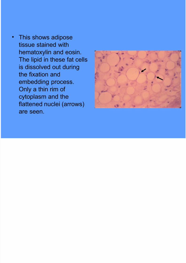
$ This sho"s adipose
tissue stained "ith
hematoxylin and eosin.
The lipid in these fat cells
is dissolved out durinthe fixation and
embeddin process.
Only a thin rim of
cytoplasm and theflattened nuclei (arro"s)
are seen.
Page 11
7/21/2019 Connective tissue images.ppt
http://slidepdf.com/reader/full/connective-tissue-imagesppt 11/54
$ This sho"s adevelopin adiposecell (arro") stained
"ith hematoxylin andeosin. Note the rinof cytoplasm and therounded nucleussurroundin a lipiddroplet. *cidophiliccollaen fibers areseen around the cell.
Page 12
7/21/2019 Connective tissue images.ppt
http://slidepdf.com/reader/full/connective-tissue-imagesppt 12/54
$ This sho"s adipose
cells preserved so
that the lipid is
retained in the celland stained "ith
osmic acid% hence the
really dar,
appearance of thecells.
Page 13
7/21/2019 Connective tissue images.ppt
http://slidepdf.com/reader/full/connective-tissue-imagesppt 13/54
$ This sho"s loose
connective tissue (CT)
"ithin a mucosal fold in
the all bladder.
Numerous collaen fibersand abundant cells can
be seen in the connective
tissue. The cells cannot
be readily identified atthis manification. (0hat
type of epithelium is seen
here1)
CT
Page 14
7/21/2019 Connective tissue images.ppt
http://slidepdf.com/reader/full/connective-tissue-imagesppt 14/54
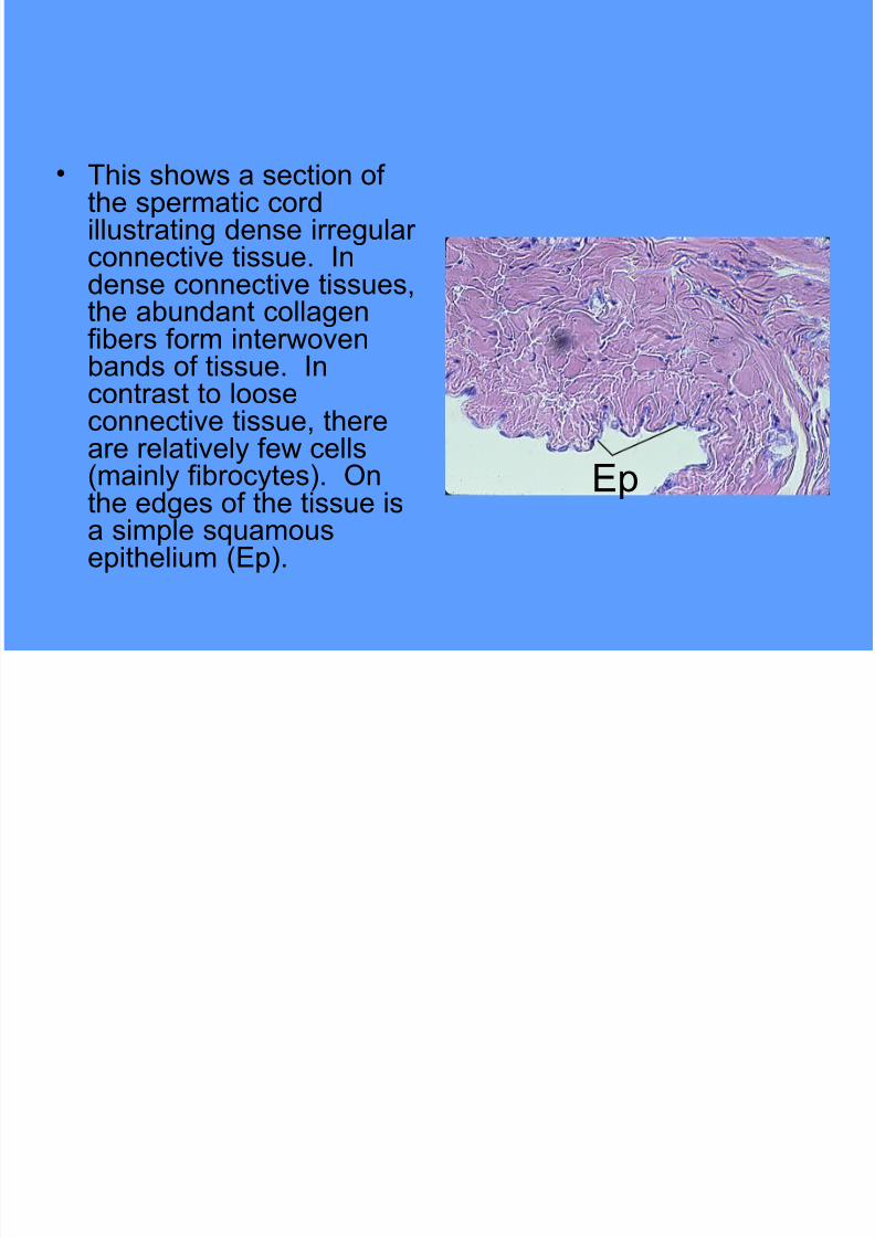
$ This sho"s a section ofthe spermatic cordillustratin dense irreularconnective tissue. Indense connective tissues%
the abundant collaenfibers form inter"ovenbands of tissue. Incontrast to looseconnective tissue% there
are relatively fe" cells(mainly fibrocytes). Onthe edes of the tissue isa simple s&uamousepithelium (Ep).
Ep
Page 15
7/21/2019 Connective tissue images.ppt
http://slidepdf.com/reader/full/connective-tissue-imagesppt 15/54
$ This sho"s denseirreular connectivetissue at hihermanification than in the
previous slide. Thebundles of bro"nishcollaen fibers can beseen coursin in differentdirections and different
planes. * fe" thinner%dar,er stainin elasticfibers can be seen in thissection (arro"s).
Page 16
7/21/2019 Connective tissue images.ppt
http://slidepdf.com/reader/full/connective-tissue-imagesppt 16/54
$ This sho"s dense elastic
tissue of the aorta at lo"
manification. The thin
elastic fibers (stained
blac,) in the "all of theaorta (arro"s) form layers
separated by spaces.
The collaen fibers are
stained bro"n in thissection.
Page 17
7/21/2019 Connective tissue images.ppt
http://slidepdf.com/reader/full/connective-tissue-imagesppt 17/54
$ This sho"s anotherexample of denseirreular connectivetissue from the urinary
bladder at lo"manification. Thisparticular section hasmore cells than theprevious example% but still
fe"er than the looseconnective tissue seenearlier. +ost of the nucleibelon to fibrocytes.
Page 18
7/21/2019 Connective tissue images.ppt
http://slidepdf.com/reader/full/connective-tissue-imagesppt 18/54
$ This sho"s another
picture of dense
irreular connective
tissue from theurinary bladder. Note
that the collaen
bundles in the top of
the field are moreorani!ed than those
at the bottom.
Page 19
7/21/2019 Connective tissue images.ppt
http://slidepdf.com/reader/full/connective-tissue-imagesppt 19/54
$ This sho"s a section of
the penis illustratin
dense reular connective
tissue stained "ith
hematoxylin and eosin.+ost of the collaen
fibers (pin, stainin) are
oriented parallel to each
other. The nuclei offibrocytes are present.
Page 20
7/21/2019 Connective tissue images.ppt
http://slidepdf.com/reader/full/connective-tissue-imagesppt 20/54
$ This sho"s dense reular
connective tissue (CT)
from a anlion. On the
left side of the field are
elonated nuclei ofnervous tissue (NT). The
remainder of the field is
occupied by dense
collaenous fibers "ithfe"% flattened nuclei of
fibrocytes.
NT CT
Page 21
7/21/2019 Connective tissue images.ppt
http://slidepdf.com/reader/full/connective-tissue-imagesppt 21/54
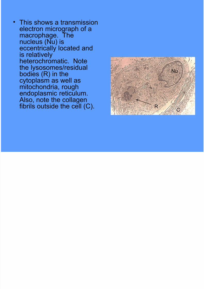
$ This sho"s a transmissionelectron microraph of a
macrophae. Thenucleus (Nu) iseccentrically located andis relativelyheterochromatic. Notethe lysosomes2residualbodies (/) in thecytoplasm as "ell asmitochondria% rouhendoplasmic reticulum. *lso% note the collaen
fibrils outside the cell (C).
Nu
C/
Page 22
7/21/2019 Connective tissue images.ppt
http://slidepdf.com/reader/full/connective-tissue-imagesppt 22/54
$ This sho"s atransmission electronmicroraph of alymphocyte. Notice thatthe nucleus (Nu)occupies a lare portionof the cell% is indentedand has a fair amount ofheterochromatin. Thecytoplasm has only a fe"
oranelles and theplasma membrane tendsto be irreular due tomovement of the cell.
Nu
Page 23
7/21/2019 Connective tissue images.ppt
http://slidepdf.com/reader/full/connective-tissue-imagesppt 23/54
C*/TI3*4E
$ Cartilae is a speciali!ed form of
connective tissue composed of cells called
chondrocytes and their surroundin
matrix. There are three types of cartilaethat are distinuished based on their
matrix characteristics.
Page 24
7/21/2019 Connective tissue images.ppt
http://slidepdf.com/reader/full/connective-tissue-imagesppt 24/54
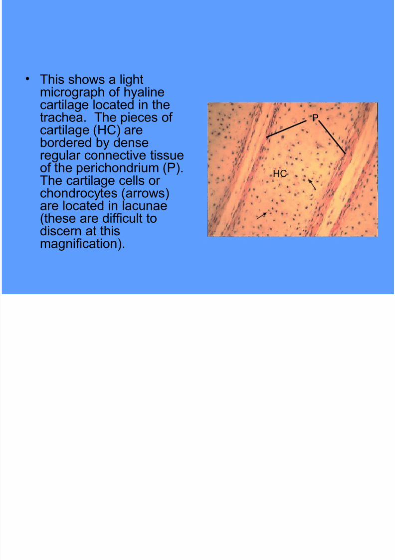
$ This sho"s a lihtmicroraph of hyalinecartilae located in thetrachea. The pieces ofcartilae (5C) arebordered by densereular connective tissueof the perichondrium ().The cartilae cells orchondrocytes (arro"s)
are located in lacunae(these are difficult todiscern at thismanification).
5C
Page 25
7/21/2019 Connective tissue images.ppt
http://slidepdf.com/reader/full/connective-tissue-imagesppt 25/54
$ This sho"s a portion ofthe previous picture athiher manificationillustratin the
chondrocytes in lacunae(arro"s). The matrixsurroundin the cells iscomposed of collaenfibers (too small too be
seen here) and othercomponents includinlycosaminolycans suchas chondroitin sulfate.
Page 26
7/21/2019 Connective tissue images.ppt
http://slidepdf.com/reader/full/connective-tissue-imagesppt 26/54
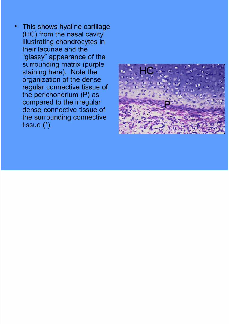
$ This sho"s hyaline cartilae(5C) from the nasal cavity
illustratin chondrocytes intheir lacunae and the6lassy7 appearance of thesurroundin matrix (purplestainin here). Note the
orani!ation of the densereular connective tissue ofthe perichondrium () ascompared to the irreulardense connective tissue of
the surroundin connectivetissue (8).
8
5C
Page 27
7/21/2019 Connective tissue images.ppt
http://slidepdf.com/reader/full/connective-tissue-imagesppt 27/54
$ This sho"s a hihermanification of hyalinecartilae from thetrachea. The
chondrocytes on theriht side of the fieldare not as mature andfill their lacunae (arro")
"hile those of the lefthave more spacesurroundin the cell.
Page 28
7/21/2019 Connective tissue images.ppt
http://slidepdf.com/reader/full/connective-tissue-imagesppt 28/54
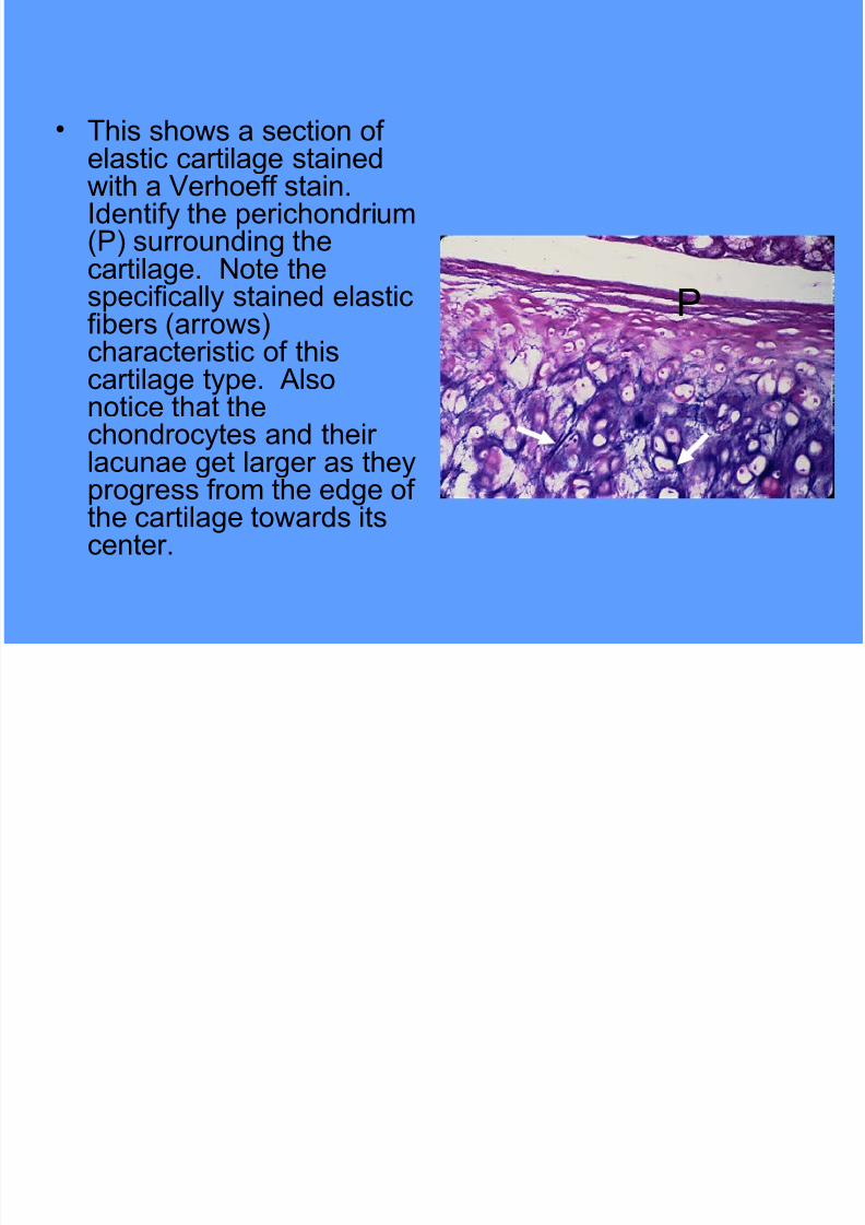
$ This sho"s a section of
elastic cartilae stained"ith a Verhoeff stain.Identify the perichondrium() surroundin thecartilae. Note the
specifically stained elasticfibers (arro"s)characteristic of thiscartilae type. *lsonotice that thechondrocytes and their
lacunae et larer as theyproress from the ede ofthe cartilae to"ards itscenter.
Page 29
7/21/2019 Connective tissue images.ppt
http://slidepdf.com/reader/full/connective-tissue-imagesppt 29/54
$ This sho"s a hiher
manification vie" of
elastic cartilae. The
elastic fibers are moreclearly seen in this
picture as are the
chondrocytes and
their lacunae.
Page 30
7/21/2019 Connective tissue images.ppt
http://slidepdf.com/reader/full/connective-tissue-imagesppt 30/54
$ This sho"s elastic
cartilae of the epilottis
stained "ith a Trichrome
stain. Note that the
collaen of theperichondrium () is
stained reen9 ho"ever%
the collaen type II "ithin
the cartilae does notstain. The elastic fibers
are stained purple
(arro"s)
Page 31
7/21/2019 Connective tissue images.ppt
http://slidepdf.com/reader/full/connective-tissue-imagesppt 31/54
$ This sho"s a liht'stainedsection of fibrocartilae.The cartilae stainsacidophilic due to the
collaen fibers. Note thatthe chondrocytes andlacunae are small andoften have some reularpattern to their
orani!ation (in ro"s).There is noperichondrium associated"ith this cartilae type.
Page 32
7/21/2019 Connective tissue images.ppt
http://slidepdf.com/reader/full/connective-tissue-imagesppt 32/54
$ This sho"s a hiher
manification vie" of
fibrocartilae illustratin
the collaen fibers "ithin
the matrix surroundinthe chondrocytes. This
type of cartilae is often
found bet"een hyaline
cartilae and denseconnective tissue.
Page 33
7/21/2019 Connective tissue images.ppt
http://slidepdf.com/reader/full/connective-tissue-imagesppt 33/54
$ #one is a speciali!ed connective tissue
"ith a minerali!ed extracellular matrix.
#one enerally consists of three cell types
includin the osteoproenitor cells%osteoblasts (osteocytes "hen
differeniated) and osteoclasts. The
follo"in slides illustrate some of thefeatures of bony tissue.
#ONE
Page 34
7/21/2019 Connective tissue images.ppt
http://slidepdf.com/reader/full/connective-tissue-imagesppt 34/54
$ This sho"s a lo"manification vie" of theend of a lon bone.
*bove the epiphysealplate (E) is spony
bone. The outer portionof the shaft is compactbone (C#)% "hich is laiddo"n by appositionalro"th from the
periosteum. Thecompact bone surroundsthe spony bone andbone marro" of the shaft.
E
C#
Page 35
7/21/2019 Connective tissue images.ppt
http://slidepdf.com/reader/full/connective-tissue-imagesppt 35/54
$ This sho"s a cross'
section of developin
bone from a fetal finer.
The outermost layer is
the connective tissue ofthe periosteum ().
Small pieces of calcified
cartilae are seen (blue
stainin) as arehypertrophyin cartilae
(arro"s).
P
Tendon
Spony #one
:evelopin
Compact #one
Page 36
7/21/2019 Connective tissue images.ppt
http://slidepdf.com/reader/full/connective-tissue-imagesppt 36/54
$ This sho"s a hiher
manification of the
previous slide
illustratin developinbone.
Spony #one
Calcified Cartilae
Page 37
7/21/2019 Connective tissue images.ppt
http://slidepdf.com/reader/full/connective-tissue-imagesppt 37/54
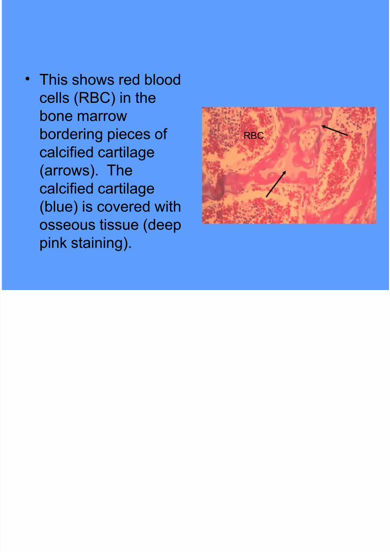
$ This sho"s red blood
cells (/#C) in the
bone marro"
borderin pieces ofcalcified cartilae
(arro"s). The
calcified cartilae
(blue) is covered "ithosseous tissue (deep
pin, stainin).
/#C
Page 38
7/21/2019 Connective tissue images.ppt
http://slidepdf.com/reader/full/connective-tissue-imagesppt 38/54
$ This sho"s a
scannin electron
microraph of bone
surface. Note the5aversian Canals
(5C)% the concentric
lamellae surroundin
these and the lacunaeof the osteocytes
(arro"s).
5C
Page 39
7/21/2019 Connective tissue images.ppt
http://slidepdf.com/reader/full/connective-tissue-imagesppt 39/54
$ This sho"s a section
of decalcified bone.
The structural
features are similar tothat in the previous
slide illustratin the
5aversian system.
5aversian Canal
Osteocytes
Page 40
7/21/2019 Connective tissue images.ppt
http://slidepdf.com/reader/full/connective-tissue-imagesppt 40/54
$ This sho"s a
transmission electron
microraph of an
osteocyte surrounded
by its lacuna. The
processes extendin
from the cell body are
located in canaliculi
(arro"s). The matrix of
the bone surrounds the
canaliculi.
Page 41
7/21/2019 Connective tissue images.ppt
http://slidepdf.com/reader/full/connective-tissue-imagesppt 41/54
$ This sho"s a section of
decalcified bone andassociated s,eletal muscle
(S+). The s,eletal muscle
is attached to the
connective tissue of the
periosteum (). The larespace is due to shrin,ae of
the periosteum durin
processin of the tissue.
Small 5aversian Canals
can be seen "ithin the bone
(arro"s).
P SM
Page 42
7/21/2019 Connective tissue images.ppt
http://slidepdf.com/reader/full/connective-tissue-imagesppt 42/54
$ This sho"s a section ofdecalcified bone
illustratin the connective
tissue of the perioosteum
(). Osteoblasts are
located alon the inner
ede of the periosteum
(arro"). These cells "ill
deposit and become
entrapped in bony matrix(appositional ro"th).
P
Compact #one
Page 43
7/21/2019 Connective tissue images.ppt
http://slidepdf.com/reader/full/connective-tissue-imagesppt 43/54
$ This sho"s another
section of decalcified
bone and associated
s,eletal muscle (S+).
Note that the osteoeniclayer of the periosteum is
&uite cellular (arro"s)
"ith basophilic
osteoblasts located at theperiphery of the bone.
SM
Page 44
7/21/2019 Connective tissue images.ppt
http://slidepdf.com/reader/full/connective-tissue-imagesppt 44/54
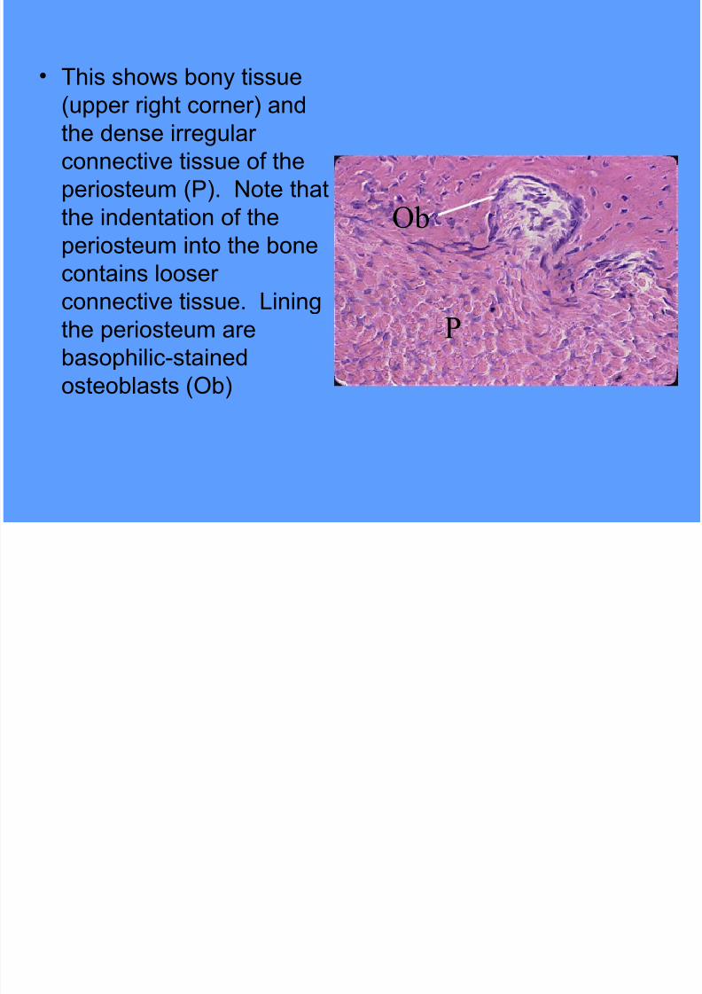
$ This sho"s bony tissue
(upper riht corner) and
the dense irreularconnective tissue of the
periosteum (). Note that
the indentation of the
periosteum into the bonecontains looser
connective tissue. 3inin
the periosteum are
basophilic'stained
osteoblasts (Ob)
P
Ob
Page 45
7/21/2019 Connective tissue images.ppt
http://slidepdf.com/reader/full/connective-tissue-imagesppt 45/54
$ This fiure sho"s
decalcified bone (#)
and dense
connective tissue of
an associated
tendon (T) attachinto the periosteum.
T
B
Page 46
7/21/2019 Connective tissue images.ppt
http://slidepdf.com/reader/full/connective-tissue-imagesppt 46/54
$ This sho"s
decalcified bone
illustratin a
resorption canalsurrounded by the
bony tissue.
Osteoclasts (arro"s)
can be seen lyinnext to the bony
tissue.
Page 47
7/21/2019 Connective tissue images.ppt
http://slidepdf.com/reader/full/connective-tissue-imagesppt 47/54
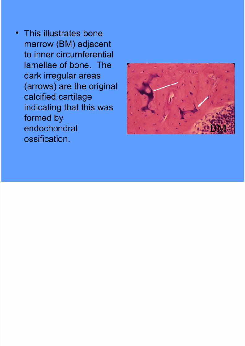
$ This illustrates bone
marro" (#+) ad;acentto inner circumferential
lamellae of bone. The
dar, irreular areas
(arro"s) are the oriinalcalcified cartilae
indicatin that this "as
formed by
endochondralossification. BM
Page 48
7/21/2019 Connective tissue images.ppt
http://slidepdf.com/reader/full/connective-tissue-imagesppt 48/54
$ This illustrates bone
marro" (#+) and the
osteoblasts (Ob)
linin bone. Theosteoblasts extend
into the beinnin of a
Vol,mann<s Canal
(VC)% "hich carriesblood vessels into the
5aversian system.
BM
Ob
VC
Page 49
7/21/2019 Connective tissue images.ppt
http://slidepdf.com/reader/full/connective-tissue-imagesppt 49/54
$ This schematic illustrates
the process of
intramembranous
ossification. +esenchymalcells form an elaborate
net"or, (=). These cells
then enlare and
differentiate into osteoblasts(>). The cells lay do"n an
intercellular matrix an form
bony spicules (?) covered by
osteoblasts.
1
2
3
Page 50
7/21/2019 Connective tissue images.ppt
http://slidepdf.com/reader/full/connective-tissue-imagesppt 50/54
$ This is a decalcified
section of the root of
a tooth and itsassociated alveolar
bone (#). The root
of the tooth is
covered by
cementum (C) and
collaen fibers
"hich anchor thetooth into the bony
tissue.
B
C
Page 51
7/21/2019 Connective tissue images.ppt
http://slidepdf.com/reader/full/connective-tissue-imagesppt 51/54
$ This sho"s a hih
voltae electron
microraph of a
relatively thic, plasticsection illustratin a
fibroblast (-i) "ith
bundles of collaen
fibers attachin to thecell surface (arro"s).
Fi
Page 52
7/21/2019 Connective tissue images.ppt
http://slidepdf.com/reader/full/connective-tissue-imagesppt 52/54
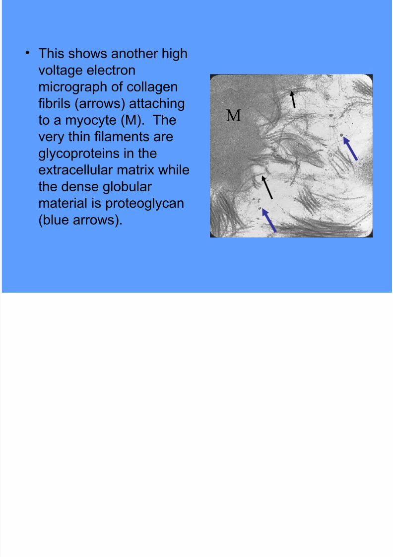
$ This sho"s another hih
voltae electronmicroraph of collaen
fibrils (arro"s) attachin
to a myocyte (+). The
very thin filaments arelycoproteins in the
extracellular matrix "hile
the dense lobular
material is proteolycan(blue arro"s).
M
Page 53
7/21/2019 Connective tissue images.ppt
http://slidepdf.com/reader/full/connective-tissue-imagesppt 53/54
$ This sho"s collaen
fibers attached to t"o
s,eletal muscle cells
(S+) and a capillary(C).
C
SM
SM
Page 54
7/21/2019 Connective tissue images.ppt
http://slidepdf.com/reader/full/connective-tissue-imagesppt 54/54
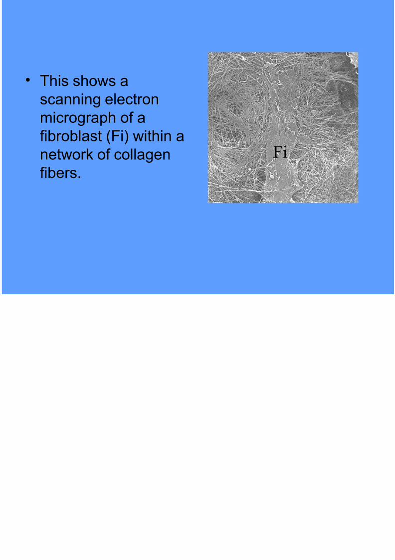
$ This sho"s a
scannin electron
microraph of a
fibroblast (-i) "ithin anet"or, of collaen
fibers.
Fi