But : Effectuer le suivi du traitement conservateur d’un patient ostéoporotique souffrant de lombalgie aiguë en raison d’une fracture lombaire avec tassement. Caractéristiques cliniques : Un homme de 74 ans souffre de lombalgies aiguës dans la région thoracolombaire survenues après avoir soulevé un objet. Une évaluation radiographique révèle une déminéralisation généralisée ainsi qu’une fracture de L1 avec tassement cunéiforme modéré. Intervention et résultat : L’approche thérapeutique conservatrice comprenait l’éducation posturale, la modification d’activités, l’électrothérapie à courants interférentiels, l’application de bandages élastiques (taping) en extension, la technique GrastonMD, et la prescription d’exercices de réadaptation. Les résultats ont notamment été mesurés par une échelle verbale de notation de la douleur, la quantité de médicaments ingérés, et le retour aux activités de la vie quotidienne (AVQs). Le patient est parvenu à une résolution à long terme des symptômes, sans récurrence de la douleur en date du suivi effectué après 12 mois. Résumé : Une combinaison de stratégies de réadaptation conservatrices peut être mise en œuvre avec succès dans le traitement de patients ostéoporotiques atteints de fractures vertébrales ostéoporotiques avec tassement d’intensité légère à modérée au niveau de la colonne lombaire.(JCCA 2012; 56(1):29–39)
m o t s c l é s : Fracture avec tassement, ostéoporose, technique GrastonMD, chiropractie, réadaptation
Objective: To chronicle the conservative treatment and management of an osteoporotic patient presenting with acute back pain resulting from a lumbar compression fracture. Clinical features: A 74-year old male presented with acute back pain in the thoracolumbar region after an episode of lifting. Radiographic evaluation revealed generalized demineralization and a moderate wedge compression fracture at L1. Intervention and outcome: The conservative treatment approach included postural education, activity modification, interferential current, taping into extension, Graston Technique®, and rehabilitative exercise prescription. Outcome measures included verbal pain rating scale, medication use, and a return to activities of daily living (ADLs). The patient attained long-term symptom resolution with no recurrence of pain at 12 month follow-up. Summary: A combination of conservative rehabilitation strategies may be successfully implemented to treat osteoporotic patients with mild to moderate osteoporotic vertebral compression fracture of the lumbar spine.(JCCA 2012; 56(1):29–39)
k e y w o r d s : compression fracture, osteoporosis, Graston Technique®, chiropractic, rehabilitation
30 J Can Chiropr Assoc 2012; 56(1)
Conservative management of a lumbar compression fracture in an osteoporotic patient: a case report
IntroductionIndividuals with osteoporosis have a greater likelihood of suffering vertebral compression fractures (VCFs), which can range from mild to severe in terms of associated pain and resultant disability.1 In the United States, it is esti-mated that at least 10 million people suffer from osteopor-osis and an additional 18 million people are at significant risk for development of the disorder. Within this affected group, it is estimated that 700,000 VCFs occur each year and approximately 70,000 result in hospitalization, with an average hospital stay per patient of 8 days.2 The risk of major osteoporotic fracture in Canada is among the high-est in the world,3 with the incidence of VCFs expected to increase as the Canadian population ages.4 The annual incidence of osteoporotic vertebral compression fractures (OVCFs) among Canadian women is currently reported to be approximately 37,000.3 Although considered a female health issue, osteoporosis is also becoming a major health concern among males.5–8
It is estimated that many OVCFs remain asymptom-atic, and that only one-third of individuals seek immedi-ate medical attention, presenting predominantly as acute back pain patients.9–12 For any given case, the diagnosis of a single OVCF increases the risk of subsequent frac-tures by a factor of five.12 Patient population studies indi-cate an increased mortality rate in patients with OVCFs that correlates with the number of involved vertebrae.13 In addition to acute pain and the risk for developing chronic pain, OVCFs may also be accompanied by other physical and emotional consequences.1,9–11 Early recog-nition, diagnosis, and conservative management can play important roles in minimizing the negative sequelae of OVCF.
This case study was conducted to evaluate the con-servative treatment and management of an osteoporotic patient presenting with acute back pain resulting from a lumbar compression fracture. Salient clinical features and diagnostic considerations are also discussed.
Case reportA 74-year old male presented with acute back pain of three days duration localized to the region of the thoracol-umbar spine. The patient explained that this pain occurred while he was lifting 30–40 lb pieces of wood. During the mid-point of a lift, with his spine forward flexed, he re-portedly heard a “pop” in his back and a sensation of
pain immediately ensued. The patient did not seek med-ical treatment following this incident. He reports that he managed his symptoms with over the counter medication (ibuprofen).
The patient rated his pain as 8/10 on the Verbal Pain Rating Scale (VPRS) where 0 is “no pain” and 10 is the “worst pain that he had ever experienced.” The pain was described as sharp and stabbing, and it was exacerbated by direct pressure over the painful area and any move-ments of the lower axial spine. He denied any radiating/referred pain symptoms into the lower extremities or dif-ficulty with bowel and bladder function. Past medical history revealed that he had been diagnosed with “mild” osteoporosis two years prior. Systems review and family health history was unremarkable. The patient was a life-long non-smoker. He did not report any previous history of disabling back injury. He indicated that he lived a very active lifestyle and walked two to four kilometres daily. His current state did not allow for him to continue his daily walking routine and he was having trouble getting a good night’s sleep due to difficulty with finding a com-fortable position.
Initial observation revealed that the patient walked slowly and moved in a guarded fashion during trans-fers. A slightly forward stooped posture was noted in the standing position. Lumbar ranges of motion were signifi-cantly restricted in all planes due to pain. Motor, reflex, and sensory testing for the lower extremities was within normal limits bilaterally. Seated straight leg raising was unremarkable bilaterally for nerve root tension signs. Percussion of the spinous processes with a reflex ham-mer revealed tenderness most notably over T11, T12, L1 and L2. Digital posterior to anterior (P-A) pressure of the spinous processes reproduced a sharp pain at these levels. Palpation revealed marked muscle spasm bilaterally in the thoracolumbar paraspinal muscles.
In consideration of the patient’s reported health history, mechanism of injury, and physical examination findings, A-P and lateral thoracic and lumbar radiographs were completed due to suspected OVCF. The radiographic examination revealed generalized demineralization and a moderate wedge compression fracture at L1. There were no other radiographic features of significance identified that would clearly explain the patient’s acute symptom presentation.
In office treatment commenced four days after initial
J Can Chiropr Assoc 2012; 56(1) 31
JA Papa
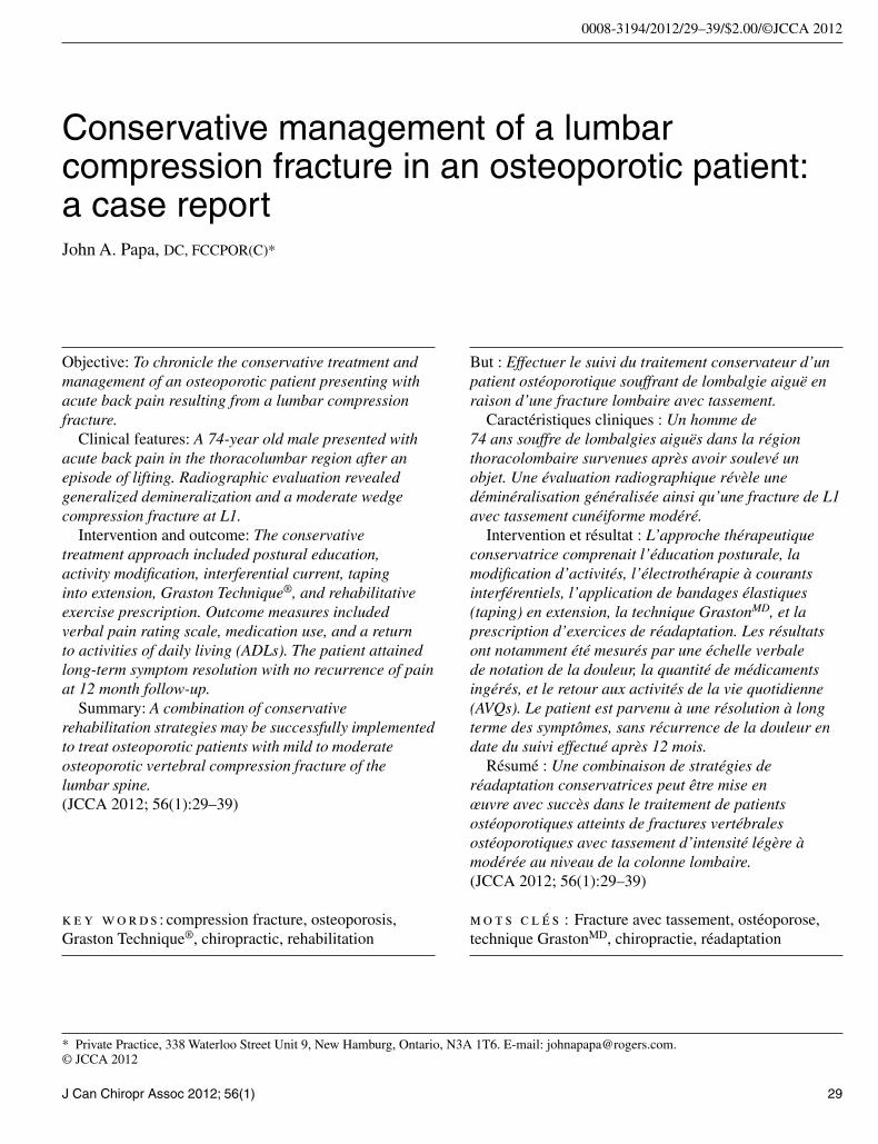
presentation. The patient was instructed after the initial assessment to maintain a neutral spine position and try to avoid forward stooped/spinal flexion movements. He was also advised to try and stay mobile and avoid pro-longed inactivity. Initial treatment was focused on provid-ing adequate pain control. This was accomplished with interferential current (IFC) applied to the hypertonic thoracolumbar paraspinal muscles, followed by taping of the thoracolumbar spine into a position of slight extension bias (Figures 1A–C). Exercises consisting of abdominal bracing, scapular setting, and gentle extension movements of the thoracolumbar region were introduced in week 3.
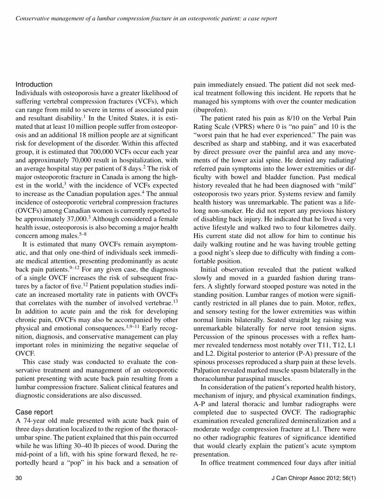
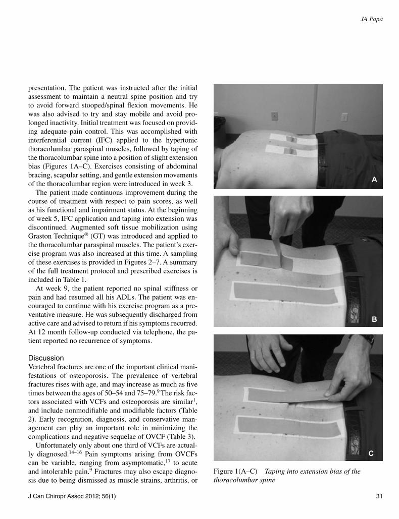
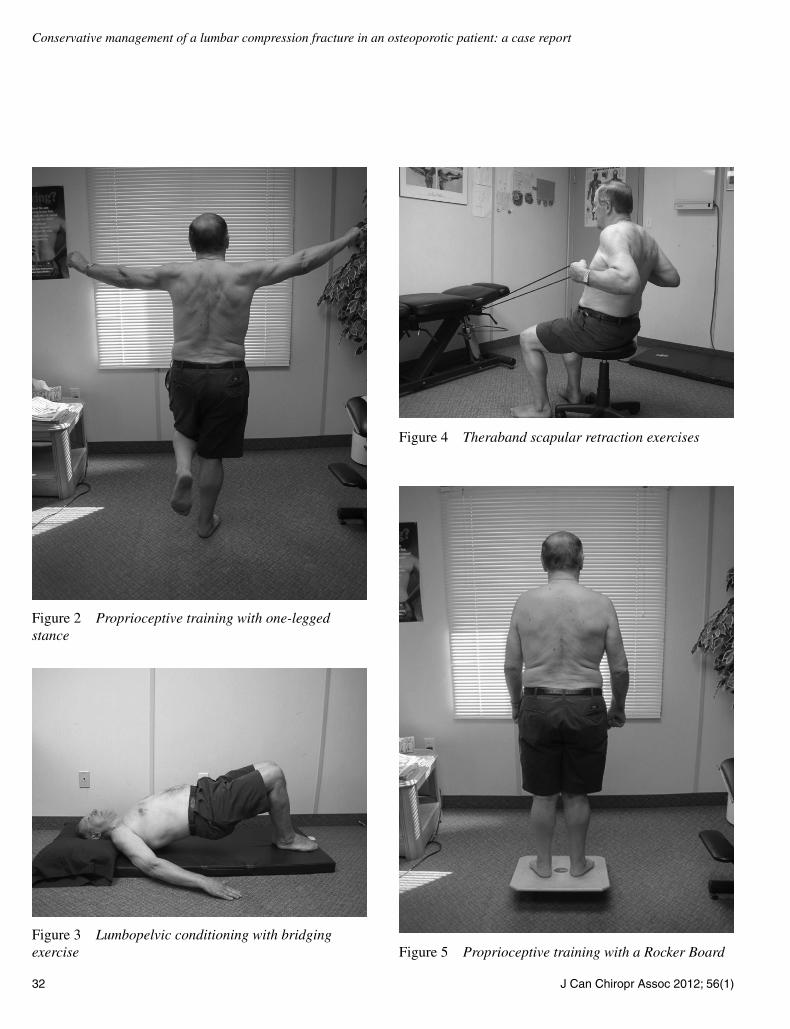
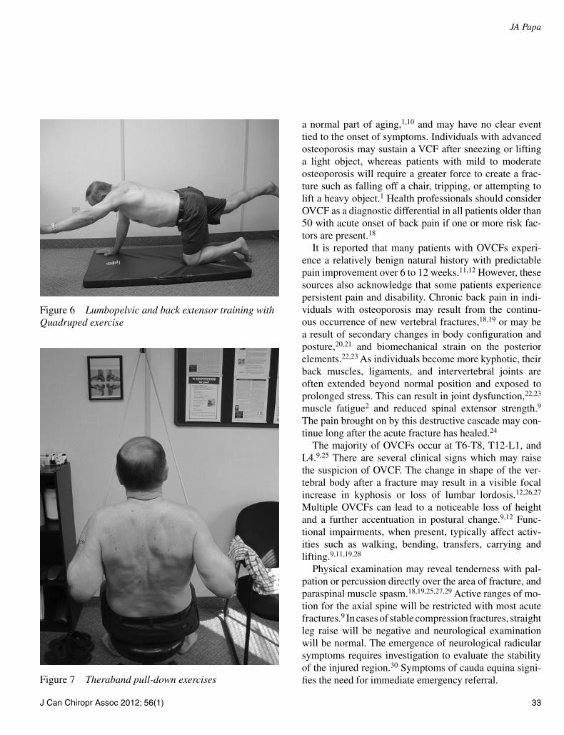
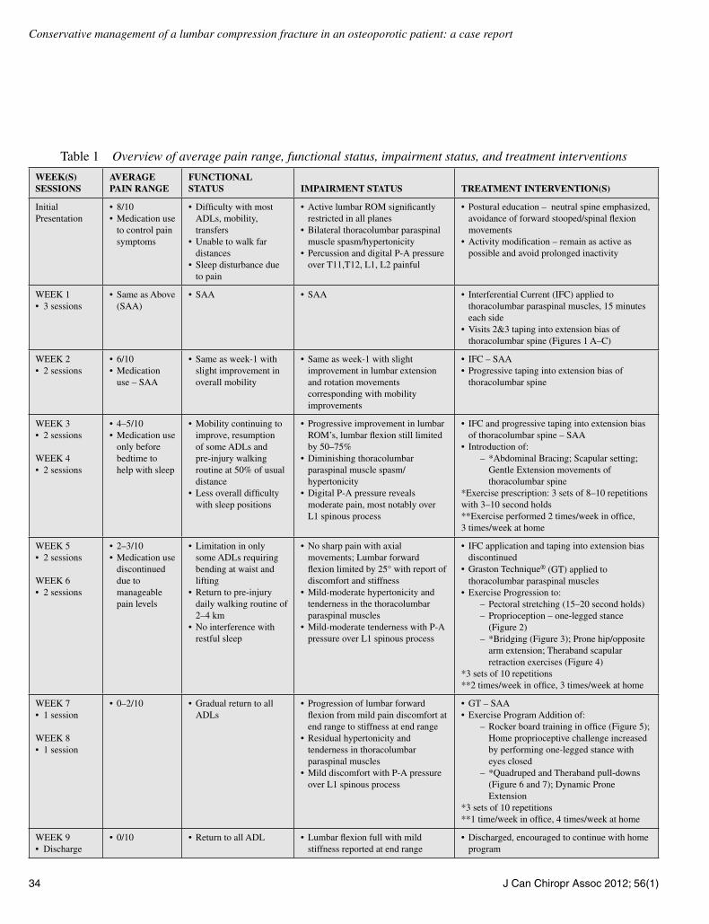
The patient made continuous improvement during the course of treatment with respect to pain scores, as well as his functional and impairment status. At the beginning of week 5, IFC application and taping into extension was discontinued. Augmented soft tissue mobilization using Graston Technique® (GT) was introduced and applied to the thoracolumbar paraspinal muscles. The patient’s exer-cise program was also increased at this time. A sampling of these exercises is provided in Figures 2–7. A summary of the full treatment protocol and prescribed exercises is included in Table 1.
At week 9, the patient reported no spinal stiffness or pain and had resumed all his ADLs. The patient was en-couraged to continue with his exercise program as a pre-ventative measure. He was subsequently discharged from active care and advised to return if his symptoms recurred. At 12 month follow-up conducted via telephone, the pa-tient reported no recurrence of symptoms.
DiscussionVertebral fractures are one of the important clinical mani-festations of osteoporosis. The prevalence of vertebral fractures rises with age, and may increase as much as five times between the ages of 50–54 and 75–79.9 The risk fac-tors associated with VCFs and osteoporosis are similar1, and include nonmodifiable and modifiable factors (Table 2). Early recognition, diagnosis, and conservative man-agement can play an important role in minimizing the complications and negative sequelae of OVCF (Table 3).
Unfortunately only about one third of VCFs are actual-ly diagnosed.14–16 Pain symptoms arising from OVCFs can be variable, ranging from asymptomatic,17 to acute and intolerable pain.9 Fractures may also escape diagno-sis due to being dismissed as muscle strains, arthritis, or
Figure 1(A–C) Taping into extension bias of the thoracolumbar spine
A
B
C
32 J Can Chiropr Assoc 2012; 56(1)
Conservative management of a lumbar compression fracture in an osteoporotic patient: a case report
Figure 2 Proprioceptive training with one-legged stance
Figure 3 Lumbopelvic conditioning with bridging exercise
Figure 4 Theraband scapular retraction exercises
Figure 5 Proprioceptive training with a Rocker Board
J Can Chiropr Assoc 2012; 56(1) 33
JA Papa
a normal part of aging,1,10 and may have no clear event tied to the onset of symptoms. Individuals with advanced osteoporosis may sustain a VCF after sneezing or lifting a light object, whereas patients with mild to moderate osteoporosis will require a greater force to create a frac-ture such as falling off a chair, tripping, or attempting to lift a heavy object.1 Health professionals should consider OVCF as a diagnostic differential in all patients older than 50 with acute onset of back pain if one or more risk fac-tors are present.18
It is reported that many patients with OVCFs experi-ence a relatively benign natural history with predictable pain improvement over 6 to 12 weeks.11,12 However, these sources also acknowledge that some patients experience persistent pain and disability. Chronic back pain in indi-viduals with osteoporosis may result from the continu-ous occurrence of new vertebral fractures,18,19 or may be a result of secondary changes in body configuration and posture,20,21 and biomechanical strain on the posterior elements.22,23 As individuals become more kyphotic, their back muscles, ligaments, and intervertebral joints are often extended beyond normal position and exposed to prolonged stress. This can result in joint dysfunction,22,23 muscle fatigue2 and reduced spinal extensor strength.9 The pain brought on by this destructive cascade may con-tinue long after the acute fracture has healed.24
The majority of OVCFs occur at T6-T8, T12-L1, and L4.9,25 There are several clinical signs which may raise the suspicion of OVCF. The change in shape of the ver-tebral body after a fracture may result in a visible focal increase in kyphosis or loss of lumbar lordosis.12,26,27 Multiple OVCFs can lead to a noticeable loss of height and a further accentuation in postural change.9,12 Func-tional impairments, when present, typically affect activ-ities such as walking, bending, transfers, carrying and lifting.9,11,19,28
Physical examination may reveal tenderness with pal-pation or percussion directly over the area of fracture, and paraspinal muscle spasm.18,19,25,27,29 Active ranges of mo-tion for the axial spine will be restricted with most acute fractures.9 In cases of stable compression fractures, straight leg raise will be negative and neurological examination will be normal. The emergence of neurological radicular symptoms requires investigation to evaluate the stability of the injured region.30 Symptoms of cauda equina signi-fies the need for immediate emergency referral.
Figure 6 Lumbopelvic and back extensor training with Quadruped exercise
Figure 7 Theraband pull-down exercises
34 J Can Chiropr Assoc 2012; 56(1)
Conservative management of a lumbar compression fracture in an osteoporotic patient: a case report
Table 1 Overview of average pain range, functional status, impairment status, and treatment interventions
Week(s) sessions
AverAge pAin rAnge
FunctionAl stAtus impAirment stAtus treAtment intervention(s)
Initial Presentation
• 8/10• Medication use
to control pain symptoms
• Difficulty with most ADLs, mobility, transfers
• Unable to walk far distances
• Sleep disturbance due to pain
• Active lumbar ROM significantly restricted in all planes
arm extension; Theraband scapular retraction exercises (Figure 4)
*3 sets of 10 repetitions**2 times/week in office, 3 times/week at home
WEEK 7• 1 session
WEEK 8• 1 session
• 0–2/10 • Gradual return to all ADLs
• Progression of lumbar forward flexion from mild pain discomfort at end range to stiffness at end range
• Residual hypertonicity and tenderness in thoracolumbar paraspinal muscles
• Mild discomfort with P-A pressure over L1 spinous process
• GT – SAA• Exercise Program Addition of:
– Rocker board training in office (Figure 5); Home proprioceptive challenge increased by performing one-legged stance with eyes closed
– *Quadruped and Theraband pull-downs (Figure 6 and 7); Dynamic Prone Extension
*3 sets of 10 repetitions**1 time/week in office, 4 times/week at home
WEEK 9• Discharge
• 0/10 • Return to all ADL • Lumbar flexion full with mild stiffness reported at end range
• Discharged, encouraged to continue with home program
J Can Chiropr Assoc 2012; 56(1) 35
JA Papa
Plain frontal and lateral radiographs are the initial imaging study obtained for a suspected VCF. Common radiographic findings associated with VCFs include a step defect, wedge deformity, disrupted vertebral end-plate, linear zone of condensation, paraspinal swelling and abdominal ileus.31 Post-fracture stability is based on the classification of Denis where the spine is divided into three columns.32 According to this model, the likeli-hood of neurological injury is high when damage occurs to more than one of these columns. VCFs involve failure of the anterior column only. The middle column is com-pletely intact and is typically characteristic of compres-sion fractures. Pathologic fractures may be identified by loss of posterior body height, pedicle or other structures, and a paraspinal mass.31 Computed tomography (CT) and magnetic resonance imaging (MRI) may be used in cases of suspected spinal cord compression, progressive neuro-logical deterioration, incrongruous neurologic or skeletal injury, unexplained neurologic deficit, or suspicion of malignancy.1,33,34
It is generally agreed upon that stable, non-malig-
nant compression fractures can be treated conservative-ly.1,10,12,35,36 An emphasis on pain control and maximizing functional outcome is important to prevent chronicity and the negative sequelae of OVCF. Even in acute cases, prolonged bed rest and inactivity should be avoided.1,12 Education in activities of daily living may include ways to minimize pain.10 In this case, the initial focus of treat-ment was to improve posture and body mechanics to re-duce the compressive loads on the spinal column.37 The patient was advised to avoid forward stooped-spinal flex-ion movements,37,38 attempt to stay mobile, and avoid prolonged inactivity.
Zambito et al.39 demonstrated that interferential cur-rent (IFC) was effective in alleviaitng both pain and disability in patients with chronic back pain due to pre-vious multiple vertebral osteoporotic fractures. Bracing has also been advocated as a pain management strategy. Bracing is believed to promote healing by stabilizing the spine,11 facilitating neuromuscular re-education, and re-ducing pain by decreasing postural flexion that causes increased loading of the painful fractured periosteum.10
Table 2 Risk Factors for Osteoporosis and Vertebral Compression Fractures (VCFs)1
Conservative management of a lumbar compression fracture in an osteoporotic patient: a case report
Progressive taping of the thoracolumbar region into ex-tension bias was utilized in this case as an alternative to bracing and well tolerated by the patient during the first four weeks of treatment.
Paraspinal muscular pressure has been found to be highly increased in the flexed standing position with load-ing in normal control groups and significantly higher in patients with osteoporosis, degenerative spondylolis-thesis and lumbar compartment syndrome.40 Hammer et al.41 demonstrated reduced pain in a patient with lumbar compartment syndrome after using GT. GT utilizes stain-less steel instruments to apply controlled microtrauma to the affected soft tissues.42 Studies suggest that this con-trolled microtrauma induces healing via fibroblast prolif-eration,43 which is necessary for soft tissue healing.43,44 Additional studies have shown clinical efficacy using GT for the treatment of various disorders with painful soft tis-sue components.42,45–50
Physical activity plays a critical role in the rehabilitation of osteoporotic patients with vertebral fractures.10,51–56 Extension or isometric back and abdominal strengthening exercises are useful and contribute to the avoidance of other fractures,10,38 whereas flexion exercises seem to be detrimental.38 Spinal extensor training has been demon-strated to help reduce pain by decreasing compressive loads and maintaining bone mineral density51,53 Proprio-ceptive exercises also appear to play a role in the rehabili-tation of OVCF. Vertebral fracture has been associated with impaired balance characteristics in the osteoporosis population.57 This may be as a result of several factors including pain, impaired muscle control and fear of fall-ing.57 Adding dynamic proprioceptive training can help reduce pain and the risk of falls in patients with kyphosis related to osteoporotic compression fracture.55
Although spinal manipulation or adjustment is a rou-tine mode of treatment administered by chiropractors, it was not utilized in this case. Osteoporosis is commonly regarded as a relative or absolute contraindication to spin-al manipulation.58 In a review of four cases, Haldeman et al.59 indicated that manipulation or adjustment of areas suspected of compression fracture may result in increased pain and prolonged patient disability. Considering that occult compression fractures may be present in any osteo-porotic patient, special care must be taken to avoid ex-acerbating the patient’s condition.
Evaluation and management of osteoporosis is an in-
tegral part in the treatment of OVCF.59 In this case, such management was deferred to the patient’s family phys-ician and naturopathic doctor as per the patient’s request. Chiropractors can play a role in educating osteoporotic or at risk patients on preventative lifestyle choices such as calcium and vitamin D supplementation, increasing weight-bearing physical activity, and limiting/avoiding consumption of caffeine, alcohol, and tobacco.60–63 Other treatment alternatives available to a patient with OVCF in-clude pain medication and epidural steroid injections.10–12 Surgical management is typically reserved for individuals with neural compression and progressive deformity with neurological deficits,12 and may include percutaneous vertebroplasty or kyphoplasty.10,36,64–66
The natural history of OVCF may have played a role in the favourable outcome of this case. However, the im-plementation of a structured rehabilitation program mini-mized the likelihood of chronicity and burden associated with OVCF, and the patient demonstrated no recurrence of pain at 12 months. With the exception of his previously diagnosed osteoporosis, this patient did not have any other co-morbidities that would have complicated recovery or limited his participation in an active exercise program. The patient also shared the belief that activity within his tolerance would be of benefit during recovery. Postural education, advice on activity modification, and pain re-lieving measures minimized prolonged immobilization and likely provided re-assurance for a patient already mo-tivated to remain active. GT was useful in decreasing the paraspinal muscle spasm and allowed the patient to par-ticipate in a progressive rehabilitation program consisting of spinal extensor training, abdominal and lumbopelvic strength training and dynamic proprioceptive training. The passive treatments used in this case were primarily utilized to support the exercise program and provide pain control during the rehabilitative process.
SummaryThis case does demonstrate the successful management of moderate OVCF of the lumbar spine using a variety of conservative interventions that can easily be employed by chiropractic practitioners. Although favourable results were obtained, it is important to note that the nature of this investigation was that of a case study involving one pa-tient. Therefore the treatment protocol utilized may not be appropriate for all individuals with OVCF. There is a pau-
J Can Chiropr Assoc 2012; 56(1) 37
JA Papa
city of quality scientific research documenting conserva-tive management for OVCF. Most of the treatment data is heavily weighted toward pharmacological and surgical interventions. Research in this field is urgently needed to deal with the ever increasing aging demographic in North America. Evaluating conservative interventions that focus on returning an individual back to ADLs in a timely man-ner and minimizing the risk of chronicity and burden as-sociated with OVCF require investigation in clinical trials with large sample sizes to determine long and short term efficacy. Furthermore, study is needed to evaluate other parameters (age, number of fractures, co-morbidities, etc.) that may predict a positive course in recovery among individuals with OVCF who attend chiropractic offices.
AcknowledgementsI would like to thank Ms. Anne Taylor-Vaisey, CMCC Reference Librarian for her assistance with searching the literature. I would also like to thank Mr. Christian Balk-ovec and Dr. Diane Grondin for their assistance with edi-ting and proof reading this manuscript.
References 1 Old JL, Calvert M. Vertebral compression fractures in the
elderly. Am Fam Physician. 2004 Jan 1; 69(1):111–6. 2 Melton LJ. Epidemiology of spinal osteoporosis. Spine.
1989; 22: 2–11. 3 Osteoporosis Canada: Towards a Fracture Free Future;
Legiehn GM, Munk PL. The current status of percutaneous vertebroplasty in Canada. Can Assoc Radiol J. 2008 Apr; 59(2):77–82.
5 American Academy of Orthopaedic Surgery. Osteoporosis and falls. Your Orthopaedic Connection 2000.
6 Kenny A, Taxel P. Osteoporosis in older men. Clin Cornerstone. 2000; 2:45–51.
7 Resch A, Schneider B, Bernecker P, Battmann A, Wergedal J, Willvonseder R, Resch H. Risk of vertebral fractures in men: relationship to mineral density of the vertebral body. Am J Roentgenol. 1995; 164:1447–50.
8 Scane AC, Sutcliffe AM, Francis RM. The sequelae of vertebral crush fractures in men. Osteoporos Int. 1994; 4:89–92.
9 Haczynski J, Jakimiuk A. Vertebral fractures: a hidden problem of osteoporosis. Med Sci Monit. 2001 Sep–Oct; 7(5):1108–17
10 Prather H, Watson JO, Gilula LA. Nonoperative management of osteoporotic vertebral compression fractures. Injury. 2007 Sep; 38 Suppl 3:S40–8.
11 Mazanec DJ, Podichetty VK, Mompoint A, Potnis A. Vertebral compression fractures: manage aggressively to prevent sequelae. Cleve Clin J Med. 2003 Feb; 70(2):147–56.
12 Kim DH, Vaccaro AR. Osteoporotic compression fractures of the spine; current options and considerations for treatment. Spine J. 2006 Sep–Oct; 6(5):479–87.
13 Cooper C, Atkinson EJ, O’Fallon WM, Melton LJ III. Incidence of clinically diagnosed vertebral compression fractures: a population based study in Rochester, Minnesota. J Bone Miner Res. 1992; 7:221–7.
14 Cooper C, O’Neill T, Silman A. The epidemiology of vertebral fractures. European Vertebral Osteoporosis Study Group. Bone. 1993; 14(Suppl 1):S89–97.
15 Kado DM, Browner WS, Palermo L, Nevitt MC, Genant HK, Cummings SR. Vertebral fractures and mortality in older women: a prospective study. Study of Osteoporotic Fractures Research Group. Arch Intern Med. 1999; 159:1215–20.
16 Lentle BC, Brown JP, Khan A, Leslie WD, Levesque J, Lyons DJ, Siminoski K,Tarulli G, Josse RG, Hodsman A; Scientific Advisory Council of Osteoporosis Canada; Canadian Association of Radiologists. Recognizing and reporting vertebral fractures: reducing the risk of future osteoporotic fractures. Can Assoc Radiol J. 2007 Feb; 58(1):27–36.
17 Orstavik R, Haugeberg G, Kvien TK, Lilleas F. Vertebral fractures in osteoporosis-silent fractures of clinical importance. Tidsskr Nor Laegeforen. 2000; 120(24):2891–4.
18 Nevitt MC, Ettinger B, Black DM, Stone K, Jamal SA, Ensrud K, Segal M, Genant HK, Cummings SR. The association of radiographically detected vertebral fractures with back pain and function: a prospective study. Ann Intern Med. 1998 May 15; 128(10):793–800.
19 Silverman SL. The clinical consequences of vertebral compression fracture. Bone. 1992; 13:S27-S31.
20 Ross PD: Clinical consequences of vertebral fractures. Am J Med. 1997; 103:30S-42S.
21 Ross PD, Davis JW, Epstein RS et al: Pain and disability associated with new vertebral fractures and other spinal conditions. J Clin Epidemiol. 1994; 47:231–239.
22 Bogduk N, MacVicar J, Borowczyk J. The pain of vertebral compression fractures can arise in the posterior elements. Pain Med. 2010 Nov; 11(11):1666–73.
23 Mitra R, Do H, Alamin T, Cheng I. Facet pain in thoracic compression fractures. Pain Med. 2010 Nov; 11(11):1674–7.
24 American Geriatrics Society. The management of chronic pain in older persons: AGS panel on chronic pain in older persons. J Am Geriatr Soc. 1998; 46: 635–51.
25 Patel U, Skingle S, Campbell GA, Crisp AJ, Boyle IT. Clinical profile of acute vertebral compression fractures in osteoporosis. Br J Rheumatol. 1991; 30:418–421.
38 J Can Chiropr Assoc 2012; 56(1)
Conservative management of a lumbar compression fracture in an osteoporotic patient: a case report
26 Francis RM, Baillie SP, Chuck AJ, Crook PR, Dunn N, Fordham JN, Kelly C, Rodgers A. Acute and long-term management of patients with vertebral fractures. QJM. 2004 Feb; 97(2):63–74.
27 Bratton RL. Assessment and management of acute low back pain. Am Fam Physician. 1999; 60:2299–308.
28 Lyles K, Gold D, Shipp KM, Pieper CF, Martinez S, Mulhausen PL. Association of osteoporotic vertebral compression fractures with impaired functional status. Am J Med. 1993; 94:595–601.
29 Langdon J, Way A, Heaton S, Bernard J, Molloy S. Vertebral compression fractures – new clinical signs to aid diagnosis. Ann R Coll Surg Engl. 2010 Mar; 92(2):163–6.
30 Heggeness MH. Spine fracture with neurological deficit in osteoporosis. Osteoporos Int. 1993 Jul; 3(4):215–21.
31 Yochum TR, Rowe LJ. Trauma, fractures and dislocations of the lumbar spine. In: Essentials of skeletal radiology, 2nd ed. Maryland, USA: Williams and Wilkins; 1996. p. 695–9 [chapter 9].
32 Denis F. The three column spine and its significance in the classification of acute thoracolumbar spinal injuries. Spine. 1983; 8(8):817–831.
33 Chapman JR, Anderson PA. Thoracolumbar spine fractures with neurologic deficit. Orthop Clin North Am. 1994 Oct; 25(4):595–612.
34 Predey TA, Sewall LE, Smith SJ. Percutaneous vertebroplasty: new treatment for vertebral compression fractures. Am Fam Physician. 2002; 66:611–5.
35 Lishchyna N, Karim R. Thoracic spine compression fractures following a snowboarding accident: a case study. JCCA. 2003; 47(2):110–115.
36 Klazen CA, Verhaar HJ, Lohle PN, Lampmann LE, Juttmann JR, Schoemaker MC, van Everdingen KJ, Muller AF, Mali WP, de Vries J. Clinical course of pain in acute osteoporotic vertebral compression fractures. J Vasc Interv Radiol. 2010 Sep; 21(9):1405–9.
37 Rohlmann A, Zander T, Graichen F, Dreischarf M, Bergmann G. Measured loads on a vertebral body replacement during sitting. Spine J. 2011 Jul 19.
38 Rapado A. General management of vertebral fractures. Bone. 1996 Mar; 18(3 Suppl):191S-196S.
39 Zambito A, Bianchini D, Gatti D, Rossini M, Adami S, Viapiana O. Interferential and horizontal therapies in chronic low back pain due to multiple vertebral fractures: a randomized, double blind, clinical study. Osteoporos Int. 2007 Nov; 18(11):1541–5.
40 Konno S, Kikuchi S, Nagaosa Y. The relationship between intramuscular pressure of the paraspinal muscles and low back pain. Spine (Phila 1976). 1994 Oct 1; 19(19):2186–9.
41 Hammer WI, Pfefer MT. Treatment of a case of subacute lumbar compartment syndrome using the Graston technique. J Manipulative Physiol Ther. 2005 Mar–Apr; 28(3):199–204.
42 Hammer WI. The effect of mechanical load on degenerated soft tissue. J Bodyw Mov Ther. 2008 Jul; 12(3):246–56.
43 Gehlsen GM, Ganion LR, Helfst R. Fibroblast responses to variation in soft tissue mobilization pressure. Med Sci Sports Exerc. 1999 Apr; 31(4):531–5.
44 Kraushaar BS, Nirschl RP. Tendinosis of the elbow (tennis elbow). Clinical features and findings of histological, immunohistochemical, and electron microscopy studies. J Bone Joint Surg Am. 1999 Feb; 81(2):259–78.
45 Hammer WI, Pfefer MT. Treatment of a case of subacute lumbar compartment syndrome using the Graston technique. J Manipulative Physiol Ther. 2005 Mar-Apr; 28(3):199–204.
46 Burke J, Buchberger DJ, Carey-Loghmani MT, Dougherty PE, Greco DS, Dishman JD. A pilot study comparing two manual therapy interventions for carpal tunnel syndrome. J Manipulative Physiol Ther. 2007 Jan; 30(1):50–61.
47 Howitt S, Wong J, Zabukovec S. The conservative treatment of Trigger thumb using Graston Techniques and Active Release Techniques. J Can Chiropr Assoc. 2006 Dec; 50(4):249–54.
48 Howitt S, Jung S, Hammonds N. Conservative treatment of a tibialis posterior strain in a novice triathlete: a case report. J Can Chiropr Assoc. 2009 Mar; 53(1):23–31.
49 Black DW. Treatment of knee arthrofibrosis and quadriceps insufficiency after patellar tendon repair: a case report including use of the graston technique. Int J Ther Massage Bodywork. 2010 Jun 23; 3(2):14–21.
50 Looney B, Srokose T, Fernández-de-las-Peñas C, Cleland JA. Graston instrument soft tissue mobilization and home stretching for the management of plantar heel pain: a case series. J Manipulative Physiol Ther. 2011 Feb; 34(2):138–42.
51 Sinaki M, Itoi E, Wahner HW, Wollan P, Gelzcer R, Mullan BP, Collins DA, Hodgson SF. Stronger back muscles reduce the incidence of vertebral fractures: a prospective 10 year follow-up of postmenopausal women. Bone. 2002 Jun; 30(6):836–41.
52 Akuthota V, Nadler SF. Core strengthening. Arch Phys Med Rehabil. 2004 Mar; 85(3 Suppl 1):S86–92.
53 Bonner FJ Jr, Sinaki M, Grabois M, Shipp KM, Lane JM, Lindsay R, Gold DT, Cosman F, Bouxsein ML, Weinstein JN, Gallagher RM, Melton LJ 3rd, Salcido RS, Gordon SL. Health professional’s guide to rehabilitation of the patient with osteoporosis. Osteoporos Int. 2003; 14 Suppl 2:S1–22.
54 Papaioannou A, Adachi JD, Winegard K, Ferko N, Parkinson W, Cook RJ, Webber C, McCartney N. Efficacy of home-based exercise for improving quality of life among elderly women with symptomatic osteoporosis-related vertebral fractures. Osteoporos Int. 2003 Aug; 14(8):677–82.
55 Sinaki M, Brey RH, Hughes CA, Larson DR, Kaufman KR. Significant reduction in risk of falls and back
J Can Chiropr Assoc 2012; 56(1) 39
JA Papa
pain in osteoporotic-kyphotic women through a Spinal Proprioceptive Extension Exercise Dynamic (SPEED) program. Mayo Clin Proc. 2005 Jul; 80(7):849–55.
56 Huntoon EA, Schmidt CK, Sinaki M. Significantly fewer refractures after vertebroplasty in patients who engage in back-extensor-strengthening exercises. Mayo Clin Proc. 2008 Jan; 83(1):54–7.
57 Greig AM, Bennell KL, Briggs AM, Wark JD, Hodges PW. Balance impairment is related to vertebral fracture rather than thoracic kyphosis in individuals with osteoporosis. Osteoporos Int. 2007 Apr; 18(4):543–51.
58 Gatterman MI. Complications of and contraindications to spinal manipulative therapy. In: Chiropractic management of spine related disorders. Baltimore, USA: Williams and Wilkins; 1990. p. 55–69 [chapter 4].
59 Sandhu SK, Hampson G. The pathogenesis, diagnosis, investigation and management of osteoporosis. J Clin Pathol. 2011 Sep 6.
60 Gielen E, Boonen S, Vanderschueren D, Sinnesael M, Verstuyf A, Claessens F, Milisen K, Verschueren S. Calcium and vitamin d supplementation in men. J Osteoporos. 2011; 2011:875249.
61 Roush K. Prevention and treatment of osteoporosis in postmenopausal women: a review. Am J Nurs. 2011 Aug; 111(8):26–35; quiz 36–7.
62 Levine JP. Identification, diagnosis, and prevention of osteoporosis. Am J Manag Care. 2011 May; 17 Suppl 6:S170–6.
64 Han S, Wan S, Ning L, Tong Y, Zhang J, Fan S. Percutaneous vertebroplasty versus balloon kyphoplasty for treatment of osteoporotic vertebral compression fracture: a meta-analysis of randomised and non-randomised controlled trials. Int Orthop. 2011 Sep; 35(9):1349–58.
65 Mpotsaris A, Abdolvahabi R, Hoffleith B, Nickel J, Harati A, Loehr C, Gerdes CH, Hennigs S, Weber W. Percutaneous vertebroplasty in vertebral compression fractures of benign or malignant origin: a prospective study of 1188 patients with follow-up of 12 months. Dtsch Arztebl Int. 2011 May; 108(19):331–8.
66 Tanigawa N, Kariya S, Komemushi A, Nakatani M, Yagi R, Kohzai M, Sawada S. Percutaneous vertebroplasty for osteoporotic compression fractures: long-term evaluation of the technical and clinical outcomes. AJR Am J Roentgenol. 2011 Jun; 196(6):1415–8.