Conservative management of pituitary tumor apoplexyTratamento conservador da apoplexia pituitária
Antonio Benigno A. Santos1, Mariana M. França1, Renata M. Hirosawa1, Mônica Marivo1, Marco A. Zanini2, Vania S. Nunes1
SUMMARYPituitary tumor apoplexy is a rare neuroendocrine syndrome resulting, in most cases, from hemorrhage or infarctation of a pre-existing pituitary adenoma. Treatment recommendations vary; some authors advocate urgent surgical decompression of the tumor, whereas others su-ggest that conservative management can lead to recovery of neuro-ophthalmologic function. We describe two patients with pituitary tumor apoplexy who had clinically non-functioning ma-croadenomas and hypopituitarism, including hypogonadism. They were treated conservatively without surgery, and achieved tumor remission. Arq Bras Endocrinol Metab. 2011;55(5):345-8
SUMáRioA apoplexia pituitária é uma rara síndrome neuroendócrina causada, na maioria dos casos, pela hemorragia ou enfarte de um adenoma pituitário preexistente. O tratamento recomen-dado é variável; alguns autores defendem a descompressão cirúrgica do tumor em regime de urgência, enquanto outros sugerem que o tratamento conservador pode levar à recuperação da função neuroftalmológica. Descrevemos os casos de dois pacientes com apoplexia pituitária que apresentaram macroadenomas clinicamente não secretores e hipopituitarismo, incluindo hipogonadismo. Ambos foram submetidos ao tratamento conservador, sem cirurgia, e houve a remissão do tumor. Arq Bras Endocrinol Metab. 2011;55(5):345-8
1 Department of Clinical Medicine, Botucatu Medical School, Universidade Estadual Paulista (Unesp), Botucatu, SP, Brazil2 Department of Neurology, Psychiatry and Psychology, Botucatu Medical School, Unesp, Botucatu, SP, Brazil
iNTRoDUCTioN
Pituitary tumor apoplexy is a rare neuroendocrine syndrome resulting, in most cases, from hemor-
rhage or infarctation of a pre-existing pituitary adeno-ma. The diagnosis is based more on clinical findings than on pathological examination, and therefore should only apply to patients with signs and symptoms of men-ingeal irritation or of compression of structures proxi-mal to the sella turcica (1).
Prevalence of pituitary tumor apoplexy has been hard to determine, principally because it is underdiag-nosed. In the majority of the epidemiological surveys, percentage of patients with the condition was calculated from the number of patients treated with surgery. Pre-valence varies between 0.6% and 10%, usually < 5%, with a mean of 2% of all surgically resected adenomas (2).
The pathophysiological mechanism involved in the genesis of apoplexy remains unclear, and it is unknown whether it involves primary hemorrhage or hemorrha-gic infarctation. The size of the adenoma appears to be the greatest risk factor; patients with macroadenomas (tumors > 1 cm) are more likely to develop this compli-cation than patients with microadenomas (tumors < 1 cm) (2-4). The reason for this difference remains unk-nown. Morphologically, the vessels of a pituitary ade-noma display characteristics of incomplete maturation and poor fenestration, and their basal membranes are often ruptured (5). These structural abnormalities can contribute to susceptibility to spontaneous hemorrha-ge, and therefore represent a potential mechanism for the occurrence of apoplexy (1).
In 60% to 80% of cases, pituitary tumor apoplexy has occurred spontaneously in an asymptomatic pa-
tient with no previous history of pituitary adenoma. However, several triggering factors have been descri-bed in 25% to 30% of cases, including cranial trauma, hypotension, anticoagulants, diabetes mellitus, throm-bocytopenia, dopamine agonists, surgeries, radio-therapy, hypertension, and dynamic testing of the hypothalamo-pituitary-adrenal axis (1,6).
Clinical presentation of pituitary tumor apoplexy varies, and includes neurological and endocrinological signs and symptoms, with headache being the most fre-quent complaint (63% to 100% of cases), followed by visual deficits (40% to 100%), cranial nerve paralysis, nausea, and vomiting (1). Less common symptoms in-clude decreased level of consciousness and, rarely, me-ningeal signs. The headache can occasionally be gene-ralized, but in most instances it is retro- or peri-orbital, and is not necessarily associated with subarachnoid he-morrhage or tumor enlargement in the pituitary (6). The mechanisms involved in the occurrence of heada-che include meningeal irritation, pressure on the dura--mater, pituitary enlargement, or involvement of the trigeminal nerve within the cavernous sinus (3).
Patients may also present with symptoms of hypo-pituitarism associated with signs of hormonal hyperse-cretion dependent on the type of pituitary adenoma. The most important hormonal deficit in clinical terms is adrenocorticotropic hormone (ACTH) deficiency, because it results in glucocorticoid deficiency, having adverse consequences in stressful conditions such as apoplexy. The majority of patients present at least par-tial hypopituitarism. Diabetes insipidus is reported in only 4% of cases, and inappropriate antidiuretic hormo-ne secretion syndrome also occurs rarely.
The best method for diagnosing hemorrhage within a pituitary adenoma is magnetic resonance imaging (MRI), although cerebral computed tomography (CT) has also been used (7).
The role of surgery in the treatment of pituitary tumor apoplexy is still controversial. Some authors re-commend urgent surgical decompression of the tumor, while others suggest that recovery of neuro-ophthal-mologic function can occur when decompression is de-layed. A retrospective analysis has indicated that delay in surgical decompression resulted in positive outcomes in some cases, particularly in stable patients with no evi-dence of progression of visual changes (8).
Here, we describe two patients with clinically non-functioning pituitary macroadenomas who developed pituitary tumor apoplexy that was treated conservatively.
CASE REPoRTSCase 1
A 38-year-old male patient was admitted to the Clini-cal Hospital of Botucatu Medical School because of a sudden intense headache in the frontal and retroocular regions, accompanied by nausea and vomiting, and a 3-week history of “blurred vision”. Physical examina-tion showed bitemporal hemianopsia and paralysis of the left sixth cranial nerve, but was otherwise unrema-rkable. His past medical history was also unremarkable.
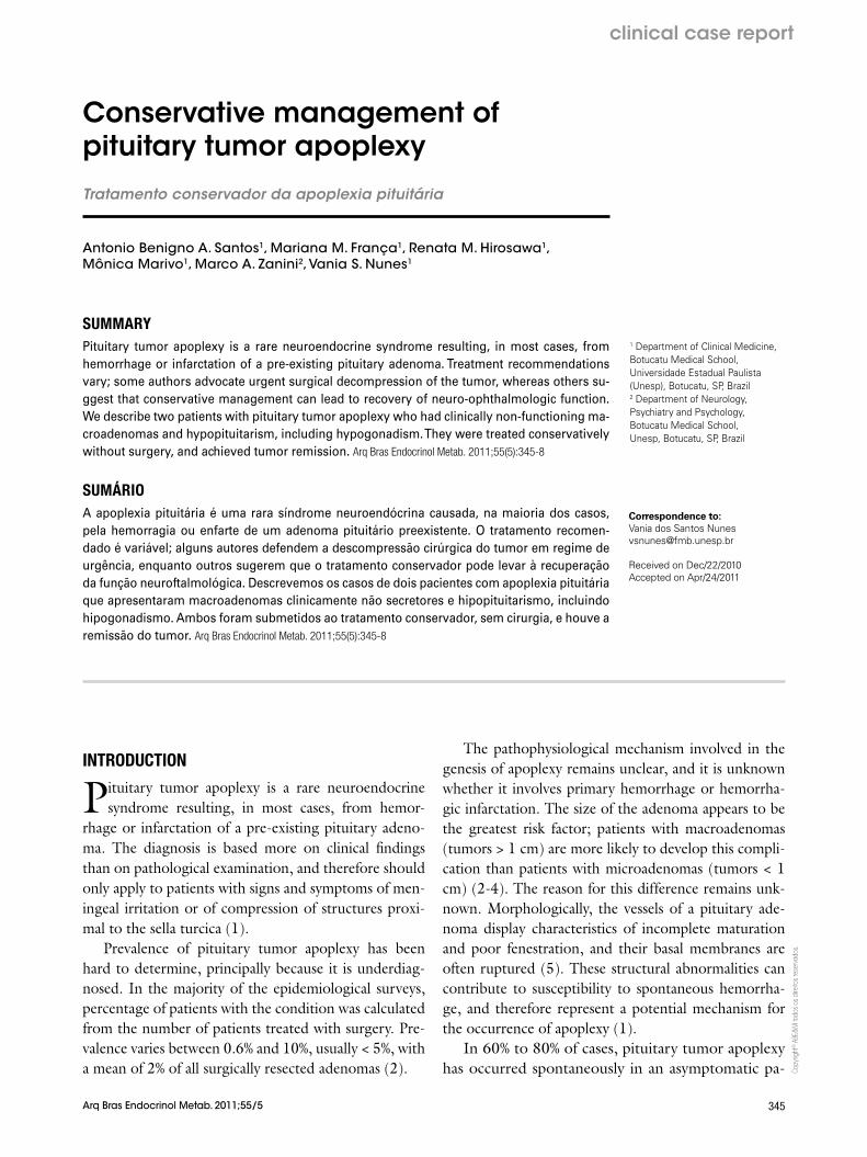
Cranial computed tomography revealed a pituitary tumor with compression of adjacent structures. Pitu-itary MRI demonstrated a solid expansive process (3.0 x 2.9 cm) with hypersignal on T1-weighted images, supra- and para-sellar extension, and optic chiasm com-pression, findings compatible with pituitary macroad-enoma and apoplexy (Figure 1). Laboratory test results showed central hypocortisolism (basal cortisol 1.21 µg/dL, reference value [RV]: 5-25 µg/dL), hypogo-nadism (total testosterone < 20 ng/dL, RV: 181-758 ng/dL; luteinizing hormone [LH] 0.28 mUI/mL, RV: 0.8-7.6 mUI/mL; follicle stimulating hormone [FSH]: 0.67 mUI/mL, RV: 0.7-11.1 mUI/mL), and central hypothyroidism (thyroid stimulating hormone [TSH] 0.05 μIU/mL, RV: 0.4-4.0 μIU/mL; free T4: 0.51 ng/dL, RV: 0.8-1.9 ng/dL), without hypersecre-tion of other hormones (growth hormone [GH] 0.14 ng/dL, RV: 0-5 ng/dL; insulin-like growth factor 1 − IGF-1 − and prolactin in normal ranges).
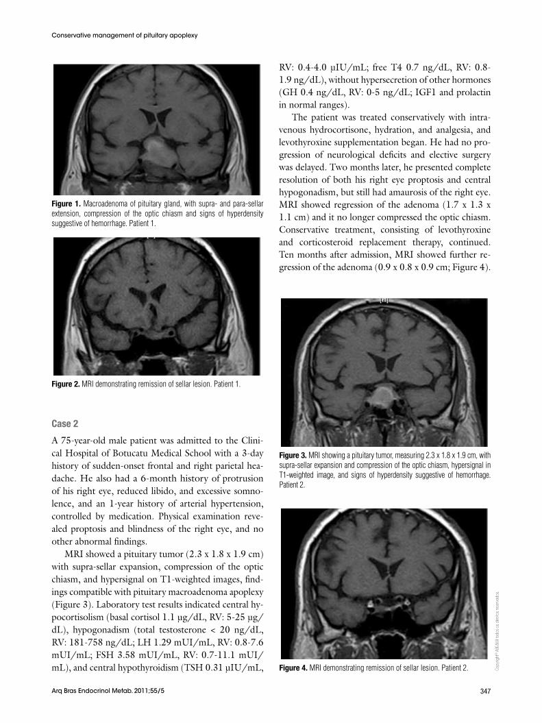
The patient was treated conservatively with intra-venous hydrocortisone, hydration, and analgesics. He was hospitalized for 20 days and remained clinically stable, with no progression of neurological deficits. After 3 months, the patient’s visual deficits and cen-tral hypogonadism were resolved. His visual fields were normal, total testosterone was 479 ng/dL, and repeti-tion of the MRI demonstrated normal pituitary gland (1.0 x 0.9 x 0.8 cm; Figure 2). He was maintained on levothyroxine treatment, but corticosteroid replace-ment therapy was discontinued to see if the deficit was resolved. Hormone replacement restarted few months later, when the patient presented symptoms of adreno-cortical deficiency and low basal cortisol level.
Two years later, the patient still needs thyroid and corticosteroid replacement therapy, but his pituitary MRIs have been normal, and he remains free of head-aches and visual deficits. His gonadotropic axis also re-mains normal (total testosterone: 521 ng/dL).
Figure 1. Macroadenoma of pituitary gland, with supra- and para-sellar extension, compression of the optic chiasm and signs of hyperdensity suggestive of hemorrhage. Patient 1.
Figure 2. MRI demonstrating remission of sellar lesion. Patient 1.
Figure 3. MRI showing a pituitary tumor, measuring 2.3 x 1.8 x 1.9 cm, with supra-sellar expansion and compression of the optic chiasm, hypersignal in T1-weighted image, and signs of hyperdensity suggestive of hemorrhage. Patient 2.
Figure 4. MRI demonstrating remission of sellar lesion. Patient 2.
Case 2
A 75-year-old male patient was admitted to the Clini-cal Hospital of Botucatu Medical School with a 3-day history of sudden-onset frontal and right parietal hea-dache. He also had a 6-month history of protrusion of his right eye, reduced libido, and excessive somno-lence, and an 1-year history of arterial hypertension, controlled by medication. Physical examination reve-aled proptosis and blindness of the right eye, and no other abnormal findings.
MRI showed a pituitary tumor (2.3 x 1.8 x 1.9 cm) with supra-sellar expansion, compression of the optic chiasm, and hypersignal on T1-weighted images, find-ings compatible with pituitary macroadenoma apoplexy (Figure 3). Laboratory test results indicated central hy-pocortisolism (basal cortisol 1.1 μg/dL, RV: 5-25 μg/dL), hypogonadism (total testosterone < 20 ng/dL, RV: 181-758 ng/dL; LH 1.29 mUI/mL, RV: 0.8-7.6 mUI/mL; FSH 3.58 mUI/mL, RV: 0.7-11.1 mUI/mL), and central hypothyroidism (TSH 0.31 μIU/mL,
RV: 0.4-4.0 μIU/mL; free T4 0.7 ng/dL, RV: 0.8-1.9 ng/dL), without hypersecretion of other hormones (GH 0.4 ng/dL, RV: 0-5 ng/dL; IGF1 and prolactin in normal ranges).
The patient was treated conservatively with intra-venous hydrocortisone, hydration, and analgesia, and levothyroxine supplementation began. He had no pro-gression of neurological deficits and elective surgery was delayed. Two months later, he presented complete resolution of both his right eye proptosis and central hypogonadism, but still had amaurosis of the right eye. MRI showed regression of the adenoma (1.7 x 1.3 x 1.1 cm) and it no longer compressed the optic chiasm. Conservative treatment, consisting of levothyroxine and corticosteroid replacement therapy, continued. Ten months after admission, MRI showed further re-gression of the adenoma (0.9 x 0.8 x 0.9 cm; Figure 4).
Two years later, the patient still needs thyroid and corticosteroid replacement therapy, but his hypogo-nadism remains in remission (free T4 1.1 ng/dL, total testosterone 311 ng/dL). He is still blind in the right eye. Follow-up pituitary MRI findings are compatible with empty sella.
DiSCUSSioN
Morbidity and mortality of patients with pituitary tu-mor apoplexy have declined over the past 6 decades. Factors that may have contributed to this include im-provement in diagnostic accuracy, more sophisticated therapeutic support, use of glucocorticoids, and refi-nement of surgical and postoperative techniques (1). Untreated patients may continue to have high morbi-dity and mortality; before the use of glucocorticoids, mortality was approximately 50% (1).
Patients with signs and symptoms of pituitary tu-mor apoplexy require thorough clinical evaluation and continuous monitoring to care for their neurological, ophthalmological, and endocrine dysfunctions. After hemodynamic stabilization and glucocorticoid repla-cement, it is important to choose the best treatment option for the improvement or stabilization of the patient’s neuro-ophthalmological deficits. Immediate surgical intervention for apoplexy remains controver-sial. Several reports showed that spontaneous neuro-logical recovery is possible, and some authors reported that patients treated conservatively had neurological and neuro-ophthalmological recovery rates similar to those of patients treated surgically (9). Other more re-cent retrospective studies have also provided evidence that nonsurgical treatment of patients with pituitary tumor apoplexy may be safe for patients who do not present visual deficits or show early evidence of recupe-ration from this condition (10,11). On the other hand, results of trans-sphenoid decompression immediately after stabilization of the patient have been very good, and recovery of neurological and pituitary function oc-curs within a period of several hours to days.
The two patients reported here achieved tumor re-mission with conservative treatment. One recovered completely from his visual deficit, and both showed normalization of at least one of the compromised pitui-tary hormone axes.
New studies with methodologies appropriate for cli-nical decision-making are needed to determine the op-timal time for surgical intervention in the treatment of pituitary tumor apolexy. Until then, the decision must be individualized, using a multidisciplinary approach that encompasses all aspects of the patient’s clinical condition.
Disclosure: no potential conflict of interest relevant to this article was reported.
7. Post MJ, David NJ, Glaser JS, Safran A. Pituitary apoplexy: diag-nosis by computed tomography. Radiology. 1980;134(3):665-70.
8. Sibal L, Ball SG, Connolly V, James RA, Kane P, Kelly WF, et al. Pi-tuitary apoplexy: a review of clinical presentation, management and outcome in 45 cases. Pituitary. 2004;7(3):157-63.
9. Maccagnan P, Macedo CL, Kayath MJ, Nogueira RG, Abucham J. Conservative management of pituitary apoplexy: a prospective study. J Clin Endocrinol Metab. 1995;80(7):2190-7.