V/rus Rrseurch. 1 ( 1984) 169-l 88 Elsevier 169 VRR 00119 Construction of human and express the adenovirus DNA binding protein gene by cotransformation with the HSV-1 tk gene Daniel F. Klessig ‘, Terri Grodzicker 2 and Vaughn Cleghon ’ ’ Depcrrrmwt of Cellulrrr, Vird und Molec~ulur B~ologv,. limrwsrrv of Utuh, Sulr Luke (‘I!\‘. UT 84122. und ’ Cold Spring Hurbor Lohorototy. Cold Spring ~rrrhor, N 1’ I 1724, U.S.A. (Accepted 31 January 1984) Summary We have introduced the DNA binding protein (DBP) gene of human adenovirus type 5 (Ad5) into high molecular weight DNA of permissive human cells by cotransformation of tk- cells with the cloned DBP and HSV-1 thymidine kinase genes. 110 tk+ cell lines were isolated after selection in HAT medium. The amount and arrangement of adenovirus sequences in the tk+ cell lines were analyzed by restriction endonuclease digestion and filter hybridization. Twelve of the 110 lines carry at least a segment of the DBP gene while only three of these contain the entire DBP gene at approximately one copy per cell. Cytoplasmic, polyadenylated DBP mRNA is made in all three cell lines though the amount is very low compared to that present in infected HeLa cells. The cell line U13-2 which contains approxi- mately l/30 the steady-state level of DBP mRNA found in infected HeLa cells produces a few percent of the amount of DBP made during the peak period of DBP synthesis in infected cells. The other two lines contain lower levels of DBP mRNA and do not synthesize detectable levels of the protein. When these DBP-tk’ cell lines are infected with adenovirus mutants containing temperature-sensitive (ts) mutations in the DBP gene, only U13-2 permits some viral DNA replication (and hence late gene expression) at the nonpermissive temperature, indicating that sufficient quanti- ties of DBP from the integrated gene are produced to allow complementation of the ts mutation in this cell line. However, growth of these ts mutants (as measured by virus production) is only partially complemented in U13-2 at the nonpermissive temperature. Key words: cotransformation, adenovirus DNA binding protein. mutant comple- mentation 016X-1702/X4/$03.00 ( 1984 Elsevier Science Publishers B.V
Transcript
V/rus Rrseurch. 1 ( 1984) 169-l 88
Elsevier
169
VRR 00119
Construction of human and express the adenovirus DNA binding protein gene
by cotransformation with the HSV-1 tk gene
Daniel F. Klessig ‘, Terri Grodzicker 2 and Vaughn Cleghon ’
’ Depcrrrmwt of Cellulrrr, Vird und Molec~ulur B~ologv,. limrwsrrv of Utuh, Sulr Luke (‘I!\‘. UT 84122. und
’ Cold Spring Hurbor Lohorototy. Cold Spring ~rrrhor, N 1’ I 1724, U.S.A.
(Accepted 31 January 1984)
Summary
We have introduced the DNA binding protein (DBP) gene of human adenovirus type 5 (Ad5) into high molecular weight DNA of permissive human cells by
cotransformation of tk- cells with the cloned DBP and HSV-1 thymidine kinase genes. 110 tk+ cell lines were isolated after selection in HAT medium. The amount and arrangement of adenovirus sequences in the tk+ cell lines were analyzed by restriction endonuclease digestion and filter hybridization. Twelve of the 110 lines carry at least a segment of the DBP gene while only three of these contain the entire DBP gene at approximately one copy per cell. Cytoplasmic, polyadenylated DBP mRNA is made in all three cell lines though the amount is very low compared to that present in infected HeLa cells. The cell line U13-2 which contains approxi- mately l/30 the steady-state level of DBP mRNA found in infected HeLa cells produces a few percent of the amount of DBP made during the peak period of DBP synthesis in infected cells. The other two lines contain lower levels of DBP mRNA and do not synthesize detectable levels of the protein. When these DBP-tk’ cell lines are infected with adenovirus mutants containing temperature-sensitive (ts) mutations
in the DBP gene, only U13-2 permits some viral DNA replication (and hence late gene expression) at the nonpermissive temperature, indicating that sufficient quanti- ties of DBP from the integrated gene are produced to allow complementation of the ts mutation in this cell line. However, growth of these ts mutants (as measured by virus production) is only partially complemented in U13-2 at the nonpermissive temperature.
Key words: cotransformation, adenovirus DNA binding protein. mutant comple- mentation
A number of multi-functional proteins from eukaryotes have been defined by a combination of genetic and biochemical studies. One of the best characterized of these is the simian virus 40 (SV40) large T antigen which exhibits separate functional domains as discerned by mapping of sets of mutants which alter one but not another of the protein’s different functions.
The human adenovirus 72 kd DNA binding protein (DBP) also performs several independent functions. Two ts mutants of DBP (Ad5ts125 and Ad5ts107), isolated by Ginsberg and colleagues (Ensinger and Ginsberg. 1972; Rubenstein and Gins- berg, 1974) have been indispensable in defining several of the functions of this protein. The ts alterations at the nonpermissive temperature diminish the single- strand DNA binding activity of the protein in vitro (van der Vliet et al., 1975) and inhibit DNA replication in vivo (Ensinger and Ginsberg, 1972: Rubenstein and Ginsberg, 1974; van der Vliet and Sussenbach, 1975; van der Vliet et al., 1977) and in vitro (Horwitz, 1978). Normal turn-off of viral early genes during the late phase of
the lytic cycle is also disrupted at the nonpermissive temperature in cells infected with the ts mutant (Babich and Nevins, 1981; Blanton and Carter, 1979; Carter and Blanton, 1978a, 1978b; Carter and Ginsberg, 1976; Nevins and Winkler, 1980; Nicolas et al., 1982). The DBP directly affects early gene turn-off as well as DNA replication since inhibition of viral DNA synthesis with either drugs or ts lesions in
other viral early genes does not affect early gene expression (Carter and Blanton, 1978a, 1978b). In addition, at the nonpermissive temperature the ts mutants trans- form rat cells in culture with an enhanced frequency compared to wild-type (wt) virus (Rubenstein and Ginsberg, 1974; Ginsberg et al., 1974: Williams et al.. 1974).
A second class of mutants which greatly enhance the ability of the virus to grow in monkey cells defines yet a second set of functions of DBP (Anderson. 1981: Klessig, 1977; Klessig and Grodzicker, 1979). In monkey cells infected with wild-type human adenovirus. the viral early genes are properly expressed (Anderson and Klessig, 1982; Feldman et al., 1966; van der Vliet and Levine. 1973) and viral DNA replication occurs normally (Friedman et al., 1970; Rapp et al., 1966; Reich et al., 1966). However, several of the late viral proteins, in particular the fiber protein. are synthesized at reduced levels (Feldman et al., 1966; Grodzicker et al.. 1974; Klessig and Anderson. 1975; Malmgren et al., 1966). The block to late gene expression appears to be due to both reduced production of several viral late mRNAs (Farber and Baum, 1978; Klessig and Anderson, 1975; Klessig and Chow, 1980) and poor translation in vivo, but not in vitro, of the residue mRNA. particularly the fiber message, made in monkey cells (Anderson and Klessig, 1983). The alteration that allows the virus to overcome this block is located in the DBP gene (Klessig and Grodzicker, 1979).
Physical mapping of the ts and host range mutations by marker rescue experi- ments and DNA sequence determination places the four known ts mutations in the 3’ half of the gene and the nine host range mutations in the 5’ half (Anderson et al.. 1983; Klessig and Quinlan, 1982; Kruijer et al.. 1981, 1982: Rice and Klessig, 1984: D.F. Klessig and D. Brough, unpublished). Thus, like the SV40 T antigen. the DBP exhibits separate functional domains.
171
In order to better understand the interrelationships between the many different functions of the DBP, a much more extensive collection of mutants must be obtained. Unfortunately, since this protein is involved in such essential viral processes as DNA replication and early and late gene expression, many potential interesting mutations will be lost because of their lethal nature. Thus, we have attempted to construct permissive human cell lines which contain and express the DBP gene for the propagation of these otherwise lethal, nonrecoverable DBP mutants.
Specific pieces of DNA can be introduced into mammalian cells by a variety of methods, including cotransformation, viral vectors, and microinjection with glass capillaries, erythrocyte ghosts, or liposomes. When mouse L tk cells are transfected with the herpes simplex virus type 1 (HSV-1) thymidine kinase (tk) gene and a variety of other unlinked DNAs (cotransformation), many of the resulting tk+ transformants also incorporate the unlinked DNA (Wigler et al., 1979b). Thus for example, $X174. pBR322, simian virus 40 (SV40), and rabbit or human /3-globin
sequence have been introduced into mouse tk cells by cotransformation (Hanahan
et al., 1980; Pelicer et al., 1978; Wigler et al., 1979b; Wold et al., 1979). We have shown that left-end segments of the human adenovirus type 2 (Ad2) genome can also be introduced into permissive tk- human cells by this method (Grodzicker and Klessig, 1980; Klessig et al., 1982). Here we describe the construction (by tk cotransformation) and characterization of three human cell lines which contain and express, at low levels, the DBP gene of adenovirus type 5 (Ad5).
Materials and Methods
Cells and viruses The 143 tk- human cell line, derived after selection with bromodeoxyuridine (K.
Huebner and C. Croce, personal communication) from the murine sarcoma virus- transformed line R970-5 (Rhim et al., 1975) was a gift from C. Croce. The RD-4 tk- human cell line (a gift from M. Nicolson) was derived from a human rhabdomyosarcoma cell which was infected with the RD114 retrovirus and selected
for the tk- phenotype in bromodeoxyuridine. The HeLa cell line was provided by J.F. Williams. All cell lines were cultivated in Dulbecco-modified Eagle medium
(Flow Labs) supplemented with 10% calf serum (gamma-globulin-free calf serum for RD-4 tk- and its cotransformants), 100 pg/ml streptomycin, 100 pg/ml penicillin and 292 pg/ml glutamine. tk+ transformants were selected and grown in Dulbecco- modified Eagle medium containing hypoxanthine (15 pg/ml), aminopterin (0.5 pg/ml), and thymidine (5 pg/ml) (HAT) and calf serum, streptomycin, penicillin and glutamine. Suspension cultures of HeLa cells were grown in Eagle F13 medium (Grand Island Biological) supplemented with 8% calf serum (Irvine Scientific). 1% fetal serum (Flow Labs). All medium supplements except serum were purchased from Sigma Chemical Co. Gamma-globulin-free calf serum was obtained from Grand Island Biological.
Ad5 and Ad2’NDl ts23 were obtained from J.F. Williams and were propagated in suspension cultures of HeLa cells. Ad5ts125 and Ad5ts107 were obtained from
172
J.F. Williams and C.S.H. Young, respectively, and were propagated in monolayers of HeLa cells at 325°C. Ad5hr404 and Ad2ts400, isolated by Klessig (Klessig and Grodzicker. 1979; Rice and Klessig, 1984). were grown in suspension cultures of HeLa cells. For measurement of virus yields from various cell lines, confluent monolayers were infected at a multiplicity of infection of 10 plaque-forming units (pfu) per cell in phosphate-buffered saline. After 60 min of incubation at 375°C. the inoculum was removed. and the monolayer was rinsed with phosphate-buffered saline and incubated in medium plus HAT for 48 h. Progeny virus were released from infected cells by freeze-thawing and sonication. Plaque titrations were per- formed as described by Grodzicker et al. (1974) at 32.5”C on HeLa cell monolayers.
Cell trunsformutiot~
143 cells were transformed by the procedure described by Wigler et al. (1979a). a modification of the DNA/calcium phosphate coprecipitation method (Graham and
van der Eb. 1973). Dishes (90 mm) of 143 tk- cells at 10% confluence were transfected with the following DNAs per dish: 5-20 ng of SalI-linearized ptk2 or the BamHI fragment of it. 150-500-fold molar excess compared to the tk gene of the SalI-linearized, DBP-containing plasmids (~730. ~1601) or the SalI-EcoRI fragment of them, and lo-15 pg of 143 tk- cell carrier DNA. Transformed cells were selected for acquisition of the HSV-1 tk gene (Bacchetti and Graham, 1977; Maitland and McDougall. 1977; Minson et al.. 1978: Wigler et al.. 1977) by HAT selection (Szybalski et al., 1962). Individual colonies were isolated by using cloning cylinders and grown into mass cultures.
RD-4 cells were transformed by a modification of the Wigler et al. (1979a) method. Dishes (60 mm) of RD-4 tk- cells at 30&50’% confluence were transfected with the following DNAs per dish: 4 ng of the BamHI fragment of ptk2, 1 pg of the SalI-EcoRI fragment of ~1605, and 5 pg of 143 tk cell carrier DNA. The monolayer of cells was treated with 0.3 ml of the precipitate solution in presence of 0.3 ml of medium for 15 min at room temperature. Then 2.5 ml of medium was added and after 6 h at 37°C in absence of HAT selection. the cells were treated by addition of dimethyl sulfoxide to the medium to a final concentration of 10% for 20 min at 37°C. After replacing the medium. the cells were incubated for 24436 h in fresh, normal medium before starting HAT selection. All experiments using recombi- nant DNA were performed in accordance with the National Institutes of Health
guidelines.
DNA preparution
DNA from tissue culture cells was prepared as described by Wigler et al. (1979b). ptk2, a pBR322 plasmid carrying a 3.6 kilobase (kb) HSV-1 BamHI fragment that contains the herpes thymidine kinase gene. was obtained from D. Hanahan. Plas- mids ~730. ~1601 and ~1605 are derived from pBR322 and contain the 59.5 map units (m.u.) to 75.9 m.u., BamHIIEcoRI fragment of Ad5hr404, Ad5ts107, wt Ad5. respectively.
Plasmid DNA was prepared according to the method of Tanaka and Weisblum. (1975). Restriction enzyme-generated fragments of viral DNA were fractionated on
173
horizontal 1% agarose gels in Tris-borate buffer (90 mM Tris base/90 mM boric acid/2.5 mM EDTA. pH 8.3) by electrophoresis at a potential of 1-2 V/cm for 16 h. The separated fragments were eluted from the gel by electrophoresis (Klessig and
Grodzicker, 1979).
Filter hybridization 10 pg of high molecular weight DNA extracted from transformed cells was
digested with 40-60 units of the different restriction endonucleases indicated (New England BioLabs or Bethesda Research Laboratories) in 100 ~1 for 337 h at 37°C. 27 pg of Ad5 or 7.5 pg of ~730 DNA was digested with appropriate restriction endonucleases in the presence of 10 pg of calf thymus DNA to generate marker fragments. This mixture contains the equivalent of one-half copy of the viral or plasmid genome per diploid cell or about one copy per 143 cell, which contains ca. 80 chromosomes. The digestion products were fractionated by electrophoresis at 1.222 V/cm for 15-20 h on horizontal 0.8 or 1% agarose gels (14
oligodeoxythymidine-cellulose columns. The
174
structure of the adenoviral RNA was determined by using a modification of the Sl nuclease mapping technique of Berk and Sharp (1977). Polyadenylated RNA selected from 500 pg of total cytoplasmic RNA obtained from 143 tk-, RD-4 tk-. or tk’ transformants was hybridized to 0.1 pg of BamHl-EcoRI-cleaved ~730 in 50 ~1. 5 pg of cytoplasmic RNA prepared 16 h post infection (hpi) from HeLa suspension cells infected with 10 pfu per cell of Ad5hr404 in the presence of 20 pg/ml cytosine arabinoside was used as a positive control. After Sl treatment, half of each sample was fractionated on an alkaline 1.2% agarose gel by electrophoresis with 10 ng of kglII-digested Ad5 DNA as size markers. The gel was treated for 60 min in 1 M Tris-HCl, pH 7.4. and 0.6 M NaCl. and the DNA transferred to a nitrocellulose
filter for 24 h in 6 X SSC. The filter was hybridized to the 59.5 m.u. to 75.9 m.u. fragment of Ad5 DNA as described above Filters were exposed to X-ray film without intensifying screens for 20 h.
Protein synthesis
Dishes (35 mm) of cells were labeled with 50 PCi of [‘5S]methionine for 2 h as described by Klessig and Anderson (1975). or 100 PCi of j2POi for 4 h after growth in phosphate-free medium for 4 h. After labeling the cells were harvested and proteins extracted in a high salt (0.65 M NaCl) and detergent (1% NP40) solution according to Crawford et al. (1982). Varying amounts of the extract were incubated for 1 h at 0°C with 10 ~1 of rabbit anti-DBP serum in 300 ~1 of RIPA buffer (10 mM Tris-HCl. pH 7.4/150 mM NaCl/O.l% SDS/l% deoxycholate/l% Triton X-100/5 mM EDTA). The immune complexes were precipitated with 50 ~1 of 10% solution of formalin-fixed Stuphylococcus aureus bacteria (Pansorbin, Calbiochem) that was further treated by boiling in 2% SDS for 5 min and washing 4 times in RIPA buffer. The labeled, precipitated proteins were analyzed by autoradiography of the 15% acrylamide gels after SDS-PAGE. The jiS signal was enhanced by soaking the polyacrylamide gels in 1 M sodium salicylate for 30 min before drying and autoradiographed for 10 days. Gels containing “P-labeled proteins were autora- diographed 6-10 h.
Western blots
After immunoprecipitation with anti-DBP serum, the proteins were fractionated on a 7.5% SDS-polyacrylamide gel and subjected to Western blot analyses using a procedure described in part by Towbin et al. (1979) and modified by W.M. Huang (personal communication). Briefly, the gel was soaked in 250 ml of transfer buffer (25 mM Tris-HCl, pH 8.3/192 mM glycine/20% (v/v) methanol) for 5-10 min. A nitrocellulose filter presoaked in transfer buffer was laid on the gel and 3 sheets of 3MM paper and a Scotch-Brite sponge (Bio-Rad) were placed on each side. The proteins were electrotransferred in a Tram-Blot Cell (Bio-Rad) for 18 h at 50 V in transfer buffer. After transfer the nitrocellulose filter was washed 5-10 min in Tris-saline buffer (10 mM Tris-HCl, pH 7.6/0.9% NaCl). incubated in 10 ml of Tris-saline buffer plus 5% bovine serum albumin (BSA) at 40°C for l-l.5 h, washed briefly in Tris-saline buffer and reacted with 5-10 ml of Tris-saline buffer plus 5% BSA and a 1 : 25 dilution of anti-DBP serum for 1 h at 37°C. The filter was washed
175
at room temperature with 250 ml of Tris-saline buffer (once for 10 min). Tris-saline
buffer plus 0.05% NP40 (twice, 20 min each), and Tris-saline buffer (once for 10 min). The filter was then reacted with 5 ml of Tris-saline buffer plus 5% BSA and ‘“51-labeled protein A of S. uuwus (2--8 X 10’ cpm/ml) for 30 min at 37°C. The washing procedure was repeated and the filter dried and autoradiographed for a few hours. Reactions with anti-serum and protein A were done in a sealed baggie or pouch.
Results
To obtain adenovirusstk cotransformants, the human cell lines 143 tk- or RD-4 tk- were transfected with both ptk2 (a plasmid carrying a 3.6 kb HSV-1 BamHI fragment that contains the tk gene; Hanahan et al., 1980) and various derivatives of pBR322 which carry the BamHI (59.5 m.u.)-EcoRI (75.0 m.u.) restriction fragment of Ad5 or mutants of it (Ad5hr404. Ad5ts107). The DBP gene is segmented containing a main body which encodes the entire protein (coordinates 61.5566; Kruijer et al., 1981, 1982) and two leaders (see Fig. 1; Berk and Sharp, 1978: Chow et al., 1979; Kitchingman et al., 1977). The second leader is located near the main body at coordinate 68 while the first leader, and presumably the promoter, is located either at coordinate 72 or 75. Early during the productive cycle of adenovirus most
-e Late mRNA
4 Early 1600 170 -50 mANA
pBR322
Fig. 1. Structure of the chimeric plasmids ~730. ~1601 and ~1605. The region of the Ad5 genome encoding the DBP is represented as a scale at the top with each segment corresponding to 1 map unit. The
structures of the early and late DBP mRNA are shown above the scale. with _,L denoting regions spliced
out of the transcripts. The adenovirus sequences in the chimeric plasmids are shown as a solid black
segment, while pBR322 sequences are indicated by a single line. The positions of several restriction
endonuclease cleavage sites are indicated by arrows. with their map coordinates on the conventional Ad5
physical map given in parenthesis.
176
DBP mRNAs contain the first leader encoded at 75 while later the majority of the mRNAs carry the leader specified at 72 (Chow et al.. 1979). Presumably this alteration in mRNA structure reflects changes in the utilization of these two promoters during the infectious cycle. bfter transfection (Wigler et al.. 1979a) with ptk2 and the DBP gene and growth in HAT medium for approximately 1 month, tk+ colonies arose at a frequency of 0.3 colonies per 2 ng of tk gene per 10” 143 tk cells. This is approximately 10 fold lower than the number of colonies obtained when sequences from the left end of the Ad2 genome rather than DBP sequences were used for cotransformation of 143 tk cells (Grodzicker and Klessig. 1980) and
lo3 fold lower than the frequency observed for transformation of mouse L tk cells
with ptk2 (Wigler et al.. 1979b). Removing the sequences of pBR322 which are
poisonous to eukaryotic cells (Lusky and Botchan, 1981) before cotransformation raised the frequency to 4-5 colonies per 2 ng of tk gene per 10h 143 cells. Eleven of the 103 tk+ 143 transformants analyzed contained part of the DBP gene while only two of these carried the entire gene.
With the RD-4 tk cells and plasmid fragments devoid of the poisonous sequences, the frequency of tk+ transformation was 0.7 colonies per 2 ng of tk gene
per 10h cells. One of the RD-4 tk’ transformants characterized carried the entire DBP gene.
In summary, three potentially useful cell lines were constructed each carrying a different variant of the DBP gene. The E5 cell line was constructed by transfecting
143 tk- cells with ptk2 and ~730 plasmid DNAs both linearized with Sal]. ~730 was derived from pBR322 by the insertion of the 59.5-75.9 m.u., BamHI-EcoRI frag- ment of Ad5hr404 (Fig. 1). Ad5hr404 is a mutant of Ad5 with an altered DBP that allows it to grow efficiently in monkey cells (Klessig and Grodzicker, 1979). U7-5b resulted from cotransformation of 143 tk cells with (i) the BamHI fragment of ptk2 that contains the HSV-1 tk gene and (ii) the SalI-EcoRl fragment of ~1601 that contains the DBP gene (Fig. 1). Both of these fragments are devoid of the poisonous sequences. ~1601 is identical to ~730 except the insert was derived from Ad5ts107. U13-2 was isolated after transfection of RD-4 tk- cells with the BamHl fragment of ptk2 and the SalI-EcoRI fragment of ~1605. ~1605 is identical to ~730 except the insert was derived from wild-type Ad5. The molar ratios of the DBP gene to the tk
gene were 500 : 1 (E5), 150 : 1 (U7-5b), and 250 : 1 (U13-2).
Amount und arrungement of udenocirus sequences in the tk cotrunsformunts
The adenovirus sequences in the tk+ cell lines were analyzed by restriction
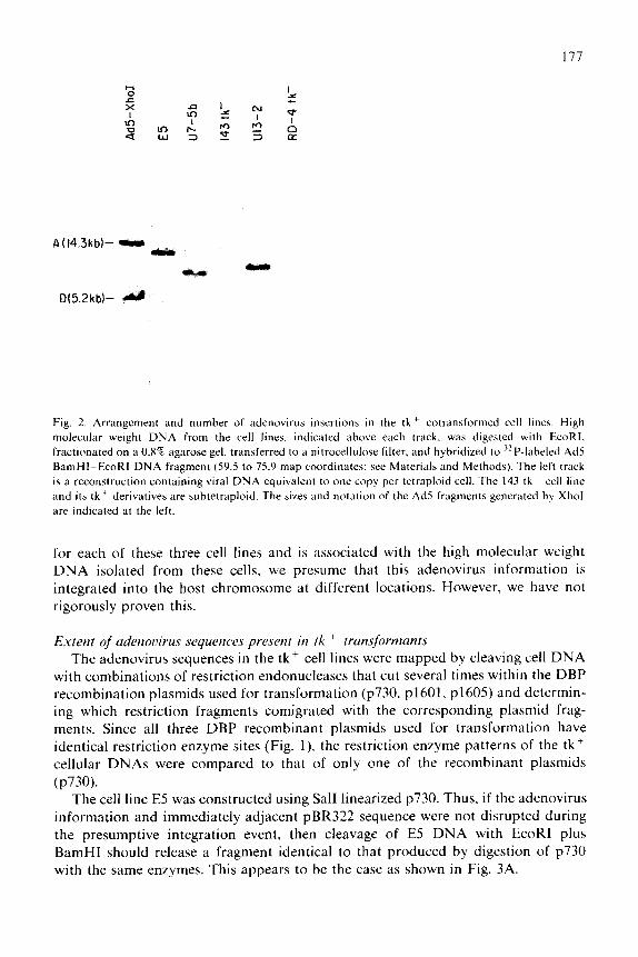
enzyme digestion and filter hybridization (Southern, 1975). Cell DNA from the tk ’ lines were digested with EcoRI, an endonuclease which cuts at one of the junctions between adenovirus and pBR322 sequences in the recombinant plasmids. Analysis of the EcoRI digestion products by electrophoresis, transfer. and filter hybridization using the nick-translated Ad5 BamHI-EcoRI fragment (59.5-75.9 m.u.) as probe showed that three of the lines had a single insertion of adenovirus sequences (Fig. 2). The intensity of the bands compared to reconstruction indicates that the adenovirus sequences were present in approximately one copy per cell.
Since the restriction fragment which contains adenovirus sequences differs in size
177
A(14.3kbh -JMM _
D62kb)-
Fig. 2. Arrangement and number of adenovirus insertions in the tk’ cotransformed cell lines. High molecular weight DNA from the cell lines. indicated above each track. was digested with EcoRI. fractionated on a 0.8% agarose gel. transferred to a nitrocellulose filter. and hybridized to 32P-labeled Ad5
BarnHI-EcoRI DNA fragment (59.5 to 75.9 map coordinates; see Materials and Methods). The left track
is a reconstruction containing viral DNA equivalent to one copy per tetraploid cell. The 143 tk cell line and ita tk ’ derivatives are subtetraploid. The sizes and notation of the Ad5 fragments generated by Xhol
are indicated at the left.
for each of these three cell lines and is associated with the high molecular weight DNA isolated from these cells. we presume that this adenovirus information is integrated into the host chromosome at different locations. However, we have not
rigorously proven this.
Extent of udenovirus sequences present in tk + trunsfornmnts
The adenovirus sequences in the tkf cell lines were mapped by cleaving cell DNA with combinations of restriction endonucleases that cut several times within the DBP recombination plasmids used for transformation (~730. ~1601, ~1605) and determin- ing which restriction fragments comigrated with the corresponding plasmid frag- ments. Since all three DBP recombinant plasmids used for transformation have identical restriction enzyme sites (Fig. l), the restriction enzyme patterns of the tk+ cellular DNAs were compared to that of only one of the recombinant plasmids
(P730). The cell line E5 was constructed using Sal1 linearized ~730. Thus, if the adenovirus
information and immediately adjacent pBR322 sequence were not disrupted during the presumptive integration event, then cleavage of E5 DNA with EcoRI plus BamHI should release a fragment identical to that produced by digestion of ~730 with the same enzymes. This appears to be the case as shown in Fig. 3A.
17X
A
BamHI-EcaRI BamHI-HindD
61.3-71.4)
KpnI
Fig. 3. Mapping of the wral sequences in the tk ’ cotranaformed cell lines. In panels A, B and C‘ IO pg of
high molecular weight DNA from each cell line indicated above each track was digested wth the
restriction rndonucleaara shown below the panel. The cloned DNA wa Cractlonatrd on 0.X or 1’4 agaroae
gels. transferred to nitrocellulose filters. and hybridized to the “P-labeled 50.5 10 75.9 mu. fragment of
Ad5 DNA (see Materials and Methods). The left track in each panel IS a recon~tructlon containing ~730
DNA equivalent to one copy per tetraploid cell. The map coordinate:, on the Ad5 gcnome of telltale
hands are shown on the right. The positions of cleavage sites on p7?0 are shown In Fig. 1.
In contrast, the U7-5b and U13-2 cell lines were isolated after transfection with the EcoRI-Sal1 fragment of ~1601 and ~1605, respectively (see Fig. 1). Because the partial EcoRI site at one end of this fragment presumably is not regenerated during the integration into the host DNA, cleavage of U7-5b or U13-2 DNA with EcoRI plus BamHI does not yield the 59.5-75.9 m.u. fragment originally cloned into the plasmids (data not shown). Cleavage of the tk+ cell DNA with BamHI plus Hind111 shows that the adenovirus sequences from one end of the cloned fragment (BamHI; 59.5 m.u.) are intact through the Hind111 site at 72.X m.u. in U7-5b and U13-2 as
well as E5 (Figs. 1 and 3B). In cell lines U7-5b and U13-2 integration of plasmid DNA presumably occurred by recombination of host DNA with adenovirus se- quences located between the EcoRI site at 75.9 and the Hind111 site at 72.8 at one end of the plasmid and pBR322 sequences at the other end. A second band complementary to the probe (59.5-75.9 m.u. fragment) is seen with E5 and Ul3-2 DNA after digestion of cell DNA with BamHI and HindIII. This is probably the junction fragment which contains host DNA (and pBR322 in the case of E5) and the residual adenovirus sequences between 75.9 and 72.8 that were not lost during integration. Presumably in U7-5b too much of this sequence has been lost to
179
efficiently hybridize to the probe. Analysis with restriction enzymes KpnI (Figs. 1 and 3C) and BglII (data not shown) indicates that the internal organization of these adenovirus sequences is maintained in these three tk+ cell lines. In sum, E5 contains the entire DBP gene including both the early and late promoters. The DBP gene with at least its late promoter at 72. and possibly also the early promoter at 75. is intact within the U7-5b and U13-2 cellular DNA.
Virul RNAs in the cotrunsfortned cell lines
The amount and structure of the DBP mRNA present in these cells were determined by using a modification of the Sl nuclease mapping technique of Berk and Sharp (1977). To detect the low concentration of the viral RNAs in these lines, cytoplasmic, polyadenylated RNA from the cotransformed cells was first annealed to the unlabeled 59.5-75.9 m.u. BamHI-EcoRI fragment of ~730. The RNA-DNA duplexes were treated with Sl nuclease. fractionated on alkaline agarose gels.
3160 (E)
1920 (G)
Fig. 4. Sl mapping of adenovirus RNA from the tk’ cotransformed cell lines. Polyadenylated. cyto-
plasmic RNAs from the cell lines indicated above each track were hybridized to BamHI~EcoRi cleaved
~730 DNA. After Sl nuclease treatment and fractionation on an alkaline agarose gel. the DNA was
transferred to nitrocellulose filters and hybridized to the “P-labeled 59.5 to 75.9 m.u. fragment of Ad5
DNA (see Materials and Methods). 5 pg of cytoplasmic RNA extracted from Ad5hr404-infected HeLa
cells 16 h after infection in the presence of cytosine arabinoside was used as a positive standard and ib
shown in the track marked ‘infected HeLa’. RNA from the parental tk- 143 and RD-4 lines as well as
yeast tRNA served as negative controls. Marker Ad5 fragments generated by BglII. along with their size
in base pairs. are indicated on the right. The size of the DNA fragment complementary to the main body
of the DBP mRNA is denoted with an arrow (see Fig. 1 for the structure of DBP mRNA).
180
transferred to nitrocellulose filters and hybridized to the labeled 59.5575.9 m.u. fragment of Ad5.
One prominent band corresponding to a 1580 nucleotide RNA is seen with RNA from the three tk’ cell lines as well as Ad5hr404-infected HeLa cells (Fig. 4). This size is in excellent agreement with that reported by Berk and colleagues (Berk et al.. 1979: Berk and Sharp, 1978) for the main body of the DBP mRNA encoded between 61.5 and 66 (see Fig. 1). As expected. the parental 143 and RD-4 tk lines do not contain this RNA.
The concentrations of the DBP mRNA in the tk’ transformed cells are much lower than in infected HeLa cells. A comparison of the band intensities obtained using 100 times more of the RNA isolated from the cotransformants than of RNA prepared from HeLa cells early after infection suggests that the concentration of DBP mRNA is approximately 150, 400 and 1200 fold lower in U13-2. U7-56 and
E5. respectively (Fig. 4). Because the conditions for infection of HeLa cells were optimized for maximum early viral mRNA levels (i.e.. RNA was extracted 16 hpi from HeLa cells infected in the presence of the DNA synthesis inhibitor cytosine arabinoside), this comparison exaggerates the difference in amount of viral mRNA in the infected versus cotransformed cells. Nonetheless, only a very small amount of DBP message is present in these cells (estimated at 2-20 molecules per cell using Flint and Sharp’s (1976) number of ca. 300-400 molecules per cell determined for RNA isolated at 8 hpi in the presence of cytosine arabinoside and assuming that the level of DBP RNA is approximately 5 fold higher at 16 hpi).
To determine whether the very low level of expression of DBP genes in these cells might be due to methylation, the cotransformant DNA was analyzed with HpalI and Mspl. These two endonucleases are isoschizomers that differ in their ability to cut at their common recognition site depending on whether it is methylated (Mann and Smith, 1977). No difference in the cleavage pattern of adenovirus sequences produced by this pair of enzymes could be detected (data not shown), suggesting that methylation. at least at the HpaII/MspI sites, is not responsible for the poor expression of the DBP gene in these cells.
DBP in the cotrunsformed cell lines
The synthesis of the Ad5 DBP, a highly phosphorylated protein (Axelrod, 1978: Klein et al., 1979; Levinson and Levine, 1977; Levinson et al., 1977: Linne et al.. 1977; Linne and Philipson, 1980; Russell and Blair. 1977). was followed by labeling the cotransformed cell lines with [ “SS]methionine or “PO: ~. and immunoprecipitat- ing the protein with anti-DBP serum. The synthesis of the DBP cannot be detected in E5 or U7-5b (Fig. 5). However, a small amount is produced in the U13-2 cell line. Comparisons of the intensity of the band on autoradiograms of gels in which varying amounts of immunoprecipitated, AdS-infected HeLa cell extracts were used as standards indicate that the DBP is synthesized at about 1/20th to 1/30th the rate seen at the peak of DBP production in an infected HeLa cell (16-24 hpi). The level of phosphorylation of DBP appears to be similar in the cotransformed U13-2 line and AdS-infected HeLa cells; however, the protein made in U13-2 migrates slightly slower in SDSPAGE.
lnfecled HeLo - I_
DBP DBP-
DBP-
Fig. 5. Synthesis and phosphorylation of DBP in the tk+
[ “S]methionine for 2 h or ” Lotransformed cells. After labeling cells with
P for 4 h. proteins were extracted in high salt and detergent. immunoprecipi-
tated with anti-DBP serum. and fractionated on 15% SDS-polyacrylamide gels by electrophoresis before
autoradiography (see Materials and Methods). The autoradiograms shown in panels A and B are from the
same experiment in which cells were labeled with [“S]methionine (panel A) or “P (panel B). The cell
lines are denoted above each track. Varying amounts of AdS-infected HeLa cell extracts prepared 16 hpi
(panels A. B and C) or 24 hpi (panel C) were used as standards. The relative amounts of infected cells
versus tk+ cotransformed cells and their parental cell lines used for analysis are indicated by the
fractional number above the infected HeLa cell tracks. In panel C the amoun’t of 3’P-labeied DBP in the
Ul3-2 cell line and its parental RD-4 tk line is compared to an equal number (or l/10 the number) of infected HeLa cells extracted at 16 or 24 hpi. The position of DBP is indicated with an arrow. The high
background of proteins seen in panels A and B was eliminated in subsequent experiments (e.g. panel C)
by boiling the Stuphbcoccus ~lureus bacteria in SDS before use in the immunoprecipitation.
The amount of DBP made in U13-2 appears to reflect the level of DBP mRNA. While the synthesis of the protein is reduced approximately 20-30 fold in these cells compared to infected HeLa cells, the relative level of DBP messenger is 30 fold lower. This latter calculation assumes that the steady-state level of DBP mRNA is enhanced 5 fold above the normal infected cell level when the infectious cycle is maintained in the early phase by blockage of viral DNA replication with cytosine arabinoside. Our inability to detect the DBP in E5 or U7-5b. even though a small amount of mRNA can be seen in these cell lines, probably reflects the lower sensitivity of the immunoprecipitation analysis versus Sl mapping.
The low rate of synthesis of DBP in U13-2 was reflected in the steady-state level of DBP as determined by Western blot analyses. The amount of DBP accumulates to less than one percent of that seen in AdZ-infected HeLa cells 24 hpi (Fig. 6). It is unclear why the rate of accumulation of DBP is even slower than the rate of its
1x2
Infected HeLo I,
IXP-
Fig. 6. Accumulation of DBP in Ul3-2. Extract from Adl’-lnfwted HeLa cdl\ or un~nftxteJ RI>-4 or
Ul3-2 crlls were immunoprecipitateJ with anti-DBP wrum and fractionatrd h\ SIX~PAGL:. Thr
fractionated protein WI.\ ~lcctrotranaferred to nitrocellulo~e fdters and reactrd fir\t with ant)-DBP xrum
and then ‘~51-labeled protein A from S. crurerr~. Varying amounts of AdZ-infected HeLn crll extract\
prepared 24 hpl were used 3s standards.
synthesis in these cells when compared to infected HeLa cells. Perhaps the absence in these cells of replicating viral DNA to which DBP normally binds reduces its stability.
To determine whether the cotransformed cells would be useful in constructing and propagating adenovirus mutants carrying an altered DBP gene, the lines were tested for their ability to complement the growth of mutants which carried tempera- ture-sensitive alterations in the DBP gene (Ad5ts125, Ad2ts400 and Ad2 + ND1 ts23). At the nonpermissive temperature the DBP ts mutants fail to (i) replicate their DNA and (ii) express their late genes since DNA replication is a prerequisite to late gene expression (Thomas and Mathews, 1980). To test if U13-2 was producing sufficient quantities of wt DBP from its integrated copy of the DBP gene to allow DNA replication and late gene expression of the ts mutants, HeLa. RD-4 tk and U13-2 cells were infected with various wt and ts mutants of adenovirus at the nonpermis- sive temperature. Viral late proteins are not produced in HeLa or RD-4 tk cells infected with the three ts mutants (Fig. 7). In contrast. U13-2 does support the expression of late genes of the ts mutants. but the rate of synthesis of these late proteins is lower for the ts mutants than for the wt adenovirus.
In similar experiments the growth of the viruses on the various cell lines was
183
Fig. 7. Complementation of viral late gene expression in Ul3-2 after inl’ection with IS mutant at the
nonpermlssive temperature. The three cell lines were infected at 39.S”C with the viruses indicated ahove each track. 30 hpi the infected cells were labeled with [ ‘5S]mrthionine t”or I h. and the newly ~yntheaurd
proteins were analyzed hy SDS-PAGE and autoradiography. The numbers on the right denote wme 01
the viral late protein.\.
TABLE 1
COMPLEMENTATION OF ts MUTANTS IN THE tk-DBP COTRANSFORMED HUMAN CELL
LINES
Cells were infected at a multiplicity of 10 pfu per cell and incubated at 32.5OC for 5 days or 39.5”C for 2
days. Virus yield was determined by plaque titration at 32.5”C on’monolayers of HeLa cells.
~’ Virus yield is shown for a single experiment. Additional experiments were performed. and while there
was some variability in the results. the general trend wab the same. i.e. U13-2 supported the growth of
Ad2+ ND1 ts23 at 39.5”C quite well hut showed only marginal complementation of AdZts400 and
Ad5ta125.
1x4
tested by analyzing the yield of virus after one infectious cycle. HeLa. 143 tk , KD-4 tk and E5 failed to support the growth of DBP ts mutants at the nonpermis- sive temperature (Table 1). The replication of ts mutants at the restrictive tempera- ture was, in general. complemented on U13-2 cells but the level of complementation was low and quite variable. For example, in two experiments the yield of Ad2+NDlts-23 at the nonpermissive versus permissive temperature was enhanced several hundred fold on U13-2 cells compared to growth on the parental RD-4 tk cells. In a third experiment, however, this enhancement was only 10 fold. Even less complementation was seen for Ad5ts125 and Ad2ts400. In some experiments growth of these two mutants at the restrictive temperature was elevated as much as 10 fold in U13-2 compared to RD-4 tk but at other times little or no complementation was
observed. U7-5b was not tested for its ability to support Ad5ts125 growth because the
integrated copy of the DBP gene in this line carries a ts mutation.
Discussion
Three tk ’ human cell lines have been constructed which contain the DBP gene of adenovirus. While all three lines express this gene at the RNA level, only one line (U13-2) makes detectable levels of the protein. The rate of synthesis of DBP in U13-2 is only a few percent of that seen at the peak times of DBP production after infection of HeLa cells with wt adenovirus (16-24 hpi), and the amount of DBP accumulated represents an even smaller fraction of that seen in infected cells. This level of wt DBP is sufficient to allow expression at the nonpermissive temperature of the late genes, and thus presumably also DNA replication. of ts mutants with an altered DBP gene. However. the synthesis of the late proteins is reduced compared to infection with wt Ad5 or Ad2. and this is reflected in the generally low and variable level of complementation of virus production. The routinely high level of complementation (up to 500 x ) observed with Ad2+NDl ts23. but not Ad2ts400 or AdSts125, is consistent with the observation that this virus is less restricted for its
growth at the nonpermissive temperature than the other two mutants even on HeLa cells. Presumably, at the restrictive temperature the Ad2+NDl ts23 DBP retains some activity, and this together with a small amount of wt DBP provided by U13-2 is sufficient to allow a productive infection.
These observations suggest that U13-2 may be useful for the propagation of some. but not all, DBP mutants. For example, it is unlikely that mutants with large deletions covering most of the DBP gene could be grown in U13-2 cells since such mutants would retain little. if any. DBP activity. The U13-2 cell line as well as the
parental RD-4 tk cell line also have the drawback that they are poorer hosts for growth of wt adenovirus than HeLa cells.
While the amount of DBP synthesized in Ul3-2 cell is only a few percent of that seen in infected cells. this still represents a significant amount of gene activity. Late during infection (36 hpi) ca. IO’ molecules of DBP accumulate per HeLa cell. In U13-2 cells, we estimate there are lo3 ~10” molecules per cell. However. this protein
185
is being produced from a single gene copy while during an infection hundreds or even thousands of DBP gene copies may be serving as templates for trancription. Thus, the activity on a per gene basis of the integrated DBP gene may be as high as those present on the infecting viral genomes.
Several observations suggest that the DBP is toxic to these human cells. First, the number of tk+ cell lines obtained when cotransforming with the DBP gene was ca. 10 fold lower than when the Ela and Elb genes of adenovirus were used as the cogene (Grodzicker and Klessig, 1980). Second, while 20-25% of the tk+ cotrans- formed lines contained the Ela and El b genes in previous experiments, less than 3% (3/110) of the DBP-tk cotransformants contained the intact DBP gene in these studies. Third, on one occasion, after 6-12 months of continuous passage of the E5 line in HAT medium, the cells began to grow much more rapidly and at the same
time the number of cells carrying the DBP gene decreased, i.e. the amount of DBP gene dropped below one copy per cell.
If. as suspected, DBP is toxic, there would be a strong selection against cells which produced large quantities of it. This would explain why all the cell lines so far constructed express the DBP gene only at moderate to low levels. To construct a viable cell line which can synthesize the high level of DBP probably required to complement all DBP mutant viruses, we have recently placed the DBP gene under control of the glucocorticoid hormone-inducible promoter of mouse mammary tumor viruses (Lee et al., 1981). A large number of cell lines which contain this chimeric DBP gene sequence is currently being characterized (D.F. Klessig. D. Brough and V. Cleghon, unpublished data).
Acknowledgements
This work was supported by grants from the N.I.H. (A117317) and the National Cancer Institute (CA13106) and a Searle Scholarship to D.F.K. from the Chicago Community Trust.
References
Anderson. C.W. (1981) Spontaneous mutants of the adenovirus-SV40 hybrid Ad2+ND3 that grow
efficiently in monkey cells. Virology 111. 263-269.
Anderson, C.W.. Hardy. M.M.. Dunn. J.. and Klessig, D.F. (1983) Independent spontaneous mutants of
adenovirus type 2-simian virus 40 hybrid Ad2+ND3 that grow efficiently in monkey cells possess
identical mutations in the adenovirus type 2 DNA-binding protein gene. J. Virol. 4X. 31-39.
Anderson, K.P., and Klessig, D.F. (1982) Synthesis of human adenovirus early RNA species is similar in
productive and abortive infections of monkey and human cells. J. Virol. 42. 74X-754.
Anderson. K.P.. and Klessig. D.F. (19X3) Posttranscriptional block to synthesis of a human adenovirus
capsid protein in abortively infected monkey cells. J. Mol. Appl. Genet. 2. 31-43.
Axelrod. N. (197X) Phoaphoproteins of adenovirus 2. Virology X7. 366-383.
Babich. A.. and Nevins. J.R. (19X1) The stability of early adenovirua mRNA is controlled by the viral 72k
DNA binding protein. Cell 26. 371-379.
186
Bacchetti, S.. and Graham, F.L. (1977) Transfer of the gene for thymidine kinaae to thymidine
kinase-deficient human cells by purified herpes simplex viral DNA. Proc. Natl. Acad. Sci. U.S.A. 74.
1590-1594.
Berk. A.J.. and Sharp. P.A. (1977) Sizing and mapping of early adenoviru\ mRNAs by gel electrophoreais
of Sl endonuclease-digested hybrids. Cell 12. 721-732.
Berk, A.J.. and Sharp, P.A. (1978) Structure of the adenovirus 2 early mRNAa. Cell 14, 6955711.
Berk, A.J.. Lee, F.. Harrison, T.. Willlams. J. and Sharp. P.A. (1979) Pre-early adenovirua 5 gene product
regulates synthesis of early viral messenger RNAs. Cell 1’7. 935944.
Blanton. R.A.. and Carter, T.H. (1979) Autoregulation of adenovirus type 5 early gene expression. 111.
Transcription studies on isolated nuclei. J. Viral. 29. 458-465.
Carter. T.H.. and Blanton. R.A. (197Xa) Possible role of the 72.000 dalton DNA binding protein on
regulatton of adenovirus type 5 early gene expression. J. Viral. 25. 6644674.
Carter, T.H.. and Blanton, R.A. (1978b) Autoregulation of adenovirua type 5 early gene expression. II.
Effect of temperature-sensitive early mutations on virus RNA accumulation. J. Viral. 2X. 450-456.
Carter. T.H., and Ginsberg. H.S. (1976) Viral transcription of KB cells infected by temperature-sensitive
‘early’ mutants of adenovirus type 5. J. Viral. 1X. 1566166.
Chow, L.T.. Broker. T.R. and Lewis, J.B. (1979) Complex splicing patterns of RNAa from the early
regions of adenovirus 2. J. Mol. Biol. 134, 265-303.
Crawford, L.. Leppard. K.. Lane, D. and Harlow. E. (19X2) Cellular proteins reactive with monoclonal
antibodies directed against simian virus 40 T antigen. J. Viral. 42. 612-620.
Denhardt. D.T. (1966) A membrane-filter technique for the detection of complementary DNA. Biochem.
Biophys. Res. Commun. 23. 641-646.
Ensinger. M.J.. and Ginsberg. H.S. (1972) Selection and preliminary characterization of temperature-sen-
sitive mutants of type 5 adenovirua. J. Viral. 10. 32X-339.
Farber, M.S.. and Baum. SC. (197X) Transcription of adenovirus RNA in permiasiv,e and nonpermiwve
infections. J. Virol. 27, 1366148.
Feldman. L.A.. Butel, J.S. and Rapp. F. (1966) Interaction of a simian papovavirus and adcnovirusrs. I.
Induction of adenovirus tumor antigen during abortive infection of simian cells. J. Bacterial. 91.
X13-818.
Flint, S.J.. and Sharp. P.A. (1976) Adcnovirua transcription. V. Quantttation of viral RNA sequences in
adenovirus 2-infected and transformed cells. J. Mol. Biol. 106. 7499771.
Friedman. M.P.. Lyons, M.J. and Ginsberg. H.S. (1970) Biochemical consequences of type 2 adenovirus
and simian virus 40 double infections in African green monkey kidney cells. J. Virol. 5. 5866597.
Ginsberg. H.S., Ensinger, M.J., Mayer, R.S. and Lundholm. A.J. (1974) Cell tranaformatton: a study 01
regulation with types 5 and 12 adenovirus temperature-sensitive mutants. Cold Spring Harbor Symp.
Quant. Biol. 39. 419-426.
Graham, F.L.. and Van der Eb. A.J. (1973) Transformation of rat cells by DNA of human adenovirus 5.
Virology 54. 5366539.
Grodzicker. T.. and Kleasig. D.F. (1980) Expression of unselected adenovirue genes in human cells
cotransformed with the HSV-1 tk gene and adenovirus 2 DNA. Cell 21. 4533463.
Grodzicker. T.. Anderson. C.W.. Sharp, P.A.. and Sambrook. J. (1974) Conditional lethal mutants of
adenovirus 2-simian virus 40 hybrids. 1. Host range mutants of Ad2+ NDl. J. Virol. 13, 1237-1244.
Hanahan. D.. Lane, D.. Lipsich, S.. Wigler, M. and Botchan. M. (1980) Characteristics of an SV40-plaa-
mid recombinant and its movement into and out of the genome of a murine cell. Cell 21, 127-139.
Horwitz, MS. (197X) Temperature-sensitive replication of H5tsl25 adenovtrus DNA in vitro. Proc. Natl.
Acad. Sci. U.S.A. 75. 4291-4295.
Kitchingman. G.R., Lai, S.P. and Westphal. H. (1977) Loop structures in hybrids of early RNA and the
separated strands of adenovirus DNA. Proc. Natl. Acad. Sci. U.S.A. 74. 439224395.
Klein H., Maltzman. W. and Levine, A.J. (1979) Structure function relationships of the adenovirus DNA binding protein. J. Biol. Chem. 254. 11051-11060.
Klessig. D.F. (1977) Isolation of a variant of human adenovirus serotype 2 that multiplies efficiently on
monkey cells. J. Viral. 21, 1243-1246.
Klessig. D.F., and Anderson, C.W. (1975) Block to multiplication of adenovirua aerotype 2 in monkey
cells. J. Viral. 16. 1650-166X.
187
Klesaig. D.F.. and Chow. L.T. (1980) Incomplete splicing and deficient accumulation of fiber messenger
RNA on monkey cells infected by human adenovirua type 2. J. Mol. Biol. 139. 221-242.
Klessig. D.F.. and Grodzicker. T. (1979) Mutations that allow human Ad2 and Ad5 to express late genes
on monkey cells map on the viral gene encoding the 72K DNA binding protein. Cell 17. 9577966.
Kleasig. D.F.. and Quinlan, M.P. (19X2) Genetic evidence for separate functional domains on the human
adenovtrus specified. 72kd. DNA hinding protein. J. Mol. Appl. Genet. 1. 263-272.
Klessig. D.F., Quinlan. M.P. and Grodzicker. T. (19X2) Proteins containing only half of the coding
informatton of early region lb of adenovirus are functional in human cells transformed with the
herpes simplex virus type I thymidine kinase gene and adenovirus type 2 DNA. J. Viral. 41. 423-434.
KruiJer. W.. Van Schaik. F.M.A. and Sussenbach, J.S. (1981) Structure and organization of the gene
coding fx the DNA binding protein of adenovirus type 5. Nucl. Acids Res. 9, 4439-4457.
KruiJer, W.. Van Schaik, F.M.A. and Sussenhach. J.S. (19X2) Nucleotide sequence of the gene encoding
adenovirua type 2 DNA binding protein. Nucl. Acids Res. 10. 4493-4500.
Lee, R.. Mulligan. R.. Berg. P. and Ringold. G. (1981) Glucocorttcoida regulate expression of dihydro-
Levinson. A.. and Levine. A.J. (1977) The isolation and identification of the adenovirus group C tumor
antigens. Virology 76, 1~ 11.
Levinson. A.D.. Pastel. E.H. and Levine. A.J. (1977) In viva and in vitro phosphorylation of the
adenovirua type 5 single strand specific DNA binding protein. Vtrology 79. 114-159.
Linne. T., and Phtlipson. L. (1980) Further characterization of the phosphate moiety of the adenovirus
type 2 DNA binding protein. Eur. J. Biochem. 103. 2599270.
Linne. T.. Jornvall. H. and Philipson. L. (1977) Purification and characterization of the phosphorylated
DNA hinding protein from adenovirus type 2 infected cells. Eur. J. Biochem. 76. 4X1-490.
Lusky, M.. and Botchan. M. (1981) Inhibition of SV40 replication in simian cells by specific pBR322
DNA sequences. Nature (London) 293. 79-X1.
Maitland. N.J.. and McDougall. J.K. (1977) Biochemical transformation of mouse cells by fragments of
herpes simplex virus DNA. Cell 11. 2333241.
Malmgren, R.A.. Rahson. A.S.. Carney. P.G. and Paul, F.J. (1966) Immunofluorescence of green monkey
kidney cells infected with adenovirus 12 and with adenovirus 12 plus simian virus 40. J. Bacterial. 91.
262-265.
Mann, M.B.. and Smith, H.Q. (1977) Specificity of Hpall and Haelll DNA methylases. Nucl. Acids Res.
4. 42llI4221.
Minson. A.C., Wildy, P.. Bochan. A. and Darby, G. (197X) Introduction of the herpes simplex virus
thymidine kinase gene into mouse cells using virus DNA or transformed cell DNA. Cell 13, 5X1-587.
Mullins. J.I.. Casey. J.W.. Nicolson. M.Q. and Davidson. N. (1980) Sequence organization of feline
leukemia virus DNA in infected cells. Nucl. Acids Res. X. 32x7-3305.
Newna. J.R.. and Winkler. J.J. (1980) Regulation of early adenovirus transcription: a protein product of
early region 2 specifically represses region 4 transcription. Proc. Natl. Acad. Sci. U.S.A. 77.
1x93-19x7.
Nicolas. J.C., Ingrand. D.. Sarnou,. P. and Levine. A.J. (19X2) A mutation in adenovirus type 5 DNA
hinding protein that fails to autoregulate the production of the DNA binding protein. Virology 122.
4X1-485.
Pelicer. A., Wigler, M. and Axel. R. (197X) The transfer and stable integration of the HSV thymidine
kinase gene into mouse cells. Cell 14. 1333141.
Rapp. R.. Feldman. L.A. and Mandel. M. (1966) Synthesis of virus deoxyribonucleic acid during abortive
infection of simian cells by human adenoviruses. J. Bacterial. 92. 931-936.
Reich. P.R.. Baum. S.G.. Rose. J.A.. Rowe. W.0. and Weissman, SM. (1966) Nucleic acid homology
studies of adenovirus type 7.SV40 interactions. Proc. Natl. Acad. Sci. U.S.A. 55, 3366341.
Rhim. J.S., Cho, H.Y. and Huehner. R.J. (1975) Non-producer human cells induced by murine sarcoma virus. Int. J. Cancer 15. 23329.
Rice. S.. and Klessig. D.F. (1984) The function(s) provided by the adenovirus specified DNA binding
protein required for viral late gene expression is independent of the protein’s role in viral DNA
replication. J. Viral. 49. 35-49.
Rubenstein. F.E.. and Ginsberg, H.S. (1974) Intervirology 3. 170-174.
Russell. W.C.. and Blair. G.E. (1977) Polypeptide phosphorylation in adenovirus-infected cells. J. Gen. Viral. 34. 19-35.
Southern, E.M. (1975) Detection of specific sequences among DNA fragments separated by gel electro-
phoresis. J. Mol. Biol. 98. 5033517.
Szybalaki. W.. Szybalski. E.H. and Ragni. G. (1962) In: Genetic studies with human cell lines. Nat].
Cancer Inst. Monograph 7.
Tanaka. T.. and Weisblum. B. (1975) Construction of a colicin El-R factor composite plasmtd in vitro;
means for amplification of deoxyribonucleic acid. J. Bacterial. 121. 354-362.
Thomas. G.P. and Mathews. M.B. (1980) DNA replication and the early to late transitton in adenowrus Infection. Cell 22, 523-533.
Towbin. H.. Stahelin. T. and Gordon, J. (1979) Electrophoretrc transfer of proteins from polyacrylamrde
gels to nitrocellulose sheets. Proc. Natl. Acad. Sci. U.S.A. 76. 4350-4354.
Van der Vliet. P.C. and Sussenbach. J.S. (1975) An adenovirus type 5 gene function required for imtintion
of viral DNA replication. Virology 67. 415-426.
Van der Vliet. P.C.. and Levme. A.J. (1973) DNA-binding proteins specific for cells infected by
adenovirua. Nature 246, 170-l 74.
Van der Vliet. P.C.. Levine, A.J.. Ensinger. M.J. and Ginsberg. H.S. (1975) Thermolabile DNA-bindmg
proteins from cells infected with a temperature-sensitive mutant of adenovirus defective tn viral DNA
synthesis. J. Viral. 15. 348-354.
Van der Vliet, P.C., Zandberg, J. and Jansz, H.S. (1977) Evidence for a function of the adenovirus
DNA-bmding protein on initiation of DNA syntheses as well as in elongatton of nascent DNA charm.
Virology 80. 98-l 10.
Wigler. M., Silverstein. S.. Lee. L.-S.. Pellicer. A.L.. Cheng. L.-C. and Axel. R. (1977) Transfer of purified
herpes virus thymidine kinase gene to cultured mouse cells. Cell 11, 223-232. Wigler, M.A.. Pellicer. A., Silverstein. S.. Axel. R.. Urlaub, G. and Chasin. L. (1979a) DNA-mediated
transfer of the adenoaine phosphoribosyltransferase locus into mammalian crlla. Proc. Natl. Acad. %I.
U.S.A. 76. 137331376.
Wigler, M.. Sweet, R.. Slim, G.K.. Weld. B.. Pelicer. A.. Lacy. T.. Maniatis, T.. Silverstein. S.. and Axel. R. (1979b) Transformation of mammalian cells with genes from procaryotes and eucaryotes. Cell 16,
777-785.
Williams. J.F.. Young, C.S.H. and Austin. P.E. (1974) Genetic analysis of human adenovirus type 5 in
permissive and nonpermissive cells. Cold Spring Harbor Symp. Quant. Biol. 39. 427-437.
Wold. B.. Wigler. M.. Lacy, E.. Mania&. T., Silversmith. S. and Axel. R. (1979) Introduction and
expression of a rabbit /3-globin gene in mouse fibroblasts. Proc. Natl. Acad. Sci. U.S.A. 76.