79

––––––––––––––––––––––––––––––––––––––––––––––––––––––––––––––––––––––––––
Contents & Summaries INTRODUCTION
GENERAL NEUROSCIENCE
–––––––––––––––––––––––––––––––––––––––––––––––––––––––––––––––––––––––––– 1
Letter From the Editors Journal Leadership page 6
A New Era of Humankind: Exploring the Relationship Between Neurotechnology, Cognitive
Liberty, and Government
Shriya Challam pages
7-10
Everyone has heard of mind-reading techniques; but did you know that the technology for it is already here?
Brain-Reading technology is currently being developed at record-speed, however, there needs to be a real
consideration of its dangers. What may appear to be science-�ction has already been used in criminal justice
systems. Shriya Challam, in A New Era of Humankind: Exploring the Relationship Between
Neurotechnology, Cognitive Liberty, and Government, explores the e�ects of neurotechnology on society, and
the issues revolving around neuroethics. In the article, the author takes us back to how some outdated
legislation may not protect us from the dangers of the future. Challam discusses the need for people to be
aware of the possibilities of brain scans that can breach our inner thoughts and pose major privacy concerns.
If you have ever thought about science-�ction, this article is a must-read in the new IYNA Journal!
Religion, Spirituality, and the Mind: The History and Contributions Toward Neurotheology
Suraj Sivaraja pages
11-16
In a world where 80% of the population believes in the existence of god and other spiritual beings, graduates
and scholars explore and make breakthroughs in the emerging �eld of neurotheology. It has already been
ascertained that religion has facilitated the mind to promote new forms of thought and beliefs, and allowed
us to grow our knowledge in both an academic and a spiritual manner. The article explores the contributions
of signi�cant neuropathologists like James Ashbrook, Dr. Andrew Newberg, and Mark Waldman to modern
neurotheology, a discipline of science drawing from ethology, neurophysiology, and related sciences. From
using SPECT scans to distinguish which parts of the brain are being used during certain activities to
publications that talk about how the concept of God manipulates negative emotional stress (stress, anxiety,
and depression), these di�erent �ndings establish the enigmatic relationship(s) between the brain and
spiritual beliefs. What does the increased activity in the attention center located in the frontal lobe of
meditating Tibetian Buddhists say about them? What is the di�erence in brain activity between theists and
atheists? What is the connection between fundamentalism and extreme behaviors such as anger or hatred?
Do devotional practices like praying ultimately reduce the aging process itself? Tune in to �nd answers to
these questions and to explore the fascinating world of neurotheology!
Exploring the Ethics of Nootropics From an Adolescent Perspective
Harish Rajan pages
17-20
Did you know that half of all drug users are under the age of 18? Substance abuse can start from a young

––––––––––––––––––––––––––––––––––––––––––––––––––––––––––––––––––––––––––
DISEASE AND DISORDERS
–––––––––––––––––––––––––––––––––––––––––––––––––––––––––––––––––––––––––– 2
age and is often caused by emotional distress and anxiety. In Exploring the Ethics of Nootropics From an
Adolescent Perspective, author Harish Rajan discusses the use of performance-enhancing drugs which often
results from high academic pressure. Due to competitive environments, some students resort to taking
nootropics, or drugs that enhance brain activity to the student's academic advantage. A similar instance can
be seen in sports when athletes take non-prescribed drugs with the intent to better their athletic ability. Yet, it
is these same drugs that are often medically prescribed to adolescents who su�er from ADHD, anxiety, and
many other neurological disorders. Because of the various uses of nootropics, it is di�cult to de�ne the line
between neuroethical and abuse. Read more about the neuroethics of nootropics in the latest IYNA journal
issue!
The Dynamic Mind: An Overview of Neuroplasticity Shrika Vejandla pages
21-25
Everyone has heard of mind-reading techniques; but did you know that the technology for it is already here?
Brain-Reading technology is currently being developed at record-speed, however, there needs to be a real
consideration of its dangers. What may appear to be science-�ction has already been used in criminal justice
systems. Shriya Challam, in A New Era of Humankind: Exploring the Relationship Between
Neurotechnology, Cognitive Liberty, and Government, explores the e�ects of neurotechnology on society, and
the issues revolving around neuroethics. In the article, the author takes us back to how some outdated
legislation may not protect us from the dangers of the future. Challam discusses the need for people to be
aware of the possibilities of brain scans that can breach our inner thoughts and pose major privacy concerns.
If you have ever thought about science-�ction, this article is a must-read in the new IYNA Journal!
The Potential E�ects of Pandemic-Induced Isolation Nora Mehler pages
26-28
One minute you’re living your normal life and the next you’re in limited contact with your peers and your
new norm is vastly di�erent. Does that sound familiar? Due to the current COVID-19, our daily social
interactions have transitioned to online methods of communication through Zoom, FaceTime, Messenger,
and so on. Although this seems normal to us now, studies have shown that this replacement harms most
people’s mental and physical health. Such include in�ammatory illness, mental health diseases, and so on.
Read on to learn more about these long-lasting e�ects.
Models and Biomarkers of Multiple Sclerosis Shamsudeen Suleiman pages
29-33
Approximately 1 million people in the United States alone live with Multiple Sclerosis, the most common
chronic neurological disorder that a�ects young adults. The condition is characterized by damage to the
myelin sheath of neurons, e�ectively disrupting the brain's signaling to the body. In Models and Biomarkers
of Multiple Sclerosis, Shamsudeen Suleiman details the experimental demyelination models of the disease
that allow researchers to further understand how Multiple Sclerosis works. Suleiman additionally covers the
essential biomarkers that are needed to assess how patients respond to new drugs. This comprehensive article
––––––––––––––––––––––––––––––––––––––––––––––––––––––––––––––––––––––––––
RESEARCH
–––––––––––––––––––––––––––––––––––––––––––––––––––––––––––––––––––––––––– 3
explores the tools researchers are using to develop potential drugs to combat MS. Check it out in the new
IYNA Journal issue!
Capgras Syndrome: An Ethical Review Divya Venkataraman pages
34-37
There are so many diseases in our world that it’s hard to keep track of all of them - even their names are hard
to memorize at times. Consider this one: Capgras syndrome. Have you ever heard of it? Chances are you
probably might not have and that is because in and of itself it is rare and some might say mysterious.
Capgras Syndrome, known as Capgras delusion, is the irrational belief that a person or place has been
replaced with an exact duplicate. There are several ethical debates surrounding the measures taken to treat
such patients and there has been a consensus on the right of doing so. Read on to �gure out how this unfolds!
Brain-Derived Neurotrophic Factor (BDNF) and Apolipoprotein (APOE): Impacts on Alzheimer’s
Disease (AD)
Katherine Wei pages
38-43
In 2018 alone, 122019 people died from Alzheimer’s, making it the 6th leading cause of death in the United
States. This makes the understanding of this condition crucial. This is not as easy as it sounds as the nervous
system is very complicated and there is no clear path. However, genes have been identi�ed that play a
potential role; in Brain-Derived Neurotrophic Factor (BDNF) and Apolipoprotein (APOE): Impacts on
Alzheimer’s Disease (AD), Katherine Wei describes how the understanding of the genes BDNF and APOE
show potential pharmacological and clinical treatments for the disease. Over the course of the article, the
author explains how the expression of BDNF can play a role in the production of beta-amyloid proteins
which is a key marker for Alzheimer’s. For anyone interested in neuroscience, this article is a must-read for
those who want to learn about one of the biggest neurological diseases in their lifetime.
Precision Medicine in the Diagnosis, Care, and Prognosis of Multiple Sclerosis
Helen Kim pages
44-51
When you think of neurological disorders, what comes to mind? Alzheimer’s, Parkinson’s, brain tumors?
Although these are all commonly recognized, another important neurological autoimmune disease is known
as Multiple Sclerosis or MS, and it impacts over 200,000 Americans each year. MS is immune-driven
demyelination of neurons that a�ect limbs, cause pain, tingling, numbness, and other potentially
life-threatening conditions. However, what if we �nd a way to eliminate this pain by making sure that all
individuals get the care they need? Well, the solution to this lies in precision medicine: a forefront of interest
among neuroscience researchers. This type of treatment focuses on individual patients’ unique needs and is
based on genes, history, as well as provides for an unrivaled system of solutions. Read on to learn more about
how this revolutionary advancement can change the fact of treating neurological disorders!
The mTOR Signaling Pathway: An Overview Allen Chau pages
52-56

––––––––––––––––––––––––––––––––––––––––––––––––––––––––––––––––––––––––––
–––––––––––––––––––––––––––––––––––––––––––––––––––––––––––––––––––––––––– 4
When you think of a pathway that may be crucial for many brain functions, the mTOR signaling pathway
should be looked at. mTOR, or the mammalian target of rapamycin, is a protein kinase that regulates
cellular processes in brain cells and beyond. In the article “The mTOR Signaling Pathway: An Overview”,
Allen Chau goes on to explain what mTOR is all about. The importance of mTOR is bigger than just being
an enzyme in the human body. mTOR is in two main subunits, mTORC1 and mTORC2; these complexes
play major roles in the body. mTORC1 has been linked to transcription and translation in the cell cycle,
while mTORC2 has been linked to creating the cytoskeleton and cell proliferation. In addition to these simple
processes, mTOR has been linked with being involved in both learning and memory. It has also been linked
with diseases such as Alzheimer’s and Parkison’s which have devastated millions. This overview of the kinase
is a must-read, check it out in the new IYNA Journal!
Forgetful Fearless Rodents and the Potential of the Retrosplenial Cortex
Michael Palumbo pages
57-61
Have you ever wondered why you remember painful events and never want to repeat them? This is due to
the Retrosplenial Cortex(RSC) in the brain. Often unheard of, the RSC may play a crucial role in the
mapping of the brain by scientists, as well as, treatment in certain forms of amnesia. In the article,
“Forgetful Fearless Rodents and the Potential of the Retrosplenial Cortex,” author Michael Palumbo
summarizes the game-changing discoveries made by Dartmouth researchers regarding the RSC. From
describing the experiments where the conclusions were drawn to showing the real-world applications for
uncovering the RSC’s potential pathway. This article summarizes a major step in the human understanding
of our neural cortex. Check out this article and more in the new IYNA Journal Issue!
Electrical Stimulation: The Cure For Paralysis Arya Reddy pages
62-65
Does the likelihood of giving a dose of electricity to your body shock you? Well, it shouldn’t if it is
administered by a physician/therapist. FES (Functional Electrical Stimulation) is the method of delivering a
healthy amount of electrical current in a controlled procedure to stimulate weakened or impaired
neuromuscular systems to revive muscle control. FES can be used to restore function to the upper and lower
extremities, improving trunk posture control and even prevent pressure ulcers. The key component of the FES
is the electronic microprocessor-based stimulator that determines when and how the stimulus is delivered. By
linking the stimulator to neuromuscular systems, pulses can be administered through a series of electrodes
aiding in activities such as sitting, standing, and walking. The article “Electrical Stimulation: The Cure for
Paralysis” gives an overview of FES and proceeds to inspect the technology, current risks, and drawbacks with
a magnifying lens. If this interests you, tune in to �nd this article in the recent publication of the IYNA
Journal!
Animal Brains: Neuroscience Sheds Light on the Problem With Comparing Human and Animal
Behavior
Kaoru Hirayama pages
66-69
When posed about the question of “Are humans similar to animals or not?” in a neuroscience context, what
would one answer? Neuroscience has used the help of animals to understand di�erent phenomenons and

––––––––––––––––––––––––––––––––––––––––––––––––––––––––––––––––––––––––––
–––––––––––––––––––––––––––––––––––––––––––––––––––––––––––––––––––––––––– 5
workings of the human brain, but the author proposes that there may be a bias in assuming that animals
can/can’t feel emotions. From the Bobtail Squid to Chimpanzees and Voles, animals have provided an
immense quantity of knowledge in order to understand how the human brain works and also have given
insights into certain cognitive functions. The practice of anthropomorphism had led us to assign human-like
behaviors to animals - like the cunning trickster wolf in “ Little Red Riding Hood.” But can this possibly
indicate that they do indeed have similar models of social behavior and intelligence as us? Modern
neuroscience research has given clues to answer this question: Our mammalian brains function similarly to
other mammals and the presence of a unique neuron correlated with social behavior (von Economo neuron -
VEN) in humans and as well as apes and whales! It can be agreed upon that human brains are di�erent
from animal brains because of each one’s individuality and the complexity of their brains, yet we still have a
few questions to ponder about. Is the environmental cause of the behavior considered to be signi�cant when
talking about the di�erence in human and animal brains? Does the oxytocin pattern underlying in voles’
brain explaining monogamy, tell us that they feel and understand feelings of love? If the above questions
make you ponder, tune in to �nd the answers in the article “Animal Brains: Neuroscience Sheds Light on the
Problem With Comparing Human and Animal Behavior” featured in the latest IYNA journal!
A Review of Commercially Available EEG Headsets Cleah Winston pages
70-76
Have you ever wanted to play a video game using just your mind? With new Electroencephalography (EEG)
headsets becoming more commercially available, thought-controlled video games are becoming a reality.
EEG measures the electrical activity of your brain, detected by small electrodes sitting on the scalp. The
concept of the diagnostic test has been harnessed to create accessible devices that can power brain-computer
interfaces, Bluetooth communication, virtual reality, and can even control emotions! As EEG technology
becomes cheaper and more available to the general public, "citizen scientist" research has become far more
prevalent, with recent studies using EEG to determine music recommendations and create neural-in�uenced
art. At the same time, researchers are using the new technology to make advances in treating ADHD,
administering proper doses for pain management, studying addiction, and more. All of this can be found in
A Review of Commercially Available EEG Headsets by Cleah Winston in the latest IYNA Journal issue!
CONTRIBUTORS PAGE pages 77

––––––––––––––––––––––––––––––––––––––––––––––––––––––––––––––––––––––––––
・ INTRODUCTION ・
–––––––––––––––––––––––––––––––––––––––––––––––––––––––––––––––––––––––––– Letter From the Editors
Journal Leadership
Dear Readers,
Welcome to the third installment in the fourth season of the IYNA Journal! Now that March has �nally begun, spring is just around the corner, a welcome invitation after snowstorms gripped much of the United States in the past few weeks. Especially in places like Texas, many people su�ered under frigid conditions with no power, and we would like to send our condolences to everyone who has been a�ected by any severe weather patterns around the world as of late. As vaccines continue to be distributed around the world, a return to normality seems slightly more within reach. During this unprecedented time, we thank you profusely for taking the time to read our latest issue! One change you might have noticed is that we have added brief summaries under each article title in the table of contents. These summaries were written by our newly recruited team of journalists, and the summaries are there for you to take advantage of if you’re in a rush or if you simply want a quick litmus test to gauge your interest in any of the articles we have to o�er!
We have worked hard at producing more high-quality articles for everyone to read and at encouraging a growing number of high school students from around the world to submit short, informal literature reviews about underreported topics in neuroscience. We continue to be blown away by the variety and creativity in the topical choices made by submitting authors, and we’ve hand-picked a special few to showcase in this month’s journal. Congratulations to Katherine Wei (“Brain-Derived Neurotrophic Factor (BDNF) and Apolipoprotein (APOE): Impacts on Alzheimer’s Disease (AD)”), Shrika Vejandla (“The Dynamic Mind: An Overview of Neuroplasticity”), and Shamsudeen Suleiman (“Models and Biomarkers of Multiple Sclerosi”) for having their articles chosen by the journal leadership team to be featured in this issue!
We would like to recognize all of our dedicated editors and our new Journal Artist-in-Residence Jenna Mackenroth, who designed the front and back cover, for helping us make this issue the success that it is. You can see all of their names and positions on our Contributors page. If you have any questions, comments, or suggestions for us, please feel free to contact us at [email protected]. We hope you enjoy reading this issue as much as we enjoyed editing it!
Best Regards, Sojas Wagle - IYNA Journal Editor-In-Chief Annie Pan - Head of Assembly Shyam Soundararajan - Managing Editor Kareena Thakur, Ashvin Kumar, Kunal Dhirani, Anca-Mihaela Vasilica, Gasser Alwasify, Sampath Rapuri - Senior Editors
–––––––––––––––––––––––––––––––––––––––––––––––––––––––––––––––––––––––––– 6
––––––––––––––––––––––––––––––––––––––––––––––––––––––––––––––––––––––––––
・ GENERAL NEUROSCIENCE ・
–––––––––––––––––––––––––––––––––––––––––––––––––––––––––––––––––––––––––– A New Era of Humankind: Exploring the Relationship Between Neurotechnology,
Cognitive Liberty, and Government Shriya Challam
Abstract
With the ever-growing neurotechnology market, we are entering a new era in
which our mental privacy, the right of an individual to control their own
thoughts and consciousness, is at risk. The government should enforce strict
legislation to ensure technologies are not used for immoral purposes. It is
imperative that the government acts now; waiting for the judicial system to
rule on future breaches of cognitive privacy is irresponsible. It is the duty of
the government to protect our basic human rights by preventing companies
from having undue access to a person’s cognitive data. People should be given
the choice of participating in the usage of neurotechnology. This is
particularly relevant in the context of the criminal justice system as these
technologies risk self-incrimination. Governments must ensure every person’s
basic right to be secure in their own mind while welcoming new scienti�c
advancements.
Neurotechnology
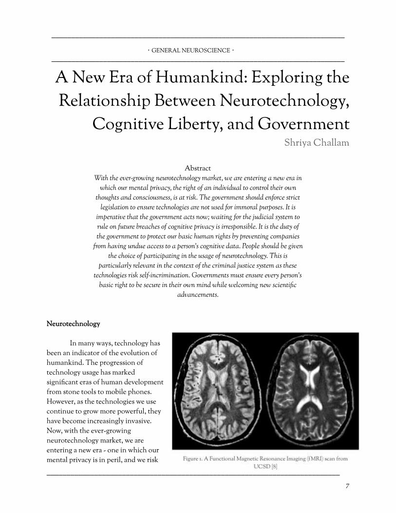
In many ways, technology has been an indicator of the evolution of humankind. The progression of technology usage has marked signi�cant eras of human development from stone tools to mobile phones. However, as the technologies we use continue to grow more powerful, they have become increasingly invasive. Now, with the ever-growing neurotechnology market, we are entering a new era - one in which our mental privacy is in peril, and we risk
–––––––––––––––––––––––––––––––––––––––––––––––––––––––––––––––––––––––––– 7

––––––––––––––––––––––––––––––––––––––––––––––––––––––––––––––––––––––––––
losing the cognitive liberties we take for granted.
Neurotechnology, technologies whose purpose is to interpret or alter neural function, can be used to vastly improve people’s lives. For example, deep brain stimulation treatments are used to alleviate symptoms of Parkinson’s Disorder and brain-computer interfaces have allowed individuals with ALS to communicate [1]. However, when these technologies are used to extract information, therefore possibly revealing unwanted thoughts, there is potential to invade the subject’s cognitive privacy. Function Magnetic Resonance Imaging (fMRI) tracks and records brain activity; by using a machine learning algorithm to compare patterns in the patient’s neural activity to a database of neural activity patterns, the machine can infer some aspects of the content such as lie detecting and quantifying characteristics [2].
There are many situations in which this can become extremely invasive - how will the
commercial availability of lie detectors a�ect our personal relationships? How will the ability to quantify character traits a�ect employment? Because of this potential for infringement of privacy, the government should enforce strict rules and regulations to ensure technologies are not used for immoral purposes.
Governmental Action
If the government does not set clear boundaries soon, we could see widespread
infringements of cognitive privacy. Social media and streaming companies, such as Facebook and Net�ix, already track our data to decipher which advertisements and recommendations cater to our preferences [5]. With the advent of neurotechnology, these companies could use our brain activity to harvest our innermost thoughts, providing a near-perfect method of marketing to the customer. At the moment, there is nothing stopping large companies from harnessing these technologies as they become more readily available. Even if customers consent to data collection, a majority of people are unaware of the range and potential uses of the information gathered about them. More troubling still is the risk that these companies will sell their customers’ data to a third party, as evidenced by the Facebook/Cambridge Analytica scandal in 2018, where the two companies used Facebook users’ data to create psychological pro�les of voters. This is why the government must step in to protect its citizens.
Some may argue that additional regulations are not necessary since the judiciary system
already protects our 4th Amendment right to privacy. However, this argument is misguided; although the 4th Amendment does ensure the right to privacy, neurotechnology opens the door to many new and unprecedented cases of privacy breaches. Rati�ed in 1791, the 4th Amendment was originally added to the constitution to protect citizens from unreasonable searches and seizures. The current threat neurotechnologies pose to privacy is far greater than in 1791, making the 4th Amendment outdated in many ways. Simply waiting for the judicial system to rule on future breaches of cognitive privacy is irresponsible; litigation is a slow process, and by the time these cases work their way up to the Supreme Court, millions of people might already have had their cognitive privacy violated. Furthermore, once a person’s cognitive data has been taken, the damage may be
–––––––––––––––––––––––––––––––––––––––––––––––––––––––––––––––––––––––––– 8

––––––––––––––––––––––––––––––––––––––––––––––––––––––––––––––––––––––––––
irreparable - data collection is not a two-way system, and that person’s information will have already been used in countless algorithms and machine-learning models. It is imperative then that the government set some fundamental restrictions to forbid certain abuses of people’s cognitive privacy. This will also establish a ground for precedents when later cases do come up.
The principle which mandates the government to actively protect its citizens’ privacy is well
established. Article 12 of the Universal Declaration of Human Rights by the United Nations states that “no one shall be subjected to arbitrary interference with [his or her] privacy” [3]. Therefore, it is the duty of the government to place restrictions on the application and commercial distribution of neurotechnology to prevent companies from having undue access to a person’s cognitive data. One could imagine an independent set of neurocognitive rights enshrined into law that would ensure that the sanctity of the mind is preserved.
Legislative Action
An essential aspect of this new set of neurocognitive legislation is the protection of a
person’s right to choose whether they want to use these technologies. This is particularly relevant in the context of the criminal justice system. The 5th Amendment states that a person will not “be compelled in any criminal case to be a witness against himself” - however, the use of neurotechnology could force a person to incriminate themselves [4]. Memory detection methods can give insight into the defendant’s prior knowledge of certain elements of a crime. According to Scienti�c American, in 2008, an Indian woman was convicted of murder and sentenced to life imprisonment on the basis of a brain scan; the judge claimed that the scan showed she had “experiential knowledge” about the crime [5]. Although such technologies are currently not used conclusively to make verdicts in the United States, as technologies used to detect lies, gauge the defendant’s familiarity with details of the crime, or diagnose psychological disorders become more prevalent and accurate, they may play much larger roles in the criminal justice system in the near future. All of these technologies giving access to a person’s mind could force them to be a witness against themselves. As such, the government must ensure that people are given the choice of participating in the usage of neurotechnology.
Implications
Neurotechnology
will revolutionize what society looks like, from the criminal justice system to the potential creation of superhumans, and the possibilities are only growing. A market report predicts that the worldwide market for
–––––––––––––––––––––––––––––––––––––––––––––––––––––––––––––––––––––––––– 9

––––––––––––––––––––––––––––––––––––––––––––––––––––––––––––––––––––––––––
neurotechnology is expected to cross $19 billion USD by the end of 2026 [6].
With this growth, it’s evident that neurotechnology is only going to become more present in our lives, and this calls for the government to address the urgent questions of neuroethics. Governments must ensure every person’s basic right to be secure in their own mind while welcoming the scienti�c advancements that continue to be made around neurotechnology as they bring about a new era of human life.
–––––––––––––––––––––––––––––––––––––––––––––––––––––––––––––––––––––––––––––––––––
–––––––––––––––––––––––––––––––––––––––––––––––––––––––––––––––––––––––––– 10
References
[1] Muller, Oliver & Rotter, Stefan. (13/12/2017). Neurotechnology: Current Developments and Ethical Issues. https://www.ncbi.nlm.nih.gov/pmc/articles/PMC5733340/. Retrieved: 17/07/2020.
{5] Ienca, Marcello. (01/10/2017). Preserving the Right to Cognitive Liberty. Scienti�c American. https://www.scienti�camerican.com/article/preserving-the-right-t o-cognitive-liberty/. Retrieved: 17/07/2020.
[2] Stanford Encyclopedia of Philosophy. (16/02/2016). Neuroethics. https://plato.stanford.edu/entries/neuroethics/. Retrieved: 17/07/2020.
[6] Watson, John. (09/03/2020). Neurotechnology Market to Reach USD 19 Billion By 2026 | Cisco Systems Inc., BMC Software Inc., ABB Limited, Dell Inc., Fujitsu Ltd. Medgadget Inc. https://www.medgadget.com/2020/03/neurotechnology-market-t o-reach-usd-19-billion-by-2026-cisco-systems-inc-bmc-software-i nc-abb-limited-dell-inc-fujitsu-ltd.html. Retrieved: 17/07/2020.
[3] United Nations General Assembly. (10/12/1948). Universal Declaration of Human Rights. Retrieved: 17/07/2020.
[7] Deep Knowledge Analytics Neurotech Division. (2019). NeuroTech Global Industry Landscape Overview 2019. http://analytics.dkv.global/data/pdf/NeuroTech/NeuroTech-Land scape-Overview-Teaser.pdf. Retrieved: 02/01/2021.
[4] U.S. Const. amend. V. Retrieved: 17/07/2020. [8] What is fMRI? https://cfmriweb.ucsd.edu/Research/whatisfmri.html. Retrieved 02/01/2021.

––––––––––––––––––––––––––––––––––––––––––––––––––––––––––––––––––––––––––
・ GENERAL NEUROSCIENCE ・
––––––––––––––––––––––––––––––––––––––––––––––––––––––––––––––––––––––––––
Religion, Spirituality, and the Mind: The History and Contributions Toward
Neurotheology
Suraj Sivaraja
Introduction Derived from the pre�x “Neuro” which refers to the nervous system and
“Theology” meaning “the study of religion and spiritual beliefs,”
Neurotheology studies the relationship(s) between the brain and our spiritual
beliefs. This �eld of study is interdisciplinary and relies on aspects of
philosophy, cognitive science, neuroscience, psychology, anthropology, and
more [1]. Neurotheology is so diverse that it is centralized in studying subjects,
including how certain types of information are stored in the brain, how
stimuli are localized in di�erent areas of the brain, and (most importantly)
what hormones and behavior patterns are produced by the nervous system as
a result of religious motivation [2]. The purpose of this article is to provide a
general understanding of how religion has played a major role in
neuroscience and the major contributions to neurotheology in history. Insight
on modern research, such as contrasts between the brain of atheists and
theists, will also be discussed.
The Major Religions Through History and Their In�uences on Science
Since the beginning of civilization, humans have been
involved in religious practices and rituals. Religion has not only enabled the mind to promote new forms of thoughts and beliefs but has also motivated our species to expand knowledge in both an academic and spiritual manner. Throughout history, the world has seen di�erent aspects of religious and spiritual beliefs playing a key role in everything from agriculture to warfare and even medical sciences. For instance, the religion of Hinduism in the Indus Valley conducted rituals and used plants to cure many psychological disorders [3].
–––––––––––––––––––––––––––––––––––––––––––––––––––––––––––––––––––––––––– 11

––––––––––––––––––––––––––––––––––––––––––––––––––––––––––––––––––––––––––
Correspondingly, many other civilizations developed their rituals and medicinal practices to combat diseases and disorders in all forms. By the �fth century, Christianity practically ruled Europe, causing the Church and its followers to question science. In the year 1053, the Roman Catholic Church and the Eastern Orthodox Church formed as a result of the Great Schism [4]. From the 16th to 17th centuries, Europe and many other parts of the world began to enter a stage in history known as the Scienti�c revolution [5]. This was the age when scientists started to devise counter-arguments against the classic greek view of the world, providing a new meaning to science. During these years, Christianity had begun to spread all across the world, and by the 19th century - when major medical advances were being made - Christianity and Islam had a major in�uence in a majority of countries on the continents of Eurasia and Africa [6]. The 20th and 21st centuries had a massive in�uence on the way people looked at the world. These times brought about the apex in scienti�c and technological research, making many people lose trust in their religious leaders and communities. This caused a decrease in the number of people a�liated with religious groups; however, over 80% of the world’s population still believes in the existence of god and other spiritual beings [5][7].
A Brief Overview of the Origin and History of Neurotheology
The ideas that founded general Neuroscience from the 19th to 20th centuries were also responsible for bringing about the dawn of neurotheology. Neurotheology originates from two strands of science (along with a few others): ethology, the study of animal behavior, and neurophysiology, the study of the functioning of the nervous system [2].
–––––––––––––––––––––––––––––––––––––––––––––––––––––––––––––––––––––––––– 12

––––––––––––––––––––––––––––––––––––––––––––––––––––––––––––––––––––––––––
I. Ethology During the 19th century, scientists began to increase their knowledge of how animals function and behave, resting their basic knowledge o� of Charles Darwin’s Theory of Evolution. As the study itself began to evolve, scientists were becoming more interested in how animals reacted in certain lab simulated conditions, giving birth to a new branch of science known as Ethology. The science was founded by Karl von Frisch, Konrad Lorenz, and Nikolaas Tinbergen. They were also the key �gures who worked together to establish the fundamentals of ethology. The research conducted by these scientists helped give rise to more specialized �elds including neuroethology [2].
II. Neurophysiology and Related Sciences
Neurophysiology’s founding fathers are John C. Eccles, Alan L. Hodgkin, Andrew F. Huxley, and Charles S. Sherrington, all of whom worked to expose the ionic mechanisms in the neural membrane and proved that inhibition as a coordinating way of thinking in the nervous system [9]. As neurophysiologists began to study behavioral stimuli and cognitive functions of the brain, major topics of interest such as electroencephalography, electromyography, and neuroplasticity recently motivated emerging scientists to integrate studies of both neurophysiology and behavioral neuroscience [2]. Along with neurophysiology, neuropsychology and neurophilosophy were major contributors to the study of neurotheology. Neuropsychology is the collective term used for the study of how the brain and the nervous system comprehend and produce cognitive and behavioral functions. Among the many scientists involved in neuropsychological research, Ralph Reitan is thought to be a major �gure in the world of neuropsychology. He has published over 320 scienti�c papers and has played a major role in addiction and brain dysfunction research [8]. Neurophilosophy is the study of how relevant philosophical theories are in a neuroscienti�c context. Similar to Reitan, in 1986, Patricia Church helped build the fundamental stepping stones for neurophilosophy by publishing a book, o�cially coining the term [9].
The dawn of Neurotheology did not occur until a neuroscience-avid theologian by the name
of James Ashbrook began to study how the brain reacts to and comprehends religious and spiritual beliefs such as supernatural beings. Ashbrook was also responsible for coining the term “neurotheology” when referring to this unique new study [10].
Major Contributions to Modern Neurotheology: The Works of Andrew Newberg
Dr. Andrew Newberg is a neuroscientist at Thomas Je�erson University and Hospital’s
Marcus Institute of Integrative Health. He is also known to be the pioneer of neurotheology and has written 10 books on his views on the relationship between religion and the brain [11]. Newberg and his team have used Single Photon Emission Computed Tomography (SPECT) to measure the volume of blood �ow circulating throughout the brain. By using SPECT scans, neuroscientists can distinguish which parts of the brain are being used during certain activities. Newberg and his team
–––––––––––––––––––––––––––––––––––––––––––––––––––––––––––––––––––––––––– 13

––––––––––––––––––––––––––––––––––––––––––––––––––––––––––––––––––––––––––
used this form of neuroimaging to study the brain of people in di�erent religious groups. When examining the brain of an active Tibetian Buddhist’s brain and the brain of a meditating Buddhist, Newberg discovered that there was reduced activity in the parietal lobe, especially in the area that is responsible for comprehending orientation [11]. This proved that Tibetian Buddhists are capable of block sensory and cognitive input in certain parts of the brain during meditation [11].
In addition to this �nding, as shown in �gure 3, the Buddhists also have increased activity in
the attention center located in the frontal lobe. Newberg has also conducted studies on the brains of atheists (as shown in �gure 4) and has presented �ndings to support that the atheist mind has less of an ability to focus attention on certain things for an established period. For instance, he and his team examined the neural activity in the mind of an active atheist and the mind of a meditating atheist, only to discover that there was a decrease in attention span during the period of meditation [11]. Dr.Newberg not only made many major discoveries in neurotheology but has also been featured in the news, the Dr. Oz Show, and many other forms of media platforms to share his unique �ndings with the rest of the world [11].
Newberg, Waldman, and How God Changes Your Brain
Mark Waldman is a therapist and a faculty member of the Executive MBA program at Loyola Marymount University in Los Angeles, where he teaches brain-based techniques to improve leadership and communication. His research on spirituality, meditation, awareness, and brains has been published in many journals around the world and has appeared on PBS TV, Canadian National Television, and National Public Radio to aid in coaching. Perhaps a notable contribution by Waldman in this �eld of research is a partnered publication written with Newberg, How God
Changes Your Brain [12]. This collaboration is a major �gure that manifests great knowledge acquired by both authors. The major subject that is explored in this book is how the concept of god manipulates negative emotional tension such as stress, anxiety, and depression [13].
Similar to Newberg’s ideologies behind improved neural activity within the brain as a result
of meditation, the two neuroscientists elaborate by proposing a great decrease in emotional tension
–––––––––––––––––––––––––––––––––––––––––––––––––––––––––––––––––––––––––– 14
––––––––––––––––––––––––––––––––––––––––––––––––––––––––––––––––––––––––––
as a bene�t of devotional practices such as praying, ultimately even reducing the aging process [13]. Additionally, the authors were able to identify that fundamentalism is a phenomenon that is engraved within many individuals, evoking extreme behaviors, such as anger and hatred, towards other social groups which can ultimately damage the function of certain behaviourally stimulated neural circuits. In contrast to this harsh reality, the two say that prayer and religious attachment has the potential to manipulate the views and perceptions of how the surrounding world functions [13].
Why Is This Field of Study So Important?
The concept of God and spiritual beings play a large role in how humans function
individually and as a collective species. As the world excels, it is important to conduct research and understand the neurological and psychological impact that religion and spiritual beliefs have on society. As stated earlier in the passage, over 80% of the world’s population still believes in the existence of god and other spiritual beings [7]. Recently, graduates and well-established scholars have shown great interest in studying this new �eld of neuroscience, and every day scientists are being able to answer some of the most important questions regarding the psychological impact of religion. There have also been many recent breakthroughs in neurotheology such as identifying the foundational beliefs of Jewish peoples based on the teachings of the Torah and other scriptures have helped shape the general scope and provide a de�nition for this unique science [11]. In the near future, it is expected that the study of neurotheology will become a major branch of neuroscience and play a key role in answering some of the most pressing questions about how our brain responds to hormonal and behavior stimuli induced by our perceptions of religion, maybe even in�uencing the way humans view the religious ideology altogether [2].
––––––––––––––––––––––––––––––––––––––––––––––––––––––––––––––––––––––––––––––––––– References
–––––––––––––––––––––––––––––––––––––––––––––––––––––––––––––––––––––––––– 15
{1} Sayadmansour, Alireza. (2014). “Neurotheology: The Relationship between Brain and Religion.” Iranian Journal of
Neurology , Tehran University of Medical Sciences, www.ncbi.nlm.nih.gov/pmc/articles/PMC3968360/ . Retrieved: 20/08/2020.
[8] Grant, Igor, and Robert K Heaton. (12/2015) “Ralph M. Reitan: A Founding Father of Neuropsychology.” Archives of Clinical
Neuropsychology: the O�cial Journal of the National Academy of
Neuropsychologists , Oxford University Press, www.ncbi.nlm.nih.gov/pmc/articles/PMC4675827/ . Retrieved: 21/08/2020.
[2] Ewert, Jörg-Peter. (01/01/1970 ). “Neuroethology.” SpringerLink , Springer, Dordrecht, link.springer.com/referenceworkentry/10.1007/978-1-4020-8265-8_76 9. Retrieved: 21/08/2020.
[9] Bickle, John, et al. (06/08/2019) “The Philosophy of Neuroscience.” Stanford Encyclopedia of Philosophy , Stanford University, plato.stanford.edu/entries/neuroscience/. Retrieved: 21/08/2020.
[3] “OVERVIEW OF INDIAN HEALING TRADITIONS: History and Science of Indian Systems of Knowledge.”, NCBI,
www.ncbs.res.in/HistoryScienceSociety/content/overview-indian- healing-traditions . Retrieved: 20/08/2020.
[10] Shukla, Samarth, et al. (07/2013) “Neurotheology-Matters of the Mind or Matters That Mind?” Journal of Clinical and
Diagnostic Research: JCDR , JCDR Research and Publications (P) Limited, www.ncbi.nlm.nih.gov/pmc/articles/PMC3749673/ . Retrieved: 21/08/2020.
[4] Cunningham, Lawrence, and Francis Christopher Oakley. (15/05/2020). “Roman Catholicism.” Encyclopædia Britannica , Encyclopædia Britannica, Inc.,
[11] Newberg, Andrew. Andrew Newberg, www.andrewnewberg.com/ . Retrieved: 21/08/2020.

––––––––––––––––––––––––––––––––––––––––––––––––––––––––––––––––––––––––––
–––––––––––––––––––––––––––––––––––––––––––––––––––––––––––––––––––––––––– 16
www.britannica.com/topic/Roman-Catholicism#ref257668 . Retrieved: 20/08/2020.
[5] Osler, Margaret J., and Stephen G. Brush. (26/11/2019). “Scienti�c Revolution.” Encyclopædia Britannica , Encyclopædia Britannica, Inc., www.britannica.com/science/Scienti�c-Revolution . Retrieved: 20/08/2019.
[12] “Andrew Newberg, M.D. and Mark Waldman | Psychology Today Canada.” Psychology Today , Sussex Publishers, www.psychologytoday.com/ca/experts/andrew-newberg-md-and- mark-waldman. Retrieved: 16/11/2020.
[6] Stefon, Matt, and Henry Chadwick. (20/09/2019) “Christianity and World Religions.” Encyclopædia Britannica , Encyclopædia Britannica, Inc., www.britannica.com/topic/Christianity/Christianity-and-world-re ligions . Retrieved: 21/08/2020.
[13] Newberg, Andrew, and Mark R. Waldman. “How God Changes Your Brain by Andrew Newberg, M.D., Mark Robert Waldman.” PenguinRandomhouse.com , Ballantine Group, www.penguinrandomhouse.com/books/120938/how-god-changes -your-brain-by-andrew-newberg-md-and-mark-robert-waldman/9 780345503428/. Retrieved: 16/11/2020.
{7} Hackett, Conrad, and David McClendon. (31/05/2020). “World's Largest Religion by Population Is Still Christianity.” Pew Research Center, www.pewresearch.org/fact-tank/2017/04/05/christians-remain-worl ds-largest-religious-group-but-they-are-declining-in-europe/ . Retrieved: 20/08/2020.

––––––––––––––––––––––––––––––––––––––––––––––––––––––––––––––––––––––––––
・ GENERAL NEUROSCIENCE ・
––––––––––––––––––––––––––––––––––––––––––––––––––––––––––––––––––––––––––
Exploring the Ethics of Nootropics From an Adolescent Perspective
Harish Rajan
Introduction In our academically competitive world, some students take arti�cial
stimulants to improve their cognitive abilities in order to ultimately improve
their grades. According to the FDA, nootropics are still a gray area [1].
Although they are not fully approved, many of them are available as
supplements and foods. This article analyses the ethics of consuming
nootropics by students.
What Are Nootropics?
The word “nootropics" �nds its origin in the Greek words nous , meaning mind, and tropein, meaning turning. Nootropics are supplements that can enhance cognitive performance and memory. They do so by altering the levels of neurotransmitters in the brain to improve mental alertness, focus, and boost energy levels. Some are synthetic compounds such as piracetam, and others are natural substances such as the common Chinese ingredient ginseng. The lack of evidence on the long-term e�ects of these nootropics on the brain has contributed to a glaring absence in the regulation of nootropics consumption. In contrast, the intake of anabolic steroids to increase muscle mass and strength is banned by many professional sports leagues, the International Olympic Committee (IOC), and the National Collegiate Athletic Association (NCAA). According to the National Institute on Drug Abuse, it appears that the abuse of Adderall to enhance brain memory and cognitive performance is reported to be at least 11.1% among college students [2].
Cinematic In�uences
Many Hollywood movies have been produced related to the topic of nootropics. “Limitless" discussed the pill “Moda�nil", which helped Bradley improve his cognitive acuity [3]. Another �lm “Lucy", claimed that human brains operate at only 10% capacity, and showed the main character, Scarlett, taking “CPH4" to activate the other 90% [4]. It is surreal to believe that with smart pills, one would need to employ only a fraction of normal e�orts and beat the rest. These kinds of unproven, false claims can attract innocent students who want to excel in their studies towards the use of nootropics.
–––––––––––––––––––––––––––––––––––––––––––––––––––––––––––––––––––––––––– 17

––––––––––––––––––––––––––––––––––––––––––––––––––––––––––––––––––––––––––
Natural vs. Synthetic
Drinking co�ee to receive bene�ts from the natural brain-boosting ca�eine supplements, which helps circulate cortisol and adrenaline, is common among the global population. Tea has the amino acid L-Theanine, which helps to produce alpha brain waves, associated with the state of relaxation and improved mental alertness and arousal. These are all beverages consumed by the common man without always having an awareness of the
additional compounds that can supercharge productivity. Lecithin, another fat substance found naturally in soybean and egg yolk, has been shown to improve brain capacity. Because these substances are legal and widely available, the ethics of their consumption does not arise as a topic of conversation. Adding nitrates via celery concentrate to meat, as a preservative to extend shelf life, enables the food industry to label that food product as "natural". On the other hand, if synthetic sodium nitrates are added, that food product is no longer labeled “natural”. The �nal result and the human impact of the two are essentially the same. These ambiguous guidelines mirror those seen regarding the regulation of nootropics and make the question of ethics complicated.
Prescription Drugs
One study showed that 5 to 35% of students at colleges had used Adderall without a
prescription to improve their cognitive abilities at some point [5]. Suppose doctors prescribe these drugs to treat students with neuropsychiatric disorders like ADHD and anxiety, is it unethical for non-prescribed students to take the same pills to excel in their academics? The answer is similar to that of any medically unnecessary drug. Taking these pills without a prescription often leads to other health issues. These stimulants can raise blood pressure and constrict the blood vessels to cause cardiovascular problems [6].
Contemporary research leaves the role of nootropics with open questions. Given the long
list of side e�ects, including memory loss, dizziness, depression, and anxiety, these drugs' illegal use is clearly problematic. However, nootropics do cure many psychological disorders. Francis Fukuyama, in his book “Our Posthuman Future: Consequences of the Biotechnology Revolution",
–––––––––––––––––––––––––––––––––––––––––––––––––––––––––––––––––––––––––– 18

––––––––––––––––––––––––––––––––––––––––––––––––––––––––––––––––––––––––––
wrote: “The original purpose of medicine is to heal the sick, not turn healthy people into gods". Banning these drugs outright for therapeutic purposes would not be right, but stricter regulation and criteria for a prescription could mitigate abuse outside their legitimate medical purpose.
Professional Sports
A parallel and e�ectively managed problem can be explored in the world of professional
sports. Not long ago, the tennis star Maria Sharapova was banned by ITF for consuming non-prescribed Meldonium under the Tennis Anti-Doping Program. Although the same Meldonium is used to treat ischemia, a condition where there is a lack of blood �ow because of heart failure, the o�ender was using it for athletic advancement [7]. If these types of steroids were not strictly regulated in professional sports, it would be hard for the viewers to di�erentiate between real ability and drug-enabled success. They violate the spirit of the game by giving an unfair advantage to the contestants. The World Anti-Doping Agency (WADA) took the right step to regulate these stimulants strictly. It is justi�ed to apply the same thinking to academic institutions for nootropics, preventing these drugs from threatening academic integrity.
Of course, no academic institution should allow the abuse of smart pills or bio-hacks on
campus. Unlike in professional athletics, where athletes can regularly test for drugs, it is not reasonable to expect the schools and colleges to test for the use of these substances. It becomes the students' ethical and moral responsibility to abstain from the use of synthetic nootropics. In a practical sense, however, that does not seem to be working out. A paper in The Journal of Clinical Psychiatry highlighted that Adderall misuse is highest among 18 to 25-year-olds who get medication without a doctor's prescription using friends or family help [8].
Conclusion: More Questions Than Answers
The obstacles seen in enforcing a level academic playing �eld among the students present
many questions related to the use of neuroethics. Where is the line for making these drugs hard enough for a healthy person to get, but easy enough for someone who needs them for medical reasons? Exploring the abuse of nootropics in young minds is both fascinating and frightening. The ethics of use, access, regulation, and enforcement are essential to keep at the top of the mind as we strive for greater equality and safety in modern-day academic settings.
–––––––––––––––––––––––––––––––––––––––––––––––––––––––––––––––––––––––––––––––––––
–––––––––––––––––––––––––––––––––––––––––––––––––––––––––––––––––––––––––– 19
References
[1] (09/10/2018). “Which dietary supplements ingredients are on FDA’s radar?”. Nutritional Outlook Vol. 21 No. 7, Volume 21, Issue 7. https://www.nutritionaloutlook.com/view/which-dietary-supple ments-ingredients-are-fdas-radar. Retrieved: 01/05/2021.
[6] Amy Laskowski. (05/05/2008) “The Other Side of Adderall". Boston University. http://www.bu.edu/articles/2008/the-other-side-of-adderall. Retrieved: 11/09/2020.
––––––––––––––––––––––––––––––––––––––––––––––––––––––––––––––––––––––––––
–––––––––––––––––––––––––––––––––––––––––––––––––––––––––––––––––––––––––– 20
[2] (09/13/2019). “Drug and Alcohol Use in College-Age Adults in 2019”. National Institute on Drug Abuse; National Institutes of Health; U.S. Department of Health and Human Services . https://www.drugabuse.gov/drug-topics/trends-statistics/infograp hics/drug-alcohol-use-in-college-age-adults-in-2018. Retrieved: 01/05/2021.
[7] Mitchell, Libby. (03/08/2016). “What is Meldonium?”. University of Utah Health. https://healthcare.utah.edu/healthfeed/postings/2016/03/sharapo va.meldonium.php. Retrieved: 06/14/2020.
[3] (07/08/2019). “The all-too-understandable urge to buy a better brain”. Kaitlyn Ti�any, Vox . https://www.vox.com/the-goods/2019/7/8/18772467/nootropics-sil icon-valley-brain-�tness-goop-smart-drugs. Retrieved: 06/24/2020.
[8] (02/16/2016). “Adderall abuse on the rise among young adults, Johns Hopkins study suggests”. Hub: Johns Hopkins University. https://hub.jhu.edu/2016/02/16/adderall-abuse-rising-young-adult s/. Retrieved: 06/17/2020.
[4] (07/25/2014). “Movie Review: 'Lucy' Starring Scarlett Johansson and Morgan Freeman”. David Blaustein, abc news . https://abcnews.go.com/Entertainment/movie-review-lucy-starri ng-scarlett-johansson-morgan-freeman/story?id=24715951. Retrieved: 01/05/2021.
[9] (02/09/2018). “What a Lifetime of Adderall Does to Your Brain". Ian Lecklitner. https://medium.com/mel-magazine/what-a-lifetime-of-adderall-d oes-to-your-brain-5beba7c2af7e. Retrieved: 01/05/2021.
[5] Levin, Elea. “Easy Access, Pressure on Students Contributes to Increase in Non-Prescribed Adderall Use. Department of Psychology”. https://psych.wisc.edu/news/easy-access-pressure-on-students-co ntributes-to-increase-in-non-prescribed-adderall-use/. Retrieved: 06/24/2020.

––––––––––––––––––––––––––––––––––––––––––––––––––––––––––––––––––––––––––
・ GENERAL NEUROSCIENCE ・
––––––––––––––––––––––––––––––––––––––––––––––––––––––––––––––––––––––––––
The Dynamic Mind: An Overview of Neuroplasticity
Shrika Vejandla
Introduction Neuroplasticity, a phenomenon describing the brain's capacity to change and
adapt, refers to the morphological changes in the brain that occur due to an
individual’s interactions with the environment. Throughout the course of
one’s life, the brain develops synapses and circuits between neurons that
reorganize to respond to an individual’s adapting needs. This process allows
one to learn and adapt to new experiences. Neuroplasticity is also vital in
higher cognitive functioning, including processes relevant to memory and
learning. This indicates neuroplasticity can act as the basis for cognitive and
physical rehabilitation practices that work to rebuild connections among
neurons [1]. This article provides an overview of the background and
neurobiology of neuroplasticity, as well as its applications.
A Brief History
The term “neuroplasticity” was �rst coined in 1948 by Polish neuroscientist Jerzy Konorski to describe changes in neuronal structure. However, this term was not widely used until the 1960s [2]. Until the 1960s, researchers took part in the notion that changes in the brain could only occur during infancy and childhood [2]. They believed that the brain’s state was mostly permanent by early adulthood [3].
In the 1920s, Karl Lashley found evidence of changes in the neural pathways of rhesus
monkeys [3]. By the 1960s, researchers began to explore cases in which older adults who had su�ered strokes could regain functioning, demonstrating that the brain was a lot more malleable than they had previously believed [3].
Due to modern advances in technology, researchers can get a look at the brain's intricate
inner mechanisms. As the study of contemporary neuroscience �ourished, researchers were able to conclusively demonstrate that people are not limited to the mental abilities they are born with, and that those facing damage to their brains are indeed capable of remarkable change.
Synaptic Plasticity
–––––––––––––––––––––––––––––––––––––––––––––––––––––––––––––––––––––––––– 21

––––––––––––––––––––––––––––––––––––––––––––––––––––––––––––––––––––––––––
The brain establishes a series of neural pathways when it is engaged in new experiences and
learning. These neural pathways, known as circuits, are routes made of interconnecting neurons. These routes form in the brain typically through daily emphasis and practice. The neurons within a neural pathway communicate with each other through synapses, and these communication pathways have the profound ability to regenerate throughout one’s life. Each time we gain new knowledge, experience, or exposure to a given thing, through repeated practice, the synaptic communication between the relevant circuit of neurons is strengthened [4]. Strengthened connections indicate that the electrical signals between neurons travel more e�ciently when using a new pathway.
For instance, when a birdwatcher tries to recognize a bird, relationships are being established
between neurons. Neurons located in the visual cortex determine its color, the auditory cortex identi�es its tune, and the hippocampus (responsible for memory) recalls the name of the bird from these observations [4]. Revisiting this neural circuit and re-establishing neuronal transmission between the implicated neurons at each new attempt is what enhances the e�ciency of synaptic transmission [4]. Communication between the relevant neurons is then facilitated, improving the speed of the related cognitive function [4]. Thus, synaptic plasticity is likely the pillar in which the brain’s malleability resides.
Neuroplasticity Adaptations
There exist four main types of neuroplasticity adaptations. Long-term potentiation
describes the strengthening of synapses through recurring activities such as studying information or practicing motor skills [5]. This type of neuroplasticity is strongly associated with learning and memory. On the other hand, long-term depression describes the weakening of synapses that are not being used in a coordinated, well-timed manner. This typically occurs when synapses �red by neurons are not �red within 20ms in a coordinated way between the presynaptic and postsynaptic terminals [5]. In addition, neuroplasticity research has studied long-term depression’s role in memory loss for neurological disorders such as Alzheimer’s Disease [5]. Synaptogenesis describes the creation of new neural connections, as described in the overview of synaptic plasticity [5]. This occurs when the brain is exposed to new environments and experiences.
Unlike synaptic plasticity, which
enhances communication at the synaptic sites between existing neurons, neurogenesis is the
–––––––––––––––––––––––––––––––––––––––––––––––––––––––––––––––––––––––––– 22

––––––––––––––––––––––––––––––––––––––––––––––––––––––––––––––––––––––––––
creation of new neurons in regions of the brain such as the hippocampus (memory formation) and olfactory bulb (odor input). Neurogenesis occurs at high rates in the young brain and can continue, albeit minimally, into adulthood [5].
Neurogenesis occurs when stem cells located in the dentate gyrus of the hippocampus and
possibly in the prefrontal cortex, divide into two separate cells: a stem cell, and a cell that will form into a neuron. The newly formed neurons migrate to regions of the brain where they are needed, and thus have the ability to allow the brain to replenish its own supply of neurons. Animal and human research has shown that sudden neuronal death (for example after stroke) is a trigger for neurogenesis [4].
Types of Experiences
There exist two types of plasticity that shape the developing brain. Experience-independent
plasticity describes everything that occurs within the brain during the prenatal developmental phase. This is when neuronal connections and brain formation are processes driven by complex genetic instructions. During this phase, neurons that �re together make some structures stronger and parts of the brain more prominent than others, whereas those that do not coordinate will die out. For instance, a lack of visual stimuli in the critical stages subsequent to birth may lead to impediments in the processing of input from the visual cortex, possibly resulting in eye disorders such as amblyopia (where both eyes are unable to align and function in unison) [7]. This shows a lack of experience-independent plasticity [7].
Experience-dependent plasticity helps neurons form synapses independent of other
processes that may be occurring in the brain [7]. For example, the formation of the retinal ganglion of the eyes consists of axons coming from the retina, which are initially sending branches for both eyes but gradually form their own neurons for each branch. The axons of each pathway coming from the retina �re synapses that eventually create neural circuits independent of those in the other eye. Experience-dependent plasticity is often seen in a�ected brain morphology when di�erent situations occur, such as moving to a new region, learning di�cult math problems, or su�ering from injury. These daily challenges either increase or decrease synapses, and shape the morphology of the brain while they are functioning [8].
–––––––––––––––––––––––––––––––––––––––––––––––––––––––––––––––––––––––––– 23
––––––––––––––––––––––––––––––––––––––––––––––––––––––––––––––––––––––––––
Applications
There are a multitude of applications of neuroplasticity within the clinical context, which
have both yielded results and shown potential in serving as strong interventions [9].
For example, transcranial magnetic stimulation (TMS) is an application that employs an extracranial magnetic coil to induce current in the cerebral cortex. Continuous e�ects of low frequency (<1 Hz) repetitive transcranial magnetic stimulation or theta burst transcranial magnetic stimulation lead to the suppression of cortical excitability in healthy subjects, while intermittent, high frequency (>1 Hz) e�ects of repetitive TMS leads to facilitation [9]. In addition, transcranial direct current stimulation (tDCS) also uses two scalp electrodes to induce low-amplitude direct currents strong enough to penetrate the brain and modify membrane potentials, which in�uences neuronal excitability without the active depolarization of neurons [9]. Both techniques can produce e�ects that last beyond the period of stimulation, insinuating the induction of plasticity due to the in�uence upon neuronal excitability.
In addition, deep brain stimulation (DBS) uses electrical stimulation to induce
neuroplasticity and produce behavioural changes through implanted electrodes. Two hypothesized mechanisms of action are that DBS creates a functional lesion via inhibition within the stimulated region and, and also that DBS activates the neuronal network connected to the stimulated region, which leads to the modulation of pathological network activity [9]. The former mechanism is consistent with the immediate e�ects of some applications of DBS (such as the e�ects on motor function in Parkinson's disease) [9]. The latter is likely to be more consistent with gradual e�ects that are induced by DBS as opposed to immediate ones (such as circuit retraining), which are also seen in neuropsychiatric disorders [9]. The most notable of these disorders include treatment-resistant depression (TRD) and treatment-refractory obsessive-compulsive disorder (OCD).
Directions for future research in clinical applications may include tailoring plasticity-based
therapies based on individual patient measurements such as the distribution of disease [9]. Therapies under study to promote neuroplasticity have been examined one at a time, however with greater understanding these therapies can be expedited by being examined in combination (such as task-speci�c plasticity training coupled with stem cell therapy) [9]. A better understanding of treatment mechanisms at every level as well as the underlying neurobiology of neuroplasticity will improve the development of both preventive and therapeutic interventions.
Closing Words
Neuroplasticity allows for rehabilitation techniques to foster improved functional outcomes
in age-related neurological conditions. Thus, the brain’s malleability can be manipulated in both the healthy and diseased brain, and using its ability to create and lay down new pathways can play a large role in rehabilitation as well as the quality of life. It is clear that the human understanding of the nature of brain development has advanced a long way in the past century, though we have begun
–––––––––––––––––––––––––––––––––––––––––––––––––––––––––––––––––––––––––– 24

––––––––––––––––––––––––––––––––––––––––––––––––––––––––––––––––––––––––––
to understand the contributing factors and mechanisms that regulate this development. Understanding these mechanisms is necessary in �nding treatments for neurodevelopmental disorders to initiate early intervention that will reverse the otherwise anticipated pathology.
–––––––––––––––––––––––––––––––––––––––––––––––––––––––––––––––––––––––––––––––––––
–––––––––––––––––––––––––––––––––––––––––––––––––––––––––––––––––––––––––– 25
References
[1] Keci, Andromeda. (25/02/2009). Role of Rehabilitation in Neural Plasticity. Macedonian Journal of Medical Sciences. https://www.ncbi.nlm.nih.gov/pmc/articles/PMC6542405/. Retrieved: 30/11/2020.
[6] Biga, Lindsay. (2019). Anatomy and Physiology: 1st Edition Oregon State University. https://open.oregonstate.education/aandp/chapter/12-4-communi cation-between-neurons/. Retrieved: 30/11/2020.
[2] Zieliński, Kazimierz. (2006). Jerzy Konorski on Brain Associations. Acta Neurobiologiae Experimentalis. https://pubmed.ncbi.nlm.nih.gov/16617679/. Retrieved: 27/20/2020.
{7] Hensch, Takao. (16/07/2018). Critical Periods in Amblyopia. Visual Neuroscience. https://www.ncbi.nlm.nih.gov/pmc/articles/PMC6047524/. Retrieved: 31/10/2020.
[3] Nadine, Weidman. (1999). Constructing Scienti�c Psychology: Karl Lashley's Mind-Brain Debates. Cambridge University Press. https://muse.jhu.edu/article/4690. Retrieved: 27/10/2020.
{8] Iuculano, Teresa. (30/09/2015). Cognitive Tutoring Induces Widespread Neuroplasticity and Remediates Brain Function in Children with Mathematical Learning Disabilities. Nature Communications. https://www.nature.com/articles/ncomms9453. Retrieved: 30/11/2020.
[4] Kolb, Bryan. (2011). Searching for Factors Underlying Cerebral Plasticity in the Normal and Injured Brain. The Journal of Communication Disorders. https://pubmed.ncbi.nlm.nih.gov/21621219/. Retrieved: 08/08/2020.
{9] Cramer, Steven. (11/04/2011). Harnessing Neuroplasticity for Clinical Applications. Brain: A Journal of Neurology. https://www.ncbi.nlm.nih.gov/pmc/articles/PMC3102236/#B211. Retrieved: 31/10/2020.
[5] Gillick, Bernadette. (01/10/2012). Neuroplasticity: An Appreciation From Synapse to System. The Archives of Physical Medicine and Rehabilitation. https://www.emotiv.com/glossary/neuroplasticity/. Retrieved: 31/10/2020.

––––––––––––––––––––––––––––––––––––––––––––––––––––––––––––––––––––––––––
・ DISEASE AND DISORDERS ・
––––––––––––––––––––––––––––––––––––––––––––––––––––––––––––––––––––––––––
The Potential E�ects of Pandemic-Induced Isolation
Nora Mehler
Abstract As a result of the COVID-19 pandemic, many of our everyday social
interactions have become virtual. The replacement of face-to-face
communications with a remote equivalent, combined with general social
distancing measures, could be harmful to people’s mental and physical
well-being. Studies have shown that most people prefer in-person to online
relationships, while chronic loneliness can increase one’s risk for
in�ammatory illnesses. With no end to the pandemic in sight, these facts
must be considered so as to minimize the negative mental consequences of
long-term social distancing procedures.
Modern-Day Loneliness
Even though nearly a year has passed since the beginning of the coronavirus pandemic, it is still impossible to have social gatherings and interactions as we did before. This situation a�ects everyone di�erently, but the lack of face-to-face interactions and interpersonal closeness is taking a toll. In the United Kingdom, 24% of adults and 44% of people aged eighteen to twenty-four reported feeling lonely when surveyed in April, compared to the 10% and 16%, respectively, who reported those feelings in a survey prior to the lockdown period [5]. Although restrictions have been eased since the beginning of the shutdown, the general theme remains the same. Life everywhere has been impacted by the pandemic, with the most drastic changes being in the realm of social interactions.
Importance of Social Interactions
By nature, humans are social creatures, reliant on interactions with one another for the
maintenance of social and emotional well-being. Loneliness and social isolation, especially when sustained for long periods of time, are detrimental to our mental health as they are directly linked to depression and hastened mental decline [1]. In fact, a study found that, when shown pictures of pleasant objects, lonely individuals show greater activation of the ventral striatum, which is involved in feelings of reward and the release of dopamine. On the other hand, non-lonely people had stronger activation of the ventral striatum when they saw pictures of pleasant people [7]. A lack of dopamine release resulting from a lack of pleasant social stimulation can have detrimental long-term
–––––––––––––––––––––––––––––––––––––––––––––––––––––––––––––––––––––––––– 26

––––––––––––––––––––––––––––––––––––––––––––––––––––––––––––––––––––––––––
e�ects in terms of both mood and neurological function, as dopamine is strongly associated not only with feelings of happiness and reward but also with motor control. For young children, interactions with peers is essential in developing language and social skills [2]. The elderly are also at risk; in fact, people who believe they are socially isolated were found to have higher levels of infection-�ghting myeloid cells, which can lead to an increased risk of chronic disease [3]. Not only that, but long-term perceived loneliness can alter the way in which people perceive stimulation, as can be seen in a study using fMRI imaging led by J. T. Cacioppo. In this study, researchers found that there was more activation in the visual cortex of lonely individuals than non-lonely individuals when they were shown pictures of unpleasant social situations, which suggests that lonely individuals pay more attention to negative stimuli in social situations and therefore experience social interactions in such a way that is di�erent from non-lonely people [7]. Because loneliness can a�ect the brain at a biological level and alters the brain’s responses to stimuli, the long-term loss of normal and ful�lling social interactions has the potential to negatively a�ect an entire generation of children and adolescents who require social interactions for growth and development.
Online Versus In-Person Interactions
Of course, the vast majority of people are able to remain connected to their friends and family throughout the pandemic with texts, calls, and FaceTime calls. In terms of schooling, teachers and students are able to see one another and interact over platforms such as Zoom. But is that enough? A 2010 study of digital versus in-person relationships, published by Oregon State University, revealed that those who interacted in-person felt a greater connection to their partner and had more positive interactions [6]. Since 2010, our reliance on technology has only grown and has become even more signi�cant in light of the COVID-19 crisis. However, signi�cant daily screen-time also has a negative impact on mental function. When the blue light from screens o�sets the circadian rhythm and leads to a decrease in
melatonin levels at night, REM sleep is interrupted. REM sleep is necessary for transforming new information into memories because it is a period of heightened brain activity, and therefore important in processing and ultimately remembering information taught in school [8]. For over half a year, technology has been the primary method of extended social interaction. However, if
–––––––––––––––––––––––––––––––––––––––––––––––––––––––––––––––––––––––––– 27

––––––––––––––––––––––––––––––––––––––––––––––––––––––––––––––––––––––––––
in-person interactions are more valuable in terms of minimizing loneliness, everyone should be concerned about the collective mental health of our nation.
In Terms of the Pandemic
In general, people value face-to-face interactions, as they allow for unobstructed
communication and connection. The restrictions resulting from the COVID-19 pandemic have created massive changes in what those interactions look like. With the e�ects of chronic loneliness and social isolation in mind, it is extremely important to consider the long-term consequences of our remote lives in the event that we have many months before a safe, e�ective vaccine is available. It will be extremely interesting to see what, if any, changes are there in reported loneliness and correlating illnesses as the course of the pandemic and social distancing continues into the foreseeable future. For example, is the e�ect of loneliness on dopamine levels enough to put lonely individuals at a greater risk for diseases such as Parkinson’s Disease, which are theorized to be caused by a lack of dopamine? The psychological impact of social distancing is very important because loneliness can a�ect the biochemistry of the brain, which ultimately impacts mental function as a whole.
–––––––––––––––––––––––––––––––––––––––––––––––––––––––––––––––––––––––––––––––––––
–––––––––––––––––––––––––––––––––––––––––––––––––––––––––––––––––––––––––– 28
References
[1] Novotney, Amy. (05/2019). The risks of social isolation. American Psychological Association. https://www.apa.org/monitor/2019/05/ce-corner-isolation. Retrieved: 09/17/2020.
[5] Loneliness during coronavirus. Mental Health Foundation. (11/09/2020). https://www.mentalhealth.org.uk/loneliness-during-coronavirus. Retrieved: 09/17/2020.
[2] Coplan, Robert J. and Kimberly A. Arbeau. (2009). Peer Interactions and Play in Early Childhood. Handbook of Peer Interactions, Relationships, and Groups. 143–161. Retrieved: 09/17/2020.
[6] Okdie, Bradley M. et al. (2011). Getting to know you: Face-to-face versus online interactions. Computers in Human
Behavior . 153–159. https://liberalarts.oregonstate.edu/sites/liberalarts.oregonstate.ed u/�les/psychology/research/okdie_guadagno_bernieri_geers_mclarn ey-vesotski_2011.pdf. Retrieved: 09/17/2020.
[3] Cole, Steven W. et al. (08/12/2015). Myeloid di�erentiation architecture of leukocyte transcriptome dynamics in perceived social isolation. https://www.pnas.org/content/112/49/15142. Retrieved: 09/17/2020.
[7] Cacioppo, Stephanie et al. (11/30/2016). Toward a neurology of loneliness. https://www.ncbi.nlm.nih.gov/pmc/articles/PMC5130107/. Retrieved: 11/18/2020.
[4] Functional MRI. (05/03/2016). https://www.ptb.de/cms/en/ptb/fachabteilungen/abt8/�-81/ag-81 2/fmri-812.html. Retrieved: 12/27/2020.
[8] Tähkämö, L., Partonen, T., & Pesonen, A. K. (02/2019). Systematic review of light exposure impact on human circadian rhythm. Chronobiology international , 36 (2), 151–170. https://doi.org/10.1080/07420528.2018.1527773. Retrieved: 12/27/2020.

––––––––––––––––––––––––––––––––––––––––––––––––––––––––––––––––––––––––––
・ DISEASE AND DISORDERS ・
––––––––––––––––––––––––––––––––––––––––––––––––––––––––––––––––––––––––––
Models and Biomarkers of Multiple Sclerosis
Shamsudeen Suleiman
Abstract Multiple Sclerosis (MS) is a progressive demyelinating disease that is
characterised by demyelination, perivascular in�ammation, oligodendrocyte
depletion, astroglia proliferation and remyelination. Generally, the aetiology
of MS is still unclear, although many environmental factors and the
interaction of multiple genes have been proposed to play a role in the disease.
MS is usually diagnosed in people between the ages of 20 and 40, and is
more common in females than in males (almost 3:1). There are several
established experimental demyelination models that, to some extent, re�ect
the heterogeneity of MS and are therefore seen as suitable to study MS
pathogenesis [1]. These models include immune-mediated, virus-induced and
toxin-induced models. Biomarkers are crucial for evaluating and assessing
the normal biological, pathogenic, and pharmacological response to
therapeutic interventions. Knowledge of the molecular basis of this disease
through various models and biomarkers is essential to ascertaining and
pinpointing the pathway and in turn, developing drugs that can help treat or
ameliorate this disease, improving the quality of life of patients.
Models of Multiple Sclerosis (MS)
There are several established experimental demyelination models that, to a certain extent, re�ect the heterogeneity of MS and are therefore seen as �tting to study MS pathogenesis. These models include immune-mediated, virus-induced, and toxin-induced models. Experimental autoimmune encephalitis (EAE) is, by far, the most explored model for studying various aspects of autoimmunity in MS pathology. Virus-induced demyelination models support the hypothesis that some environmental factors, such as viral infections, are involved in MS and may be a trigger for the disease. Toxin-induced demyelination models are exploited in the evaluation of the demyelination/remyelination process in the relative absence of immune cells, even though these ways of damaging the myelin do not bear a resemblance to features of the demyelination seen in MS [1][2].
Toxin-Induced Demyelination Models
–––––––––––––––––––––––––––––––––––––––––––––––––––––––––––––––––––––––––– 29
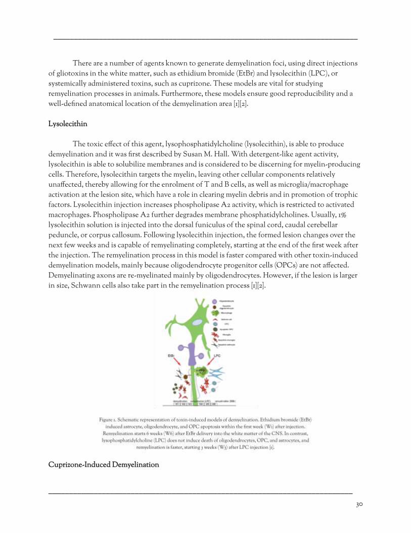
––––––––––––––––––––––––––––––––––––––––––––––––––––––––––––––––––––––––––
There are a number of agents known to generate demyelination foci, using direct injections
of gliotoxins in the white matter, such as ethidium bromide (EtBr) and lysolecithin (LPC), or systemically administered toxins, such as cuprizone. These models are vital for studying remyelination processes in animals. Furthermore, these models ensure good reproducibility and a well-de�ned anatomical location of the demyelination area [1][2].
Lysolecithin
The toxic e�ect of this agent, lysophosphatidylcholine (lysolecithin), is able to produce
demyelination and it was �rst described by Susan M. Hall. With detergent-like agent activity, lysolecithin is able to solubilize membranes and is considered to be discerning for myelin-producing cells. Therefore, lysolecithin targets the myelin, leaving other cellular components relatively una�ected, thereby allowing for the enrolment of T and B cells, as well as microglia/macrophage activation at the lesion site, which have a role in clearing myelin debris and in promotion of trophic factors. Lysolecithin injection increases phospholipase A2 activity, which is restricted to activated macrophages. Phospholipase A2 further degrades membrane phosphatidylcholines. Usually, 1% lysolecithin solution is injected into the dorsal funiculus of the spinal cord, caudal cerebellar peduncle, or corpus callosum. Following lysolecithin injection, the formed lesion changes over the next few weeks and is capable of remyelinating completely, starting at the end of the �rst week after the injection. The remyelination process in this model is faster compared with other toxin-induced demyelination models, mainly because oligodendrocyte progenitor cells (OPCs) are not a�ected. Demyelinating axons are re-myelinated mainly by oligodendrocytes. However, if the lesion is larger in size, Schwann cells also take part in the remyelination process [1][2].
Cuprizone-Induced Demyelination
–––––––––––––––––––––––––––––––––––––––––––––––––––––––––––––––––––––––––– 30

––––––––––––––––––––––––––––––––––––––––––––––––––––––––––––––––––––––––––
Figure 2 depicts a model of demyelination and remyelination that has been utilised to tease apart the speci�c mechanisms that contribute towards oligodendrocyte death, oligodendrocyte precursor cell (OPC) migration, di�erentiation, and remyelination. Cuprizone-induced demyelination was �rst described in the late 1960s using Swiss mice. Protocols now widely use C57BL/6 mice. Nonetheless, methods have also been developed for rats, which are particularly useful for imaging studies due to their larger brain size. Cuprizone causes detailed oligodendrocyte death and successive demyelination; however, the mechanism is not fully known. It is a copper ion (Cu2+) chelator disturbing cellular metabolism. Mega-mitochondria are observed in the liver of mice following cuprizone intoxication. The presence of these mega-mitochondria indicates a disturbance in metabolic tasks that ultimately results in oligodendrocyte death. The dosage of cuprizone needed is dependent on the strain, age and sex of the mouse. Cuprizone is mixed with the feed at a concentration of 0.2%–0.6% (w/w) for 6 weeks to induce extensive demyelination throughout the brain, of which the corpus callosum is the most widely studied. Intoxication in SJL mice results in a di�erent demyelinating pattern than that seen in C57BL/6 mice. This is further in�uenced by gender. Mice present with a time dependent weight loss and important behavioural and motor de�ciencies [2][3].
Biomarkers of Multiple Sclerosis
In 1998, the National Institutes of Health (NIH) defined biomarkers as “a characteristic that
is objectively measured and evaluated as an indicator of normal biological processes, pathogenic processes, or pharmacologic responses to therapeutic intervention.” The World Health Organization (WHO), the United Nations, and the International Labour Organization jointly defined a biomarker as “any substance, structure, or process that can be measured in the body or its products and influence or predict the incidence of outcome or disease.” This detailed definition included e�ects of treatments, interventions, and environmental exposures, such as chemicals or nutrients [4][5][6].
Classi�cations of Biomarkers in MS
–––––––––––––––––––––––––––––––––––––––––––––––––––––––––––––––––––––––––– 31

––––––––––––––––––––––––––––––––––––––––––––––––––––––––––––––––––––––––––
Biomarkers are characterized for reasons of systematic approach, and according to their pathophysiological implication in MS pathogenesis.
1. Genetic-Immunogenetic Biomarkers
The TOB-1 gene has a role in opposition to T-cell multiplication, keeping autoreactive cells
in a latent state. Its decreased expression leads to a more intense immune response (higher percentage of Th1 and Th17 cells and a lower percentage of T-regulatory cells). TOB-1 polymorphisms represent an independent factor in�uencing the progression from clinically isolated syndrome (CIS) to clinically de�nite multiple sclerosis (CDMS) [6].
2. Laboratorial Biomarkers
Biomarker of Neuroprotection
Vitamin D plays a potential pathogenic role in MS and can be seen in previous
epidemiological studies that showed correlation between latitude and sun exposure with relative risk for developing the disease. It suppresses Th1 immune response in multiple levels and enables the production of many neurotrophic factors. 25-Hydroxy vitamin D levels in untreated MS patients inversely correlate with radiologic disease activity. Recently, a vitamin response element (VDRE) was recognized close to the HLA-DRB1∗1501 coding area, with the help of genomics. Vitamin D exhibits an inhibitory role in MS, also at a genetic level, by interacting with VDRE [6].
Demyelination Biomarkers
Myelin Basic Protein (MBP) and its fragments are found in great quantities in the
Cerebrospinal �uid (CSF) of most MS patients during a relapse (80%). A signi�cant correlation of decrease in CSF-MBP, contrast-enhancement in MRI, and clinical disability in response to treatment with methylprednisolone suggests a relationship between in�ammation and myelin breakdown in MS [6].
Biomarkers of Glial Activation Dysfunction
Glial Fibrillary Acidic Protein (GFAP) is a structural protein of the astrocytes whose CSF
levels increase in association with gliosis-astrocytosis. High CSF values have been found in Secondary Progressive Multiple sclerosis (SPMS) patients, but rarely in Relapsing Remitting Multiple sclerosis (RRMS) patients, and seem to correlate well with disability progression. CSF-GFAP levels are signi�cantly higher during Neuromyelitis Optica (NMO) relapse, in comparison with MS relapse, and show adequate connection with clinical improvement and disability progression in NMO [6].
Biomarkers of Remyelination Repair
–––––––––––––––––––––––––––––––––––––––––––––––––––––––––––––––––––––––––– 32

––––––––––––––––––––––––––––––––––––––––––––––––––––––––––––––––––––––––––
An example is the Brain-Derived Neurotrophic Factor (BDNF), studies have shown that there exists a Lower CSF-BDNF level in SPMS patients in relation to RRMS patients. These Low BDNF levels are considered to contribute in demyelination and axonal damage progress. An increased BDNF production was observed in Glatiramer Acetate responders, correlating well with clinical improvement [4][6].
Summary
Multiple sclerosis (MS) is a whimsical disease that a�ects the central nervous system,
disrupting the �ow of information within the brain and between the brain and the body. Understanding the mechanism underlying this disease is important for researchers and science communicators to develop therapeutic remedies, thereby e�ectively disseminating their �ndings to the public.
–––––––––––––––––––––––––––––––––––––––––––––––––––––––––––––––––––––––––––––––––––
–––––––––––––––––––––––––––––––––––––––––––––––––––––––––––––––––––––––––– 33
References
[1] Bjelobaba, I., Begovic-Kupresanin, V., Pekovic, S., and Lavrnja, I. (2018). Animal models of multiple sclerosis: Focus on experimental autoimmune encephalomyelitis. Journal of
Neuroscience Research , 96 (6), 1021–1042. https://doi.org/10.1002/jnr.24224. Retrieved: 15/02/2018.
[4] Jelčić, I., and Martin, R. (2010). Biomarkers in Multiple Sclerosis. Blue Books of Neurology , 35 (C), 120–146. https://doi.org/10.1016/B978-1-4160-6068-0.00006-1. Retrieved: 25/12/2009.
[2] J. van der Star, B., Y.S. Vogel, D., Kipp, M., Puentes, F., Baker, D., and Amor, S. (2012). In Vitro and In Vivo Models of Multiple Sclerosis. CNS & Neurological Disorders - Drug Targets , 11 (5), 570–588. https://doi.org/10.2174/187152712801661284. Retrieved: 11/08/2012.
[5] KatrinPaap, B. (2013). Molecular Biomarkers in Multiple Sclerosis. Journal of Clinical & Cellular Immunology , 01 (S10). https://doi.org/10.4172/2155-9899.s10-009. Retrieved: 25/02/2013.
[3] Burrows, D. J., McGown, A., Jain, S. A., De Felice, M., Ramesh, T. M., Sharrack, B., and Majid, A. (2019). Animal models of multiple sclerosis: From rodents to zebra�sh. Multiple Sclerosis
Journal , 25 (3), 306–324. https://doi.org/10.1177/1352458518805246. Retrieved: 15/10/2018.
[6] Waschbisch, A., Atiya, M., Schaub, C., Derfuss, T., Schwab, S., Lee, D. H., … Linker, R. A. (2013). Aquaporin-4 antibody negative recurrent isolated optic neuritis: Clinical evidence for disease heterogeneity. Journal of the Neurological Sciences , 331 (1–2), 72–75. https://doi.org/10.1016/j.jns.2013.05.012. Retrieved: 02/06/2013.

––––––––––––––––––––––––––––––––––––––––––––––––––––––––––––––––––––––––––
・ DISEASE AND DISORDERS ・
––––––––––––––––––––––––––––––––––––––––––––––––––––––––––––––––––––––––––
Capgras Syndrome: An Ethical Review Divya Venkataraman
Abstract
Imagine waking up one morning to the smell of fresh baked cookies from
your loving mother. Now, imagine seeing your mother, but thinking that she
is an imposter, someone who looks exactly like your mother, but isn’t. “My
parents looked funny the other day… I think they’ve been replaced by
imposters,” Janet, a patient of Capgras Syndrome said [1]. Needless to say,
this syndrome is surrounded by numerous ethical controversies. This article
explores the causes and symptoms of Capgras Syndrome and the possible
treatments that have been proposed, to then review the ethical and societal
concerns behind this peculiar syndrome.
Etiology and Symptomatology
Many patients of Capgras Syndrome have numerous areas of damage in their brain. However, scientists like Dr. VS Ramachandran have realized that the damage in Capgras patients isn’t usually in the speci�c parts of the brain responsible for face recognition (like the fusiform gyrus), but instead in the connections between those areas (e.g. fusiform gyrus to the amygdala) [2]. In fact, Dr. Ramachandran states that the connection between the fusiform gyrus and amygdala must have been damaged, since the connection allows humans to associate speci�c emotions to the face recognized (e.g. love for mother). However, because this connection is severed in Capgras patients, as displayed in Figure 1, they can recognize the face as their mother’s, but cannot sense any
–––––––––––––––––––––––––––––––––––––––––––––––––––––––––––––––––––––––––– 34

––––––––––––––––––––––––––––––––––––––––––––––––––––––––––––––––––––––––––
emotional connection to their mother. To justify this lack of emotion, their brain concludes that their mother is an imposter [3].
As mentioned above, the most astonishing symptom of Capgras Syndrome is the mind
believing that all loved ones are imposters. However, patients also tend to think that pets, distant people, or even objects are imposters. This recognition of people and things is so strong that no recorded amount of evidence can tell any patient otherwise. It was thought that this peculiar syndrome stems from violence, but recent research has suggested that this likely originates from other neurological conditions like Alzheimer’s disease [2].
Treatment
Unfortunately, there aren’t many treatments for this disease. Some drugs used to treat dementia such as donepezil, rivastigmine, and galantamine have helped with the syndrome by reducing symptoms. Additionally, antipsychotic medications like olanzapine and pimozide can ease delusions. Antidepressants have also helped in some cases [2].
However, the main treatment used is therapy. The �rst type is habilitation therapy, which is when loved ones try to understand what the patient is going through and be in the patients’ shoes. This allows loved ones to empathize with the patient more, which in turn prevents harsh interactions between the two. Habilitation therapy also says that one should never argue with or correct a Capgras syndrome patient, especially since the patient believes the loved one to be an imposter, and arguing will only emphasize this even more. Finally, the last part of Habilitation therapy is the idea of letting the patient know that you are there for them after understanding what they are going through. However hard this may be, a loved one’s empathy will suppress some of the hatred that patients feel. [6] The second type is validation therapy, which gives the patient a sense of safety. However, this is only used if the patient had previously thought that the imposter was dangerous. Finally, there is family counseling, which has helped in a few reported cases, by reducing the anxiety and fear of the patients [2].
Ethical and Social Implications
The most obvious issue is that many people don’t know that Capgras Syndrome exists. To allow this syndrome to reach the medical attention it deserves, more awareness needs to be spread about this psychiatric delusion and how to help [3].
Unfortunately, society tends to disregard diseases that are seen as delusions, and Capgras Syndrome is a victim of this unjust societal and ethical law. Many people and doctors who haven’t seen someone with Capgras Syndrome, which is likely much of the population,
–––––––––––––––––––––––––––––––––––––––––––––––––––––––––––––––––––––––––– 35
––––––––––––––––––––––––––––––––––––––––––––––––––––––––––––––––––––––––––
categorize Capgras Syndrome as ‘delusional’. This in turn is detrimental to Capgras Syndrome’s progress in society. Delusional diseases like Capgras Syndrome are considered to be treated by only psychotherapeutic methods. Although this has been proven wrong time and time again, since the syndrome doesn’t have much societal awareness, this maintains to be on the ethical radar [4]. Due to this, there is a societal issue surrounding patients with Capgras Syndrome. Because of the rarity of the disease, those who have it tend to be ridiculed or ignored. Not many people are educated about this syndrome and tend to categorize the patient as “crazy”.
The last ethical issue surrounds treatments. Since there aren’t many treatments available, and the ones that are there are multi-purposed (initially meant for something else), many have a hard time believing that Capgras Syndrome needs to be taken seriously [4]. They believe that if scientists aren’t �nding treatments for it, it is not worth their attention. What they fail to understand is that scientists are working on treatments, but due to a relatively decreased understanding of Capgras Syndrome, treatments are hard to �nd. This explains the necessity of spreading awareness of Capgras Syndrome.
Discussion
Capgras Syndrome, contrary to popular belief, is a serious disorder that needs more recognition. Patients not only su�er from the medical symptoms of this syndrome, but the consequences due to the ethical and societal issues as well. Yet, the syndrome is not getting the attention it deserves [5]. Due to a lack of emotional ability, kids see their parents as an imposter, out to get them. Adults are unable to love their kids, seeing them as imposters. To a patient, all of their loved ones, who usually help and support them, have been taken over by imposters. This syndrome can be very detrimental, and needs more awareness across the globe.
There are also many ways through which society can help this issue. To begin, people can spread the word about this syndrome by telling someone about it. One person can make a huge di�erence in the progression of the disease. Also, promote the syndrome rather than putting it down. Capgras Syndrome already has a negative stigma in the community, and society should actively be trying to obliviate that pessimism, one person at a time .
–––––––––––––––––––––––––––––––––––––––––––––––––––––––––––––––––––––––––––––––––––
–––––––––––––––––––––––––––––––––––––––––––––––––––––––––––––––––––––––––– 36
References
[1] Carol W. Berman, M. D. (04/09/2011). Ever Thought Your Loved
One Was An Impostor? Understanding Capgras Syndrome . https://www.hu�post.com/entry/capgras-syndrome_b_888854. Retrieved: 04/07/2020.
[4] BCA-clinic, & BCA-lab. (01/11/2019). Why Morgellons Disease Is
Still Not Taken Seriously... And Why It Really Should Be - BCA-Clinic . https://www.bca-clinic.com/why-morgellons-disease-is-still-not-t aken-seriously-and-why-it-really-should-be/. Retrieved: 09/07/2020.
[2] Bhandari, S. (06/01/2020). Capgras Syndrome (Impostor Syndrome):
Symptoms, Treatment & More . WebMD. https://www.webmd.com/mental-health/impostor-syndrome-cap gras. Retrieved: 04/07/2020.
[5] RJ;, B. (08/1983). Capgras' syndrome . https://pubmed.ncbi.nlm.nih.gov/6869616/. Retrieved: 12/07/2020.

––––––––––––––––––––––––––––––––––––––––––––––––––––––––––––––––––––––––––
–––––––––––––––––––––––––––––––––––––––––––––––––––––––––––––––––––––––––– 37
[3] Ramachandran, V. S. (03/2007). Transcript of "3 clues to
understanding your brain" . TED. https://www.ted.com/talks/vs_ramachandran_3_clues_to_understan ding_your_brain/transcript. Retrieved: 05/07/2020.
[6] Bier, D. (08/10/2018). Dementia and Capgras Syndrome: Handling Behavior and Emotional Fallout. Psych Central. https://psychcentral.com/lib/dementia-and-capgras-syndrome-ha ndling-behavior-and-emotional-fallout/. Retrieved: 14/12/2020.
––––––––––––––––––––––––––––––––––––––––––––––––––––––––––––––––––––––––––
・ DISEASE AND DISORDERS ・
––––––––––––––––––––––––––––––––––––––––––––––––––––––––––––––––––––––––––
Brain-Derived Neurotrophic Factor (BDNF) and Apolipoprotein (APOE): Impacts on Alzheimer’s Disease (AD)
Katherine Wei
Abstract The purpose of this review is to understand how the downregulation of
brain-derived neurotrophic factor (BDNF) may elevate the accumulation of
beta-amyloid protein and impact the pathology of Alzheimer’s Disease (AD).
This review also identi�es possible treatments that can increase BDNF levels
to prevent cognitive impairment and the relationship between BDNF and
other AD-related genes like apolipoprotein (APOE). Single nucleotide
polymorphisms (SNPs) in the BDNF gene may lead to its loss-of-function and
subsequent deteriorating e�ects on cognition. The lack of BDNF expression
has a damaging e�ect because of its bene�cial neurotrophic supply,
prevention of beta-amyloid production, and inhibition of beta-amyloid’s
neurotoxicity. When genotyping di�erent AD patients, a correlation between
BDNF, APOE, and AD were found. The combined treatment of
cerebrolysin and donepezil can be used in AD patients to increase BDNF
levels and improve cognition. There is a promising connection between
BDNF and carriers of the E4 allele of APOE, as increasing BDNF levels
will increase neurotrophic supply, which could help with preventing AD.
Introduction
Alzheimer’s Disease (AD) was discovered in 1906 when Dr. Alois Alzheimer recognized the unusual brain tissue of one of his female patients, Auguste Deter, who exhibited delusional qualities and cognitive/memory impairment [1]. Even though AD was the cause of 121,404 deaths in 2017 and is the 6th leading cause of death in the U.S., the amount of AD research is small compared to cancer research and other diseases [2]. The symptoms that we associate with AD now are very similar to Deter’s: memory loss, confusion, di�culty completing familiar tasks, and inability to understand basic images and words [3]. Current researchers have discovered that the accumulation of proteins like beta-amyloid and tau in neural tissue has been the root cause of the disease, but exactly how they are formed and what exact genes play into AD pathology is still unknown. There are many genes associated with AD such as the amyloid precursor protein (APP), apolipoprotein (APOE), and phospholipase D3 (PLD3) [1]. One particular gene called the brain-derived neurotrophic factor
–––––––––––––––––––––––––––––––––––––––––––––––––––––––––––––––––––––––––– 38

––––––––––––––––––––––––––––––––––––––––––––––––––––––––––––––––––––––––––
( BDNF ) gene has shown promise alongside the APOE for potential avenues of further exploration of AD pathology.
APOE Alleles
Studies conducted with pluripotent stem cells and clustered regularly interspaced short
palindromic repeats (CRISPR) have allowed us to get more insight, showcasing a strong correlation between some genes and late-onset AD [4]. For instance, the apolipoprotein (APOE) gene, which functions in the transport of brain cholesterol and promotion of lipoprotein clearance from circulation, has been heavily studied recently. It has three alleles: E2, E3, and E4. The E2 allele is considered protective and has a worldwide frequency of 4.2%, while E3 is the most common allele with a frequency of 77.9%. Finally, the E4 allele has been found to be the strongest risk factor associated with AD with a 13.7% worldwide frequency but a ~40% frequency with patients who have AD. Since identifying the risk of the development of AD-related to APOE4, researchers have studied a lot about the gene’s function, structure, and sequence. All three alleles have one or two di�erent amino acid substitutions [5]. Between E2 and E3, out of the 299 amino acids, they have a single amino acid di�erence at 158 where E3 has arginine while E2 has cysteine. Between E3 and E4, the amino acid di�erence lies in position 112 where E3 has cysteine and E4 has arginine. Between E2 and E4, there is a double amino acid di�erence in 158 and 112 where E2 has cysteine in both locations, while E4 has arginine [5]. Infants that carry the APOE4 gene have been found to have less gray matter than normal infants. Less gray matter typically translates to limited communication between neurons and other cells [6].
BDNF’s Role in APOE
Recently, there has been interest in identifying factors related to the APOE gene to further
understand risk factors for AD. One study found that the brain-derived neurotrophic factor (BDNF) gene was signi�cantly downregulated in APOE4 genotypes that expressed increased AD pathology. Using cDNA sequence, northern blot analysis, and in situ hybridization, BDNF mRNA had the highest concentration in the hippocampus, followed by the cerebral cortex [7]. Despite being heavily concentrated in those areas, BDNF is widespread across the nervous system, found in the spinal cord, superior colliculus, primary sensory neurons, and retinal ganglion cells. When analyzing the adult mouse, the BDNF gene was most prevalent in the central nervous system. Additionally, when BDNF levels were compared to nerve growth factor (NGF), another neurotrophic factor, BDNF mRNA showed signi�cantly higher levels of expression, despite BDNF and NGF mRNA’s striking similarities in regional and cellular localization [7]. There was a strong association between a SNP, the E6K or the Glu6Lys, in the BDNF gene which may have caused the loss-of-function. However, additional research should be performed to support this �nding [4]. The downregulation of the BDNF gene worsened the AD pathology. Thus, it can be inferred that increasing the BDNF levels could alleviate some of the symptoms and cognitive decline associated with AD.
–––––––––––––––––––––––––––––––––––––––––––––––––––––––––––––––––––––––––– 39

––––––––––––––––––––––––––––––––––––––––––––––––––––––––––––––––––––––––––
A possible reason why the downregulation of BDNF exacerbates AD pathology is because BDNF’s neurotrophic supply improves cognition in patients. This means that lower BDNF levels will thus lead to lower cognition. Another possibility is how BDNF prevents the generation of beta-amyloid, the protein that builds up the brain causing AD, through an innate process called the non-amyloidogenic pathway [8]. It is also possible that BDNF inhibits and lessens the neurotoxicity of beta-amyloid thus attenuating AD pathology like the previous possibility [9]. BDNF de�ciency is caused by a variety of factors, such as metal dyshomeostasis, shortage in NGF support, malfunctional Aβ monomers, and toxic Aβ oligomers. These factors could all attribute to AD pathology [10].
Yu-Hui Liu et al. performed another study to determine the relationship between APOE4
and BDNF levels under the assumption that they are both important factors related to the development of AD [11]. In this experiment, 120 normal patients and 110 patients with AD were examined for their BDNF levels and the APOE4 gene. By using restriction fragment length polymorphism (RFLP) to genotype ApoE and enzyme-linked immunosorbent assay (ELISA) to detect the serum BDNF levels, they found that those carrying one or more copies of the APOE4 gene had the lowest BDNF levels compared to those without any copies of the APOE4 gene and the normal patients. Additionally, there was no signi�cant di�erence in BDNF levels between those who carried one or two copies of APOE4 [11]. The association between APOE4, BDNF, and AD was further explored through a series of tests and statistical analyses. Through the use of a regression model where APOE E4 was the independent variable and AD was the dependent variable, an association between the two factors (AD and carrying APOE E4) was found. The second analysis used BDNF as the independent variable which indicated a relationship between BDNF and APOE E4 carriers with AD. Using di�erent tests on AD patients, the researchers found that patients with AD tended to score lower on the mini-mental state examinations (MMSE). The MMSE is a test used to evaluate cognitive function by analyzing aspects such as memory, attention, and orientation. AD patients also had higher clinical dementia rating (CDR), higher activities of daily living (ADL), and higher prevention of disability scores (POD). These di�erent tests were then used in a univariate general linear model which showed that carrying the APOE E4 gene tended to a�ect ADL scores but not CDR and POD. However, the interaction between APOE E4 carriers and BDNF levels were further analyzed and found to a�ect MMSE scores. This interaction implies the possibility of APOE regulating BDNF which may correlate with the overall development of AD [11]. BDNF supplies the brain with neurotrophic support, and consequently, a de�ciency in BDNF will directly negatively impact brain integrity. The �nal conclusion reached was that APOE4 may impact BDNF metabolism, but an experiment with a larger sample size needs to be conducted in order to con�rm this hypothesis.
Increasing BDNF Levels as Treatment
After recognizing that low BDNF levels are correlated to a higher risk of AD, researchers
then began e�orts to increase BDNF levels in patients. Anton Alvarez et al. performed an experiment comparing the use of cerebrolysin, donepezil, and combined therapy with both cerebrolysin and donepezil on patients carrying the APOE4 gene. Their goal was to determine if
–––––––––––––––––––––––––––––––––––––––––––––––––––––––––––––––––––––––––– 40
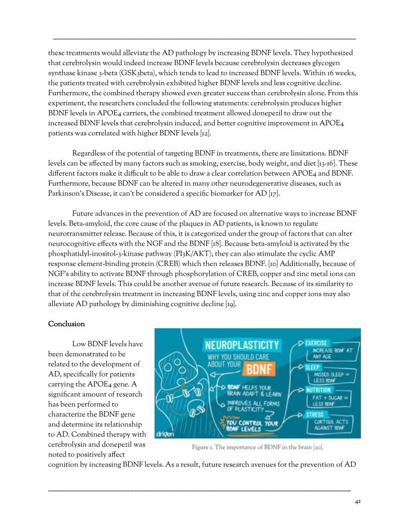
––––––––––––––––––––––––––––––––––––––––––––––––––––––––––––––––––––––––––
these treatments would alleviate the AD pathology by increasing BDNF levels. They hypothesized that cerebrolysin would indeed increase BDNF levels because cerebrolysin decreases glycogen synthase kinase 3-beta (GSK3beta), which tends to lead to increased BDNF levels. Within 16 weeks, the patients treated with cerebrolysin exhibited higher BDNF levels and less cognitive decline. Furthermore, the combined therapy showed even greater success than cerebrolysin alone. From this experiment, the researchers concluded the following statements: cerebrolysin produces higher BDNF levels in APOE4 carriers, the combined treatment allowed donepezil to draw out the increased BDNF levels that cerebrolysin induced, and better cognitive improvement in APOE4 patients was correlated with higher BDNF levels [12].
Regardless of the potential of targeting BDNF in treatments, there are limitations. BDNF
levels can be a�ected by many factors such as smoking, exercise, body weight, and diet [13-16]. These di�erent factors make it di�cult to be able to draw a clear correlation between APOE4 and BDNF. Furthermore, because BDNF can be altered in many other neurodegenerative diseases, such as Parkinson’s Disease, it can't be considered a speci�c biomarker for AD [17].
Future advances in the prevention of AD are focused on alternative ways to increase BDNF
levels. Beta-amyloid, the core cause of the plaques in AD patients, is known to regulate neurotransmitter release. Because of this, it is categorized under the group of factors that can alter neurocognitive e�ects with the NGF and the BDNF [18]. Because beta-amyloid is activated by the phosphatidyl-inositol-3-kinase pathway (PI3K/AKT) , they can also stimulate the cyclic AMP response element-binding protein (CREB) which then releases BDNF. [10] Additionally, because of NGF’s ability to activate BDNF through phosphorylation of CREB, copper and zinc metal ions can increase BDNF levels. This could be another avenue of future research. Because of its similarity to that of the cerebrolysin treatment in increasing BDNF levels, using zinc and copper ions may also alleviate AD pathology by diminishing cognitive decline [19].
Conclusion
Low BDNF levels have
been demonstrated to be related to the development of AD, speci�cally for patients carrying the APOE4 gene. A signi�cant amount of research has been performed to characterize the BDNF gene and determine its relationship to AD. Combined therapy with cerebrolysin and donepezil was noted to positively a�ect cognition by increasing BDNF levels. As a result, future research avenues for the prevention of AD
–––––––––––––––––––––––––––––––––––––––––––––––––––––––––––––––––––––––––– 41

––––––––––––––––––––––––––––––––––––––––––––––––––––––––––––––––––––––––––
are focused on additional ways to increase BDNF levels. As of now, the bene�ts of BDNF are highlighted by its ability to strengthen di�erent aspects of brain function.
––––––––––––––––––––––––––––––––––––––––––––––––––––––––––––––––––––––––––––––––––– References
–––––––––––––––––––––––––––––––––––––––––––––––––––––––––––––––––––––––––– 42
[1] National Institute on Aging. (2003). “Alzheimer's Disease Fact Sheet.” PsycEXTRA Dataset . https://www.nia.nih.gov/health/alzheimers-disease-fact-sheet. Retrieved: 06/23/2020.
[11] Yu-Hui Liu, Shu-Sheng Jiao, Ye-Ran Wang, Xian-Le Bu, Xiu-Qing Yao, Yang Xiang, Qing-Hua Wang, Lin Wang, Juan Deng, Jing Li, Xin-Fu Zhou, Hua-Dong Zhou & Yan-Jiang Wang (2015). Associations Between ApoEε4 Carrier Status and Serum BDNF Levels—New Insights into the Molecular Mechanism of ApoEε4 Actions in Alzheimer’s Disease. Mol Neurobiol 51, 1271–1277. https://doi.org/10.1007/s12035-014-8804-8 . Retrieved: 06/26/2020.
[2] Kenneth D. Kochanek, M.A., Sherry L. Murphy, B.S., Jiaquan Xu, M.D., and Elizabeth Arias, (2019). “Deaths: Final Data for 2017.” National Vital Statistics Reports , vol. 68. www.cdc.gov/nchs/data/nvsr/nvsr68/nvsr68_09-508.pdf . Retrieved: 06/23/2020.
[12] X. Anton Alvarez, Irene Alvarez, Olalla Iglesias, Ignacio Crespo, Jesus Figueroa, Manuel Aleixandre, Carlos Linares, Elias Granizo, Manuel Garcia-Fantini, Jose Marey, Eliezer Masliah, Stefan Winter, Da�n Muresanu, Herbert Moessler (2016). Synergistic Increase of Serum BDNF in Alzheimer Patients Treated with Cerebrolysin and Donepezil: Association with Cognitive Improvement in ApoE4 Cases, International Journal of
Neuropsychopharmacology , 19(6). https://doi.org/10.1093/ijnp/pyw024 . Retrieved: 06/26/2020.
[3] Constantine G. Lyketsos , Maria C. Carrillo , J. Michael Ryan , Ara S. Khachaturian , Paula Trzepacz , Joan Amatniek , Jesse Cedarbaum , Robert Brashear , David S. Miller (2011). “Neuropsychiatric Symptoms in Alzheimer's Disease.” Alzheimer's
Association , 7(5), 532-9. https://doi.org/10.1016/j.jalz.2011.05.2410 . Retrieved: 06/24/2020.
[13] Tae-Suk Kim, Dai-Jin Kima, Heejin Lee, Yong-Ku Kim (2007). “Increased Plasma Brain-Derived Neurotrophic Factor Levels in Chronic Smokers Following Unaided Smoking Cessation.” Neuroscience Letters , 423(1), 53-7. https://doi.org/10.1016/j.neulet.2007.05.064 . Retrieved: 06/26/2020.
[4] Yuan-Ta Lin, Jinsoo Seo, Fan Gao, Tak Ko, Bruce A. Yankner Li-Huei Tsai . (2018). APOE4 Causes Widespread Molecular and Cellular Alterations Associated with Alzheimer's Disease Phenotypes in Human iPSC-Derived Brain Cell Types. Neuron , 98 (6), 1141–1154.e7. https://doi.org/10.1016/j.neuron.2018.05.008 . Retrieved: 06/24/2020.
[14] Michiko Nakazato, Kenji Hashimoto, Eiji Shimizu, Chikara Kumakiri, Hiroki Koizumi, Naoe Okamura, Makoto Mitsumori, Naoya Komatsu, Masaomi Iyo (2003). Decreased levels of serum brain-derived neurotrophic factor in female patients with eating disorders. Biol Psychiatry 54(4):485–490. https://doi.org/10.1016/S0006-3223(02)01746-8 . Retrieved: 06/27/2020.
[5] Chia-Chen Liu, Takahisa Kanekiyo, Huaxi Xu & Guojun Bu (2013). Apolipoprotein E and Alzheimer disease: risk, mechanisms and therapy. Nature reviews. Neurology , 9 (2), 106–118. https://doi.org/10.1038/nrneurol.2012.263 . Retrieved: 06/24/2020.
[15] Molteni, R., Barnard, R.J., Ying, Z., Roberts, C.K., & Gomez-Pinilla, F. (2002). A high-fat, re�ned sugar diet reduces hippocampal brain-derived neurotrophic factor, neuronal plasticity, and learning. Neuroscience. 112(4):803–814. https://doi.org/10.1016/S0306-4522(02)00123-9 . Retrieved: 06/27/2020.
[6] Dean III, Douglas et al. (2014). Brain Di�erences in Infants at Di�erential Genetic Risk for Late-Onset Alzheimer Disease: A Cross-sectional Imaging Study. JAMA Neurol. 71(1):11–22. https://doi.org/10.1001/jamaneurol.2013.4544. Retrieved: 06/24/2020.
[16] Hideaki Soya, Toru Nakamura, Custer C. Deocarisa, Akiyo Kimpara, Miho Iimura, Takahiko Fujikawa, Hyukki Chang, Bruce S. McEwen, Takeshi Nishijima (2007). “BDNF Induction with Mild Exercise in the Rat Hippocampus.” Biochemical and
Biophysical Research Communications , 358(4), 961-7. https://doi.org/10.1016/j.bbrc.2007.04.173 . Retrieved: 06/27/2020.
––––––––––––––––––––––––––––––––––––––––––––––––––––––––––––––––––––––––––
–––––––––––––––––––––––––––––––––––––––––––––––––––––––––––––––––––––––––– 43
[7] Magdalena Hofer, Sonia R.Pagliusi, Andreas Hohn, Joachim Leibrock and Yves-Alain Barde (1990). “Regional Distribution of Brain-Derived Neurotrophic Factor MRNA in the Adult Mouse Brain.” EMBO , 9(8), 2459-2464. www.ncbi.nlm.nih.gov/pmc/articles/PMC552273/pdf/emboj00235-0 106.pdf . Retrieved: 06/25/2020.
[17] Breno Satler Diniz & Antonio Lucio Teixeira (2011). Brain-Derived Neurotrophic Factor and Alzheimer’s Disease: Physiopathology and Beyond. Neuromol Med 13, 217–222. https://doi.org/10.1007/s12017-011-8154-x . Retrieved: 06/28/2020.
[8 ] Emma Thornton, Robert Vink, Peter C. Blumbergs, Corinna Van Den Heuvel (2006). “Soluble Amyloid Precursor Protein α Reduces Neuronal Injury and Improves Functional Outcome Following Di�use Trau matic Brain Injury in Rats.” Brain Research , 1094(1), 38-46. https://doi.org/10.1016/j.brainres.2006.03.107 . Retrieved: 06/25/2020.
[18] Efrat Abramov, Iftach Dolev, Hilla Fogel, Giuseppe D Ciccotosto, Eyal Ru� & Inna Slutsky (2009). Amyloid-β as a positive endogenous regulator of release probability at hippocampal synapses. Nat Neurosci 12, 1567–1576. https://doi.org/10.1038/nn.2433 . Retrieved: 06/28/2020.
[9] Sandor Arancibia, Silhol, M., Mouliere, F., Me�re, J., Hollinger, I., Maurice, T., & Tapia-Arancibia, L. (2008). “Protective E�ect of BDNF against Beta-Amyloid Induced Neurotoxicity in Vitro and in Vivo in Rats.” Neurobiology of Disease , 31(3), 316-326. https://doi.org/10.1016/j.nbd.2008.05.012 . Retrieved: 06/25/2020.
[19] Giuseppe Pandini , Cristina Satriano ,Adriana Pietropaolo, Fiorenza Gianì , Alessio Travaglia , Diego La Mendola , Vincenzo G. Nicoletti and Enrico Rizzarelli (2016). The Inorganic Side of NGF: Copper(II) and Zinc(II) A�ect the NGF Mimicking Signaling of the N-Terminus Peptides Encompassing the Recognition Domain of TrkA Receptor. Frontiers in neuroscience , 10 , 569. https://doi.org/10.3389/fnins.2016.00569 . Retrieved: 06/28/2020.
[10] Stefania Zimbone , Irene Monaco , Fiorenza Gianì , Giuseppe Pandini , Agata G. Copani , Maria Laura Giu�rida , Enrico Rizzarelli (2018). Amyloid Beta monomers regulate cyclic adenosine monophosphate response element binding protein functions by activating type-1 insulin-like growth factor receptors in neuronal cells. Aging cell , 17 (1), e12684. https://doi.org/10.1111/acel.12684 . Retrieved: 06/25/2020.
[20] Rossouw, Jurie. (2012). “ Neuroplasticity - Why you should care about your BDNF.” Driven . https://home.hellodriven.com/neuroplasticity-bdnf-resilience.ht ml. Retrieved: 07/01/2020.

––––––––––––––––––––––––––––––––––––––––––––––––––––––––––––––––––––––––––
・ DISEASE AND DISORDERS ・
––––––––––––––––––––––––––––––––––––––––––––––––––––––––––––––––––––––––––
Precision Medicine in the Diagnosis, Care, and Prognosis of Multiple Sclerosis
Helen Kim
Abstract Multiple sclerosis (MS) is an immune-driven demyelination of neurons
leading to neurological defects. It is currently one of the leading causes of
neurological disability in young adults. With the advent of genotyping and
advanced technology, precision medicine has risen to the forefront of MS
research. Due to the heterogeneity of clinical expression, disease progression,
and response to drugs displayed by patients diagnosed with MS, precision
medicine could be the key to formulating and tailoring treatment that can
not only provide symptomatic relief but also reverse MS. Precision medicine
structures healthcare around individual patients’ unique needs, taking into
account their varying genes, lifestyles, and disease progression. This review
paper will explore current and potential diagnostic measures for MS, weigh
the methods in which the likely outcome and progression of MS can be
predicted, and analyze how MS patients can be monitored. This paper will
also provide examples of the current trends and limitations of using precision
medicine for MS and conclude that a universalized system for MS data
collection and analysis is imperative to revolutionize clinical MS care.
Background
Multiple sclerosis (MS) is a chronic, in�ammatory disease of the brain and spinal cord where the immune system attacks the myelin covering nerve �bers [1]. While most patients present with paresthesias, optic neuritis, diplopia, ataxia, vertigo, and muscle weaknesses due to axonal damage [2], MS patients exhibit highly heterogeneous symptoms [3]. While the cause is unknown, the onset of MS is suspected to be due to a combination of factors, such as an abnormal immune response, environmental factors like low vitamin D and smoking, previous infection of the Epstein-Barr virus, and genetic factors [1].
A�ecting approximately 400,000 people in the United States and 2.1 million people
worldwide, MS is the most common disabling neurological disease of people in their 20s and 30s [4]. Twice as many women are a�ected as men [4]. According to the McDonald criteria, the key
–––––––––––––––––––––––––––––––––––––––––––––––––––––––––––––––––––––––––– 44

––––––––––––––––––––––––––––––––––––––––––––––––––––––––––––––––––––––––––
requirement for diagnosing an individual with MS is the presence of neurological damage that is disseminated in time and space [5].
Currently, there is a lack of therapies that can cure or e�ectively modify the disease. In other
words, the lack of disease-modifying treatments indicates that an individual with MS cannot eliminate their illness. As a result, symptomatic treatments are provided to target the early in�ammatory process, prevent neurodegeneration, and potentially improve the patient’s quality of life by providing symptomatic relief to acute episodes [2]. Furthermore, because symptoms of MS present during early adulthood, patients continue to live with this long-term disease for decades [6]. Therefore, research must discover more therapies and potential cures to relieve debilitation for the increasing number of people with MS and ease the �nancial burden on the healthcare system. With the advent of genotyping, precision medicine could be the key to �nding disease-modifying therapies that are individualized for patients living with widely heterogeneous forms and patterns of MS.
Precision Medicine
According to the Precision Medicine Initiative, precision medicine is an emerging strategy for disease treatment and prevention. Precision Medicine enables tailored strategies that account for an individual’s lifestyle, genome sequence, health history, microbiome composition, and other unique characteristics [7]. From treating cancer patients to �ghting rare inherited diseases, precision medicine is increasingly at the forefront of medicine.
In MS, where clinical expression and treatment response is highly variable from case to case,
focusing on individual patients is not just e�ective, but essential. Precision medicine for MS starts with an accurate diagnosis so that prognosis, treatment, and monitoring can follow an evidence-based framework. A hybrid of clinical and biological data is used to construct the framework.
Precision medicine for MS would not have been relevant twenty years ago when treatment
options were very limited. However, increasing treatment options and a more informed patient population calls for personalized care. Additionally, personalized medicine aids in �nding the optimal balance between e�ective management and has minimized the risk of adverse e�ects [8].
Current Problem
Predicting the likely outcome of MS, or providing a prognosis, is currently lacking. This is not ideal because all treatment comes with a cost, whether that be the literal �nancial cost, decreased quality of life, or side e�ects as a result of nonspeci�c and excessive treatment. The health care team can only provide the best treatment plan when all information about the patient’s genetics and medical information are known.
–––––––––––––––––––––––––––––––––––––––––––––––––––––––––––––––––––––––––– 45

––––––––––––––––––––––––––––––––––––––––––––––––––––––––––––––––––––––––––
Additionally, despite advancements in research, there is no current curative treatment for progressive MS. Exploration is active but unsuccessful in identifying new speci�c biomarkers for MS that could reveal potential drug and diagnostic markers [3]. The purpose of this review paper is to summarize the trends and limitations of current research on the use of precision medicine in MS and propose a rede�ned direction for future MS research. A review of current papers published on precision medicine in MS reveals a need for (1) big data collection in MS care and (2) greater access to tools for precision medicine.
Diagnosis of Multiple Sclerosis
In a 2016 paper, Gafson et al. presented the latest approaches to diagnosing MS for patients exhibiting unconventional symptoms. The paper e�ectively informs the community of MS researchers and practitioners of a summary of successful, unsuccessful, and potential diagnosis methods.
In most cases, clinical symptoms, laboratory tests, and imaging are used for diagnosing MS
syndrome. However, relying on only these steps cannot accurately rule out the potential of other diseases. Additionally, it is imperative to consider the various clinical and immunopathological subtypes of MS when diagnosing individual patients, which often cannot be discerned with simple laboratory tests [9].
The paper found that in a high percentage of clinics, cerebrospinal �uid examinations are
utilized to identify distinct sub-syndromes of MS. High levels of astrocyte-derived chitinase 3-like protein 1 (CHI3L1) are often associated with a strong prediction of primary progressive (PP) MS [8]. In other cases, to further con�rm and specialize in the diagnosis, auto-antibody testing was used. These tests identify the type of idiopathic demyelinating disorders in a patient. For example, Neuromyelitis Optica spectrum disorders (NMOSD) are accurately identi�ed by serum antibodies against aquaporin [8]. This study highlighted the potential of combining both clinical and biomarker data when giving an early diagnosis and o�ering speci�c therapeutic advice to MS patients.
The Rise of PET Imaging
Poutiainen et al. presented a novel approach to precision medicine in MS by using positron emission tomography (PET) technology to detect in�ammation and reactive astrocytes in the nervous system [3]. PET imaging is a non-invasive and precise imaging method that has recently shown potential in enhancing the early diagnosis of MS [10]. PET imaging provides functional information of molecular biology, allowing follow up of disease progression and treatment response [10].
The particular research study primarily focused on the modulation of di�erent receptor
systems and activation of glial cells, which serves an important function in the in�ammatory aspect of MS (Figure 1). Findings suggested positive results with tracing the P2X7 receptor, adenosine receptors, cholinergic activity, cannabinoid receptors CB2, metabotropic glutamate receptors, and
–––––––––––––––––––––––––––––––––––––––––––––––––––––––––––––––––––––––––– 46

––––––––––––––––––––––––––––––––––––––––––––––––––––––––––––––––––––––––––
more [3]. These explorations, though positive, are not entirely validated. The paper acknowledges that though PET imaging is a powerful method for dynamic imaging, the full potential is not yet seen due to the lack of validated tracers. However, the multitude of ongoing PET imaging studies reveals that precision medicine could become more e�ective in MS. Overall, information from various biomarkers and imaging studies can be used for not just disease diagnosis, but potential prognosis.
Identi�cation of Prognostic Factors
A 2015 paper published in the neurology journal Brain exploring high, medium, and low impact prognostics factors for developing MS is notably one of the few articles focused on the prognostic aspect of MS precision medicine [11]. While many advances have been made in this area, there are still limitations. For instance, magnetic resonance imaging (MRI) measures by gadolinium contrast enhancement or T2-hyperintense load have been valuable in predicting the risk of clinically de�nite disease [11]. Researchers are heavily reliant on MRI measures of disease activity in relation to age and sex as the primary method of prognostic diagnosis [12]. However, recent literature studies have found that the sensitivity of MRI can be as low as 35% [12]. Other epidemiological research studies have suggested that obesity, serum vitamin D, exposure to sunlight, and other lifestyle factors, like smoking, can impact prognosis [13]. However, this statement is limited as models that de�ne quantitative interactions with individual susceptibilities are absent [13]. For instance, how could it be known if the impact of obesity is higher in people carrying a certain allele or with early presentation of the disease?
To overcome the limitations that come from many confounding variables, a study by Tintore
et al. in 2015 and many other researchers identi�ed and strati�ed baseline characteristics of subjects. Some categories included demographic, biological, clinical, and radiological characteristics. Various statistical tests such as t-test and chi-squared test were employed to analyze the data and evaluate the
–––––––––––––––––––––––––––––––––––––––––––––––––––––––––––––––––––––––––– 47

––––––––––––––––––––––––––––––––––––––––––––––––––––––––––––––––––––––––––
high, medium, and low impact prognostic factors for developing MS. In short, the research study expressed that because there was no highly accurate predictive marker; precision medicine for MS is currently built on the foundation of analyzing multiple markers [11].
Precision Medicine Through Monitoring
The absence of a strong prospective marker turns neurologists to treatment monitoring and personalized medicine for patients. This is best seen in a 2012 study that focused on the monitoring of the natalizumab treatment, which is therapy through titers of anti-JC virus antibody [14]. Following this, physicians have been able to record treatment duration and previous history of immunosuppressive therapy in order to predict the patients’ risk of progressive multifocal leukoencephalopathy (PML) [14]. This personalization of medicine was internationally recognized when PML was identi�ed as a complication.
Though monitoring for risk was shown to be successful, monitoring for e�ectiveness has not
yet seen a breakthrough. Additional biomarkers are being researched actively, but most reports are based on smaller sample sizes and have later failed to replicate. For example, researcher Kroksveen reported that 180 have proposed the success of the CSF MS biomarkers [15]. However, only 5% of the reports were validated [15]. More research that quanti�es the success of treatment for individual patients is important as it would allow physicians to change therapy plans as needed.
A Need for Data Collection in MS Care
One of the limitations to progressing MS research is the lack of data collection. Without data that reveals how MS progresses in patients and how patients respond to treatment, it becomes even more di�cult to identify biomarkers and other prognostic factors. Moreover, the potential that new and powerful technology like PET imaging cannot be exploited for furthering precision medicine in MS.
As mentioned earlier, MS is a long-term disease that patients must endure for decades. A
large amount of important medical data accumulates throughout the year. Therefore, much of the gathered information such as symptoms, diagnostic measures, and therapeutic measures is susceptible to being lost. Even in the case of documentation, the responses to immunomodulatory therapy are not easily quanti�able. Moreover, psychological symptoms (like depression and fatigue) and other potential data sets (like urology and neuroradiology) are more often than not left out [16]. In order to account for all these challenges, there needs to be a complex documentation platform and process.
Many studies have been calling for a comprehensive electronic database system.
Additionally, MS experts are increasingly recommending the use of scales to quantify MS observations like the Expanded Disability Status Scale (EDSS) and the Multiple Sclerosis Functional Composite (MSFC) [17]. Employing scales such as these would allow easier sharing of information and would potentially advance and mobilize research at a faster and more accurate pace.
–––––––––––––––––––––––––––––––––––––––––––––––––––––––––––––––––––––––––– 48

––––––––––––––––––––––––––––––––––––––––––––––––––––––––––––––––––––––––––
Despite the bene�ts of implementing a universal standard when collecting MS data,
implementation into clinical practice has proven di�cult. Di�ering practices from clinic to clinic was often a barrier for MS databases [18]. However, major sources of MS big data have recently appeared to gain more traction. With the use of clinical registries, electronic health record data, and administrative databases, more medical imaging, biomarkers, and other points of data are accessible and can easily be collected.
Computational Analysis
Along with an expanded database, machine learning algorithms should be integrated into the pro�ling and diagnosis of patients with MS. Dr. Ariel Miller, an expert in Neuroimmunology, emphasizes that three key information concepts will need to be integrated to successfully transition to personalized medicine in MS:
(1) Integrating genomic, molecular, and epigenetic data about each patient in a uni�ed
framework
(2) E�ectively analyzing the data using complex queries and data mining methods
(3) Applying computational procedures that predict the patient’s response to treatment based on their genomic make-up, epigenetic tendencies, and environmental data” (Figure 2) [19]
Current barriers to big data analysis include a shortage of high-quality clinical data, legal and
regulatory aspects of patient data privacy, and failure to employ newer MS techniques to the clinical
work�ow [20]. To make precision medicine a reality in MS, more advances must be made in bioinformatics and biological computation systems to process large sets of complex data.
–––––––––––––––––––––––––––––––––––––––––––––––––––––––––––––––––––––––––– 49
––––––––––––––––––––––––––––––––––––––––––––––––––––––––––––––––––––––––––
Greater Access to Tools and Approaches for Personalized Medicine in MS
The modern MS community, including researchers, practitioners, industry, payers, and patients, is not on a standardized system. One country's approach to personalized medicine of MS may be completely di�erent from another country’s approach. For example, the observation of MS disease phenotypes varies widely from patient to patient; therefore, assessment of the symptoms varies widely from clinic to clinic. There is no standardization when monitoring patients. However, the Multiple Sclerosis Performance Test (MSPT) has recently emerged at the forefront of MS research, showing promise in providing a better platform for assessing patients [21]. The MSPT is a computer program that attempts precise measurements of MS severity on observations of factors like manual dexterity, visual function, walking speed, and cognitive processing speed [21]. If universalized, the MSPT may bear fruitful progress in personalized MS care.
Another great way to stimulate progress in MS research would be the widespread sharing of
anonymized, individual subject level data. Transparency in viewing and studying individual data, including clinical trial reports, would be a practical step towards personalized medicine. This would allow for more dynamic datasets and a healthcare system that continuously improves.
Addressing the wider entirety of the healthcare �eld, sharing the best of practices, and
striving for the latest breakthroughs will support equal access to medicine. Helping all MS patients receive individualized and cost-optimal treatment is an essential objective to bear in mind.
Conclusion
MS is multifaceted. Every patient is di�erent, and there are countless factors to account for
when optimizing a treatment option for an individual. The prevalence of MS is increasing, but there is relatively little data to personalize treatments and increase cost-e�ectiveness.
Future research should concentrate on areas that have limited MS research. First, data
collection and data analysis models must become the forefront of MS research. Computational models will aid in studying the many di�erent potential biomarkers for diagnosis and prognosis. In addition, data and development must be shared on a standardized system. Coordination in the MS community would not only increase the e�ectiveness of research but also strengthen the quality of treatment to more patients debilitated by MS.
–––––––––––––––––––––––––––––––––––––––––––––––––––––––––––––––––––––––––––––––––––
–––––––––––––––––––––––––––––––––––––––––––––––––––––––––––––––––––––––––– 50
References
[1] (12/06/2020). http://www.mayoclinic.org/diseases-conditions/multiple-sclerosis /symptoms-causes/syc-20350269. Retrieved: 31/08/2020.
{12] Tillema, J. (2013). Neuroradiological evaluation of demyelinating disease. https://pubmed.ncbi.nlm.nih.gov/23858328/. Retrieved: 31/08/2020.
[2] Goldenberg, M. (03/2012). Multiple sclerosis review. http://www.ncbi.nlm.nih.gov/pmc/articles/PMC3351877/.
[13] Ascherio, A. (2007). Environmental risk factors for multiple sclerosis. Part II: Noninfectious factors.

––––––––––––––––––––––––––––––––––––––––––––––––––––––––––––––––––––––––––
–––––––––––––––––––––––––––––––––––––––––––––––––––––––––––––––––––––––––– 51
Retrieved: 31/08/2020. https://pubmed.ncbi.nlm.nih.gov/17492755/. Retrieved: 31/08/2020.
[3] Poutiainen, P. (15/09/2016). Precision Medicine in Multiple Sclerosis: Future of PET Imaging of In�ammation and Reactive Astrocytes. http://www.ncbi.nlm.nih.gov/pmc/articles/PMC5023680/. Retrieved: 31/08/2020.
[14] Bloomgren, G. (17/05/2012). Risk of Natalizumab-Associated Progressive Multifocal Leukoencephalopathy: NEJM. https://www.nejm.org/doi/full/10.1056/nejmoa1107829 Retrieved: 31/08/2020.
[4] Dilokthornsakul, P. (15/03/2016). Multiple sclerosis prevalence in the United States commercially insured population. https://www.ncbi.nlm.nih.gov/pmc/articles/PMC4799713/. Retrieved: 31/08/2020.
[15] Kroksveen, A. (2014). Cerebrospinal �uid proteomics in multiple sclerosis. https://pubmed.ncbi.nlm.nih.gov/25526888/ Retrieved: 31/08/2020.
[5] AJ, T. (01/06/2018). McDonald criteria. https://www.mstrust.org.uk/a-z/mcdonald-criteria. Retrieved: 31/08/2020.
[16] Ross, A. (2012). Assessing relapses and response to relapse treatment in patients with multiple sclerosis: A nursing perspective. https://pubmed.ncbi.nlm.nih.gov/24453746/. Retrieved: 31/08/2020.
[6] Ziemssen, T. (02/08/2016). Multiple sclerosis: Clinical pro�ling and data collection as prerequisite for personalized medicine approach. http://www.ncbi.nlm.nih.gov/pmc/articles/PMC4971685/. Retrieved: 31/08/2020.
[17] Ebers, G. (2008). Disability as an outcome in MS clinical trials. https://pubmed.ncbi.nlm.nih.gov/18480462/. Retrieved: 31/08/2020.
[7] White House Precision Medicine Initiative. https://obamawhitehouse.archives.gov/precision-medicine. Retrieved: 31/08/2020.
[18] Rauch, A. (2008). How to apply the International Classi�cation of Functioning, Disability and Health (ICF) for rehabilitation management in clinical practice. https://pubmed.ncbi.nlm.nih.gov/18762742/.Retrieved: 31/08/2020.
[8] Gafson, A. (2017). Personalised medicine for multiple sclerosis care. https://pubmed.ncbi.nlm.nih.gov/27672137/. Retrieved: 31/08/2020.
[19] Miller, A. (2008). Translation towards personalized medicine in Multiple Sclerosis. https://pubmed.ncbi.nlm.nih.gov/18789804/. Retrieved: 31/08/2020.
[9] Polman, C. (02/2011). Diagnostic criteria for multiple sclerosis: 2010 revisions to the McDonald criteria. https://www.ncbi.nlm.nih.gov/pmc/articles/PMC3084507/ Retrieved: 31/08/2020.
[20] Raghupathi, W. (2014). Big data analytics in healthcare: Promise and potential. https://pubmed.ncbi.nlm.nih.gov/25825667/. Retrieved: 31/08/2020.
[10] (25/08/2020). http://www.mayoclinic.org/tests-procedures/pet-scan/about/pac-2 0385078. Retrieved: 31/08/2020.
[21] Rudick, R. (2014). The Multiple Sclerosis Performance Test (MSPT): An iPad-based disability assessment tool. https://pubmed.ncbi.nlm.nih.gov/25046650/. Retrieved: 31/08/2020.
[11] Tintore, M. (21/04/2015). De�ning high, medium and low impact prognostic factors for developing multiple sclerosis. https://academic.oup.com/brain/article/138/7/1863/253729. Retrieved: 31/08/2020.

––––––––––––––––––––––––––––––––––––––––––––––––––––––––––––––––––––––––––
・ RESEARCH ・
––––––––––––––––––––––––––––––––––––––––––––––––––––––––––––––––––––––––––
The mTOR Signaling Pathway: An Overview
Allen Chau
Introduction The mammalian target of rapamycin, or mTOR for short, is a protein kinase
that helps control many processes that generate or use large amounts of
energy and nutrients. This protein is a central component of the mTOR
signaling pathway, which senses and integrates a variety of environmental
cues to regulate organismal development and homeostasis. mTOR is
particularly crucial in the nervous system, where it is responsible for neuron
development and the synaptic plasticity that leads to learning and memory
formation [1]. Additionally, several studies have shown that mTOR activity is
altered in degenerative pathological states of the nervous system such as
Alzheimer’s and Parkinson's disease. This article discusses mTOR signaling
in detail, as well as its role in neuronal development, plasticity, and
neurodegenerative diseases.
General Overview of mTOR Signaling
The mTOR signaling pathway acts as the master regulator of cell metabolism, growth, proliferation and survival. This is a direct result of mTOR kinase activity being directly modulated in response to several stimuli such as hormones, trophic factors (which promote cell survival), cellular stress and cell energy status [1]. When these varied signals converge together at mTOR, the result is a balance between all of the catabolic and anabolic processes in the cell. mTOR is a key component of the functional nervous system, as it has been shown to regulate survival and development in neurons and to play an important role in learning and memory formation [4]. Deregulated mTOR signaling is implicated in a variety of neurodegenerative diseases from Parkinson’s disease to Alzheimer’s disease. Therefore, a deeper understanding of the mTOR signaling pathway would allow us to understand why abnormal brain development sometimes occurs and become more e�ective in therapeutically targeting mTOR to provide more promising treatments to these devastating disorders.
mTOR Complexes
–––––––––––––––––––––––––––––––––––––––––––––––––––––––––––––––––––––––––– 52

––––––––––––––––––––––––––––––––––––––––––––––––––––––––––––––––––––––––––
In mammalian cells, mTOR acts as a catalytic subunit of two distinct complexes: mTOR complex I (mTORC1) and mTOR complex II (mTORC2). Both of these complexes localize in di�erent parts of the brain cell, thus a�ecting their function and activation mechanisms. mTORC1 is involved in the control of a range of cellular processes including transcription, translation, autophagy and the cell cycle [1]. It also serves as a nutrient or energy detector and controls protein synthesis. This complex has been taken advantage of by bodybuilders in order to gain muscle mass in a short amount of time. In fact, steroids are so e�ective for bodybuilders as testosterone is a powerful activator of mTORC1. mTORC2, although less understood than mTORC1, is known to control cell survival and proliferation while also regulating the organization of the actin cytoskeleton [1]. Additionally, mTORC2 has been observed to regulate other protein kinases as well. The two mTOR complexes are also distinguished from each other by their core components. mTORC1 is de�ned by three core components: mTOR, Raptor (regulatory protein associated with mTOR), and mLST8 (protein coded by mLST8 gene). Raptor essentially acts as a sca�old for mTOR to recruit and modify speci�c substances [2]. In contrast, mLST8 associates with the catalytic domain of mTORC1 and directly stabilizes the active site of mTOR. Like mTORC1, mTORC2 also contains mTOR and mLST8. Instead of Raptor, however, mTORC2 contains Rictor, which has a similar role to Raptor, enhancing substrate speci�city of mTOR toward mTORC2 [2].
Important Inhibitors of mTOR
It should be noted mTOR activity can be regulated through mTOR inhibitors, mainly
rapamycin and its derivatives (hence the name mammalian target of rapamycin). Rapamycin inhibits mTOR by binding to a domain separate from the catalytic site to block a subset of mTOR functions [3]. Speci�cally, rapamycin interferes with growth-promoting cytokine signaling [3]. However, there are other small molecules that have been shown to be capable of binding to the catalytic site such as the PI3Ks, a lipid kinase family whose catalytic site resembles that of mTOR [3]. While rapamycin directly inhibits mTORC1, mTORC2 is characterized by its insensitivity to acute rapamycin treatment [3]. This is most likely due to the fact that Rictor is insensitive to rapamycin.
mTOR in Neuronal Development
mTOR is expressed at high levels in the brain, mainly in neurons but also in glial cells.
Although the exact mechanisms are not completely understood yet, regulated and coordinated activities of mTORC1 and mTORC2 are essential for the normal development of neurons and the brain itself [2]. During brain development, mTOR promotes the extension of dendrites and axons. More speci�cally, mTORC1 activation induces the extension of dendrite and axon plasma membranes, while mTORC2 facilitates the direction of axon growth and neuritis path�nding. Recent studies have shown mTOR to be one of the many protein kinases important for establishing proper dendrite branching patterns. Prolonged inhibition of mTOR activity due to chronic rapamycin treatment resulted in a decrease in the total number of dendritic branches and shrinkage of total dendritic areas of hippocampal neurons cultured in vitro [4]. Long-term application of rapamycin also resulted in a decrease in the number of dendritic spines, which are tiny structures protruding from the dendritic shaft that help transduce electrical messages [4]. Conversely, a
–––––––––––––––––––––––––––––––––––––––––––––––––––––––––––––––––––––––––– 53

––––––––––––––––––––––––––––––––––––––––––––––––––––––––––––––––––––––––––
dramatic increase of mTOR activity in the brain can also result in serious complications of neural development, including increased dendrite branching, higher numbers of immature protrusions of the dendrites, and a decrease in the density of mature dendritic spines [4]. Therefore it becomes evident that regulated and coordinated activities of mTORC1 and mTORC2 must be necessary for the normal development of neurons and the brain.
mTOR in Learning and Memory Formation
In addition to brain development, mTOR is crucial for synaptic plasticity, and therefore,
plays an important role in the process of learning and memory via protein synthesis-dependent strengthening of synapses [5]. Memory is ‘stored’ through intricately regulated interactions of neuronal networks of the nervous system. The synapse is the essential cellular unit of memory and is the connection between neurons. These connections are ‘plastic’, meaning that the physiological responsiveness or the ‘strength’ of the synaptic connection is modi�able [5]. When synaptic plasticity is compromised, it becomes tremendously di�cult to modify subsequent thoughts, feelings, and behavior. This was illustrated in an experiment conducted in a study where Mongolian gerbils were prompted to learn how to discriminate between two di�erent sequences of sounds in order to avoid toxic stimuli [5]. Inhibition of mTOR in the auditory cortex of tested animals via rapamycin injections resulted in a prevention of the consolidation of long-term memory [5]. These and other reports clearly suggest that the complete blockage of mTOR activity is detrimental to synaptic plasticity, which in turn impairs learning and memory formation. There is also a large body of evidence indicating that hyperactive mTOR signaling also has detrimental e�ects on di�erent forms of learning and memory [6]. For example, a study involving neurons in the hippocampus demonstrated that long-term memory de�cits can be associated with an overactivation of mTOR signaling and an imbalance in protein synthesis [6]. Overall, it seems that there may be a set level for
–––––––––––––––––––––––––––––––––––––––––––––––––––––––––––––––––––––––––– 54

––––––––––––––––––––––––––––––––––––––––––––––––––––––––––––––––––––––––––
mTOR signaling that is optimal for learning and memory, and any alterations leading to an increase or decrease in mTOR signaling outside that set level may result in detrimental e�ects on learning and memory.
mTOR in Neurodegenerative Disorders
Neurodegenerative disorders are characterized by the accumulation of misfolded proteins,
neuroin�ammation, autophagy impairment, and metabolic disturbances associated with cognitive and/or physical decline through extensive neuronal loss in certain brain areas. Several lines of evidence suggest that mTOR signaling might be altered in several neurodegenerative disorders including Parkinson’s Disease (PD) and Alzheimer’s Disease (AD).
AD is by far the most common form of dementia and accounts for an estimated 60-80% of all
cases [7]. mTOR signaling intersects with AD pathology in several aspects, suggesting its potential role as a contributor to disease progression. AD brains are characterized by a dramatic increase of the amyloid-beta and tau proteins, which aggregate and form two hallmarks of the disease: amyloid-beta plaques and neuro�brillary tangles [7]. Because the activity of both proteins is controlled by mTOR, this suggests that mTOR activity is elevated in AD brains. The relationship between amyloid beta and mTOR has been analyzed in animal models of AD. In a study involving transgenic mice, the scientists showed that high levels of amyloid-beta can indirectly activate the mTOR pathway through a closely related pathway called the PI3K/AKT pathway [8]. This resembles a positive feedback loop, in which the slow buildup of amyloid-beta in the aging human brain induces further amyloid-beta synthesis and can culminate in the conditions seen in an AD brain. Additionally, postmortem studies from human AD brains also indicate a link between mTOR signaling and tau neuropathology. Hyperactive mTOR signaling may facilitate tau accumulation by increasing its translation and phosphorylation, thus allowing tau to become insoluble and form aggregates within neurons and glial cells [9]. Indeed, experiments in primary neurons of mice have shown that blocking mTOR activity reduces tau phosphorylation by activating a protein phosphatase, which has the ability to remove a phosphate group from its substrate [9].
PD is the second most common neurodegenerative disorder after AD, and it's characterized
by a loss of midbrain dopaminergic neurons in the substantia nigra pars compacta [7]. Another hallmark of PD is the presence of alpha-synuclein intracytoplasmic inclusions identi�ed in Lewy bodies (abnormal protein aggregates that occur in nerve cells) [10]. It is estimated that PD a�ects about 1-2% of all people 65 years and older [7]. Individuals su�ering from PD display symptoms of motor instability as well as a decline in cognitive function. The causation of PD is unknown; the major risk factor is age though like AD. In recent years, evidence has accumulated that mTOR signaling is altered in PD progression. For example, mTOR activation has been shown to be decreased in the PD brain and in PD mouse models, which can contribute to translational control deregulation of protein synthesis. In neuroblastoma cells, it was also observed that abnormal proteins in the Lewy bodies induced the inhibition of the mTOR pathway and the autophagic process due to the accumulation of reactive oxygen species [10]. In mice, similar results were seen as a reduction in mTOR activation was associated with the PD learning and memory impairments.
–––––––––––––––––––––––––––––––––––––––––––––––––––––––––––––––––––––––––– 55

––––––––––––––––––––––––––––––––––––––––––––––––––––––––––––––––––––––––––
However, there has recently been some controversy over whether the role of mTOR in PD is actually neurotoxic or neuroprotective. Studies have found that inhibiting mTOR with rapamycin prevents the harmful side-e�ects of L-DOPA (the leading treatment) without compromising the potency of L-DOPA [8]. These results suggest that inhibition of mTOR in PD may be bene�cial.
Conclusion
Our present understanding of the various roles that mTOR plays in the nervous system is
dramatically increasing. Considerable progress is also being made in understanding changes in mTOR activity accompanying brain disease. Nonetheless, we are still far from understanding how mTOR modi�cations lead to disease progression. One major focus of current mTOR research involves addressing whether particular molecules of the complex signaling pathway can improve the therapeutic targeting of mTOR in medicine. Although rapamycin and its derivatives have been successful in the context of treating symptoms in neurodegeneration and partially restoring synaptic plasticity, there are clear limitations to their utility. Future research is planned to speci�cally target only certain parts of mTOR so that the survival functions of the individual are not compromised [2]. Ultimately, such insights may enable the selective targeting of mTOR signaling to unlock the full potential of this remarkable pathway.
–––––––––––––––––––––––––––––––––––––––––––––––––––––––––––––––––––––––––––––––––––
–––––––––––––––––––––––––––––––––––––––––––––––––––––––––––––––––––––––––– 56
References
[1] Swiech, L., Perycz, M., Malik, A., & Jaworski, J. (10/08/2008). Role of mTOR in physiology and pathology of the nervous system. Biochimica et Biophysica Acta (BBA) . 1784(1), 116-132. Retrieved: 09/09/20.
[6] Pereyra, M., Katche, C., de Landeta, A.B., Medina, J. (08/06/2018). mTORC1 controls long-term memory retrieval. Scienti�c Reports. 8-10. Retrieved: 09/09/20.
[2] Saxton, R., Sabatini, D. (09/03/2017). mTOR Signaling in Growth, Metabolism, and Disease. Cell. 960-976. Retrieved: 09/09/20.
{7] Perluigi, M., Domenico, F., Butter�eld, D. (DD/MM/YYYY). mTOR signaling in aging and neurodegeneration:At the crossroad between metabolism dysfunction and impairment of autophagy. Neurobiology of Disease. 39-49. Retrieved: 09/09/20.
[3] Ballou, L., Lin, R. (07/11/2008). Rapamycin and mTOR kinase inhibitors. Journal of Chemical Biology. 27-36. Retrieved: 09/09/20.
[8] Schirooie, S., Nabavi, S.F., Dehpour, A.R., Belwal, T., Habtemariam, S., Arguelles, S., Sureda, A., Daglian, M.m Tomczyk, M., Sobaro-Sanchez, E., Xu, S., Nabavi, S.M. (2016). Targeting mTORs by omega-3 fatty acids: A possible novel therapeutic strategy for neurodegeneration. Pharmacological Research. 85-104. Retrieved: 09/09/20.
[4] Takei, N., Nawa, H. (23/04/2014). mTOR signaling and its roles in normal and abnormal brain development. Frontiers in molecular neuroscience. 28-35. Retrieved: 09/09/20.
[9] Oddo, S. (01/01/2012). The role of mTOR signaling in Alzheimer disease. Frontiers in Bioscience (Scholar Edition). 941-52. Retrieved: 09/09/20.
[5] Hoe�er, C., Klann, E.. (2010). mTOR signaling: At the crossroads of plasticity, memory, and disease. Trends in neuroscience. 67-75. Retrieved: 09/09/20.
[10] Lan, A., Chen, J., Zhao, Y., Chai, Z., Hu, Y. (27/05/2016). mTOR signaling in Parkinson’s Disease. Neuromolecular Medicine. 19(1):1-10. Retrieved: 09/09/20.
––––––––––––––––––––––––––––––––––––––––––––––––––––––––––––––––––––––––––
・ RESEARCH ・
––––––––––––––––––––––––––––––––––––––––––––––––––––––––––––––––––––––––––
Forgetful Fearless Rodents and the Potential of the Retrosplenial Cortex
Michael Palumbo
Abstract Previous studies have found that both the hippocampus and retrosplenial
cortex work in conjunction to consolidate long-term contextual fear memory.
These are memories associated with a speci�c location that invoke a fear
response. However, research from Dartmouth College provides evidence for a
new map of the circuits involved in memory [4]. This particularly pertains to
those involved in anterograde and retrograde fear-related amnesias. This
study is characterized by two key experiments involving rats, shocking, and
freezing; this could be a major breakthrough in neuroanatomy in the
mapping of the cortex.
Introduction
When an individual �nds himself in a familiar setting (or context), he will act based on what produces the better outcome [6]. He is afraid of the more painful outcome. He had burnt himself on a hot stove before. He had accidentally cut himself with a sharp knife before. These familiar settings evoke what is known as a contextual fear memory, which primes someone to fear due to a speci�c context. This is all thanks to the Retrosplenial Cortex (RSC), a part of the brain near both hippocampi. Each hippocampus is located in the middle portion of the cerebral hemisphere [1]. The hippocampus was thought to aid in preventing retrograde as well as anterograde context fear amnesia [4]. Retrograde is the type of amnesia that one typically thinks of when picturing amnesia; it is when a person forgets what has occurred before the brain was damaged. In contrast, anterograde amnesia is the inability to create new, long-term memories following the damage [1]. Previous studies revealed that strong enough stimuli would prevent anterograde amnesia in severely damaged hippocampi, but would not stop retrograde amnesia. These �ndings suggest that some other portion of the brain is responsible for fear-induced anterograde amnesia [4]. Thanks to the researchers at the Geisel School of Medicine at Dartmouth College, new evidence suggests that the RSC is the primary cerebral mass responsible for these forgetful fear phenomena, which acts independently of the hippocampus.
These researchers conducted two key experiments on rats using strong shocks to test how
major cuts (also called lesions) to the RSC would a�ect contextual fear memory. They hypothesized
–––––––––––––––––––––––––––––––––––––––––––––––––––––––––––––––––––––––––– 57

––––––––––––––––––––––––––––––––––––––––––––––––––––––––––––––––––––––––––
that anterograde and retrograde context amnesia would result. This means that placing rats in the same area where they were repeatedly shocked would not induce fear. They were absolutely correct.
Each experiment had two parts. One for post-training lesions (A) and one for pre-training
lesions (B). Training is just another word for shocking. Surgery was performed on all the rats using a heavy anesthetic and the RSC was damaged (these are the lesions). Cutting the RSC after training measures retrograde amnesia while cutting it before training measures anterograde amnesia. In both experiments, they included controls to test how their �ndings on lesioned rats di�ered from those with “sham” lesions. This means the cuts on this sham group would have no e�ect on the RSC or the brain as a whole.
Experiment 1A
In 1A, prior to surgery, the rats in both groups (sham
and lesioned) were measured for baseline fear in similarly designed cages. Then, they were repeatedly shocked and their fear was measured. However, a big problem remains: how does one measure fear after shocking? Rats have a tendency to experience total motor incapacitation when frightened [4]. Naturally, the researchers took advantage of this. In other words, they timed how long rats spent frozen in place.
As one can imagine, both groups were terri�ed to
relatively the same degree. The two groups were then placed back in these same cages a few days following surgery. Without shocking, their fear was measured to gage how well they remembered the context and surroundings of the shocking area. Unsurprisingly, the lesioned rats seemed fearless as ever while the sham rats were frozen for the duration of the observation period. The lesioned rats possessed no memory of fearing this cage nor the events which took place here. This is retrograde amnesia at its �nest [4].
Experiment 1B
In 1B, these same two groups of rats were then retrained in the same cages and showed
similar degrees of fear during the shocks. The researchers waited a few days to see if the rats could consolidate this memory to be used. Once again, only the sham rats showed signs of fear when placed back in these same cages. The lesioned rats had no painful memories of this cage, even when they experienced zero cerebral damage between this point and the training. This is anterograde amnesia at work [4].
These results indicate that even with strong, thought-provoking shocks, these rats simply
could not remember how much pain their surroundings once brought to them. This was solid
–––––––––––––––––––––––––––––––––––––––––––––––––––––––––––––––––––––––––– 58

––––––––––––––––––––––––––––––––––––––––––––––––––––––––––––––––––––––––––
evidence that the RSC, unlike the hippocampus, is responsible for the anterograde amnesia seen in strong context fear conditioning [4].
But there is more.
Experiment 2A
Experiment 2A followed a similar path to 1A. The only di�erences were that there were new
rats and that the researchers conducted tests with cues. This means that every time a rat was shocked, the same pitch (or tone) played in their cages. Once again, the lesioned and sham rats were placed in the same cages as when they were shocked a few days after surgery. As expected, only the sham rats froze. However, about three weeks later, the researchers put both groups in new cages and played the same pitches. To their surprise, both groups experienced similar, high degrees of fear. This led them to conclude that the lesions had no e�ect on cued fear memory, but rather, merely a�ected context fear retrograde amnesia [4].
Experiment 2B
As with 1B, in 2B these same rats
were then retrained with the pitches. Two weeks later, they were put in the same cages. As expected, only the sham rats showed signs of freezing. However, three weeks later, both groups showed equal amounts of freezing with the same pitch in new cages, suggesting that the lesions had no e�ect on anterograde cued fear memory either [4].
Analysis
These �ndings o�er strong evidence that the RSC is the primary chief orchestrator of
contextual fear memory, not cued fear memory. It was previously reasoned that there exists a singular pathway responsible for fear context memory that was also linked in some form to auditory fear memory [2].
Within the hippocampus, there is a trisynaptic circuit. The �rst part of this circuit lies
outside the hippocampus in the entorhinal cortex which contains grid cells. These cells create a coordinate-like system in our brains that helps us remember the layout of speci�c locations. This heavily pertains to context and thus context fear. Axons from the entorhinal cortex project to the dentate gyrus within the hippocampus. The next part of the circuit is the CA3 cells and then the
–––––––––––––––––––––––––––––––––––––––––––––––––––––––––––––––––––––––––– 59

––––––––––––––––––––––––––––––––––––––––––––––––––––––––––––––––––––––––––
CA1 which project out of the hippocampus [1]. When these CA1 cells are damaged, activity in the RSC is halted [5]. This suggests that the RSC and hippocampus are very much connected. However, following lesions to the hippocampus, the patterns of memory observed were not identical to those
of the RSC. When rats were given strong training, they experienced only retrograde amnesia in lesioned hippocampi, but a lesioned RSC produced both anterograde and retrograde amnesia with the same training [4]. This suggests that this known circuit within the hippocampus is involved in fear context memory, but when damaged, something else is able to compensate, allowing for the consolidation of new fear context memories. The dissociation from the hippocampal memory patterns suggests that the RSC has a separate circuit within it as well as this primary circuit [7]
When the hippocampus receives damage, strong enough shocks to the rat can still produce
fear context memories because this slightly less e�ective alternative pathway that still exists within the RSC, compensates [4]. The rat may have forgotten the context of fear-related events prior to surgery, but it is still able to form new memories using this alternative pathway. However, damaging the RSC results in the inability to formulate or recall context fear memories. This suggests that nothing is able to produce or retain these fear memories - this is why the rats have forgotten the context [4].
The primary pathway within the hippocampus likely projects to the RSC for processing, but
is not connected to the RSC’s separate alternative pathway. Damage to the RSC results in the inability to process new fear memories or remember old ones entirely because both the primary and alternative pathways are impaired [4]. This is independent of memories associated with auditory fear as shown in experiment two. Lesions in the RSC bear no signi�cance over fear audition and thus a separate circuit must exist for fear memories involving audition [3].
Conclusion
Armed with this newfound knowledge of neuroanatomy, the medical community has the
ability to create new drugs and surgical procedures to aid those a�icted by context fear amnesia. Additionally, this brings us new knowledge about a part of the cortex which was relatively unheard of prior to this study. The RSC does not echo in the minds of scientists and laymen the same way the hippocampus does. Scientists are now one step closer to mapping out the cortex and deciphering the mysteries of the mind.
–––––––––––––––––––––––––––––––––––––––––––––––––––––––––––––––––––––––––– 60
––––––––––––––––––––––––––––––––––––––––––––––––––––––––––––––––––––––––––
However, with every breakthrough study, there is criticism. The researchers reported that they received some backlash for the set up of the experiments. For instance, the rats observed in the post-training lesion groups were the exact same as those used for pre-training groups. As a result, it is possible that these rats could have regained their memories to some degree after being shocked because of the numerous, complex mechanisms at work [8]. However, the researchers rebut saying that they were not looking for exact values of memory retention, but rather, they searched for a sharp, signi�cant decline in fear of memory retention. This is precisely what was seen [4].
Unfortunately, there still needs to be some more research done to con�rm the speci�c
mechanisms underlying these pathways within the cortex and the hippocampus. All of this information is still brand new. More peer-reviewed studies must be conducted to con�rm these �ndings before this knowledge can be applied to the medical industry.
Until then, just remember to thank the retrosplenial cortex for helping to avoid boiling
co�ee, sharp knives, or 400-degree cookie sheets .
–––––––––––––––––––––––––––––––––––––––––––––––––––––––––––––––––––––––––––––––––––
–––––––––––––––––––––––––––––––––––––––––––––––––––––––––––––––––––––––––– 61
References
[1] Bear, M. F., Ph.D, Connors, B. W., Ph.D, & Paradiso, M. A., Ph.D. (2016). Neuroscience (4th ed.). The Structure of the Nervous System. 192-263. Memory Systems. 824-862. Retrieved: 06/22/2020.
[5] K.Z. Tanaka, A. Pevzner, A.B. Hamidi, Y. Nakazawa, J. Graham, B.J. Wiltgen, Cortical representations are reinstated by the Hippocampus during memory retrieval, Neuron 84 (2) (2014) 347–354. https://doi.org/10.1016/j.neuron.2014.09. 037. Retrieved: 08/22/20.
[2] B.J. Wiltgen, M.J. Sanders, S.G. Anagnostaras, J.R. Sage, M.S. Fanselow, Context fear learning in the absence of the hippocampus, J. Neurosci. 26 (20) (2006) 5484–5491, https://doi.org/10.1523/JNEUROSCI.2685-05.2006. Retrieved: 11/20/20.
[6] M.E. Bouton, The Multiple Forms of “context” in Associative Learning Theory. The Mind in Context, (2010), pp. 233–258. Retrieved: 11/20/20.
[3] C.S. Keene, D.J. Bucci, Contributions of the retrosplenial and posterior parietal cortices to cue-speci�c and contextual fear conditioning, Behav. Neurosci. 122 (1) (2008) 89–97, https://doi.org/10.1037/0735-7044.122.1.89. Retrieved: 11/20/20.
[7] M.S. Fanselow, From contextual fear to a dynamic view of memory systems, Trends Cogn. Sci. 14 (1) (2010) 7–15. , https://doi.org/10.1007/BF03001163. Retrieved: 08/22/20.
[4] Danielle I. Fournier , Meghan C. Eddy , Nicole E. DeAngeli , Roman Huszár,1 , David J. Bucc. (2019). Retrosplenial cortex damage produces retrograde and anterograde context amnesia using strong fear conditioning procedures. Elsevier , 1-9. https://doi.org/10.1016/j.bbr.2019.111920 . Retrieved: 07/24/2020.
[8] V. Laurent, A.R. Marchand, R.F. Westbrook, The basolateral amygdala is necessary for learning but not relearning extinction of context conditioned fear, Learn. Mem. 15 (5) (2008) 304–314. Retrieved: 11/20/20.

––––––––––––––––––––––––––––––––––––––––––––––––––––––––––––––––––––––––––
・ RESEARCH ・
––––––––––––––––––––––––––––––––––––––––––––––––––––––––––––––––––––––––––
Electrical Stimulation: The Cure For Paralysis
Arya Reddy
Abstract Functional Electrical Stimulation (FES) is an emerging technology that
sends electrical impulses throughout the body or a speci�c part to stimulate
muscle movement. This technology has improved in recent years, helping to
reverse the e�ects of paralysis. The electronic micro-based processor is the
primary component of FES in terms of determining when and how the
stimulus is delivered. As of right now, FES is being used for the upper
extremity, lower extremity, bowls, and it is even being applied to bikes to
allow people to ride bikes. FES can also be used for the regeneration of
bladder and respiratory functions and the prevention of pressure ulcers. This
article discusses FES in the following aspects: its mechanism, applications,
limitations, and scope in the future as a means of medical practice and
prosthetics.
Spinal Cord Injury (SCI)
A spinal cord injury occurs when the spinal cord or nerves situated at the end of the spinal canal are damaged. This causes loss of sensation and strength at the sites below the injury. The severity of the injury is referred to as complete or incomplete based on whether there is a total loss of sensation and motor skills, or if there is still some sensation or functions. Paralysis from a spinal cord injury is referred to as tetraplegia and paraplegia. Tetraplegia occurs when the arms, hands, trunk, legs, and pelvic organs are a�ected by the injury, whereas paraplegia only a�ects the trunk, legs, and pelvic organs. Some common causes of spinal cord injuries are motor vehicle accidents, alcohol use, acts of violence, falls, sports and recreation injuries, and other preexisting conditions {3}.
History of Functional Electrical Stimulation
Luigi Galvani was an Italian scientist born in 1737. He obtained a degree from Bologna
Medical School and made many contributions and achievements in the �eld, including his research on the genitourinary tract of birds and human anatomy. Galvani’s most notable discovery was discovering that a frog muscle could be made to contract by putting an iron wire on its muscle and a copper wire on its nerve. He created an instrument in which the nerve of the frog was attached to a
–––––––––––––––––––––––––––––––––––––––––––––––––––––––––––––––––––––––––– 62

––––––––––––––––––––––––––––––––––––––––––––––––––––––––––––––––––––––––––
single metal electrode, and a separate metal electrode was attached to the muscle of the frog and realized that when a voltage was applied to it, an animal body performed convulsive motions. This discovery led to the spark of functional electrical stimulation and many other discoveries such as bioelectricity {5}.
Functional Electrical Stimulation (FES)
Functional electrical stimulation (FES) is the practice of delivering a healthy amount of
electrical current in a controlled fashion to activate weakened or impaired neuromuscular systems in an attempt to restore lost muscle control. Neuro-prosthesis is a technology that stimulates the nervous system through electrical stimulation. This initiates physiological-like activation of the preserved peripheral nerves, supplying neurologically disabled people with functional regeneration of separate body organs {2}. FES succeeds when it applies electrical impulses to restore or improve the function of paralyzed muscles. The use of FES became more common after a college student who was paraplegic was able to stand up, and walked to get her diploma. Nan Davis performed this seemingly impossible feat in 1983 at Wright University. From this event, FES progressed and developed into the modern technology we know it as today {4}.
Mechanism of FES
Electric current is the activating agent
of both nerves and muscle �bers. However, FES is only used to speci�cally activate nerve �bers, as a much smaller level of current is needed to produce an action potential in a nerve than is required for muscle depolarization. The key component of the FES system is the electronic microprocessor-based stimulator that determines when and how the stimulus is delivered. It has channels linked to the neuromuscular system for the transmission of individual pulses through a series of electrodes. It contains programs such as sitting, standing, and walking and aims to produce a series of impulses for these programs that mimic the synaptic stimuli that would otherwise have passed through the spinal cord to the intended peripheral nerves below the spinal cord lesion. Thus, these stimuli induce action potentials in the peripheral nerves that cause muscle contractions in the corresponding muscle �bers. The FES system's feedback control can be either
–––––––––––––––––––––––––––––––––––––––––––––––––––––––––––––––––––––––––– 63

––––––––––––––––––––––––––––––––––––––––––––––––––––––––––––––––––––––––––
open-looped or closed-looped. For basic tasks such as muscle activation, open-looped control is used and involves a consistent electrical output from the stimulator. T he conditions for electrical stimulation are changed in a closed-looped system by a computer using feedback information on muscle strength and joint posture, thereby stimulating multiple muscle groups at the same time, and leading to a mixture of muscle contractions required for a complex, sophisticated, and functional operation {2}.
Applications of FES
FES can have many applications to bene�t humans, but FES is mainly used to restore function in the upper and lower extremities. When FES is used with neuroprosthesis, it can often lead to function in the hand being restored. Similarly, when FES is used with the lower extremity, it can often lead to function in the legs. FES can also be used to improve trunk and posture control. After an SCI injury, a damaged posture might be hard to �x, but with FES technology this issue could be resolved. Pressure ulcers are also sometimes caused by an SCI. A pressure ulcer could be classi�ed as a deep tissue injury (DTI). Early use of FES can prevent pressure ulcers {1}. Some other examples include FES bikes, bladder or bowel FES, upper extremity FES, walking with FES, and cyberkinetics. FES bikes make a stationary leg-cycle called an ergometer to pedal individuals with little to no leg movement. FES can also stimulate bladder or bowel function in paralyzed patients, and these implants have proven to improve control of these organs in the majority of patients. FES has also been applied to the upper extremities to improve function in the hands and arms, with devices such as Parastep having been shown to improve the function of leg muscles {4}.
Limitations of FES
There are two FES device implementations that are scienti�cally designed to support SCI patients according to their needs. Cardiovascular conditioning and the treatment of muscular atrophy by exercise have clinical applications, while essential body functions missing due to SCI are supported by practical applications. Examples include ambulation and locomotive aid in cases of paraplegia, respiratory assistance or hand grip in cases of quadriplegia, and electro-ejaculation, which is the automatic voiding of the intestines or bladder. Many commercial as well as research-based FES instruments have been produced in various centers around the world for other therapeutic and practical uses {2}.
Future Scope of FES
While recent advancements and improvements in the development of the FES system have
paved the way for SCI patients to be provided with some functionality and functions, FES itself still has many inherent drawbacks at present, and further analysis is needed to restore the missing function safely, fully, and e�ectively. There are various problems that need to be solved before the SCI population can use them on a daily basis. As a product of CNS dysfunction, FES promises a new age of recovery and provides tremendous optimism for patients who are in a wheelchair or su�ering from ambulatory di�culties. In the near future, however, total motion and function are
–––––––––––––––––––––––––––––––––––––––––––––––––––––––––––––––––––––––––– 64
––––––––––––––––––––––––––––––––––––––––––––––––––––––––––––––––––––––––––
not expected to be seen. Experimental versions are under production for FES systems with implantable electrical stimulators and compact microprocessors. Intraspinal microstimulation (ISMS) is a development paradigm in which the spinal-cord-locomotor-circuits named Central Pattern Generators (CPG) are speci�cally tapped for stimulation and regeneration of limb movements. Future experiments on these neural surgical devices will concentrate on interpreting the cerebral motor cortex's expected motion trajectories, as well as the use of this signal to regulate the FES devices. Hybrid neural-prosthetics are being studied and can contribute to the development of a neurological attachment to these neural prosthetic devices from the cerebral motor cortex. In other words, FES is constantly improving and having ground-breaking discoveries {2}.
Conclusion
SCI can cause paralyzation, which is the loss of sensation and strength at the sites below the
injury. In an e�ort to regain lost power, functional electrical stimulation (FES) is the process of administering a healthy amount of electrical current doses in a controlled manner to stimulate the compromised or damaged neuromuscular system. FES is a technology that could revolutionize the way we view paralysis. It can lead to advanced prosthetics that can revert function back to the muscles and many more impeccable applications. As of now, this technology has developed into FES bikes, bladder or bowel FES, upper extremity FES, walking with FES, and cyberkinetics . The triggering agent in both nerves and muscle �bers is the electric current. FES, however, is used only to stimulate nerve �bers directly, since a much lower level of current is needed to create an action potential in a nerve than is required for muscle depolarization.
–––––––––––––––––––––––––––––––––––––––––––––––––––––––––––––––––––––––––––––––––––
–––––––––––––––––––––––––––––––––––––––––––––––––––––––––––––––––––––––––– 65
References
[1] Ho, Chester et al. (01/05/2014). Functional Electrical Stimulation and Spinal Cord Injury. https://www.ncbi.nlm.nih.gov/pmc/articles/PMC4519233/. Retrieved: 10/22/2020.
[4] Functional Electrical Stimulation. https://www.christopherreeve.org/living-with-paralysis/rehabilita tion/functional-electrical-stimulation. Retrieved: 11/06/2020.
[2] Hamid, Samar & Hayek, Ray. (17/09/2008). Role of electrical stimulation for rehabilitation and regeneration after spinal cord injury: an overview. https://www.ncbi.nlm.nih.gov/pmc/articles/PMC2527422/ Retrieved: 10/22/2020.
{5] Luigi Galvani. https://www.famousscientists.org/luigi-galvani/. Retrieved: 11/06/2020.
[3] (17/09/2019). Spinal Cord Injury. https://www.mayoclinic.org/diseases-conditions/spinal-cord-inju ry/symptoms-causes/syc-20377890. Retrieved: 10/23/2020.
[6] Controlled Functional Electrical Stimulation for Rehabilitation Purposes. https://www.mpi-magdeburg.mpg.de/1377984/Controlled-Functio nal-Electric-Stimulation-for-Rehabilitation-Purposes. Retrieved: 12/10/2020.
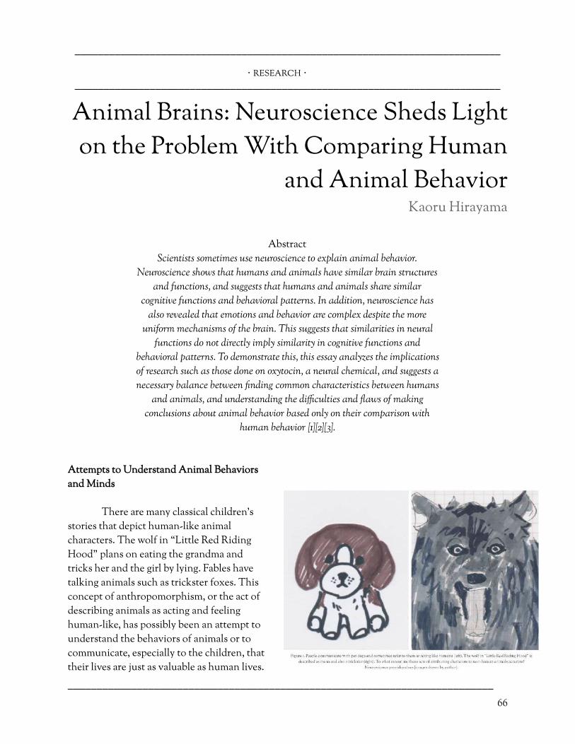
––––––––––––––––––––––––––––––––––––––––––––––––––––––––––––––––––––––––––
・ RESEARCH ・
––––––––––––––––––––––––––––––––––––––––––––––––––––––––––––––––––––––––––
Animal Brains: Neuroscience Sheds Light on the Problem With Comparing Human
and Animal Behavior Kaoru Hirayama
Abstract
Scientists sometimes use neuroscience to explain animal behavior.
Neuroscience shows that humans and animals have similar brain structures
and functions, and suggests that humans and animals share similar
cognitive functions and behavioral patterns. In addition, neuroscience has
also revealed that emotions and behavior are complex despite the more
uniform mechanisms of the brain. This suggests that similarities in neural
functions do not directly imply similarity in cognitive functions and
behavioral patterns. To demonstrate this, this essay analyzes the implications
of research such as those done on oxytocin, a neural chemical, and suggests a
necessary balance between �nding common characteristics between humans
and animals, and understanding the di�culties and �aws of making
conclusions about animal behavior based only on their comparison with
human behavior [1][2][3].
Attempts to Understand Animal Behaviors and Minds
There are many classical children’s
stories that depict human-like animal characters. The wolf in “Little Red Riding Hood” plans on eating the grandma and tricks her and the girl by lying. Fables have talking animals such as trickster foxes. This concept of anthropomorphism, or the act of describing animals as acting and feeling human-like, has possibly been an attempt to understand the behaviors of animals or to communicate, especially to the children, that their lives are just as valuable as human lives.
–––––––––––––––––––––––––––––––––––––––––––––––––––––––––––––––––––––––––– 66

––––––––––––––––––––––––––––––––––––––––––––––––––––––––––––––––––––––––––
However, anthropomorphism also has its �aws. In recent years, the growing �eld of neuroscience has provided a way of explaining animal behavior and emotions in addition to observing the behavior of animals. Neuroscience can contribute to explaining the direct biological reason of behavior and emotions, not just the environmental factors observed to correlate with certain behaviors. These new studies have revealed the complexity of human and animal behavior, and simply explaining animal behavior in terms of whether or not they are similar to humans has become an irresponsible and inaccurate method.
Neuroscience in Support of the Comparison Method
Researchers have attempted to identify what makes humans behave di�erently from
animals. For example, they try to answer the questions of why humans seem to have a higher level of intelligence and emotions than animals, and why they are able to understand languages that they use. However, as more studies are being conducted, there is more evidence pointing to the idea that humans are in fact similar to animals. The amygdala processes feelings of fear, pain, and aggression [4]. The prefrontal cortex is responsible for complex decision-making such as thinking in di�erent categories [5]. Oxytocin in the brain is responsible for recognizing known individuals such as family and friends, which is necessary for complex social behavior to occur [1]. As far as biological explanations of behavior indicate, brains function similarly in other mammals as well, giving rise to similar social behavior and intelligence in several other species other than humans. This is possible evidence in support of anthropomorphism. Even when scientists found a unique neuron correlated with social behavior, the von Economo neuron (VEN), which at �rst was thought to be a unique characteristic of the human brain, they discovered the same type of neurons in other social animals such as great apes and whales [6][7].
An Alternative Interpretation of the Neuroscience of Animals
The �eld of neuroscience has
been improving thanks to this comparison between humans and animals, whether by comparing humans to animals to understand animal behavior or vice versa. Though, one should not be content with understanding animal behavior from just one’s own perspectives. In other words, no human in the world has ever experienced the mind of an animal, so for example, observing friendly behavior between animals and simply assuming that they have empathy, may be �awed.
–––––––––––––––––––––––––––––––––––––––––––––––––––––––––––––––––––––––––– 67

––––––––––––––––––––––––––––––––––––––––––––––––––––––––––––––––––––––––––
The distribution of oxytocin receptors in the brain correlates with vole monogamy, while oxytocin is also released in humans during communicating with loved ones [2][3]. Despite their similarity in oxytocin in the brain, scientists cannot fully determine whether the voles understand feelings of love, or whether oxytocin merely a�ects voles to prefer one partner over the others [8]. Thus, neuroscience alone cannot explain behavior, no matter its accuracy and improvements. In addition to understanding the science of the brain, scientists need to understand the environmental cause of behavior and emotions, such as how and where the individuals grew up, social norms of their communities, and events leading up to the emotion and behavior [9].
While neuroscience illustrates many similarities in animal and human behavior,
observations of environmental factors reveal their di�erences. Humans live in safe and comfortable houses, but animals do not. Humans can go to hospitals, and leave history for the next generation, not only by direct communication but also by writing. What is di�erent and what is similar is far too complex to keep track of. Furthermore, if oxytocin cannot provide evidence to show whether one understands love, then one cannot know if animals can love and hate, and it would be �awed to simply assume that they can. Interestingly, this also suggests that it is �awed to assume that other people understand love just as one does. After all, no one has ever experienced the emotions of people other than oneself. In the same way, understanding human behavior and comparing it to animals can only explain so much. Knowing one’s own thoughts does not guarantee that one will know all the emotions and behaviors that human beings are capable of experiencing. That is why people talk about biases getting in one’s way. Ultimately, using anthropomorphism to say that animals are also very emotional like humans is forcing them one’s biased thoughts about what emotions should be like. On the other hand, it is also a bias to simply assume that animals do not have emotions just because they are not like humans. Instead, one needs to understand that the brains of animals and humans may work completely di�erently, and it may be di�cult to understand an animal's viewpoint from a human's viewpoint. The comparison should be left aside.
Conclusion
While people get excited that neuroscience can explain the mind, whilst in truth, it reveals
that current neuroscience cannot explain everything in detail. “So, are humans similar to animals or not?” is a question that should be asked with great care in today’s world. What is necessary is a balance between understanding the general pattern of behavior, and understanding that there are great individual di�erences in behavior and emotions. Perhaps all of this is already agreed upon, but one must always come back and remember the complexity of the brain and its individuality between species as well as between single individuals, especially in a world where improvements in science are revealing laws that explain the whole universe, and researchers are working to understand the fundamental reasons why humans and other animals think and behave in certain ways.
–––––––––––––––––––––––––––––––––––––––––––––––––––––––––––––––––––––––––––––––––––
–––––––––––––––––––––––––––––––––––––––––––––––––––––––––––––––––––––––––– 68
References

––––––––––––––––––––––––––––––––––––––––––––––––––––––––––––––––––––––––––
–––––––––––––––––––––––––––––––––––––––––––––––––––––––––––––––––––––––––– 69
[1] Numan, Michael and Larry Young. (7/6/2015). Neural mechanisms of mother-infant bonding and pair bonding: Similarities, di�erences, and broader implications. Hormones and Behavior. 98–112. Retrieved: 7/7/2020.
[6] Allman, John, Nicole Tetreault, Atiya Hakeem, Kebreten Manaye, Katerina Semendeferi, Joseph Erwin, Soyoung Park, Virginie Goubert, and Patrick Hof. (29/5/2010). The von Economo neurons in frontoinsular and anterior cingulate cortex in great apes and humans. Brain Structure and Function. 495–517. Retrieved: 7/7/2020.
[2] Insel, Thomas and Lawrence Shapiro. (1/7/1992). Oxytocin receptor distribution re�ects social organization in monogamous and polygamous voles. Proceedings of the National Academy of Sciences of the United States of America. 5981–5985. Retrieved: 9/7/2020.
{7] Butti, Camilla, Chet Sherwood, Atiya Hakeem, John Allman, and Patrick Hof. (20/3/2009). Total number and volume of Von Economo neurons in the cerebral cortex of cetaceans. The Journal of Comparative Neurology. 243–259. Retrieved: 7/7/2020.
[3] Algoe, Sara, Laura Kurtz, and Karen Grewen. (2/10/2017). Oxytocin and Social Bonds: The Role of Oxytocin in Perceptions of Romantic Partners’ Bonding Behavior. Psychological Science. 1763–1772. Retrieved: 9/7/2020.
[8] Churchland, Patricia. (2019). Conscience: The Origins of Moral Intuition. Getting Attached. 49. Retrieved: 7/7/2020.
[4] Rizvi, Tilat, Matthew Ennis, Michael Behbehani, and Michael Shipley. (1/1/1991). Connections between the central nucleus of the amygdala and the midbrain periaqueductal gray: Topography and reciprocity. The Journal of Comparative Neurology. 121–131. Retrieved: 7/7/2020.
[9] Sapolsky, Robert. (2017). Behave: The Biology of Humans at Our Best and Worst. Introduction. 4–5. Retrieved: 7/7/2020.
[5] Freedman, David, Maximilian Riesenhuber, Tomaso Poggio, and Earl Miller. (12/1/2001). Categorical Representation of Visual Stimuli in the Primate Prefrontal Cortex. Science. 312–316. Retrieved: 7/7/2020.

––––––––––––––––––––––––––––––––––––––––––––––––––––––––––––––––––––––––––
・ RESEARCH ・
––––––––––––––––––––––––––––––––––––––––––––––––––––––––––––––––––––––––––
A Review of Commercially Available EEG Headsets
Cleah Winston
Abstract The rise of the citizen scientist is being spurred on by new access to
research-grade hardware at publically a�ordable prices. In this article, we
speci�cally discuss various electroencephalography (EEG) hardware systems
that are now commercially available and their impact on research within
and outside of academic institutions. Although previous-generation
research-grade EEG recording devices were expensive and not portable, new
EEG headsets are becoming more a�ordable and portable. This article
discusses various research studies that have used newer, accessible EEG
devices to cover a variety of research topics that range from a focus on
medical disorders to cognition to thought-controlled video games and
neurally-responsive art.
What Is Electroencephalography?
Electroencephalography (EEG) was �rst introduced in 1924 and revolutionized neuroscience by enabling a non-invasive recording of both human and animal brains during awake, conscious activity. EEG measures electrical activity from the surface of the brain using electrodes placed on the scalp (Figure 1). The electrical activity recorded by the EEG scalp electrodes is caused by neurons generating action potentials. An action potential refers to a single electrical �ring of an individual neuron, the small impulse that forms the basis of all communication between neurons. It takes
from thousands to millions of concurrent action potentials across the brain to create an electrical signal large enough for an EEG system to detect it. The ability to record neural signals by electrodes placed noninvasively on the scalp has been remarkably useful for learning about brain abnormalities as well as healthy neural functioning [1].
A Brief History of EEG
–––––––––––––––––––––––––––––––––––––––––––––––––––––––––––––––––––––––––– 70

––––––––––––––––––––––––––––––––––––––––––––––––––––––––––––––––––––––––––
Around 1780, an Italian scientist named Luigi Galvani conducted a revolutionary
experiment in which he connected the nerves of a dead frog to an electrical wire. In doing so, Galvani became the �rst scientist to explore the e�ect of electrical stimulation on animal muscle tissue. Galvani found that electrical stimulation caused the frog’s leg to move. This discovery paved the way to electroencephalography. After Galvani, neuroscientists continued to expand their understanding of electricity in the brain and peripheral nervous systems of animals. Although many scientists were critical in the invention of the EEG, a German man named Hans Berger is considered to be the father of modern electroencephalography. Building on past experiments, Berger used electrodes, an electrometer, and a galvanometer to create the �rst EEG. By placing electrodes onto the scalp and needles into the scalp, he recorded human brain waves. He was the �rst to do so and he called the waves he recorded Alpha and Beta, the �rst two letters of the Greek alphabet. Following this landmark experiment, Grey Walter created a toposcope which used a greater density of electrodes and cathode-ray tubes to obtain higher quality signals. Since then, EEG technology has advanced considerably and can now be used as a medical tool in addition to other diverse applications [2].
Modern EEG Recording Hardware: Research Grade
While Dr. Walter’s 1957 EEG boasted 37 electrodes, modern research-grade EEG systems use
up to 256 electrode channels. To ensure consistency between experiments and to target speci�c regions of the brain, these electrodes are carefully positioned on the scalp, often held in place using a geodesic head cap, a mesh, or a rigid grid. In some experiments, each electrode temporarily adheres directly to the scalp.
Neural signals are very small in magnitude after passing through the layers of protection
around the brain, including the thick bone of the skull. As a result of this attenuation, quality EEG recording requires ampli�cation, which has become an integral part of the EEG hardware.
Moreover, the skull dampens not only the magnitude, but also the range of frequencies of neural signals that can be recorded. The higher frequency signals are the most dampened, or attenuated, by the skull. The quality of the EEG recordings is also dependent on the sampling rate of the EEG hardware. The sampling rate is how many times
–––––––––––––––––––––––––––––––––––––––––––––––––––––––––––––––––––––––––– 71
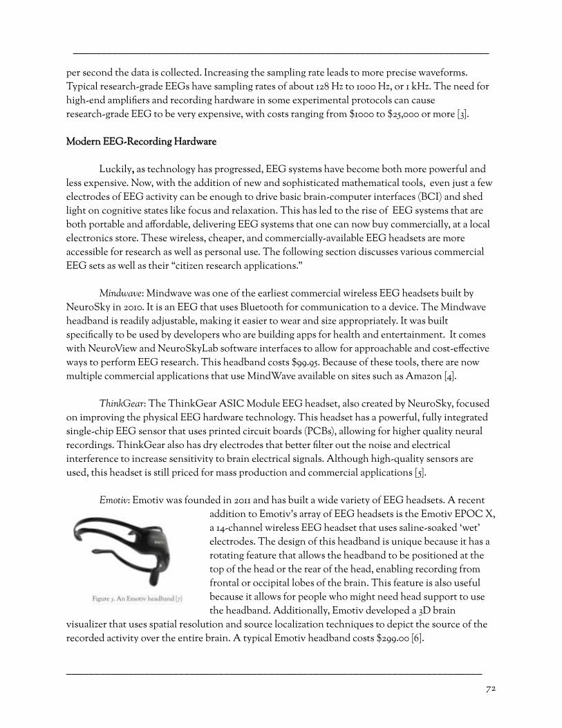
––––––––––––––––––––––––––––––––––––––––––––––––––––––––––––––––––––––––––
per second the data is collected. Increasing the sampling rate leads to more precise waveforms. Typical research-grade EEGs have sampling rates of about 128 Hz to 1000 Hz, or 1 kHz. The need for high-end ampli�ers and recording hardware in some experimental protocols can cause research-grade EEG to be very expensive, with costs ranging from $1000 to $25,000 or more [3].
Modern EEG-Recording Hardware
Luckily , as technology has progressed, EEG systems have become both more powerful and
less expensive. Now, with the addition of new and sophisticated mathematical tools, even just a few electrodes of EEG activity can be enough to drive basic brain-computer interfaces (BCI) and shed light on cognitive states like focus and relaxation. This has led to the rise of EEG systems that are both portable and a�ordable, delivering EEG systems that one can now buy commercially, at a local electronics store. These wireless, cheaper, and commercially-available EEG headsets are more accessible for research as well as personal use. The following section discusses various commercial EEG sets as well as their “citizen research applications.”
Mindwave : Mindwave was one of the earliest commercial wireless EEG headsets built by
NeuroSky in 2010. It is an EEG that uses Bluetooth for communication to a device. The Mindwave headband is readily adjustable, making it easier to wear and size appropriately. It was built speci�cally to be used by developers who are building apps for health and entertainment. It comes with NeuroView and NeuroSkyLab software interfaces to allow for approachable and cost-e�ective ways to perform EEG research. This headband costs $99.95. Because of these tools, there are now multiple commercial applications that use MindWave available on sites such as Amazon [4].
ThinkGear : The ThinkGear ASIC Module EEG headset, also created by NeuroSky, focused
on improving the physical EEG hardware technology. This headset has a powerful, fully integrated single-chip EEG sensor that uses printed circuit boards (PCBs), allowing for higher quality neural recordings. ThinkGear also has dry electrodes that better �lter out the noise and electrical interference to increase sensitivity to brain electrical signals. Although high-quality sensors are used, this headset is still priced for mass production and commercial applications [5].
Emotiv : Emotiv was founded in 2011 and has built a wide variety of EEG headsets. A recent
addition to Emotiv’s array of EEG headsets is the Emotiv EPOC X, a 14-channel wireless EEG headset that uses saline-soaked ‘wet’ electrodes. The design of this headband is unique because it has a rotating feature that allows the headband to be positioned at the top of the head or the rear of the head, enabling recording from frontal or occipital lobes of the brain. This feature is also useful because it allows for people who might need head support to use the headband. Additionally, Emotiv developed a 3D brain
visualizer that uses spatial resolution and source localization techniques to depict the source of the recorded activity over the entire brain. A typical Emotiv headband costs $299.00 [6].
–––––––––––––––––––––––––––––––––––––––––––––––––––––––––––––––––––––––––– 72
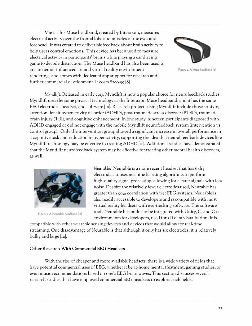
––––––––––––––––––––––––––––––––––––––––––––––––––––––––––––––––––––––––––
Muse : This Muse headband, created by Interaxon, measures electrical activity over the frontal lobe and muscles of the eyes and forehead. It was created to deliver biofeedback about brain activity to help users control emotions. This device has been used to measure electrical activity in participants' brains while playing a car driving game to decode distraction. The Muse headband has also been used to create neural-in�uenced art and virtual reality environment renderings and comes with dedicated app support for research and further commercial development. It costs $209.99 [8].
Myndlift : Released in early 2015, Myndlift is now a popular choice for neurofeedback studies.
Myndlift uses the same physical technology as the Interaxon Muse headband, and it has the same EEG electrodes, headset, and software [10]. Research projects using Myndlift include those studying attention de�cit hyperactivity disorder (ADHD), post-traumatic stress disorder (PTSD), traumatic brain injury (TBI), and cognitive enhancement. In one study, nineteen participants diagnosed with ADHD engaged or did not engage with the mobile Myndlift neurofeedback system (intervention vs control group). Only the intervention group showed a signi�cant increase in overall performance in a cognitive task and reduction in hyperactivity, supporting the idea that neural feedback devices like Myndlift technology may be e�ective in treating ADHD [11]. Additional studies have demonstrated that the Myndlift neurofeedback system may be e�ective for treating other mental health disorders, as well.
Neurable : Neurable is a more recent headset that has 6 dry electrodes. It uses machine learning algorithms to perform high-quality signal processing, allowing for clearer signals with less noise. Despite the relatively fewer electrodes used, Neurable has greater than 90% correlation with wet EEG systems. Neurable is also readily accessible to developers and is compatible with most virtual reality headsets with eye-tracking software. The software tools Neurable has built can be integrated with Unity, C, and C++ environments for developers, used for 3D data visualization. It is
compatible with other wearable sensing devices and devices that would allow for real-time streaming. One disadvantage of Neurable is that although it only has six electrodes, it is relatively bulky and large [12].
Other Research With Commercial EEG Headsets
With the rise of cheaper and more available headsets, there is a wide variety of �elds that
have potential commercial uses of EEG, whether it be at-home mental treatment, gaming studies, or even music recommendations based on one’s EEG brain waves. This section discusses several research studies that have employed commercial EEG headsets to explore such �elds.
–––––––––––––––––––––––––––––––––––––––––––––––––––––––––––––––––––––––––– 73
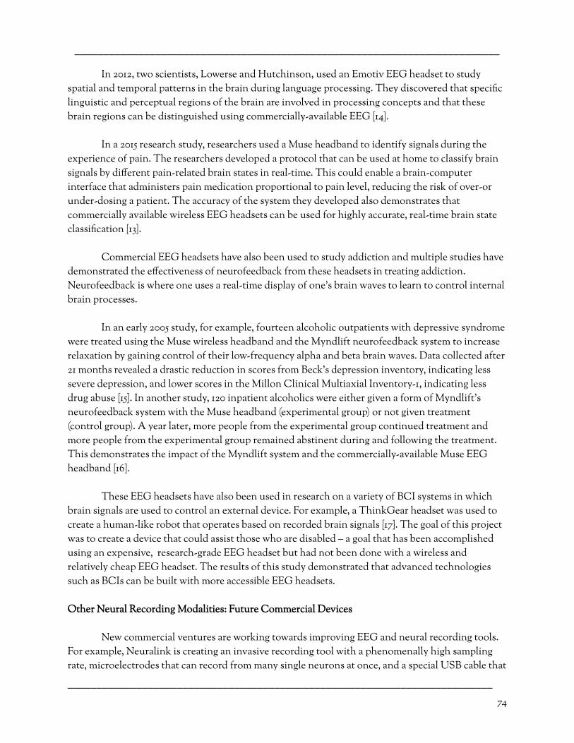
––––––––––––––––––––––––––––––––––––––––––––––––––––––––––––––––––––––––––
In 2012, two scientists, Lowerse and Hutchinson, used an Emotiv EEG headset to study spatial and temporal patterns in the brain during language processing. They discovered that speci�c linguistic and perceptual regions of the brain are involved in processing concepts and that these brain regions can be distinguished using commercially-available EEG [14].
In a 2015 research study, researchers used a Muse headband to identify signals during the
experience of pain. The researchers developed a protocol that can be used at home to classify brain signals by di�erent pain-related brain states in real-time. This could enable a brain-computer interface that administers pain medication proportional to pain level, reducing the risk of over-or under-dosing a patient. The accuracy of the system they developed also demonstrates that commercially available wireless EEG headsets can be used for highly accurate, real-time brain state classi�cation [13].
Commercial EEG headsets have also been used to study addiction and multiple studies have
demonstrated the e�ectiveness of neurofeedback from these headsets in treating addiction. Neurofeedback is where one uses a real-time display of one’s brain waves to learn to control internal brain processes.
In an early 2005 study, for example, fourteen alcoholic outpatients with depressive syndrome
were treated using the Muse wireless headband and the Myndlift neurofeedback system to increase relaxation by gaining control of their low-frequency alpha and beta brain waves. Data collected after 21 months revealed a drastic reduction in scores from Beck’s depression inventory, indicating less severe depression, and lower scores in the Millon Clinical Multiaxial Inventory-1, indicating less drug abuse [15]. In another study, 120 inpatient alcoholics were either given a form of Myndlift’s neurofeedback system with the Muse headband (experimental group) or not given treatment (control group). A year later, more people from the experimental group continued treatment and more people from the experimental group remained abstinent during and following the treatment. This demonstrates the impact of the Myndlift system and the commercially-available Muse EEG headband [16].
These EEG headsets have also been used in research on a variety of BCI systems in which
brain signals are used to control an external device. For example, a ThinkGear headset was used to create a human-like robot that operates based on recorded brain signals [17}. The goal of this project was to create a device that could assist those who are disabled – a goal that has been accomplished using an expensive, research-grade EEG headset but had not been done with a wireless and relatively cheap EEG headset. The results of this study demonstrated that advanced technologies such as BCIs can be built with more accessible EEG headsets.
Other Neural Recording Modalities: Future Commercial Devices
New commercial ventures are working towards improving EEG and neural recording tools.
For example, Neuralink is creating an invasive recording tool with a phenomenally high sampling rate, microelectrodes that can record from many single neurons at once, and a special USB cable that
–––––––––––––––––––––––––––––––––––––––––––––––––––––––––––––––––––––––––– 74

––––––––––––––––––––––––––––––––––––––––––––––––––––––––––––––––––––––––––
allows for full bandwidth streaming from the electrodes [18]. This company has also built a neurosurgical robot for placing their tiny, small, and �exible electrode threads into the brain - currently of pigs, but the vision is to make these implants available to humans someday. Currently, the surgical robot has microscopic precision to prevent damage to vascular tissue of the brain and to target highly speci�c brain regions.
Another upcoming venture involves functional near-infrared spectroscopy (fNIRS). This
non-invasive technology measures changing hemoglobin concentrations by measuring certain waves emitted by the brain to get a re�ned EEG signal. Recently there has been an almost exponential growth of research studies that use fNIRS for brain imaging. Also, they have a relatively low cost, are easy to make, and are adjustable for various purposes. This points to the idea that the fNIRS might soon be ready to be used in commercial settings [19].
Conclusion
Commercially available wireless EEG headsets have now been on the market for over 15 years
and much progress has been made. Though none of them are perfect, each of the wireless EEG headsets discussed here has unique qualities that make them very useful. For example, while the ThinkGear module features high-quality EEG sensors, Emotiv has a distinctive setup for the design of the headband that makes it more accessible and allows for the EEG reading to give more information. While Myndlift o�ers an advanced neurofeedback system that has proved itself in various research studies, Mindwave has focused more on arming developers with an accessible programming environment and a suite of research tools to promote the development of novel commercial applications. To date, commercially available wireless headsets have revealed promising avenues for citizen research and rapid development of human self-interaction, as with neurofeedback. Undoubtedly, research in the coming years will further reduce the costs and improve the performance of these headsets.
–––––––––––––––––––––––––––––––––––––––––––––––––––––––––––––––––––––––––––––––––––
–––––––––––––––––––––––––––––––––––––––––––––––––––––––––––––––––––––––––– 75
References
[1 ] (15/04/2020). EEG (electroencephalogram). Mayo Clinic. https://www.mayoclinic.org/tests-procedures/eeg/about/pac-2039 3875#:~:text=An%20electroencephalogram%20(EEG)%20is%20a,li nes%20on%20an%20EEG%20recording. Retrieved: 09/07/2020.
[11] Steiner, Naomi. (01/03/2014). In-School Neurofeedback Training for ADHD: Sustained Improvements From a Randomized Control Trial. Pediatrics. Vol. 133, Issue 3. Retrieved: 12/07/2020.
[2] Farnsworth, Bryn. (18/10/2016). Making Waves – The Past, Present, and Future of EEG Research. IMOTIONS. https://imotions.com/blog/history-of-eeg/ . Retrieved : 12/06/2020.
[12] Strickland, Eliza. Startup Neurable Unveils the World's First Brain-Controlled VR Game. IEEE Spectrum. https://spectrum.ieee.org/the-human-os/biomedical/bionics/brai ny-startup-neurable-unveils-the-worlds-�rst-braincontrolled-vr-g ame. Retrieved: 09/06/2020.
[3] (05/07/2016). The Anatomy of an EEG Cap. IMOTIONS. https://imotions.com/blog/eeg-cap/ . Retrieved : 13/06/2020.
[13] Karydis, Thrasyvoulos. (07/2015). Self-calibrating protocols enhance wearable EEG diagnostics and consumer applications. ACM Digital Library. https://dl.acm.org/doi/10.1145/2769493.2769581. Retrieved: 11/06/2020.

––––––––––––––––––––––––––––––––––––––––––––––––––––––––––––––––––––––––––
–––––––––––––––––––––––––––––––––––––––––––––––––––––––––––––––––––––––––– 76
[4] Brainwave Sensing Headset. Neurosky Store. https://store.neurosky.com/pages/mindwave . Retrieved : 03/07/2020.
[14] Louwerse, Max. (16/10/2012). Neurological Evidence Linguistic Processes Precede Perceptual Simulation in Conceptual Processing. US National Library of Medicine National Institutes of Health. https://www.ncbi.nlm.nih.gov/pmc/articles/PMC3488936/ . Retrieved: 12/07/2020
[5] BioSensors / EEG Headsets. Neurosky. http://neurosky.com/biosensors/eeg-sensor/biosensors/ . Retrieved: 22/08/2020.
[15] Saxby, E. (09/1995) 1Alpha-theta brainwave neurofeedback training: an e�ective treatment for male and female alcoholics with depressive symptoms. National Library of Medicine. https://pubmed.ncbi.nlm.nih.gov/8801245/. Retrieved: 17/06/2020.
[6] The Science behind our Technology. Emotiv. https://www.emotiv.com/our-technology/ . Retrieved: 22/08/2020.
[16] Scott, William. (31/03/2005). E�ects of an EEG biofeedback protocol on a mixed substance abusing population. PubMed. https://pubmed.ncbi.nlm.nih.gov/16161729/ . Retrieved: 14/07/2020.
[7] Emotiv Insight. Emotiv. https://www.emotiv.com/insight/ . Retrieved: 09/09/2020.
[17] Shyaa, Nabeel. (23/04/2018). Electroencephalography (EEG) Based Mobile Robot Control through an Adaptive Brain Robot Interface. American Scienti�c Research Journal for Engineering, Technology, and Sciences (ASRJETS). Vol 42 No 1. Retrieved: 15/07/2020.
[8] Neurofeedback EEG Device. choosemuse. https://choosemuse.com/how-it-works/?utm_source=google&utm _medium=cpc&utm_campaign=BoF_USA_Search_Brand&utm_ter m=%2Bmuse%20%2Bheadband--b&utm_content=Muse_Headban d--458528407858?gclid=CjwKCAjwtNf6BRAwEiwAkt6UQk6BU LScga0WmLWZEJeZAhUlT0lsrCJQEy1vPyzM2A-5frOmxtTO8B oC2aMQAvD_BwE . Retrieved: 17/06/2020.
[18] Interfacing with the Brain. Neuralink. https://neuralink.com/approach/. Retrieved: 07/09/2020.
[9] Muse: The Brain Sensing Headband. Digital Health. https://www.digital.health/new-products/interaxon-muse . Retrieved: 17/06/2020.
[19] Domenico, Stefano. Functional Near-Infrared Spectroscopy: Proof of Concept for Its Application in Social Neuroscience. ScienceDirect. Pages 169-173. Retrieved: 17/07/2020.
[10] What’s the Science behind MyndLift. MyndLift. https://www.myndlift.com/neurofeedback-science . Retrieved: 18/05/2020.

––––––––––––––––––––––––––––––––––––––––––––––––––––––––––––––––––––––––––
Contributors Page
IYNA EDITING TEAM:
CONTRIBUTING AUTHORS:
IYNA BOARD OF DIRECTORS:
ADVISORY BOARD:
–––––––––––––––––––––––––––––––––––––––––––––––––––––––––––––––––––––––––– 77
Editor-In-Chief: Sojas Wagle
Head of Assembly: Annie Pan
Managing Editor: Shyam Soundararajan
Journal Artist-in-Residence Jenna Mackenroth
Senior Editors: Kareena Thakur, Kunal Dhirani, Anca-Mihaela Vasilica, Ashvin Kumar, Gasser Alwasify, Sampath Rapuri
Junior Editors: Bhavya Boddu, Haris Rana, Lori Saxena, Rod Moore, Sneha Nadella, Thelaxini Reginold, Vaishnavi Kode, Johnny Yue
Journalists: Divyash Shah, Mariya Meleganich, Rhea Ray, Sai Snigdha Kodali
Featured Writers: Katherine Wei, Shrika Vejandla, Shamsudeen Suleiman
Writers: Shriya Challam, Suraj Sivaraja, Harish Rajan, Nora Mehler, Divya Venkataraman, Helen Kim, Allen Chau, Michael Palumbo, Arya Reddy, Kaoru Hirayama, Cleah Winston
Board Chair: Khayla Black
Vice Chair: Nipun Gorantla
Secretary: Sarah Iqbal
Chief Executive O�cer: Brian Lee
Board Members: Allen Chau, Sofía Vaca Narvaja, Lara Ressin, Marisol Arau
Founder & Advisory Board: Dr. Norbert Myslinski


