PDF of this chapter (https://emcrit.org/wp-content/uploads/2020/03/COVID-19-EMCrit-Project.pdf) (or create customized PDF (https://emcrit.org/ibcc/about-
guide/#pdf) )
biology(back to contents) (#top)
basics
COVID-19 is a non-segmented, positive sense RNA virus.COVID-19 is part of the family of coronaviruses. This contains:
(i) Four coronaviruses which are widely distributed and usually cause the common cold (but can cause viral pneumonia in patients withcomorbidities).(ii) SARS and MERS – these caused epidemics with high mortality which are somewhat similar to COVID-19. COVID-19 is most closelyrelated to SARS.
It binds via the angiotensin-converting enzyme 2 (ACE2) receptor located on type II alveolar cells and intestinal epithelia (Hamming 2004(https://www.ncbi.nlm.nih.gov/pubmed/15141377) ).
This is the same receptor as used by SARS (hence the technical name for the COVID-19, “SARS-CoV-2”).When considering possible therapies, SARS (a.k.a. “SARS-CoV-1”) is the most closely related virus to COVID-19.
COVID-19 is mutating, which may complicate matters even further (�gure below). Virulence and transmission will shift over times, in wayswhich we cannot predict. New evidence suggests that there are roughly two different groups of COVID-19. This explains why initial reportsfrom Wuhan described a higher mortality than some more recent case series (Tang et al. 2020 (https://academic.oup.com/nsr/advance-
article/doi/10.1093/nsr/nwaa036/5775463#.XmA64GbsBuI.twitter) ; Xu et al 2020 (https://www.bmj.com/content/368/bmj.m606) ).(Ongoing phylogenetic mapping of new strains can be found here (https://nextstrain.org/ncov) .)
nomenclature used in this chapter
Technically, the virus is supposed to be called “SARS-CoV-2” and the clinical illness is called “COVID-19.” This gets confusing, so for thischapter the term COVID-19 will be used to refer to both entities.The term “SARS” will be used to refer to the original SARS virus from 2003 (which has currently been renamed SARS-CoV-1).
(1) ARDSThe primary pathology is ARDS, characterized by diffuse alveolar damage (e.g. including hyaline membranes). Pneumocytes with viralcytopathic effect are seen, implying direct virus damage (rather than a purely hyper-in�ammatory injury; Xu et al 2/17)(https://www.thelancet.com/action/showPdf?pii=S2213-2600%2820%2930076-X) .
(2) Cytokine stormEmerging evidence suggests that some patients may respond to COVID-19 with an exuberant “cytokine storm” reaction (with featuresof bacterial sepsis or hemophagocytic lymphohistiocytosis (https://emcrit.org/pulmcrit/sepsis-hlh-overlap-syndrome-shlhos/) ).Clinical markers of this may include elevations of C-reactive protein and ferritin, which appear to track with disease severity andmortality (Ruan 3/3/20 (https://link.springer.com/article/10.1007/s00134-020-05991-x) ).
stages of illness ??
There seem to be different stages of illness that patients may move through.(#1) Replicative stage – Viral replication occurs over a period of several days. An innate immune response occurs, but this responsefails to contain the virus. Relatively mild symptoms may occur due to direct viral cytopathic effect and innate immune responses.(#2) Adaptive immunity stage – An adaptive immune response eventually kicks into gear. This leads to falling titers of virus. However,it may also increase levels of in�ammatory cytokines and lead to tissue damage – causing clinical deterioration.
This progression may explain the clinical phenomenon wherein patients are relatively OK for several days, but then suddenly deterioratewhen they enter the adaptive immunity stage (e.g. Young et al. 3/3/2020 (https://jamanetwork.com/journals/jama/fullarticle/2762688) ).This has potentially important clinical implications:
Initial clinical symptoms aren't necessarily predictive of future deterioration. Sophisticated strategies may be required to guide risk-strati�cation and disposition (see below section on prognosis (#prognosis) ).Anti-viral therapies might need to be deployed early to work optimally (during the replicative stage).Immunosuppressive therapy (e.g. low-dose steroid) might be best initiated during the adaptive immune stage (with a goal of bluntingthis immunopathologic response slightly, in the sickest patients). But this is purely speculative.
transmission(back to contents) (#top)
large droplet transmission
COVID-19 transmission can occur via large droplet transmission (with a risk limited to ~6 feet from the patient)(Carlos del Rio 2/28(https://jamanetwork.com/journals/jama/fullarticle/2762510) ).This is typical for respiratory viruses such as in�uenza.Transmission via large droplet transmission can be prevented by using a standard surgical-style mask.
airborne transmission ??
It's controversial whether COVID19 can be transmitted via an airborne route (small particles which remain aloft in the air for longer periods oftime). Airborne transmission would imply the need for N95 masks (“FFP2” in Europe), rather than surgical masks. This controversy isexplored further in Shiu et al 2019 (https://emcrit.org/wp-content/uploads/2020/03/tada2019.pdf) .Airborne precautions started being used with MERS and SARS out of an abundance of caution (rather than any clear evidence thatcoronaviruses are transmitted via an airborne route). This practice has often been carried down to COVID19.Guidelines disagree about whether to use airborne precautions:
The Canadian Guidelines (https://www.canada.ca/en/public-health/services/diseases/2019-novel-coronavirus-infection/health-professionals.html) and WorldHealth Organization guidelines (https://www-who-int.ezproxy.uvm.edu/publications-detail/infection-prevention-and-control-during-health-care-when-novel-
coronavirus-(ncov)-infection-is-suspected-20200125) both recommend using only droplet precautions for routine care of COVID19 patients. However, both of these guidelines recommend airborne precautions for procedures which generate aerosols (e.g. intubation,noninvasive ventilation, CPR, bag-mask ventilation, and bronchoscopy).The United States CDC recommends (https://www-cdc-gov.ezproxy.uvm.edu/coronavirus/2019-ncov/infection-control/control-recommendations.html?
CDC_AA_refVal=https%3A%2F%2Fwww.cdc.gov%2Fcoronavirus%2F2019-ncov%2Fhcp%2Finfection-control.html) using airborne precautions all the timewhen managing COVID19 patients.
Using airborne precautions for all patients who are de�nitely or potentially infected with COVID19 will likely result in rapid depletion of N95masks. This will leave healthcare providers unprotected when they actually need these masks for aerosol-generating procedures.
In the context of a pandemic, the Canadian and WHO guidelines may be more sensible in countries with �nite resources (i.e. most locales). However, infection control is ultimately local, so be sure to follow your hospital's guidance regarding this.
contact transmission (“fomite-to-face”)
This mode of transmission has a tendency to get overlooked, but it may be incredibly important. This is how it works:(i) Someone with coronavirus coughs, emitting large droplets containing the virus. Droplets settle on surfaces in the room, creating athin �lm of coronavirus. The virus may be shed in a variety of other bodily �uids as well (e.g. sputum, nasal secretions, stool, saliva,urine, and blood) – so there are a variety of other ways that an infected person could shed virus into the environment.(ii) The virus persists on fomites in the environment. Human coronaviruses can survive on surfaces for up to about a week (Kampf etal 2020 (https://www.ncbi.nlm.nih.gov/pubmed/32035997) ). It's unknown how long COVID-19 can survive in the environment, but it might beeven longer (some animal coronaviruses can survive for weeks!).(iii) Someone else touches the contaminated the surface hours or days later, transferring the virus to their hands.(iv) If the hands touch the oropharynx or nasopharynx, this will result in transmission of infection.
Any effort to limit spread of the virus must block contact transmission. The above chain of events can be disrupted in a variety of ways:(a) Regular cleaning of environmental surfaces (e.g. using 70% ethanol or 0.5% sodium hypochlorite solutions; for details see Kampf etal 2020 (https://emcrit.org/wp-content/uploads/2020/02/[email protected]_.2020.01.022-2.pdf) and CDC guidelines (https://www-cdc-
CDC_AA_refVal=https%3A%2F%2Fwww.cdc.gov%2Fcoronavirus%2F2019-ncov%2Fhcp%2Finfection-control.html) ).(b) Hand hygiene (high concentration ethanol neutralizes the virus and is easy to perform, so this might be preferable if hands aren'tvisibly soiled)(Kampf 2017 (http://www.fha.org/�les/JohnW/EM/Ethanol-hand-sanitizer-and-HAV.pdf) ).(c) Avoidance of touching your face. This is nearly impossible, as we unconsciously touch our faces constantly. The main bene�t ofwearing a surgical mask could be that the mask acts as a physical barrier to prevent touching the mouth or nose.
Any medical equipment could become contaminated with COVID-19 and potentially transfer virus to providers (e.g. stethoscope earpiecesand shoes). A recent study found widespread deposition of COVID-19 in one patient's room, but fortunately this seems to be removable bycleaning with sodium dichloroisocyanurate (Ong et al 2020 (https://jamanetwork.com/journals/jama/fullarticle/2762692) ).
asymptomatic transmission
Asymptomatic transmission could potentially occur in two ways.(#1) Transmission despite a lack of symptoms seems to be possible (Carlos del Rio 2/28 (https://jamanetwork.com/journals/jama/fullarticle/2762510)
).(#2) An additional carrier state could occur in patients who have clinically recovered from the virus, but continue shedding the virus.
A recent study found that after convalescence, patients may continue to have a positive pharyngeal PCR for COVID-19 for weeks (Lan2/27 (https://jamanetwork.com/journals/jama/fullarticle/2762452) ). However, the clinical signi�cance of these PCR results is unknown. Convalescing patients probably have a low viral load and relatively low risk of transmission.CDC guidance (https://www.cdc.gov/coronavirus/2019-ncov/hcp/clinical-guidance-management-patients.html) is vague on how long patients withknown COVID-19 should be isolated. It may be advisable to obtain two paired RT-PCR tests (one of the nasopharynx and one of thepharynx), with each pair collected >24 hours apart, prior to discontinuing precautions.
R⌀
R⌀ is the average number of people that an infected person transmits the virus to.If R⌀ is <1, the epidemic will burn out.If R⌀ = 1, then epidemic will continue at a steady pace.If R⌀ >1, the epidemic will increase exponentially.
Current estimates put R⌀ at ~2.5-2.9 (Peng PWH et al, 2/28 (https://bjanaesthesia.org/article/S0007-0912(20)30098-2/pdf) ). This is a bit higher thanseasonal in�uenza.R⌀ is a re�ection of both the virus and also human behavior. Interventions such as social distancing and improved hygiene will decrease R⌀.
Control of spread of COVID-19 in China proves that R⌀ is a modi�able number that can be reduced by effective public healthinterventions.The R⌀ on board the Diamond Princess cruise ship was 15 – illustrating that cramped quarters with inadequate hygiene will increaseR⌀ (Rocklov 2/28 (https://academic.oup.com/jtm/advance-article/doi/10.1093/jtm/taaa030/5766334) ).
personal protective equipment (PPE)(back to contents) (#top)
gear
(1) Contact precautions (waterproof gown and gloves)(2) Some sort of mask (discussed above in the transmission (#transmission) section)
N95 mask or a powered, air-purifying respiratory (“PAPR”)Surgical mask for patients not undergoing aerosol-generating procedures (based on WHO & Canadian guidelines)
(3) Goggles or eye shieldNote: The exact gear used is probably less important than using it correctly.
applying and removing PPE (donning & do�ng)
Understanding how to put on (don) and remove (doff) personal protective equipment is extremely important (especially if contacttransmission is a dominant mode of transmission).Removing soiled PPE is the most critical and di�cult aspect.Applying and removing PPE should ideally be practiced before patients arrive (e.g. using simulation).The video below describes how to use PPE (you may skip the �rst 5 minutes).
some pearls about personal protective equipment
Pay attention to the junction between gloves and gowns. The gown should be tucked into the gloves (leaving no gap in-between). Usinggloves with extended cuffs facilitates this (similar to sterile surgical gloves). Gloves with long cuffs may facilitate removal of the gown andgloves as a single unit (see 12:30 in the above video if this doesn't make sense).When removing PPE, always start by �rst applying alcohol-based hand sanitizer to your gloves.After fully removing PPE, sanitize hands and wrists with alcohol-based hand sanitizer again.
screening & selection for investigation(back to contents) (#top)
key considerations include:
(1) Recent travel to affected areas.Areas with community-based transmission are increasing rapidly.The incubation time is up to 14 days, so travel within that window is relevant.
(2) Contact with anyone with known COVID-19 (de�ned as a prolonged period of time spent <6 feet apart).(3) As community acquisition emerges, broader testing will be needed. This will be based on a more detailed clinical evaluation, weighing:
i) How well patients meet the clinical features of Coronavirus (e.g. laboratory and imaging features explored further below).
NETEC: Personal Protective Equipment for COVID-19NETEC: Personal Protective Equipment for COVID-19
ii) Presence or absence of alternative diagnoses (e.g. if patient tests positive for in�uenza, this would make it less likely that theysimultaneously contracted in�uenza and coronavirus).
approach to isolation and testing
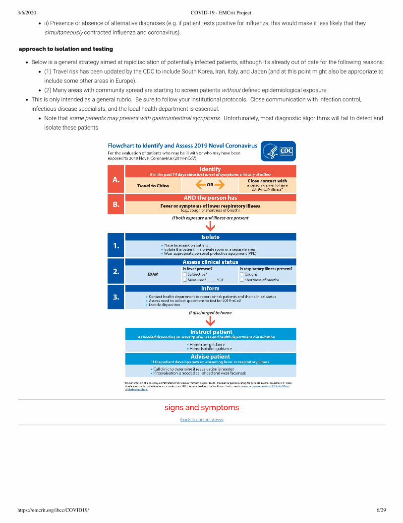
Below is a general strategy aimed at rapid isolation of potentially infected patients, although it's already out of date for the following reasons:(1) Travel risk has been updated by the CDC to include South Korea, Iran, Italy, and Japan (and at this point might also be appropriate toinclude some other areas in Europe).(2) Many areas with community spread are starting to screen patients without de�ned epidemiological exposure.
This is only intended as a general rubric. Be sure to follow your institutional protocols. Close communication with infection control,infectious disease specialists, and the local health department is essential.
Note that some patients may present with gastrointestinal symptoms. Unfortunately, most diagnostic algorithms will fail to detect andisolate these patients.
signs and symptoms(back to contents) (#top)
3/6/2020 COVID-19 - EMCrit Project
https://emcrit.org/ibcc/COVID19/ 7/29
symptoms
COVID-19 may cause constitutional symptoms, upper respiratory symptoms, lower respiratory symptoms, and, less commonly,gastrointestinal symptoms.Sources disagree about the exact rate of fever. This may relate to:
(1) Methodology used in various studies (i.e. whether they recorded the initial temperature, or whether there was a fever at any pointduring the patient's hospitalization).(2) Patients included in various series (sicker patients may be more likely to present with fever).Regardless of the exact numbers – absence of a fever does not exclude COVID-19.
traditional physical examination
This is generally unrevealing.Patients may have hypoxemia without signs of respiratory distress (“silent hypoxemia”)(Xie et al 3/2(https://link.springer.com/article/10.1007/s00134-020-05979-7) ).~2% may have pharyngitis or tonsil enlargement – but of course this is an entirely nonspeci�c �nding (Guan et al 2/28(https://www.nejm.org/doi/pdf/10.1056/NEJMoa2002032?articleTools=true) ).The rate of abnormal chest auscultation is unclear (but lung sonography is likely more accurate; more on this below).
typical disease course
Incubation is a median of ~4 days (interquartile range of 2-7 days), with a range up to 14 days (Carlos del Rio 2/28(https://jamanetwork.com/journals/jama/fullarticle/2762510) ).Typical evolution of severe disease (based on analysis of multiple studies by Arnold Forest (https://www.youtube.com/watch?
Dyspnea ~ 6 days post exposure.Admission after ~8 days post exposure.ICU admission/intubation after ~10 days post exposure.
labs(back to contents) (#top)
complete blood count
WBC count tends to be normal.Lymphopenia is common, seen in ~80% of patients (Guan et al 2/28 (https://www.nejm.org/doi/pdf/10.1056/NEJMoa2002032?articleTools=true) , Yanget al 2/21 (https://www.thelancet.com/action/showPdf?pii=S2213-2600%2820%2930079-5) ).Mild thrombocytopenia is common (but platelets are rarely <100). Lower platelet count is a poor prognostic sign (Ruan et al 3/3(https://link.springer.com/article/10.1007/s00134-020-05991-x) ).
coagulation studies
Coagulation labs are generally fairly normal upon admission, although elevated D-dimer is commonly seen (table above).Disseminated intravascular coagulation may evolve over time, correlating with poor prognosis (�gure below)(Tang et al. 2020(https://www.ncbi.nlm.nih.gov/pubmed/32073213) ).
ProcalcitoninCOVID-19 does not appear to increase the procalcitonin. For example, the largest series found that procalcitonin levels were <0.5 in95% of patients (Guan et al 2/28 (https://www.nejm.org/doi/pdf/10.1056/NEJMoa2002032?articleTools=true) ).Elevated procalcitonin may suggest an alternative diagnosis (e.g. pure bacterial pneumonia). For patients who have been admitted withCOVID-19, procalcitonin elevation may suggest a superimposed bacterial infection.
C-reactive protein (CRP)COVID-19 increases CRP. This seems to track with disease severity and prognosis. In a patient with severe respiratory failure and anormal CRP, consider non-COVID etiologies (such as heart failure).Young et al. 3/3 (https://jamanetwork.com/journals/jama/fullarticle/2762688) found low CRP levels in patients not requiring oxygen (mean 11mg/L, interquartile range 1-20 mg/L) compared to patients who became hypoxemic (mean 66 mg/L, interquartile range 48-98 mg/L).Ruan et al 3/3 (https://link.springer.com/article/10.1007/s00134-020-05991-x) found CRP levels to track with mortality risk (surviving patients hada median CRP of ~40 mg/L with an interquartile range of ~10-60 mg/L, whereas patients who died had a median of 125 mg/L with aninterquartile range of ~60-160 mg/L)(�gure below in the section on prognosis (#prognosis) ).
evaluation for competing diagnoses
PCR for in�uenza and other respiratory viruses (e.g. RSV) may be helpful. Detection of other respiratory viruses doesn't prove that thepatient isn't co-infected with COVID-19. However, an alternative explanation for the patient's symptoms might reduce the index of suspicionfor COVID-19 substantially.Conventional viral panels available in some hospitals will test for “coronavirus.”
This test does not work for COVID-19!This PCR test for “coronavirus” is designed to evaluate for four coronaviruses which usually cause mild illness.Ironically, a positive conventional test for “coronavirus” actually makes it less likely that the patient has COVID-19.
Blood cultures should be performed as per usual indications.
speci�c testing for COVID-19(back to contents) (#top)
Currently in the United States, all testing is done by state reference labs. Specimen collection and testing should be coordinated with thedepartment of health.
(1) Nasopharyngeal swab should be sent.(2) If intubated, tracheal aspirate should be performed.(3) Bronchoalveolar lavage or induced sputum are other options for a patient who isn't intubated. However, obtaining these specimens maypose substantial risk of transmission.
It's dubious whether these tests are bene�cial for the sole purpose of evaluating for coronavirus (see the section below onbronchoscopy (#bronchoscopy) ).
performance of COVID-19 RT-PCR?
Test performance is unclear; this is impossible to sort out in the absence of a de�nitive “gold standard” diagnostic test for COVID-19.Speci�city seems to be high.Sensitivity may not be terri�c.
In a case series diagnosed on the basis of clinical criteria and CT scans, the sensitivity of RT-PCR was only 60-70% (Kanne 2/28). However, it's probable that some patients diagnosed based on CT scanning had other respiratory viruses rather than COVID-19, causingthis study to under-estimate the sensitivity of the RT-PCR assay.A single negative RT-PCR doesn't exclude COVID-19 (especially if obtained from a nasopharyngeal source and if taken relatively early inthe disease course).If the RT-PCR is negative but suspicion for COVID-19 remains, then ongoing isolation and re-sampling should be considered.
imaging (POCUS, CXR, CT)(back to contents) (#top)
general description of imaging �ndings on chest x-ray and CT scan
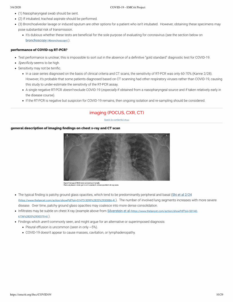
The typical �nding is patchy ground glass opacities, which tend to be predominantly peripheral and basal (Shi et al 2/24(https://www.thelancet.com/action/showPdf?pii=S1473-3099%2820%2930086-4) ). The number of involved lung segments increases with more severedisease. Over time, patchy ground glass opacities may coalesce into more dense consolidation.In�ltrates may be subtle on chest X-ray (example above from Silverstein et al (https://www.thelancet.com/action/showPdf?pii=S0140-
6736%2820%2930370-6) ).Findings which aren't commonly seen, and might argue for an alternative or superimposed diagnosis:
Pleural effusion is uncommon (seen in only ~5%).COVID-19 doesn't appear to cause masses, cavitation, or lymphadenopathy.
sensitivity and time delay in chest X-ray and CT scan
Limitations in the dataData from different studies con�ict to a certain extent. This probably re�ects varying levels of exposure intensity and illness severity(cohorts with higher exposure intensity and disease severity will be more likely to have radiologic changes).
Sensitivity of CT scanning?Guan et al. (https://www.nejm.org/doi/pdf/10.1056/NEJMoa2002032?articleTools=true) found CT abnormalities among 86% of symptomaticpatients presenting to the hospital. Likewise, Fang et al. found CT abnormalities among 50/51 patients.Among patients with constitutional symptoms only (but not respiratory symptoms), CT scan may be less sensitive (e.g. perhaps ~50%)(Kanne 2/27 (https://pubs.rsna.org/doi/10.1148/radiol.2020200527) ).
CT scan abnormalities might emerge before symptoms?Shi et al (https://www.thelancet.com/action/showPdf?pii=S1473-3099%2820%2930086-4) performed CT scanning in 15 healthcare workers who wereexposed to COVID-19 before they became symptomatic.Ground glass opaci�cation on CT scan was seen in 14/15 patients! 9/15 patients had peripheral lung involvement (some bilateral,some unilateral).Emergence of CT abnormality before symptoms could be consistent with the existence of an asymptomatic carrier state (discussedabove).
Chest X-rayLess data has focused on the sensitivity of chest X-ray than CT scan.It's fair to assume that the sensitivity of chest X-ray must be lower than CT scan.The sensitivity of chest X-ray was found to be 59% among symptomatic patients presenting to the hospital in one series (Guan et al2/28 (https://www.nejm.org/doi/pdf/10.1056/NEJMoa2002032?articleTools=true) ).
lung ultrasonography
There isn't any data available currently regarding the use of lung ultrasonography for COVID-19. However, a peripheral ground-glass patternon CT scan will generate patchy B-lines on lung ultrasonography (regardless of what the underlying disease is – this is simply a matter ofultrasound physics). This allows us to a make some educated guesses regarding lung ultrasonography �ndings:
(1) The most common pattern seen on lung ultrasonography will likely be patchy B-lines (areas with B-lines, with interspersed areas ofnormal lung in between).(2) The sensitivity of lung ultrasonography will increase in parallel with disease severity (more severe illness involves more lungsegments, which will be easier to detect on lung ultrasonography).(3) Some patients may have lung ultrasonography abnormalities before they develop symptoms (based on the peripheral distribution ofin�ltrates in asymptomatic patients described by Shi et al. (https://www.thelancet.com/action/showPdf?pii=S1473-3099%2820%2930086-4) ).(4) Some patients will probably have early, mild pneumonitis which may be detectable via ultrasonography, but not via traditional chestX-ray (ultrasonography is more sensitive for subtle, pleural-based ground glass opacity).(5) In order to achieve sensitivity, a thorough lung examination would be needed (taking a “lawnmower” approach, attempting tovisualize as much lung tissue as possible). Simple two-point lung ultrasonography examinations will miss focal ground-glass opacities.
Lung ultrasonography has some advantages in the context of a COVID-19 outbreak:Ultrasonography may be performed at the point of care (including outside the hospital). This avoids transporting the patient toradiology, with possible exposure to hospital staff.Ultrasonography could be helpful as an early detection tool in patients who are under suspicion of having COVID-19 (e.g. lungabnormalities might suggest the presence of an asymptomatic carrier state, which might indicate the need for quarantine).
all imaging modalities are nonspeci�c
All of the above techniques (CXR, CT, sonography) are nonspeci�c. Patchy ground-glass opacities may be caused by a broad range ofdisease processes (e.g. viral and bacterial pneumonias). For example, right now in the United States, someone with patchy ground-glassopacities on CT scan would be much more likely to have a garden variety viral pneumonia (e.g. in�uenza or RSV) rather than COVID-19.Imaging cannot differentiate between COVID-19 and other forms of pneumonia.Imaging could help differentiate between COVID-19 and non-pulmonary disorders (e.g. sinusitis, non-pulmonary viral illness).Ultimately, the imaging is only one bit of information which must be integrated into clinical context.
possible approach to imaging in COVID-19
Below is one possible strategy to use for patients presenting with respiratory symptoms and possible COVID-19.The temptation to get a CT scan in all of these patients should be resisted. In most cases, a CT scan will probably add little to chest X-rayand lung ultrasonography (in terms of actionable data which affects patient management).From a critical care perspective, CT scanning will likely add little to the management of these patients (all of whom will have diffusein�ltrates).
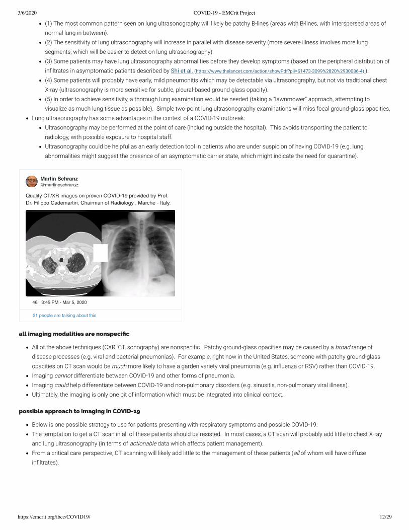
Martin Schranz@martinpschranz
Quality CT/XR images on proven COVID-19 provided by Prof. Dr. Filippo Cademartiri, Chairman of Radiology , Marche - Italy.
RSNA focus page on coronavirus (https://pubs.rsna.org/2019-nCoV#images) (contains fantastic slide show that provides an appreciation ofpossible imaging �ndings in a few minutes)
bronchoscopy(back to contents) (#top)
Risks of bronchoscopy:May cause some deterioration in clinical condition (due to instillation of saline and sedation).Enormous risk of transmission to providers.Considerable resource allocation (requires N95 respirators, physicians, respiratory therapists) – all resources which will be in slimsupply during an epidemic.
Bene�ts of bronchoscopy:Bene�t of diagnosing COVID-19 is dubious at this point (given that treatment is primarily supportive).
Bottom line on bronchoscopy?Bronchoscopy might be considered in situations where it would otherwise be performed (e.g. patient with immunosuppression withconcerns for Pneumocystis pneumonia or fungal pneumonia).Bronchoscopy should not be done for the purpose of ruling COVID-19 in or out (as this entails risk with no de�nite bene�ts)(Bouadmaet al. (https://link.springer.com/content/pdf/10.1007/s00134-020-05967-x.pdf) ).
key principle: supportive care for viral pneumonia(back to contents) (#top)
general principle: avoid COVID-19 exceptionalism
We know how to treat severe viral pneumonia and ARDS. We've been doing this for years.There is not yet any compelling evidence that the fundamentals of treating COVID-19 are substantially different fromtreating other forms of viral pneumonia (e.g. in�uenza).The essential strategy of treatment for COVID-19 is supportive care, which should be performed as it would be donefor any patient with severe viral pneumonia. For example, if you were to simply treat the patient as if they hadin�uenza (minus the oseltamivir), you would be doing an excellent job.Below are some minor adjustments on the care that we provide, which might optimize things a bit for treating COVID-19. However, overall the treatment is fundamentally the same as for treating any viral pneumonia.
background on antiviral therapy(back to contents) (#top)
No anti-viral therapy has been proven to work for COVID-19 in humans. Multiple RCTs are ongoing; hopefully they will bring us furtherinformation soon.
Whenever possible, patients should be enrolled in RCTs.Information is provided below about some of the more popular agents which are being used by some practitioners.
Inclusion in this chapter is not a recommendation to use one or more of these medications. This information is simply provided as abackground to help us understand these therapies.A focus is placed on lopinavir/ritonavir and chloroquine since these agents are currently available.Practitioners are encouraged to review available evidence and reach their own conclusions regarding whether to use thesemedications.If you have experience or new evidence or opinions on anti-viral therapy, please share it on the COVID-19 discussion page here(https://emcrit.org/pulmcrit/covid19/) .
single vs. multi-drug regimens ??
Another unknown is whether a single drug could work, or whether a combination of multiple anti-viral agents is needed.Analogous to HIV, it's possible that two or three anti-virals working in synergy might be needed. Combinations of agents could increasetoxicity however (especially cardiotoxicity).
indications for antiviral therapy: who & when ??
When ??Retrospective data from SARS suggests that earlier treatment (e.g. within 1-2 days of admission) may be more effective than reservingtherapy until severe organ failures occur (Chan 2003 (https://www.hkmj.org/abstracts/v9n6/399.htm) ). This is consistent with data fromin�uenza that suggests a �nite treatment window occurring relatively early in the disease course.
Who ??The vast majority of patients will do �ne without any therapy, so in most cases there's no need for antiviral therapy.However, waiting until patients are severely ill before initiating therapy could cause us to miss an early treatment window, during whichthe disease course is more modi�able.Predictors of adverse outcome might be useful in predicting who will do poorly and thus who might bene�t most from early anti-viraltherapy? (see section below on prognosis (#prognosis) ).
remdesivir(back to contents) (#top)
Remdesivir might be an excellent antiviral, based on a study involving in vitro and animal data with MERS (e.g. Sheahan 2020(https://www.ncbi.nlm.nih.gov/pmc/articles/PMC6954302/pdf/41467_2019_Article_13940.pdf) ).Unfortunately, remdesivir is not commercially available. Remdesivir was used on the basis of “compassionate use” for one of the �rstpatients with COVID-19 in the United States (Holshue 2020 (https://www.nejm.org/doi/full/10.1056/NEJMoa2001191) ).Remdesivir is being used in one trial (https://clinicaltrials.gov/ct2/show/NCT04280705) in the United States being sponsored by NIAID. Enrollment inthis trial is the most desirable approach to antiviral therapy (if feasible).
lopinavir/ritonavir (KALETRA)(back to contents) (#top)
general description
This is a combination of antiviral agents used in treatment of HIV (including post-exposure prophylaxis following needle-stick injury).Compared to remdesivir, lopinavir/ritonavir has the advantage that it's widely available and has an established toxicity pro�le (it does haveknown side-effects and drug interactions, but these are generally tolerable).Lopinavir/ritonavir appears to work synergistically with ribavirin. Available human data on SARS and MERS have combined these threeagents together. It's possible that a cocktail of all three drugs is required for e�cacy (potentially explaining failures of any of these agents inisolation). A recent very small study on lopinavir/ritonavir alone wasn't particularly impressive, suggesting that triple therapy withlopinavir/ritonavir/ribavirin might be necessary (Young 3/3/20 (https://jamanetwork.com/journals/jama/fullarticle/2762688) ).
Lopinavir and ritonavir are protease inhibitors, which block viral replication.Lopinavir seems to be the agent which actually acts on the virus. Ritonavir is a CYP3A inhibitor which functions primarily to reducemetabolism of lopinavir, thereby boosting lopinavir levels.
in vitro data
Lopinavir showed in vitro antiviral activity against SARS at concentration of 4 ug/ml. However, when combined with ribavirin, lopinavirappears considerably more effective (with an inhibitory concentration of 1 ug/mL (Chu et al. 2004 (https://thorax.bmj.com/content/59/3/252.long) ).For reference, the peak and trough serum concentrations of lopinavir are 10 and 5.5 ug/ml (Chu et al. 2004(https://thorax.bmj.com/content/59/3/252.long) ).
animal data
Lopinavir/ritonavir was effective against MERS-CoV in a primate animal model (Chan 2015 (https://www.ncbi.nlm.nih.gov/pubmed/26198719) ).
human data
Chu et al. 2004 (https://thorax.bmj.com/content/59/3/252.long) : Open-label before/after study on SARS.41 patients treated with lopinavir/ritonavir plus ribavirin were compared to 111 historical control patients treated with ribavirin alone. Baseline imbalances did exist between groups (patients treated with lopinavir/ritonavir had lower initial lactate dehydrogenase (LDH)levels – so they weren't as sick).Poor clinical outcomes (ARDS or death) were lower in treatment group (2.4% vs. 29%). These differences persisted in multivariablemodels, which attempted to correct for baseline imbalances between the groups.Use of lopinavir/ritonavir use correlated with a dramatic reduction in viral load (�gure above).All patients received concomitant ribavirin. The dose was 4 grams oral loading dose followed by 1.2 grams PO q8hr (or 8 mg/kg IVq8hr) for 14 days.
Chan et al. 2003 (https://www.hkmj.org/abstracts/v9n6/399.htm) : Retrospective matched multi-center cohort study on SARS75 patients treated with lopinavir/ritonavir were compared with controls (matched on the basis of sex, age, comorbidities, lactatedehydrogenase level, and use of pulse-dose steroid).Up-front treatment with lopinavir/ritonavir combined with ribavirin correlated with reduced mortality (2.3% versus 16%). However,rescue therapy with lopinavir/ritonavir (often without concomitant ribavirin) didn't seem to make any difference. The ribavirin dose was2.4 grams loading dose, followed by 1.2 grams PO q8hr (or 8 mg/kg IV q8hr) for 10-14 days.
Park et al. 2019 (https://www.ncbi.nlm.nih.gov/pubmed/30240813) : Retrospective cohort study on post-exposure prophylaxis against MERSThis is a retrospective cohort study involving 22 patients with high-risk exposure to a single MERS patient (table below). As a controlgroup, four hospitals with outbreaks of MERS were selected.Post-exposure prophylaxis consisted of a combination of lopinavir/ritonavir (400 mg / 100 mg BID for 11-13 days) plus ribavirin (2000mg loading dose, then 1200 mg q8hr for four days, then 600 mg q8hr for 6-8 days).MERS infections didn't occur in anyone treated with post-exposure prophylaxis (table below). However, the manner in which the controlgroup was selected (retrospectively selecting hospitals with MERS outbreaks) likely biased the study in favor of showing a bene�t ofpost-exposure prophylaxis.Post-exposure therapy was generally well tolerated, although most patients reported some side-effects (most commonly nausea,diarrhea, stomatitis, or fever). Laboratory evaluation shows frequent occurrence of anemia (45%), leukopenia (40%), andhyperbilirubinemia (100%).
Young et al. 3/3/2020 (https://jamanetwork.com/journals/jama/fullarticle/2762688)
Cohort study describing 16 COVID-19 patients in Singapore. Among six patients with hypoxemia, �ve were treated withlopinavir/ritonavir (200 mg/100 mg BID, which is half of the usual dose of lopinavir).Among the �ve patients, two patients deteriorated and had persistent nasopharyngeal virus carriage.Possible reasons for these underwhelming results might include: statistical underpowering, low dose of lopinavir/ritonavir, lack ofsynergistic ribavirin, and/or late initiation of therapy. For further discussion see PulmCrit blog on this study here(https://emcrit.org/pulmcrit/lopinavir/) .
Other evidence of lower quality:Lopinavir/ritonavir has been used to treat one patient with COVID-19 (Kim 2020(https://www.ncbi.nlm.nih.gov/pmc/articles/PMC7025910/pdf/jkms-35-e79.pdf) ).Lopinavir/ritonavir was reported to be effective in some case reports of MERS (Momattin 2019(https://www.ncbi.nlm.nih.gov/pubmed/31252170) ).
Lopinavir/ritonavir is currently under investigation within multiple RCTs in China (but none in the United States).
dosing
(1) Lopinavir/Ritonavir (Monograph (https://reference.medscape.com/drug/kaletra-lopinavir-ritonavir-342629) from MedScape)Standard dose (and dose used against coronaviruses) is 400 mg / 100 mg PO BID.Generally no adjustment is made in renal dysfunction.
(2) Ribavirin (Monograph (https://reference.medscape.com/drug/rebetol-ribasphere-ribavirin-342625) from MedScape)Unknown whether synergistic ribavirin is useful.The best validated regimen is probably Chu et al. 2004 (https://thorax.bmj.com/content/59/3/252.long) : 4 grams oral loading dose followed by1.2 grams PO q8hr (or 8 mg/kg IV q8hr) for 14 days.
In Chu et al. 2004 (https://thorax.bmj.com/content/59/3/252.long) , 41 patients with SARS tolerated lopinavir/ritonavir reasonably well (onepatient needed to discontinue due to doubling of transaminase levels).In Chan 2003 (https://www.hkmj.org/abstracts/v9n6/399.htm) , 75 patients with SARS were treated with lopinavir/ritonavir without reports ofsevere adverse effects.
further information
PulmCrit blog 3/4 (https://emcrit.org/pulmcrit/lopinavir/) discussing the Young study and double vs. triple therapy.Further information on this is available in a recent review by Yao TT et al. (https://onlinelibrary.wiley.com/doi/epdf/10.1002/jmv.25729)
chloroquine(back to contents) (#top)
general description
Chloroquine is generally used for treatment of malaria and amebiasis. It has anti-viral activity in vitro, but no established track record intreatment of viral disease.The toxicity pro�le seems to be acceptable (e.g. its widely used as malaria prophylaxis — albeit at a much lower dose than is currently beingconsidered for COVID-19).
mechanism of action
Chloroquine appears to work via multiple mechanisms, including:Interference with with the cellular receptor ACE2 (potentially making it particularly effective against SARS and COVID-19).Impairment of acidi�cation of endosomes, which interferes with virus tra�cking within cells.
in vitro data
In vitro data using cell lines shows that chloroquine can inhibit COVID-19 with an 50% inhibitory concentration of 1 uM, implying thattherapeutic levels could be achieved in humans (Wang 2020 (https://www.ncbi.nlm.nih.gov/pubmed/32020029) ). The 50% inhibitory concentration ofchloroquine for SARS is closer to 9 uM, suggesting that chloroquine could be more effective against COVID-19 than SARS (Al-Bari 2017(https://bpspubs.onlinelibrary.wiley.com/doi/full/10.1002/prp2.293) ).
animal data
Chloroquine failed to work in mice infected with SARS (Bernard 2006 (https://www.ncbi.nlm.nih.gov/pubmed/17176632) ).
Currently lacking.Emerging data from China suggests that chloroquine has been studied with favorable results, although precise data is lacking as of yet (Gao2020 (https://www.jstage.jst.go.jp/article/bst/advpub/0/advpub_2020.01047/_pdf/-char/en) ). An expert consensus group in China is recommending atreatment regimen of 500 mg PO twice daily for patients without contraindications . Hopefully, clinical data with chloroquine will bepublished shortly.
dosing (Monograph (https://reference.medscape.com/drug/aralen-chloroquine-phosphate-chloroquine-342687) from MedScape)
500 mg chloroquine phosphate contains 300 mg of chlorquine itself (a.k.a. chloroquine base).500 mg PO twice daily for 10 days is the regimen recommended by a group in China for patients without contraindications (Zhonghua 2020(https://www.ncbi.nlm.nih.gov/pubmed/32075365) ).May require dose adjustment in renal or hepatic dysfunction.
contraindications/cautions
Serious adverse effects may include:QT prolongation & Torsades de PointesReduction in seizure thresholdAnaphylaxis or anaphylactoid reactionNeuromuscular impairmentNeuropsychiatric disorders (potential to increase delirium?)Pancytopenia, neutropenia, thrombocytopenia, aplastic anemiaHepatitis
Common adverse reactions:Nausea/vomiting, diarrhea, abdominal painVisual disturbance, headacheExtrapyramidal symptoms
Mixed messages from China regarding how widely this is being used or recommended.Many articles don't mention chloroquine at all.A few articles strongly recommend this (Zhonghua 2020 (https://www.ncbi.nlm.nih.gov/pubmed/32075365) , Gao 2020(https://www.jstage.jst.go.jp/article/bst/advpub/0/advpub_2020.01047/_pdf/-char/en) )
Hopefully additional data will be forthcoming shortly.
oseltamavir & other neuraminidase inhibitors(back to contents) (#top)
Neuraminidase inhibitors don't seem to work against COVID-19 (Tan et al 2004 (https://www.ncbi.nlm.nih.gov/pmc/articles/PMC3323075/pdf/03-
0458.pdf) ).Initial empiric therapy with neuraminidase inhibitors could be reasonable during in�uenza season in critically ill patients, if there is concernthat the patient might have in�uenza pneumonia.
Currently, in many locations, patients presenting with viral pneumonia are much more likely to have in�uenza than COVID-19.
anti-bacterial therapy(back to contents) (#top)
initial empiric antibiotics
COVID-19 itself is not an indication for antibiotics.Initially, there may be concerns regarding the possibility of a superimposed bacterial pneumonia. When in doubt, it may be sensible to obtainbacterial cultures and procalcitonin, prior to initiation of empiric antibiotic therapy. Based on culture and procalcitonin results, antibiotics
might be discontinued in <48 hours if there isn't evidence of a bacterial infection (this is exactly the same as management of in�uenzapneumonia).
delayed bacterial superinfection
Bacterial pneumonia can emerge during the hospital course (especially ventilator-associated pneumonia in patients who are intubated).Among patients who died from COVID-19, one series found that 11/68 (16%) had secondary infections (Ruan 3/3/20(https://link.springer.com/content/pdf/10.1007/s00134-020-05991-x.pdf) ).
This may be investigated and treated similarly to other ventilator-associated pneumonias, or hospital-acquired pneumonias.
steroid(back to contents) (#top)
steroid
Steroid should not generally be used. Steroid hasn't demonstrated bene�t in prior SARS or MERS epidemics. Steroid may increase viralshedding (Lee 2004 (https://www.sciencedirect.com/science/article/pii/S1386653204001957?via%3Dihub) ).Nearly all articles recommend against the use of steroid. However, steroid may be used if there is another clear-cut indication for steroid(e.g. coronavirus plus asthma exacerbation).
WHO guidelines summary the relevant evidence regarding steroid; for further information read them here(�le:///Users/joshuafarkas1/Downloads/clinical-management-of-novel-cov%20(1).pdf) (see bottom of page 4).
ascorbic acid ??
Ascorbic acid did appear to improve mortality in the multi-center CITRIS-ALI trial (https://jamanetwork.com/journals/jama/article-abstract/2752063) . However, interpretation of this trial remains hopelessly contentious due to nearly unsolvable issues with survival-ship bias (discussed here(https://emcrit.org/pulmcrit/pulmcrit-citris-ali-can-a-secondary-endpoint-stage-a-coup-detat/) ).Extremely limited evidence suggests that ascorbic acid could be bene�cial in animal models of coronavirus (Atherton 1978(https://www.ncbi.nlm.nih.gov/pubmed/205194) ).Administration of a moderate dose of IV vitamin C could be considered (e.g. 1.5 grams IV q6 ascorbic acid plus 200 mg thiamine IV q12). This dose seems to be safe. However, there is no high-quality evidence to support ascorbic acid in viral pneumonia.
hemodynamic support(back to contents) (#top)
avoid �uid resuscitation
Patients rarely are shocked on admission (even among critically ill patients, admission blood pressure is generally normal and lactateelevations are mild-moderate)(Yang et al 2/21 (https://www.thelancet.com/action/showPdf?pii=S2213-2600%2820%2930079-5) ).
Overall, the rate of reported “sepsis” is generally low (<5%). The virus doesn't seem to generally cause a septic shock picture (but ofcourse, patients may always suffer from superimposed bacterial septic shock).
The cause of death from COVID-19 is nearly always ARDS – which may be exacerbated by �uid administration.Gentle �uid administration could be considered for patients with evidence of hypoperfusion and a history suggestive of total bodyhypovolemia (e.g. prolonged nausea/vomiting and diarrhea).More discussion on �uid therapy for COVID-19 here (https://emcrit.org/pulmcrit/coronavirus/) .
cardiomyopathy ?
COVID-19 does commonly cause troponin elevations (which generally will not represent type-I myocardial infarctions).Ruan 3/3/20 (https://link.springer.com/article/10.1007/s00134-020-05991-x) reported that ~7% of patients die of fulminant myocarditis. This may alsobe a contributing factor in ~33% of deaths.Wang 2/7 (https://jamanetwork.com/journals/jama/fullarticle/2761044) reported that arrhythmia was a cause of ICU transfer in 12% of patients.Troponin elevation seems to be a strong prognostic indicator for mortality (see prognosis (#prognosis) section below). It's unclear to whatextent this represents cardiac involvement causing death versus troponin merely being an indicator of severe global illness placing stress onthe heart. Elevated troponin levels correlate with mortality across a variety of critical illnesses.
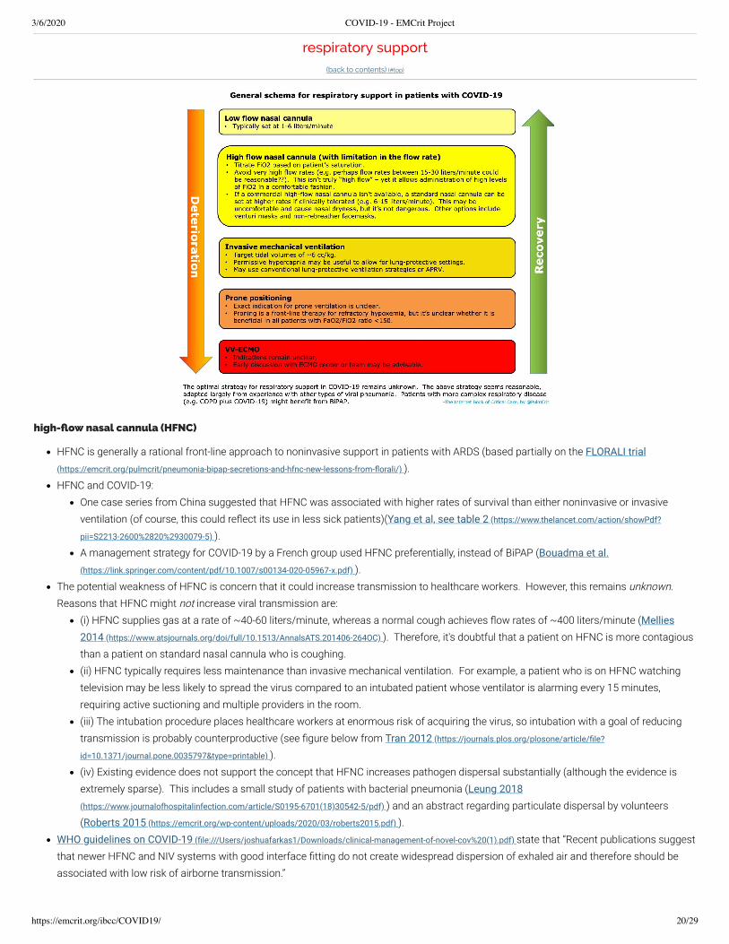
HFNC is generally a rational front-line approach to noninvasive support in patients with ARDS (based partially on the FLORALI trial(https://emcrit.org/pulmcrit/pneumonia-bipap-secretions-and-hfnc-new-lessons-from-�orali/) ).HFNC and COVID-19:
One case series from China suggested that HFNC was associated with higher rates of survival than either noninvasive or invasiveventilation (of course, this could re�ect its use in less sick patients)(Yang et al, see table 2 (https://www.thelancet.com/action/showPdf?
pii=S2213-2600%2820%2930079-5) ).A management strategy for COVID-19 by a French group used HFNC preferentially, instead of BiPAP (Bouadma et al.(https://link.springer.com/content/pdf/10.1007/s00134-020-05967-x.pdf) ).
The potential weakness of HFNC is concern that it could increase transmission to healthcare workers. However, this remains unknown. Reasons that HFNC might not increase viral transmission are:
(i) HFNC supplies gas at a rate of ~40-60 liters/minute, whereas a normal cough achieves �ow rates of ~400 liters/minute (Mellies2014 (https://www.atsjournals.org/doi/full/10.1513/AnnalsATS.201406-264OC) ). Therefore, it's doubtful that a patient on HFNC is more contagiousthan a patient on standard nasal cannula who is coughing.(ii) HFNC typically requires less maintenance than invasive mechanical ventilation. For example, a patient who is on HFNC watchingtelevision may be less likely to spread the virus compared to an intubated patient whose ventilator is alarming every 15 minutes,requiring active suctioning and multiple providers in the room.(iii) The intubation procedure places healthcare workers at enormous risk of acquiring the virus, so intubation with a goal of reducingtransmission is probably counterproductive (see �gure below from Tran 2012 (https://journals.plos.org/plosone/article/�le?
id=10.1371/journal.pone.0035797&type=printable) ).(iv) Existing evidence does not support the concept that HFNC increases pathogen dispersal substantially (although the evidence isextremely sparse). This includes a small study of patients with bacterial pneumonia (Leung 2018(https://www.journalofhospitalinfection.com/article/S0195-6701(18)30542-5/pdf) ) and an abstract regarding particulate dispersal by volunteers(Roberts 2015 (https://emcrit.org/wp-content/uploads/2020/03/roberts2015.pdf) ).
WHO guidelines on COVID-19 (�le:///Users/joshuafarkas1/Downloads/clinical-management-of-novel-cov%20(1).pdf) state that “Recent publications suggestthat newer HFNC and NIV systems with good interface �tting do not create widespread dispersion of exhaled air and therefore should beassociated with low risk of airborne transmission.”
One potential compromise might be to use HFNC with a moderate rate of �ow (e.g. 15-30 liters/minute, rather than 40-60 liters/minute). Since 15-30 liters/minute �ow is close to a baseline minute ventilation for a sick respiratory failure patient, adding this level of �ow is unlikelyto affect matters substantially.A potential limitation of HFNC during an epidemic could be exhaustion of the hospital's oxygen supply.
noninvasive ventilation (BiPAP)
The role of BiPAP is a bit dubious here.In a multicenter cohort of 302 patients with MERS coronavirus, 92% of patients treated with BiPAP failed this modality and requiredintubation (Alraddadi 2019 (https://www.ncbi.nlm.nih.gov/pubmed/30884185) ).In the FLORALI trial (https://emcrit.org/pulmcrit/pneumonia-bipap-secretions-and-hfnc-new-lessons-from-�orali/) of ARDS patients (with mostlypneumonia of various etiologies), patients randomized to BiPAP did worse compared to patients randomized to HFNC.
BiPAP could have a niche role in patients with combined syndromes (e.g. COPD plus COVID-19). For more on the selection of BiPAP vs.HFNC, see this chapter (https://emcrit.org/ibcc/support/) on noninvasive respiratory support.A helmet interface has been proposed to reduce environmental contamination (Cabrini 2020(https://www.thelancet.com/journals/lancet/article/PIIS0140-6736(20)30359-7/fulltext) ). Unfortunately, access to these devices is limited in the UnitedStates. Placement of a viral �lter in-line with the exhalation tubing could also potentially reduce contamination.
invasive ventilation
Begin with ventilator optimization (using either conventional lung-protective ventilation or APRV) for 12-18 hours.The PulmCrit guide to using APRV can be found here (#https://emcrit.org/squirt/aprv/) .Based on informal reports from Italy, patients may require relatively high airway pressures (e.g. PEEP of 16-18 cm).
For failure to respond to initial ventilator optimization (e.g. with persistent PaO2/FiO2 below 150 mm), prone ventilation may be considered. However, there are some reasons that prone ventilation might not be desirable here:
Prone ventilation demonstrated mortality bene�t in the PROSEVA trial (https://www.nejm.org/doi/full/10.1056/NEJMoa1214103) in France, in thecontext of centers which were highly experienced at prone ventilation. It's controversial whether these bene�ts would be replicated inanother RCT in a country less experienced with prone ventilation.
Prone ventilation is very labor-intensive. This would require exposing numerous healthcare providers to the patient, multiple times perday.Nevertheless, prone ventilation could be a useful intervention for truly refractory hypoxemia.
ECMO
Patients with COVID-19 are often relatively young and suffering from single-organ failure due to a reversible etiology, so many would beexcellent candidates for ECMO (probably mostly VV ECMO).Indications and timing are unclear.In an epidemic, ECMO capabilities would probably rapidly become saturated. Very thorny ethical issues could arise (e.g. how long of anECMO run is one patient allowed to have before the withdrawal of life-sustaining therapy, in order to allow the circuit to be used for anotherpatient).
awake prone ventilation
This involves a non-intubated patient on nasal cannula who prone themselves by lying on their belly.There is relatively little evidence to support this and it is useful only for highly selected patients (reviewed here (https://emcrit.org/pulmcrit/proning-
nonintubated/) ).Awake-prone ventilation could be a useful option if the availability of mechanical ventilators is exhausted.
Typically awake prone ventilation is paired with high-�ow nasal cannula, but it could also be used with a standard nasal cannula (e.g.running at ~6 L/min or a bit higher if tolerated).Consider securing the nasal cannula to the patient's face using tape or tegaderm, to prevent dislodgment when the patient moves.
intubation procedure(back to contents) (#top)
This represents a high risk for transmission to healthcare workers.Airborne precautions are indicated (e.g. N95/FFP2 masks or positive air-purifying respirators, along with full face shields and full contactprecautions).Rapid sequence intubation with no bag-mask ventilation may avoid aerosolizing particles. However, during the apneic period, a bag-valvemask with a PEEP valve could be passively held on the patient's face to maintain positive pressure in the airway and thereby prevent de-recruitment.Use of videolaryngoscopy may avoid placing the operator's face close to the patient.Attach a viral �lter to the bag-valve mask before the procedure, if possible. This should reduce the spread of viral particles out of theendotracheal tube following intubation (or during bag-mask ventilation if that is required)(Peng et al. 2/27 (https://bjanaesthesia.org/article/S0007-
0912(20)30098-2/pdf) ).Endotracheal tube con�rmation with a stethoscope could pose a risk of transferring virus to the practitioner. It could be safer to advance theendotracheal tube to a pre-calculated depth calculated based on the patient's height (see MDCalc formula (https://www.mdcalc.com/endotracheal-
tube-ett-depth-tidal-volume-calculator) here).
more information
EMCrit Wee: Airway management in COVID-19 (https://emcrit.org/emcrit/airway-covid-19/) (3/1)
">
renal failure(back to contents) (#top)
Renal failure requiring dialysis is reported in a subset of patients admitted to ICU.The exact mechanism is unclear at this point, but some conjectures may be reached based on SARS (Chu et al. 2005 (https://www.kidney-
international.org/article/S0085-2538(15)50506-1/pdf) ).SARS causes renal failure in ~7% of patients. The pathology shows acute tubular necrosis, which appears to be a re�ection ofgeneralized multi-organ failure. In some cases rhabdomyolysis may have contributed as well. Renal failure correlates with a poor
overall prognosis (92% mortality with renal failure versus 9% without). In multivariable analysis, renal failure was the strongestpredictor of mortality (more-so even than ARDS).
prognosis(back to contents) (#top)
general prognosis
(1) It remains unclear what fraction of patients are hospitalized.There may be lots of patients with mild illness who don't present to medical attention and aren't counted.The vast majority of infected patients (e.g. >80%) don't get signi�cantly ill and don't require hospitalization.
(2) Among hospitalized patients (Guan et al 2/28 (https://www.nejm.org/doi/pdf/10.1056/NEJMoa2002032?articleTools=true) )~6% of patients are admitted to ICU.~2.4% require intubation.~1.5% die.
(There are numerous sets of numbers published and they vary quite a bit. However, from the clinician's standpoint the precise numbers don'treally matter.)
Laboratory abnormalities (Xie 2020 (https://link.springer.com/article/10.1007/s00134-020-05979-7) )(1) Lymphopenia and its trends over time (prolonged or worsening lymphopenia portends poor outcome)(Chu et al. 2004(https://thorax.bmj.com/content/59/3/252.long) )(2) Higher levels of C-reactive protein and troponin (�gure below).(3) Neutrophil/lymphocyte ratio (NLR) (https://emcrit.org/pulmcrit/nlr/) appears to be a superior prognosticator when compared to eitherlymphopenia or C-reactive protein (Liu et al. pre-print (https://www.medrxiv.org/content/10.1101/2020.02.10.20021584v1.full.pdf) ). As shown in thethird �gure below, values >3 could suggest a worse prognosis.
Evidence on mortality(1) Ruan 3/3/20 (https://link.springer.com/article/10.1007/s00134-020-05991-x) described predictors and patterns of mortality (�gure below).(2) The largest series of mortality data comes from the Chinese CDC (http://weekly.chinacdc.cn/en/article/id/e53946e2-c6c4-41e9-9a9b-
fea8db1a8f51) (table below). The absolute numbers may vary depending on whether some cases were missed, but the relative impact ofvarious risk factors is probably accurate.
Patients who survive the initial phases of the illness may still require prolonged ventilator support (possibly developing some radiographicelements of �brosis)(Zhang 2020 (https://link.springer.com/article/10.1007/s00134-020-05990-y) ).As the epidemic progresses, an issue which may arise is a large volume of patients unable to wean from mechanical ventilation.
disposition(back to contents) (#top)
avoidance of unnecessary emergency department or clinic visits
Health systems should ideally be put in place to dissuade patients from presenting to the clinic or emergency department for testing to seeif they have COVID-19 (e.g. if they have mild constitutional symptoms and don't otherwise require medical attention).Korea has developed a system of drive-thru testing, which avoids exposure of other patients in the emergency department. Outdoor testingalso ensures ongoing circulation of fresh air.
ian bremmer@ianbremmer
Drive-thru coronavirus testing clinic in South Korea. Innovation drives resilience.
The vast majority of patients with coronavirus will recover spontaneously, without requiring any medical attention (perhaps >80% ofpatients).Patients with mild symptoms can generally be discharged home, with instructions to isolate themselves. These decisions should be madein coordination with local health departments, who can assist in follow-up.Features favoring home discharge may include:
Ability to understand and comply with self-isolation (e.g. separate bedroom and bathroom).Ability to call for assistance if they are deteriorating.Having household members who aren't at increased risk of complications from COVID-19 (e.g. elderly, pregnant women, or people withsigni�cant medical comorbidities).Lack of hypoxemia, marked chest in�ltrates, or other features that would generally indicate admission.
For more, see CDC interim guidance for disposition of patients with COVID-19 here (https://www.cdc.gov/coronavirus/2019-ncov/hcp/disposition-
hospitalized-patients.html) and here (https://www.cdc.gov/coronavirus/2019-ncov/hcp/guidance-home-care.html?
Delayed consideration of COVID19, leading to delayed initiation of precautions (e.g. in a patient presenting with gastrointestinal illness).Treatment of COVID19 based on Surviving Sepsis Guidelines (e.g. with 30 cc/kg �uid). This is wrong on so many levels(https://emcrit.org/pulmcrit/coronavirus/) , for example:
Broad application of 30 cc/kg �uid is often detrimental in septic shock.COVID-19 patients don't actually present with septic shock anyways.Large volume �uid is extremely dangerous in ARDS.
Inadequate attention to contact precautions (e.g. hand hygiene and sterilization of surfaces).Admission of patients to hospital for COVID19 who could be safely managed as outpatients.Use of the emergency department as a COVID-19 screening area.Be careful of making major changes to usual treatment approaches for viral pneumonia, based on limited evidence. Ultimately the key hereis simply high-quality supportive care for viral pneumonia.
Update from ESICM (https://www.esicm.org/blog/?p=2569) by David Lyness 3/2
Excellent lecture by Forest Arnold at the University of Louisville:
Going further:
Journal & Society homepages on COVID-19CDC (https://www.cdc.gov/coronavirus/index.html)
FOAMed on COVID-19WHO guidelines on �uid administration for COVID-19 are dangerous (https://emcrit.org/pulmcrit/coronavirus/) (PulmCrit)EMCrit RACC on airway management (https://emcrit.org/emcrit/airway-covid-19/) in COVID-19 (Weingart & Brian Wright)COVID-19 on RebelEM (https://rebelem.com/covid-19-the-novel-coronavirus-2019/) (Salim Rezaie)COVID-19 on St. Emlyns (https://www.stemlynsblog.org/2019-novel-coronavirus-wuhan-at-st-emlyns/) (Ashley Liebig)COVID-19 on Radiopaedia (https://radiopaedia.org/articles/covid-19?fbclid=IwAR2G1HjFlbP1aj3Inj_-HqG27NM3lx8TXE4VpTpTHUu0PYWRTJmHvIqbVAE)
(Daniel Bell)(References to some patient series listed in the tables)
Yang et al (https://www.thelancet.com/action/showPdf?pii=S2213-2600%2820%2930079-5) : 52 critically ill patients, LancetChen et al (https://www.thelancet.com/action/showPdf?pii=S0140-6736%2820%2930211-7) : 99 infected patients, LancetShi et al (https://www.thelancet.com/action/showPdf?pii=S1473-3099%2820%2930086-4) : 81 patients with CT imaging, Lancet.
The Internet Book of Critical Care is an online textbook written by Josh Farkas (@PulmCrit), an associate professor ofPulmonary and Critical Care Medicine at the University of Vermont.
EMCrit is a trademark of Metasin LLC. Copyright 2009-. This site represents our opinions only. See our full disclaimer, our privacy policy, commenting policy and here for credits