Department of Veterans Affairs Journal of Rehabilitation Research and Development Vol . 37 No . 2, March/April 2000 Pages 179—188 Continuous passive motion (CPM) : Theory and principles of clinical application Shawn W . O'Driscoll, MD, PhD and Nicholas J . Giori, MD, PhD Mayo Clinic, Rochester MN 55905 Abstract —Stiffness following surgery or injury to a joint develops as a progression of four stages : bleeding, edema, granulation tissue, and fibrosis . Continuous passive motion (CPM) properly applied during the first two stages of stiffness acts to pump blood and edema fluid away from the joint and periarticular tissues . This allows maintenance of normal periar- ticular soft tissue compliance . CPM is thus effective in pre- venting the development of stiffness if full motion is applied immediately following surgery and continued until swelling that limits the full motion of the joint no longer develops . This concept has been applied successfully to elbow rehabilitation, and explains the controversy surrounding CPM following knee arthroplasty . The application of this concept to clinical practice requires a paradigm shift, resulting in our attention being focused on preventing the initial or delayed accumulation of periarticular interstitial fluids. Key words : bleeding, continuous passive motion (CPM), edema, fibrosis, granulation tissue, joint stiffness. Address all correspondence and requests for reprints to : Shawn W. O'Driscoll, MD, PhD ; Mayo Clinic, 200 First Street SW, 3-69 Medical Sciences Building, Rochester, MN 55905 ; email : odriscoll .shawn@mayo .edu . INTRODUCTION Von Riemke, in his presidential address to the Danish Surgical Society in 1926, stated that, "All joint affections . . .should be moved . Movement should begin on the first day, should be very slow, and as much as possi- ble it should be continuous ." Salter, who invented the concept of continuous passive motion, which has come to be known as simply "CPM," derived this concept on the basis of a series of experimental investigations and well thought-out rationale . Salter and Field (1) showed in 1960 that immobilization of a rabbit knee joint under con- tinuous compression, provided by either a compression device or forced position, resulted in pressure necrosis of the cartilage . In 1965, Salter et al . (2) reported deleterious effects of immobilization on the articular cartilage of rab- bit knee joints and the resultant lesion that they termed "obliterative degeneration of articular cartilage ." Salter (3) believed that "The relative place of rest and of motion is considerably less controversial on the basis of experi- mental investigation than on the basis of clinical empiri- cism ." He reasoned that because immobilization is obviously unhealthy for joints, and if intermittent move- ment is healthier for both normal and injured joints, then 179
Transcript
Department ofVeterans Affairs
Journal of Rehabilitation Research andDevelopment Vol . 37 No . 2, March/April 2000Pages 179—188
Continuous passive motion (CPM) : Theory and principles ofclinical application
Shawn W. O'Driscoll, MD, PhD and Nicholas J . Giori, MD, PhDMayo Clinic, Rochester MN 55905
Abstract—Stiffness following surgery or injury to a jointdevelops as a progression of four stages : bleeding, edema,granulation tissue, and fibrosis . Continuous passive motion(CPM) properly applied during the first two stages of stiffnessacts to pump blood and edema fluid away from the joint andperiarticular tissues . This allows maintenance of normal periar-ticular soft tissue compliance . CPM is thus effective in pre-venting the development of stiffness if full motion is appliedimmediately following surgery and continued until swellingthat limits the full motion of the joint no longer develops . Thisconcept has been applied successfully to elbow rehabilitation,and explains the controversy surrounding CPM following kneearthroplasty . The application of this concept to clinical practicerequires a paradigm shift, resulting in our attention beingfocused on preventing the initial or delayed accumulation ofperiarticular interstitial fluids.
Address all correspondence and requests for reprints to : Shawn W. O'Driscoll,MD, PhD ; Mayo Clinic, 200 First Street SW, 3-69 Medical Sciences Building,Rochester, MN 55905 ; email : odriscoll .shawn@mayo .edu .
INTRODUCTION
Von Riemke, in his presidential address to theDanish Surgical Society in 1926, stated that, "All jointaffections . . .should be moved . Movement should begin onthe first day, should be very slow, and as much as possi-ble it should be continuous ." Salter, who invented theconcept of continuous passive motion, which has come tobe known as simply "CPM," derived this concept on thebasis of a series of experimental investigations and wellthought-out rationale . Salter and Field (1) showed in1960 that immobilization of a rabbit knee joint under con-tinuous compression, provided by either a compressiondevice or forced position, resulted in pressure necrosis ofthe cartilage. In 1965, Salter et al . (2) reported deleteriouseffects of immobilization on the articular cartilage of rab-bit knee joints and the resultant lesion that they termed"obliterative degeneration of articular cartilage ." Salter(3) believed that "The relative place of rest and of motionis considerably less controversial on the basis of experi-mental investigation than on the basis of clinical empiri-cism." He reasoned that because immobilization isobviously unhealthy for joints, and if intermittent move-ment is healthier for both normal and injured joints, then
179
180
Journal of Rehabilitation Research and Development Vol . 37 No . 2 2000
perhaps continuous motion would be even better.Because of the fatigability of skeletal muscle, andbecause a patient could not be expected to move his orher own joint constantly, he concluded that for motion tobe continuous it would also have to be passive . He alsobelieved that CPM would have an added advantage,namely that if the movement was reasonably slow, itshould be possible to apply it immediately after injury oroperation without causing the patient undue pain . Thisidea was based on the gate-control theory of pain byMelzack and Wall (4,5), that with competing afferent sen-sory stimulation, painful stimuli would be inhibited . Theconcepts, tested in patients since 1978, have proven to befeasible (6).
Originally, development and subsequent researchregarding CPM was driven primarily by the theory thatjoint motion would promote the healing and regenerationof articular cartilage (7,8) . Although cartilage healing andregeneration continues to be an active area of research,the major clinical use of CPM today is to avoid arthrofi-brosis following trauma or surgery on joints that areprone to stiffness, such as the knee, elbow, and joints ofthe hand.
Interestingly, CPM has found its greatest clinical usein rehabilitation following total knee arthroplasty, despitea relevant body of literature that appears confusing andcontradictory (9-22) . Indeed, there is substantial debateregarding whether CPM has any clinical utility at all inthis setting (11,12) . The methods of CPM applicationhave differed among many of these studies, and all havediffered in their approach to the use of postoperativeCPM from that of the inventor, Salter, who recommend-ed that it be continuous and through a full range ofmotion. To sort through the controversy and confusion, itis necessary to understand the potential implications forthe method of CPM application.
To understand how CPM could be helpful in main-taining joint range of motion following trauma or surgery,one must understand the pathophysiology of joint stiff-ness . This paper describes a theory regarding the etiologyand evolution of joint stiffness . Using this theory as aconceptual framework, the principles of CPM use in pre-venting joint stiffness are developed, and elbow rehabili-tation is described as a paradigm. In order to elucidate thepotential benefits of CPM following knee arthroplasty, acritical review of the literature is conducted, and inter-preted within the framework of the joint stiffness theory.Based on this analysis, the general principles of CPM
application and recommendations for its use followingtotal knee arthroplasty are described.
PATHOPHYSIOLOGY OF JOINT STIFFNESS
The Four Stages of Stiffness:
1. Bleeding
2. Edema
3. Granulation Tissue
4. Fibrosis
Stage I : BleedingThe first stage, occurring within minutes to hours
following articular surgery or trauma, is caused bybleeding, which results in distension of the joint capsuleand swelling of the periarticular tissues . Depending onthe individual joint, the capsule achieves a maximumpotential volume at a certain joint angle . In the knee, themaximum capacity of the joint capsule has been foundto occur at approximately 35 Q of flexion (23–26) ; in theelbow, it occurs at 80° of flexion (27) . Any attempt toflex or extend a joint beyond its position of maximumcapacity, when the joint and/or periarticular tissues aremarkedly swollen, creates extremely high hydrostaticpressures within the joint and periarticular tissues.Associated with these high pressures are severe pain anda marked increase in resistance to motion . Immediatelyfollowing injury or surgery to the joint, the natural ten-dency is to hold the joint in the position of maximumarticular volume to minimize painful stretching of thejoint capsule and the pressure of the intra-articularhematoma.
Stage 2 : EdemaThe second stage of stiffness, which occurs during
the next few hours or days, is very similar but progressesless rapidly. It is due to edema, caused by inflammatorymediators that are released by platelets and dead andinjured cells . These mediators cause nearby blood vesselsto dilate and leak plasma, resulting in swelling of theperiarticular tissues, thereby diminishing their compli-ance. With swollen and less compliant tissues surround-ing it, the joint becomes physically more difficult tomove and movement becomes more painful (24,27) . Upto this point, stiffness and loss of periarticular tissue com-
181
O'DRISCOLL and GIORI : Continuous passive motion (CPM)
pliance are simply due to the accumulation of fluid . In thenext two stages, fluid is replaced by extracellular matrixdeposition, marking a significant transition.
Stage 3: Granulation TissueThe third stage consists of the formation of granula-
tion tissue . This occurs during the first few days or weeksfollowing trauma or surgery . Granulation tissue is a high-ly vascularized, loosely organized tissue with materialproperties somewhere between a highly organized bloodclot and loose areolar fibrous tissue . As this granulationtissue appears within and surrounding the joint, the stiff-ness previously due to fluid accumulation becomesincreasingly due to the deposition of a solid extracellularmatrix.
Stage 4: FibrosisThe fourth stage of stiffness represents fibrosis.
During this stage, the granulation tissue matures, formingdense, rigid scar tissue . This scar tissue has a high con-centration of collagen type I fibers in its extracellularmatrix.
Evolution of Joint StiffnessTo understand how a joint ends up permanently stiff,
it is necessary to understand how the stiffness evolves,and how one stage ushers in the next . Let us consider theexample of a total knee arthroplasty . At the completion ofthe procedure (with the patient still anesthetized), whenthe wound has been closed, the knee has a certain rangeof motion. If one were to bring the patient back to theoperating room from the recovery room 2 hours later andreexamine the patient's knee under general anesthesia, itwould not move through the full arc of motion foundintraoperatively. This is because the accumulating bloodin and around the knee causes distention and loss of com-pliance of the periarticular tissues . However, if this bloodwere forced out of the periarticular region (or better still,not permitted to accumulate), mobility of the knee wouldimmediately be restored.
One to 2 days later, if one were to examine thepatient's knee again under general anesthesia, it wouldcertainly not move through a full arc of motion based oncurrent practices of rehabilitation following total kneearthroplasty. This loss of motion is due to accumulationof fluid, representing the second stage of stiffness,edema . It is still possible to eliminate this fluid from theperiarticular tissues, but that requires sustained "milk-ing" of the fluid away from the region of the joint .
Several days later, the knee definitely has a feel ofstiffness that cannot be overcome by milking the fluid outof the region . In this third stage of granulation tissuedeposition, extracellular matrix is being deposited in thetissues around the knee joint, causing them to thicken andgreatly lose their compliance . A knee at this stage is stillamenable to "manipulation" under anesthesia, but adegree of force is required to overcome the blockedmotion. Several weeks to months later, when fibrosis isoccurring during the fourth stage of stiffness, the extra-cellular matrix and granulation tissue are being replacedby dense, collagenous scar tissue . This provides greatresistance to mobility, and the loss of motion cannot beovercome, even with manipulation.
One tends to think of stiffness as the final stagefour—when the patient presents with an apparentlyirrecoverable loss of motion . Understanding the evolutionof stiffness, however, should readily permit one to under-stand that the ultimate goal in its prevention and treat-ment is to prevent accumulation of fluid in and around theperiarticular tissues . This is accomplished by minimizingbleeding and edema formation as well as by milking thisblood and edema fluid away from the region of the joint.By preventing intra-articular and periarticular fluid col-lection, one prevents excess deposition of granulation tis-sue and fibrosis in the periarticular tissues. By the samelogic, failure to achieve a full range of motion in theimmediate or early postoperative period, combined withpermitting the accumulation of even relatively smallamounts of periarticular blood and edema, naturally per-mits extracellular matrix and collagenous scar tissue to bedeposited, such that the full range of motion may neverbe recovered.
Principles of CPM ApplicationUsing this theory, the role of CPM in preventing
joint stiffness can be clarified . In the first few days fol-lowing injury or surgery, CPM is useful primarily to min-imize joint hemarthrosis and periarticular edema ; CPMhas been found to increase the clearance of a hemarthro-sis from a rabbit knee (28) . In the presence of a joint effu-sion, movement of the knee away from the position ofmaximum volume and compliance causes an increase inintra-articular pressure. The greater the effusion, thegreater the pressure generated at a certain degree of jointflexion (23-27). CPM causes a sinusoidal oscillation inintra-articular pressure (29), as shown in Figure 1 (30).This accelerates the clearance of a hemarthrosis (Figure2). The enhanced clearance of blood from within the joint
182
Journal of Rehabilitation Research and Development Vol . 37 No . 2 2000
Figure 1.An actual tracing of the intra-articular pressure in one knee duringCPM reveals that, with 2 ml of fluid in the joint, the pressure oscillatesin a regular sinusoidal fashion . This results in a "pumping effect"which is responsible for clearing blood and edema fluid from the jointand periarticular tissues . (Reproduced with permission fromO'Driscoll et al. J Rheumatol. 10 :360-3, 1983 .)
Figure 2.Alternate flexion and extension of the joint by CPM raises and lowersthe hydrostatic pressure in the joint and periarticular tissues resultingin a "pumping effect" that forces fluid out of the joint and periarticu-lar tissues.
(Figure 3) as well as the clearance of blood from the peri-articular tissues (Figure 4) due to CPM has been docu-mented and quantified by tracking radiolabelederythrocytes . By effectively pumping fluid away from thearea of the joint (28,30), CPM similarly prevents furtheraccumulation of edema in the periarticular soft tissues(Figure 5) . Thus, CPM is of maximum benefit andimportance in the first few hours and days followingsurgery (i.e ., the first and second stages of stiffness).CPM is less effective in the third stage of stiffness andineffectual in the fourth. Sustained stretching of the peri-articular tissues, through the use of splints, may be need-
48 Hours
7 Days
Figure 3.Effect of CPM on clearance of a hemarthrosis . CPM rapidly acceler-ates the clearance of blood from the joint in the periarticular soft tis-sues, as seen in these comparison photographs at 48 hours and 7 daysfollowing injection of 2 cc of blood into both knees of a series of rab-bits . The rabbits were treated by immobilizing one knee in a cast andmoving the other knee on a CPM machine immediately followingsurgery and then continuously for 7 days . At 48 hours, the knee thathad been immobilized in a cast (left) was still grossly bloody, where-as the opposite knee (right) treated by CPM was almost free of blood.At 7 days, the cast knee contained free blood in the joint while theCPM knee from the same rabbit was clear . In contrast to the immobi-lized knees, most of which contained small amounts of blood in thesynovium at 7 days, all of the CPM knees appeared normal.
CLEARANCE OF INDIUM-IiI-OXINE LABELLED ERYTHROCYTES FROM THE KNEE
loo
CPM
Figure 4.Treatment with CPM enhanced the rate of hemarthrosis clearance bymore than 100 percent. Values expressed as mean ;pml standard error
of the mean . (Reproduced with permission from O'Driscoll et al . Clin
Orthop 176 :305-11, 1983 .)
ed in the granulation stage, and fibrosis may only beamenable to aggressive splinting or surgical treatment.
Elbow Rehabilitation as a Model of CPM UseWe have successfully used CPM to rehabilitate
elbows following trauma and reconstructive procedures.The principles of CPM use are applied and readily appar-
d
DAYS (P<-01 ALL VALUES)
183
O'DRISCOLL and GIORI : Continuous passive motion (CPM)
PERCENTAGE OF INJECTED "l in-RBC's
TRAPPED IN THE SYNOVIUM AT 7 DAYS
CAST
PMp < .0
Figure 5.The bars represent the percentages of the injected, labeled erythrocytesthat remained trapped in the synovium after 7 days . Treatment by CPMdecreased this trapping by approximately 50 percent, which woulddecrease the thickening and inflammatory response in the periarticularsoft tissues, thereby improving compliance of the joint overall . Values areexpressed as mean;pm1 standard error of the mean . (Reproduced withpermission from O'Driscoll et al . Clin Orthop 178 :305-11, 1983 .)
ent in our standard protocol for elbow rehabilitation.Motion should commence as soon as possible followingsurgery, ideally in the recovery room . As this is notalways practical, it may be preferable to elevate the armin full extension and keep it wrapped in a compressiveJones dressing to minimize swelling until motion is start-ed. A drain is used to prevent accumulation of blood.Prior to starting CPM, all circumferential wrappings(Jones, cling, etc .) are removed and replaced with a sin-gle elastic sleeve. This is essential to prevent excessivestress being applied to the skin, which can result in shear-ing and damage to the wound.
Once CPM is started, it is necessary to utilize thefull range of motion (Figure 6) . Essentially, the periartic-ular tissues are being stretched and compressed alternate-ly in flexion and extension. By this mechanism, CPMcauses a sinusoidal oscillation in intra-articular pressure(29,30), which squeezes out excess blood and fluid and
Figure 6.Photographs taken on the same day of surgery in a young patient oper-ated on for posttraumatic arthritis and stiffness of the elbow. This wastreated by distraction interposition ar throplasty and immediate com-mencement of a full range of motion on a CPM machine. Pain wascontrolled with an axillary indwelling catheter for brachial plexusblock anesthesia . This particular machine does not permit a full rangeof motion of the elbow, which can be accomplished by augmenting themotion with wedges placed alternately beneath the wrist or elbow toenhance flexion or extension, respectively.
prevents further edema from accumulating (28) . In thefirst 24 hours, swelling due to bleeding can develop inminutes, so CPM should be continuous . Only bathroomprivileges are allowed . As the number of days followingsurgery increases, the amount of time required forswelling to develop also increases, so that longer periodsout of the machine are permitted.
CPM requires close supervision by someone skilledwith its use, so it is mandatory that the patient and fami-ly are involved and educated from the beginning regard-ing the principles of use and how to monitor the limb .
184
Journal of Rehabilitation Research and Development Vol . 37 No . 2 2000
weeks for a joint that was stiff before surgery and 1 to 2weeks for elbows requiring assistance to prevent stiffnessfrom developing.
Frequent checking and slight adjustments of position pre-vent pressure-related problems . Nurses do not alwayshave sufficient time, or sometimes the experience, to lookafter these needs . The patients and their families developa keen sense of responsibility very quickly and becomean invaluable asset to the process.
Proper use of CPM, as described in this paper,immediately raises questions and concerns regardinguncontrollable pain . Achieving satisfactory pain controlin these patients requires that we depart from traditionalteaching; rather than adjusting the motion according tothe level of pain, the analgesia is adjusted instead . This isno different than the principles of anesthesia for surgery.Some patients have more pain than others, and appropri-ate modifications need to be made for them.
There are essentially three options for pain control:1) narcotic medication, either by injection or by continu-ous infusion using a PCA (patient controlled analgesia)pump; 2) local anesthetic by continuous infusion with anindwelling catheter and infusion pump ; or 3) regionalanesthetic, by brachial plexus block anesthesia in theupper limb, or nerve blocks or epidural in the lower limb.With upper limb surgery, we favor the use of anindwelling catheter for continuous brachial plexus blockanesthesia (31-35) . This permits a range from analgesiato anesthesia by varying the dose of bupivacaine, a long-acting local anesthetic . In many cases, the dose initiallyemployed is sufficient to cause a complete or near-com-plete motor and sensory block . Motor blockade requiressplinting of the wrist to protect it . Moderate or completeanesthesia, as opposed to analgesia with minimal anes-thesia, requires careful attention to the overall status ofthe limb, as the patient's protective pain response is nolonger present.
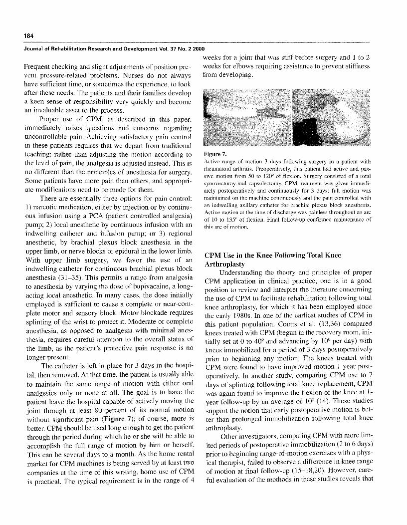
The catheter is left in place for 3 days in the hospi-tal, then removed. At that time, the patient is usually ableto maintain the same range of motion with either oralanalgesics only or none at all . The goal is to have thepatient leave the hospital capable of actively moving thejoint through at least 80 percent of its normal motionwithout significant pain (Figure 7); of course, more isbetter. CPM should be used long enough to get the patientthrough the period during which he or she will be able toaccomplish the full range of motion by him or herself.This can be several days to a month . As the home rentalmarket for CPM machines is being served by at least twocompanies at the time of this writing, home use of CPMis practical . The typical requirement is in the range of 4
Figure 7.Active range of motion 3 days following surgery in a patient withrheumatoid arthritis . Preoperatively, this patient had active and pas-sive motion from 50 to 120° of flexion . Surgery consisted of a totalsynovectomy and capsulectomy . CPM treatment was given immedi-ately postoperatively and continuously for 3 days : full motion wasmaintained on the machine continuously and the pain controlled withan indwelling axillary catheter for brachial plexus block anesthesia.Active motion at the time of discharge was painless throughout an arcof 10 to 135° of flexion . Final follow-up confirmed maintenance ofthis arc of motion.
CPM Use in the Knee Following Total KneeArthroplasty
Understanding the theory and principles of properCPM application in clinical practice, one is in a goodposition to review and interpret the literature concerningthe use of CPM to facilitate rehabilitation following totalknee arthroplasty, for which it has been employed sincethe early 1980s . In one of the earliest studies of CPM inthis patient population, Coutts et al . (13,36) comparedknees treated with CPM (begun in the recovery room, ini-tially set at 0 to 409 and advancing by 109 per day) withknees immobilized for a period of 3 days postoperativelyprior to beginning any motion. The knees treated withCPM were found to have improved motion 1 year post-operatively. In another study, comparing CPM use to 7days of splinting following total knee replacement, CPMwas again found to improve the flexion of the knee at 1-year follow-up by an average of 10 9 (14) . These studiessupport the notion that early postoperative motion is bet-ter than prolonged immobilization following total kneearthroplasty.
Other investigators, comparing CPM with more lim-ited periods of postoperative immobilization (2 to 6 days)prior to beginning range-of-motion exercises with a phys-ical therapist, failed to observe a difference in knee rangeof motion at final follow-up (15—18,20) . However, care-ful evaluation of the methods in these studies reveals that
185
O'DRISCOLL and GIORI : Continuous passive motion (CPM)
CPM was not used according to the recommendations ofthe inventor or the principles outlined above. Instead,CPM was initiated through a very limited arc of 30 to 40°and advanced by 10 to 20° per day as tolerated by thepatient . The control groups began knee range-of-motionexercises with the therapist 2 (20), 3 (16-18), or 4 to 6days (15) postoperatively. What we can conclude fromthis set of studies is that such limited motion with a CPMmachine is no more effective at achieving a final range ofmotion following total knee arthroplasty than the use ofphysical therapy in the early postoperative period.Furthermore, we can conclude that an adequate range ofmotion for typical activities of daily living can generallybe obtained with or without the use of CPM.
The impact of limited-range CPM on swelling andfluid dynamics in the operated limb is somewhat variable.In a subset of patients in Coutts' study (13), pressure inthe deep posterior compartment of the calf and venousflow velocity in the femoral vein were measured duringCPM application . These measures were found to varycyclically with the application of CPM . This finding isconsistent with the previously described animal studieson intra-articular pressure and CPM, and with the conceptdescribed by O'Driscoll et al. (30) of CPM acting as afluid pump to reduce edema in the limb. In four studies,knee circumference was measured to see if CPM usereduced knee swelling : three found a statistically signifi-cant reduction in knee circumference (9,22,37) and onedid not (20) . Perhaps relating to decreased knee swelling,studies have found CPM to facilitate a more rapidachievement of knee flexion (9,18,20) and to decrease thenumber of patients requiring postoperative knee manipu-lation (18,20,22).
Because CPM utilized in this limited way has notbeen found to reliably reduce knee swelling, one wouldpredict, based on the theory presented thus far, that CPMused in such a limited fashion would result in little or noimprovement in final range of motion . Furthermore, thestudies presented to this point do not contribute signifi-cantly to answering the question of whether or not use ofCPM as it was originally conceived might ultimatelyaffect final range of motion following total knee arthro-plasty.
Based on the discussion thus far, it could be antici-pated that if CPM were used through a greater range ofmotion or higher in the flexion arc where tissue tensionis greater, the effects would be more significant . Twostudies compared more aggressive CPM range of motionwith physical therapy . In one study, Pope and colleagues
(21) divided 53 patients into 3 groups . The first had a"traditional" course of CPM with initial range set at 0 to40°, advanced to 0 to 609 24 hours later, and then dis-continued after 48 hours . The second had a more aggres-sive course of CPM with initial range set in the recoveryroom at 0 to 70 9 and progressing to 0 to 90° 24 hourslater. This group also had the CPM removed after 48hours . In both CPM groups, traditional physical therapy,which included active knee flexion and extensionthrough the maximum possible range, was begun afterCPM was discontinued . The control group received tra-ditional physical therapy beginning on the first postoper-ative day . In this study, CPM use did not cover the extentof the second stage of stiffness and there was no differ-ence between the three groups with regard to range ofmotion at 1 year.
Because intra-articular pressure has been noted toprogressively increase as knee flexion increases, onewould expect that the pumping action of CPM would begreater with greater degrees of knee flexion . Jordan et al.(10) compared two different postoperative CPM proto-cols . In the first group, CPM was begun on the secondpostoperative day with a range of motion of 0 to 40(2 andprogressed as tolerated in 10° increments . In the secondgroup, CPM was begun in the recovery room and set at70 to 1009 of flexion . Extension was advanced by 20° onthe first postoperative day and then to full extension bythe second postoperative day. The authors found thatrange of flexion 1 year postoperatively was 1.11 9 in thefirst group and 1209 in the second group, a statisticallysignificant difference.
Thus, the literature is entirely consistent with whatwould be predicted based on the principles of CPM appli-cation formulated within the paradigm of the pathophys-iology of swelling . In other words, limited use of CPM ina range that is ineffective in the prevention or eliminationof blood and edema collection in and around the jointwould be unlikely to have any permanent impact on therange of motion following total knee arthroplasty . On theother hand, by employing greater ranges of motion or uti-lizing CPM higher in the flexion portion of the arc, thepumping effect in the periarticular tissues would beexpected to have a greater impact on soft tissue swellingand ultimate range of motion . Whether or not CPM, uti-lized through a full range of motion in the immediatepostoperative period and continued long enough to guidethe patient through the early stages, might prevent thethird and final stages of swelling has not been addressedin the literature .
186
Journal of Rehabilitation Research and Development Vol . 37 No. 2 2000
COMPLICATIONS
Complications resulting from the application ofCPM can and do occur. Most are not serious or perma-nent . The most common complication may be increasedbleeding, but rarely is this sufficient to require a transfu-sion due to the marginal increase in blood loss . Althoughfour studies found no difference in wound drainage ortransfusion requirements with CPM use following totalknee replacement (15,16,18,20), two found increasedwound drainage with CPM (19,21) . Some patients mayrequire a return to the operating room for evacuation ofhematoma under such circumstances.
The major concern in using CPM following majorjoint operations such as knee replacement relates to thewound itself. Among the literature on wound complica-tions following knee replacement, three studies did notfind an increase in wound healing complications withCPM use (14,20,22) but one study did (15) . Interestingly,an experimental study in rabbits by van Royen et al . (38)showed that wound healing was accelerated by CPMpostoperatively : skin wounds evaluated 3 weeks follow-ing arthrotomy displayed 200-percent increases instrength, stiffness, strain prior to failure, and energyabsorption prior to failure.
Our clinical experience with CPM application to theelbow suggests that CPM does not have a negative impacton healing of the wound itself . The tension generated inthe wound may be a problem if swelling is present whenCPM is initiated or if swelling is permitted to occur.Under such circumstances, CPM must not be usedthrough the full range of motion until the swelling hasbeen reduced. This is accomplished by "working" theend-ranges of motion : the patient alternates flexion andextension through a small arc of motion at the limit offlexion (and then extension) to viscoelastically milk thefluid out of the joint and periarticular tissues . Twopatients with extensive elbow procedures and large pos-terior skin flaps experienced wound dehiscence in thefirst 48 hours postoperatively, when their wounds hadbeen closed with subcuticular stitches rather than staplesor interrupted sutures.
However, there is concern about the impact on via-bility of the skin flaps, particularly over the extensor sur-faces such as the knee and the elbow . On severaloccasions, a long posterior skin flap on the elbow hasturned dark and its viability looked questionable . Thoseelbows were treated by placing them into a well-padded,cylindrical Jones dressing with an anterior plaster slab
holding the elbow in extension, then elevating the arm for2 to 4 days . None of these patients has required a proce-dure such as skin grafting or a free flap, although a fewhave had areas of necrosis that healed by secondary inten-tion without surgical treatment. A word of caution isrequired concerning the required modification to dress-ings applied to a limb being treated by CPM : no circum-ferential wrapping (cling, etc .) should be left on the limbonce the CPM is started. An elastic tube grip sleeve orfishnet is preferred as it can move freely with the joint.Failure to recognize this increases the likelihood of com-plications due to compressive and shear forces generatedagainst the wound by a restrictive non-elastic dressing.
Patients may develop nerve compression palsies dueto local pressure against the CPM device, particularly ifregional anesthetic is used and the patient or staff doesnot recognize pressures . Frequent adjustments of positionand close inspection of the set up on an ongoing basis canprevent this.
Another concern, particularly with operations of thelower limb, is deep vein thrombosis and pulmonaryembolism. Preliminary work by Coutts and colleagues(20) revealed an increase in femoral vein flow with eachCPM cycle, but this has not been shown to protect againstpulmonary embolism. One study using venograms toscreen for deep vein thrombosis showed a decreased inci-dence of thrombophlebitis below the knee but not abovethe knee (17).
Patients having knee arthroplasty are usually antico-agulated for prophylaxis against deep vein thrombosis.Although epidural anesthesia or analgesia provides excel-lent pain control, particularly with an indwelling epidur-al catheter, potential complications of prolonged epiduralanesthetic in an anticoagulated, bedridden patient are ofconcern. For this reason, we have moved away fromregional anesthetic to favor PCA pumps in these patients.The recent advent of local infusion pumps promises to beof potential benefit in these patients . Narcotic use follow-ing total knee arthroplasty and subjective sensation ofpain with and without CPM has been studied, but theresults are mixed and no clear conclusion can be drawn(9,16,20–22).
RECOMMENDATIONS FOR CPM USE
When utilized according to the principles describedin this paper, CPM acts to reduce blood and fluid accu-mulation in and around joints that have been traumatized
187
O'DRISCOLL and GIORI : Continuous passive motion (CPM)
or undergone surgery. In this way, CPM is useful inavoiding the development of subsequent joint stiffness inthe first few hours or days . Avoiding stiffness in the earlystages minimizes its chances of progression to fibrosis ofthe joint and establishment of contracture . Long-termbenefits, however, are predicated on preventing the accu-mulation of blood and/or edema fluid in the joint or peri-articular tissues . This is accomplished by the immediateapplication of a full range of passive motion on CPM, orby briefly elevating and splinting the limb in a positionthat keeps the periarticular tissues stretched before insti-tuting a full range of passive motion on CPM . In theevent that the patient is temporarily prevented from usingthe machine due to other medical or technical factors (if,for example, the machine breaks down) and periarticularswelling does occur, it must be reduced by alternatelystretching the joint at its limits of flexion and extension towork the fluid out of the periarticular region.
CPM is indicated to prevent stiffness and to main-tain motion obtained at the time of surgery, particularlyfollowing joint replacement, synovectomy, contracturerelease, excision of heterotopic ossification, and fixationof intra-articular fractures . This is particularly true forjoints that were stiff preoperatively . It is relatively con-traindicated if the soft tissue constraints (ligaments) areinsufficient, if the joint is unstable, or if rigid fixation offractures has not been attained . By following these guide-lines and adhering strictly to the principles of CPM use,one will increase the chances of obtaining maximumrange of joint motion following trauma or surgery . Itwould be anticipated that proper application of CPMwould, indeed, be cost effective, because it woulddecrease the need for physical therapy and joint manipu-lation under anesthesia, and later rehabilitation or surgi-cal intervention to treat stiffness.
CONCLUSION
The principles on which the concept of CPM isbased are twofold . First, joint motion is necessary for themaintenance of articular cartilage . Second, and relevantto the present discussion, joint homeostasis requiresmaintenance of normal periarticular soft tissue compli-ance. For CPM to accomplish this requires that themotion be full, and that the soft tissues be subjected totension immediately following surgery in order to preventswelling. It is this early maintenance of motion that is thekey determinant of the joint's long-term mobility . The
application of this concept to clinical practice requires aparadigm shift, resulting in the focus of our attention onpreventing the initial or delayed accumulation of periar-ticular interstitial fluids.
REFERENCES
1. Salter RB, Field P. The effects of continuous compression on liv-ing articular cartilage . An experimental investigation . J Bone JointSurg 1960;42-A:31-49.
2. Salter RB, McNeill OR, Carbin R . The pathological changes in artic-ular cartilage associated with persistent joint deformity. An experi-mental investigation . In : Studies of the rheumatoid diseases . ThirdCanadian conference on research in rheumatic diseases . Toronto;1965 . p . 33-47.
3. Salter RB . Motion vs . rest . Why immobilize joints? J Bone JointSurg 1982 ;64-B:251-4.
4. Melzack R, Wall PD . Evolution of pain theories . Mt AnesthesiolClin 1970;8( I ) :3-34.
5. Melzack R, Wall PD. Pain mechanisms : a new theory . Science1965 ;150(699) :971-9.
6. Salter RB, Hamilton HW, Wedge JH, Tile M, Torode IP, O'DriscollSW, et al . Clinical application of basic research on continuous pas-sive motion for disorders and injuries of synovial joints : A prelim-inary report of a feasibility study . J Orthop Res 1984 ;1 :325-42.
7. Salter RB . The physiologic basis of continuous passive motion forarticular cartilage healing and regeneration . Hand Clin1994 ;10(2) :211-9.
8. Salter RB . History of rest and motion and the scientific basis forearly continuous passive motion . Hand Clin 1996;12(1) :1-ll.
9. Montgomery F, Eliasson M. Continuous passive motion comparedto active physical therapy after knee arthroplasty : similar hospital-ization times in a randomized study of 68 patients . Acta OrthopScand 1996 ;67(1) :7-9.
10. Jordan LR, Siegel JL, Olivo JL . Early flexion routine . An alternativemethod of continuous passive motion. Clin Orthop 1995(315) :231-3.
11. Rorabeck CH. Continuous passive motion is a useful postoperativetool [see comments] . Orthopedics 1999 ;22(4) :392.
12.Dorr LD . Continuous passive motion offers no benefit to thepatient [comment] . Orthopedics 1999 ;22(4) :393.
13.Coutts RD. A conversation with Richard D . Coutts, MD. Continuouspassive motion in the rehabilitation of the total knee patient, its roleand effect [interview] . Orthop Rev 1986 ;15(3) :126-34.
14. Johnson DP, Eastwood DM . Beneficial effects of continuous pas-sive motion after total condylar knee arthroplasty . Ann R Coll SurgEngl 1992;74(6) :412-6.
15.Maloney WJ, Schulman DJ, Hangen D, Goodman SB, EdworthyS, Bloch DA . The influence of continuous passive motion on out-come in total knee arthroplasty . Clin Orthop 1990(256) :162-8.
16. Colwell CW Jr, Morris BA . The influence of continuous passivemotion on the results of total knee arthroplasty . Clin Orthop1992(276) :225-8.
17.Vince KG, Kelly MA, Beck J, Insall JN . Continuous passive motionafter total knee arthroplasty . J Arthroplasty 1987;2(4) :281-4.
18. Romness DW, Rand JA. The role of continuous passive motionfollowing total knee arthroplasty. Clin Orthop 1988 ;226 :34-7 .
188
Journal of Rehabilitation Research and Development Vol . 37 No . 2 2000
19. Kumar PJ, McPherson EJ, Dorr LD, Wan Z, Baldwin K.Rehabilitation after total knee arthroplasty : a comparison of 2rehabilitation techniques . Clin Orthop 1996(331) :93–101.
20. Ververeli PA, Sutton DC, Hearn SL, Booth RE Jr, Hozack WJ,Rothman RR . Continuous passive motion after total knee arthroplas-ty . Analysis of cost and benefits. Clin Orthop 1995(321) :208–15.
21.Pope RO, Corcoran S, McCaul K, Howie DW Continuous passivemotion after primary total knee arthroplasty . J Bone Joint Surg Br1997 ;79 :914–7.
22. McInnes J, Larson MG, Daltroy LH, Brown T, Fossel AH, Eaton HM,et al . A controlled evaluation of continuous passive motion in patientsundergoing total knee arthroplasty. JAMA 1992;268(11) :1423–8.
23. Funk DA, Noyes FR, Grood ES, Hoffman SD . Effect of flexion angleon the pressure-volume of the human knee . Arthroscopy1991 ;7(1) :86–90.
24. Jayson MIV, Dixon ASJ . Intra-articular pressure in rheumatoid arthri-tis of the knee . III. Pressure changes during joint use . Ann Rheum Dis1970 ;29 :266–8.
25. Levick JR . The influence of hydrostatic pressure on trans-synovialfluid movement and on capsular expansion in the rabbit knee . JPhysiol (Lond) 1979 ;289 :69–82.
26. Favreau JC, Laurin CA. Joint effusions and flexion deformities . CanMed Assoc J 1963 ;88 :575–6.
27.O'Driscoll SW, Morrey BF, An K-N . Intra-articular pressure andcapacity of the elbow. Arthroscopy 1990;6 :100-3.
28.O'Driscoll SW, Kumar A, Salter RB. The effect of continuous pas-sive motion on the clearance of a hemarthrosis from a synovialjoint . An experimental investigation in the rabbit . Clin Orthop1983 ;176 :305–11.
29. Breen TF, Gelbennan RH, Ackerman GN . Elbow flexion contrac-tures: Treatment by anterior release and continuous passive motion.J Hand Surg 1988 ;13–B :286–7 .
30. O'Driscoll SW, Kumar A, Salter RB . The effect of the volume ofeffusion, joint position and continuous passive motion on intra-artic-ular pressure in the rabbit knee . J Rheumatol 1983 ;10:360–3.
31. Stinson LJ, Lennon R, Adams R, Money B . The technique and effi-cacy of axillary catheter analgesia as an adjunct to distraction elbowarthroplasty : a prospective study. J Shoulder Elbow Surg1993 ;2 :182–9.
32.Brown AR, Weiss R, Greenberg C, Flatow EL, Bigliani LU.Interscalene block for shoulder arthroscopy : comparison with gener-al anesthesia. Arthroscopy 1993 ;9 :295–300.
33. Schroeder LE, Horlocker Schroeder DR. The efficacy of axillaryblock for surgical procedures about the elbow. Anesth Analg1996 ;83 :747–51.
34. Rice ASC . Prevention of nerve damage in brachial plexus block . XVIAnnual European Society of Regional Anaesthesia Congress ; 1997.London : Pennanyer, 1997 . p . 60–3.
35. Gaumann DM, Lennon RL, Wedel DJ . Continuous axillary block forpostoperative pain management . Reg Anesth 1988 ;13 :77–81.
36. Coutts RD, Borden LS, Bryan RS, Hungerford DS, Stulberg B,Thomas WH, et al . The effect of continuous passive motion on totalknee rehabilitation (abstract) . Orthop Tran 1983 ;7 :535–6.
37. Ritter M, Gandolf V, Holston K . Continuous passive motion versusphysical therapy in total knee arthroplasty . Clin Orthop1989 ;244:239-43.
38. Van Royen BJ, O'Driscoll SW, Dhert WJA, Salter RB . A comparisonof the effects of immobilization and continuous passive motion onsurgical wound healing in mature rabbits . Plast Reconstr Surg1986;78 :360–6