Journal of Physics: Conference Series OPEN ACCESS Contrast Variation Application in Small-Angle Neutron Scattering Experiments To cite this article: H B Stuhrmann 2012 J. Phys.: Conf. Ser. 351 012002 View the article online for updates and enhancements. You may also like Contrast variation in spin-echo small angle neutron scattering Xin Li, Bin Wu, Yun Liu et al. - Dual-mode phase-shifting interferometry based on iterative algorithms Qian Liu, Wen Huang and Xiaobin Yue - SANS with contrast variation study of the bacteriorhodopsin-octyl glucoside complex Yiming Mo and William T Heller - Recent citations Practical applications of small-angle neutron scattering Martin J. Hollamby - This content was downloaded from IP address 219.100.37.238 on 29/11/2021 at 00:48
Transcript
Journal of Physics Conference Series
OPEN ACCESS
Contrast Variation Application in Small-AngleNeutron Scattering ExperimentsTo cite this article H B Stuhrmann 2012 J Phys Conf Ser 351 012002
View the article online for updates and enhancements
You may also likeContrast variation in spin-echo small angleneutron scatteringXin Li Bin Wu Yun Liu et al
-
Dual-mode phase-shifting interferometrybased on iterative algorithmsQian Liu Wen Huang and Xiaobin Yue
-
SANS with contrast variation study of thebacteriorhodopsin-octyl glucoside complexYiming Mo and William T Heller
-
Recent citationsPractical applications of small-angleneutron scatteringMartin J Hollamby
-
This content was downloaded from IP address 21910037238 on 29112021 at 0048
Contrast Variation Application in Small-Angle Neutron
Scattering Experiments
H B Stuhrmann
Helmholtz-Forschungszentrum Geesthacht Germany and Institut de Biologie Structurale Jean-Pierre Ebel CEACNRSUJF Grenoble France
heinrichstuhrmannorangefr
Abstract The mathematical formalism of contrast variation is presented in terms of an expansion of spherical harmonics Early attempts of contrast variation in X-ray small-angle scattering are compared with the corresponding more versatile techniques of neutron small-angle scattering Some applications in life sciences illustrate the power of nuclear spin contrast variation
1 Introduction In the early times contrast variation aimed at the elimination of intra-particle background scattering in X-ray small-angle scattering [1] X-ray small-angle scattering from solutions of myoglobin in solvents of different electron density allowed the determination of three basic scattering functions one of them being due to the scattering from the shape of the protein molecule [2] As the shape of a molecule is described by much less parameters than its total structure the probability to find a unique molecular shape from its scattering function is greatly enhanced [3]
2 The spherical harmonics as natural co-ordinates in small-angle scattering
Spherical harmonics can be considered as the natural co-ordinates for the description of the structure of spherical viruses [4] Another reason for the use of spherical harmonics is the question of uniqueness of structure determination from small-angle scattering (SAS) In order to discuss this point a reminder of some basic equations is mandatory
We start from an expansion of )r()( ωρ=ρ rrrr and of its Fourier transform the scattering
amplitude )Q(A)(A Ω=QQQQ as a series of spherical harmonics mlY
)(Y)Q(A)(A)(Y)r()( ml0l
ml0l
mlml Ω=hArrωρ=ρ sumsuminfin
=
infin
=
QQQQrrrr ( 1 )
Q is a vector in the Fourier space and Q is its modulus ω and Ω are unit vectors in the real space and in the Fourier space respectively The intensity of small-angle scattering )(QI then is
obtained as the sum of absolute squares of the coefficients )( QA ml
SANS-YuMO User Meeting IOP PublishingJournal of Physics Conference Series 351 (2012) 012002 doi1010881742-65963511012002
Published under licence by IOP Publishing Ltd 1
sumsumsuminfin
=
infin
= minus=
equiv=0
2
0
2
2 )(2)(2)(
l
l
l
l
lm
ml QIQAQI ππ ( 2 )
The coefficients )( QA ml are the Hankel transforms of )( rmlρ They may be calculated from
the polar co-ordinates ( nnnr ϕθ ) of the N atoms of a molecule
Fig 1 Various density maps giving rise to the scattering function of a cube
SANS-YuMO User Meeting IOP PublishingJournal of Physics Conference Series 351 (2012) 012002 doi1010881742-65963511012002
2
)()(2
)()(2
)(
10
2 nnmln
N
n
l
r
l
lml
l
ml YQrjidrrQrjriQA ϕθπ
ρπ sumint
=
infin
=
== ( 3 )
lj are the spherical Bessel functions ml
m
ml YY minusminus= )1( is the conjugate complex of mlY Each
partial structure )()( ϕθρ mlml Yr gives rise to a partial scattering function 2
)(QA ml
The rotation of a single set of partial structures summinus=
l
lm
mlml Yr )()( ϕθρ by an arbitrary angle
with respect to the rest of the structure has no influence on )(QI l defined in (2) while )(rrrrρ may
change considerably [5] Some density maps of )(rrrrρ giving rise to the scattering function of a cube are shown in Fig 1 The rotation of the partial structures of the cube by l-dependent angles blurs the sharp contours of the original structure Hence the guess may be allowed that there is only one shape which may be associated with the scattering function of a cube
Let us turn to the molecular shape determination as it has been proposed 40 years ago [3] It was assumed that the surface of the shape could be described by a unique function
sumsum= minus=
=L
l
l
lm
mlml YfF0
)()( ωω The shape scattering function is developed as a power series of 2Q [3]
suminfin
==
0
222)(k
k
kC QbQI π where
( )intsum sum sum =+minusminus++
==
minus
= minus=
+minusminus++minus
ω
ωωω dYFfplkpl
ffddb ml
qq
ml
k
l
lk
p
l
lm
plk
ml
pl
mlpklpl
k )()()32()3(
)(
0 0
)32(
)3(
(4)
( 7 )
Computer simulations with model bodies indicate that the low resolution shape determination from error-free data is unique even when very limited ranges are used in the simulated curves [6]
3 Solvent contrast variation Introduction In a first paper the idea of contrast variation has been illustrated by a simulated model consisting
of a cube with a non-uniform density [1] The structure of the model is described by two terms the shape )(rrrrCρ multiplied by the contrast ρ and the internal structure )(rrrrSρ
)()()( rrrrrrrrrrrr SC ρρρρ += (5)
The contrast ρ is the difference between the average scattering density of the dissolved particle
and that of the solvent The shape )(rrrrCρ has the value 1 inside the volume excluded to the solvent
The intensity diffracted by )(rrrrρ is the absolute square of the corresponding amplitude 22
)()()( QQQQQQQQQQQQ SC AAA += ρ For randomly oriented particles we obtain from (2)
SANS-YuMO User Meeting IOP PublishingJournal of Physics Conference Series 351 (2012) 012002 doi1010881742-65963511012002
3
)()()()()()( 22
0
)(
)( QIQIQIQAQAQI SCSC
l
l
lm
S
ml
C
ml ++equiv+=sumsuminfin
= minus=
ρρρ (6 )
Among the three basic scattering functions of solvent contrast variation )(QI S is the only one
which in favorable cases can be measured directly at zero contrast 0=ρ The shape scattering
function )(QIC is dominant at high contrast The cross term )(QICS reflects the convolution
between the shape with the internal structure As int ==v
S
SdVA 0)()0()(
00 rrrrρ both )0(CSI and )0(SI
are zero
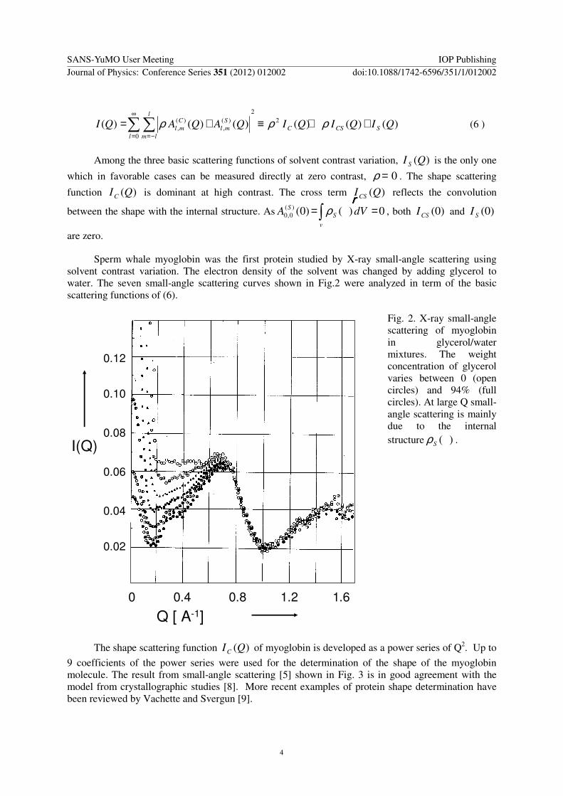
Sperm whale myoglobin was the first protein studied by X-ray small-angle scattering using solvent contrast variation The electron density of the solvent was changed by adding glycerol to water The seven small-angle scattering curves shown in Fig2 were analyzed in term of the basic scattering functions of (6)
0 04 08 12 16
Q [ A-1]
002
004
006
008
010
012
I(Q)
Fig 2 X-ray small-angle scattering of myoglobin in glycerolwater mixtures The weight concentration of glycerol varies between 0 (open circles) and 94 (full circles) At large Q small-angle scattering is mainly due to the internal structure )(rrrrSρ
The shape scattering function )(QIC of myoglobin is developed as a power series of Q2 Up to
9 coefficients of the power series were used for the determination of the shape of the myoglobin molecule The result from small-angle scattering [5] shown in Fig 3 is in good agreement with the model from crystallographic studies [8] More recent examples of protein shape determination have been reviewed by Vachette and Svergun [9]
SANS-YuMO User Meeting IOP PublishingJournal of Physics Conference Series 351 (2012) 012002 doi1010881742-65963511012002
4
1
0
-1
0 180 360
cos θ
ϕ
Fig 3 The shape function )(cos)( ϕθω FF = of myoglobin determined from the shape scattering
function )(QIC The coefficients mlf up to l=3 of )(ωF have been determined The distances of
the isohypse lines are given in Aring
The internal structure is more complicated than the shape Its elucidation is often restricted to the analysis of the variation of the apparent radius of gyration with the contrast ρ [10]
222
ρβ
ρα minus+= CRR (7)
The coefficients α and β provide an unmistakable feature of the low-resolution of complex particles in solution Particles with a high density core (eg ribosomes ferritin) will give rise to a negative α whereas particle with a low density core (eg nucleosome core particle low density lipoprotein) give rise to positive α in a spherical approximation A non-vanishing β will be due to a dipolar structure ie the centers of mass of different components do not coincide
Having said this we find ourselves in the field of neutron scattering In fact there are only very few examples of contrast variation using X-ray small-angle scattering [211] But there are considerably more applications of contrast variation in neutron scattering
4 Neutron scattering from hydrogen
Nearly all applications of contrast variation in neutron scattering use the extraordinary properties of the interaction of neutrons with the hydrogen nuclei 1H (=H) and 2H (=Deuterium) More recent applications use dependence of neutron scattering length cohb of these hydrogen isotopes on their
nuclear polarization P(H) and P(D) respectively
SANS-YuMO User Meeting IOP PublishingJournal of Physics Conference Series 351 (2012) 012002 doi1010881742-65963511012002
5
[ ][ ] cmDpPDb
cmHpPHb
coh
coh
12
12
10)(2706670)(
10)(45613740)(minus
minus
++=
+minus= (8)
This equation holds if a completely polarized neutron beam is used The polarization p of the neutron beam then will be +1 or -1 The nuclear polarization may assume values between -1 and +1 The methods for achieving neutron and nuclear polarization will be presented in the other paper
Similarly there is a strong variation of the cross section of incoherent scattering with polarization
( )[ ] 2242
2242
10201)(
1041
21
43
105)(
cmPpPD
cmPpPH
inc
inc
minus
minus
minusminus=
minusminus=
σ
σ (9)
There is no incoherent scattering for P=1
5 Solvent contrast variation by isotopic substitution
Most of the applications of neutron small-angle scattering rely on the difference between the lengths of coherent scattering of H and D This difference is large compared with scattering lengths of other elements Another fact is also important hydrogen is abundant in organic matter The variation of the scattering density of organic solvents due to isotopic substitution in neutron scattering is much larger than equivalent methods in X-ray scattering Solvent contrast variation by isotopic substitution is almost exclusively used in neutron scattering
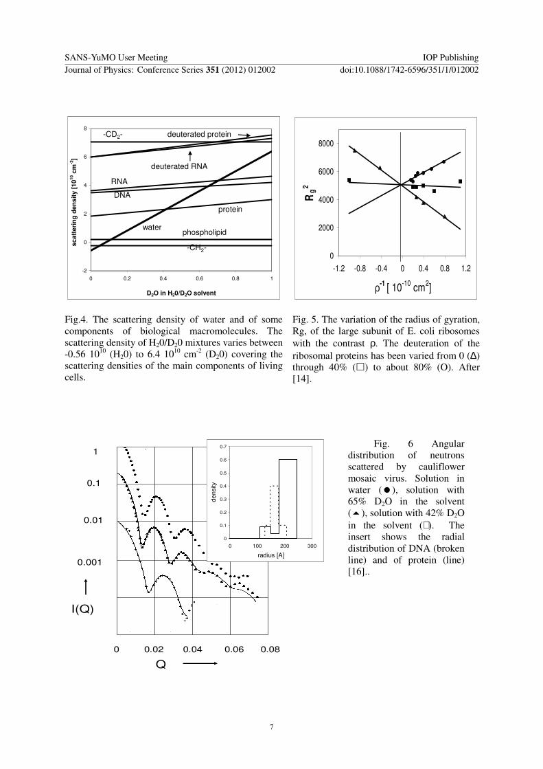
Neutron small-angle scattering in H20D20 distinguishes clearly between lipids material protein and RNADNA (Fig 4) Most of the applications aim at the internal structure of complex macromolecules As an example we cite the studies on the large subunit of Ecoli ribosomes which consists of rRNA by two third of its mass and more than thirty different ribosomal proteins The variation of the apparent radius of gyration with the D2O content of H20D20 mixtures provides a low resolution model with a high RNA content close to the centre and proteins preferring the surface region [1213] Deuteration of the rRNA confirmed this model [14] (Fig 5) The shape of this ribosomal subunit being asymmetric [15] the elucidation of a more detailed internal structure had to be left to labeling techniques using specific deuteration (see below)
The analysis of the internal structure is greatly simplified with spherical particles because of the absence of spherical harmonics with l gt 0 in (2)
2
2
0
00
2
0
2000
sin)()()()( drr
Qr
QrrdrrQrjrQI
rr
intintinfin
=
infin
=
=asymp ρρ (10)
SANS-YuMO User Meeting IOP PublishingJournal of Physics Conference Series 351 (2012) 012002 doi1010881742-65963511012002
6
-2
0
2
4
6
8
0 02 04 06 08 1
D2O in H20D2O solvent
sc
att
eri
ng
de
ns
ity
[1
010 c
m-2
]
-CD2- deuterated protein
deuterated RNA
RNA
DNA
protein
waterphospholipid
-CH2-
0
2000
4000
6000
8000
-12 -08 -04 0 04 08 12
ρ-1 [ 10
-10 cm
2]
Rg
2
Fig4 The scattering density of water and of some components of biological macromolecules The scattering density of H20D20 mixtures varies between -056 1010 (H20) to 64 1010 cm-2 (D20) covering the scattering densities of the main components of living cells
Fig 5 The variation of the radius of gyration Rg of the large subunit of E coli ribosomes with the contrast ρ The deuteration of the ribosomal proteins has been varied from 0 (∆) through 40 () to about 80 (O) After [14]
1
001
01
0001
I(Q)
0 002 004 006 008
Q
0
01
02
03
04
05
06
07
0 100 200 300
radius [A]
de
nsity
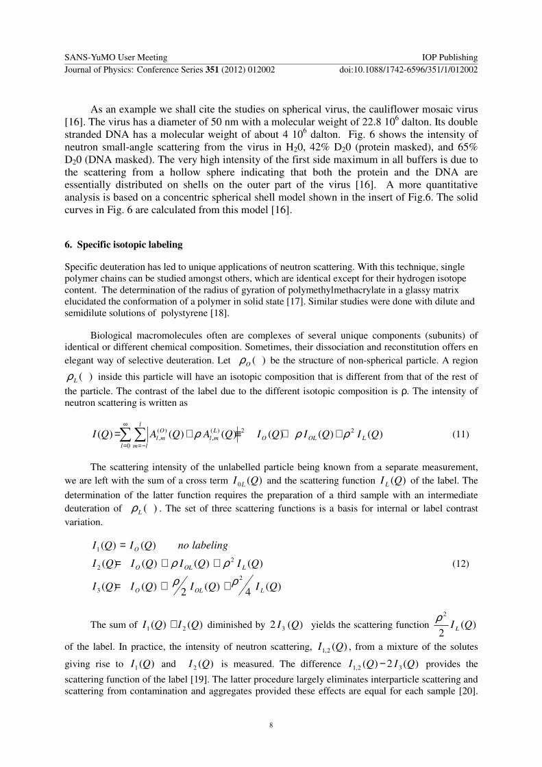
Fig 6 Angular distribution of neutrons scattered by cauliflower mosaic virus Solution in water () solution with 65 D2O in the solvent () solution with 42 D2O in the solvent (+) The insert shows the radial distribution of DNA (broken line) and of protein (line) [16]
SANS-YuMO User Meeting IOP PublishingJournal of Physics Conference Series 351 (2012) 012002 doi1010881742-65963511012002
7
As an example we shall cite the studies on spherical virus the cauliflower mosaic virus [16] The virus has a diameter of 50 nm with a molecular weight of 228 106 dalton Its double stranded DNA has a molecular weight of about 4 106 dalton Fig 6 shows the intensity of neutron small-angle scattering from the virus in H20 42 D20 (protein masked) and 65 D20 (DNA masked) The very high intensity of the first side maximum in all buffers is due to the scattering from a hollow sphere indicating that both the protein and the DNA are essentially distributed on shells on the outer part of the virus [16] A more quantitative analysis is based on a concentric spherical shell model shown in the insert of Fig6 The solid curves in Fig 6 are calculated from this model [16]
6 Specific isotopic labeling
Specific deuteration has led to unique applications of neutron scattering With this technique single polymer chains can be studied amongst others which are identical except for their hydrogen isotope content The determination of the radius of gyration of polymethylmethacrylate in a glassy matrix elucidated the conformation of a polymer in solid state [17] Similar studies were done with dilute and semidilute solutions of polystyrene [18]
Biological macromolecules often are complexes of several unique components (subunits) of identical or different chemical composition Sometimes their dissociation and reconstitution offers en elegant way of selective deuteration Let )(rrrrOρ be the structure of non-spherical particle A region
)(rrrrLρ inside this particle will have an isotopic composition that is different from that of the rest of
the particle The contrast of the label due to the different isotopic composition is ρ The intensity of neutron scattering is written as
)()()()()()( 22
0
)(
)( QIQIQIQAQAQI LOLO
l
l
lm
L
ml
O
ml ρρρ ++equiv+=sumsuminfin
= minus=
(11)
The scattering intensity of the unlabelled particle being known from a separate measurement we are left with the sum of a cross term )(0 QI L and the scattering function )(QI L of the label The
determination of the latter function requires the preparation of a third sample with an intermediate deuteration of )(rrrrLρ The set of three scattering functions is a basis for internal or label contrast variation
)(4)(2)()(
)()()()(
)()(
2
3
22
1
QIQIQIQI
QIQIQIQI
labelingnoQIQI
LOLO
LOLO
O
ρρρρ
++=
++=
=
(12)
The sum of )()( 21 QIQI + diminished by )(2 3 QI yields the scattering function )(2
2
QI L
ρ
of the label In practice the intensity of neutron scattering )(21 QI from a mixture of the solutes
giving rise to )(1 QI and )(2 QI is measured The difference )(2)( 321 QIQI minus provides the
scattering function of the label [19] The latter procedure largely eliminates interparticle scattering and scattering from contamination and aggregates provided these effects are equal for each sample [20]
SANS-YuMO User Meeting IOP PublishingJournal of Physics Conference Series 351 (2012) 012002 doi1010881742-65963511012002
8
The determination of the in situ structure of components of a complex is one of the numerous applications of the method of triple isotopic substitution (TIS) [20]
Biological macromolecules are often composed several well-defined constituents An important approach to determine their architecture starts with the measurement of the spatial correlation function of two selected components The number of different distinct pair correlation functions is increased to a point where the total structure emerges The pair of selected regions )(1 rrrrLρ and )(2 rrrrLρ is
embedded in the total structure )(rrrrOρ The corresponding amplitudes are )()( 21 QQQQQQQQ AA and
)(QQQQOA respectively Each of the two regions may be labeled ie it may have its isotope 1H
substituted by 2H Each of the labeled region may have an excess spatial scattering length distribution
described by [ ]sum minusminusn
nHbDb )()()( rrrrrrrrδ In fact four different solutions have to be prepared in order
to obtain the inter label scattering function from neutron scattering 1 the unlabelled particle 2 )(1 rrrrLρ is labeled 3 )(2 rrrrLρ is labeled 4 )(1 rrrrLρ and )(2 rrrrLρ are labeled Each of these samples
gives rise to the following scattering functions
21201022
21
222104
222
22203
211
22102
21
222)(
2)(
2)(
AAAAAAAAAAAAI
AAAAAAI
AAAAAAI
AI
O
OO
OO
O
+++++=++=
++=+=
++=+=
=
(13 )
The samples giving rise 1I and 4I are mixed and so are the samples giving rise to 2I and 3I
From the difference between 41 II + and 32 II + one obtains the cross term 212 AA This method of
mixing the samples has the great advantage that it eliminates interparticle scattering Using (3) and the
development of
sin
rr
rrQ
minusminus
as a series of spherical harmonics we obtain
sumsumsumsum sumsum
sumsum
infin
= minus= = = = =
infin
= minus=
minus
minusasympasymp
=
0 1
1 1
1)2(
)1(
)2(
)1(
)2(
)1(
)2(
)1(
0
)2(
)1(21
sin)()()()(
)()(
l
l
lm
N
n
N
n
N
n
N
n nn
nn
nmlnmlnlnl
l
l
lm
mlml
Q
QYYQrjQrj
QAQAAA
rrrrrrrr
rrrrrrrrωω
(14)
The super scripts (1) and (2) refer to the hydrogen atoms of the labels )(1 rrrrLρ and )(2 rrrrLρ respectively Representing the labels by their center of gravity one obtains
[ ] [ ]Qd
QdQIQIQIQIQI X
sin)()()()(()( 3241 asymp+minus+= (15 )
)(QI X images the spatial relationship of the pair of components in question [21] The Fourier
transform of )(QI X is the distribution of lengths of all possible vectors connecting the non-exchangeable hydrogen sites in the two labeled regions The second moment of the length distribution related to a pair of labels is equal to the sum of the squares of their radii of gyration in situ plus the
SANS-YuMO User Meeting IOP PublishingJournal of Physics Conference Series 351 (2012) 012002 doi1010881742-65963511012002
9
square of the separation between their centers [22] A table of such distances yields the spatial arrangement of the components In total 2)1( minusnn distances exist between n components In order
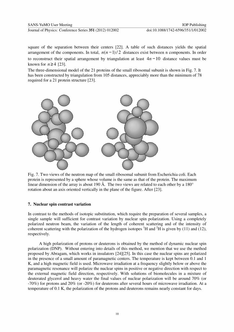
to reconstruct their spatial arrangement by triangulation at least 104 minusn distance values must be known for 4gen [23] The three-dimensional model of the 21 proteins of the small ribosomal subunit is shown in Fig 7 It has been constructed by triangulation from 105 distances appreciably more than the minimum of 78 required for a 21 protein structure [23]
Fig 7 Two views of the neutron map of the small ribosomal subunit from Escherichia coli Each protein is represented by a sphere whose volume is the same as that of the protein The maximum linear dimension of the array is about 190 Aring The two views are related to each other by a 180deg rotation about an axis oriented vertically in the plane of the figure After [23]
7 Nuclear spin contrast variation
In contrast to the methods of isotopic substitution which require the preparation of several samples a single sample will sufficient for contrast variation by nuclear spin polarization Using a completely polarized neutron beam the variation of the length of coherent scattering and of the intensity of coherent scattering with the polarization of the hydrogen isotopes 1H and 2H is given by (11) and (12) respectively
A high polarization of protons or deuterons is obtained by the method of dynamic nuclear spin polarization (DNP) Without entering into details of this method we mention that we use the method proposed by Abragam which works in insulators [24][25] In this case the nuclear spins are polarized in the presence of a small amount of paramagnetic centers The temperature is kept between 01 and 1 K and a high magnetic field is used Microwave irradiation at a frequency slightly below or above the paramagnetic resonance will polarize the nuclear spins in positive or negative direction with respect to the external magnetic field direction respectively With solutions of biomolecules in a mixture of deuterated glycerol and heavy water the final values of nuclear polarization will be around 70 (or -70) for protons and 20 (or -20) for deuterons after several hours of microwave irradiation At a temperature of 01 K the polarization of the protons and deuterons remains nearly constant for days
SANS-YuMO User Meeting IOP PublishingJournal of Physics Conference Series 351 (2012) 012002 doi1010881742-65963511012002
10
A proton spin polarized sample is obtained by destroying the deuteron polarization A deuteron spin sample is obtained by destroying the proton polarization At a temperature of 01 K differently polarized nuclear spin systems will coexist for many hours without significant change of polarization
A typical experiment of polarized neutron scattering takes 5 days [26] Loading the frozen sample and cooling the refrigerator to 1 K takes 6 hours After calibration of the proton NMR signal and neutron scattering from the sample at P=0 microwave irradiation will polarize the nuclear spins of the sample at a temperature well below 1 K The microwaves are switched off and the temperature of the sample will drop to 01 K The deuterons are depolarized Neutron scattering from the proton spin polarized sample is measured for two days The direction of the neutron spin polarization is changed each ten minutes Then the same procedure is repeated for the deuteron spin polarized sample for another two days The sample is unloaded on a Friday afternoon The data set consists of five spectra of neutron small-angle scattering The spectra of the sample at P=0 and the spectra of the proton polarized target and deuteron polarized target at two neutron beam polarizations in direction and opposite to the external field [26]
0
05
1
15
2
25
3
-1 -05 0 05 1
proton spin polarisation
sq
uare
ro
ot
of
forw
ard
scatt
eri
ng
Fig 8 The variation of zero-angle of a protein in a mixture of deuterated solvent and heavy water (11) with proton polarization
Nuclear spin contrast variation is mainly used as a method of internal contrast variation
sumsuminfin
=
=
minus=plusmn ++equiv+=
0
22
)( )()()()()()(l
lm
lm
VUVUmlml QIPQIPpQIQVPpQUQI (16 )
)()( QI + and )()( QI minus are obtained with positive and negative polarization p of the neutron
beam respectively The nuclear polarization P stands for the proton polarization P(H) or deuteron polarization P(D) )(QQQQU is the amplitude of the sample at P=0 With deuterated solvents )0(U will be negative With increasing proton spin polarization P(H) the amplitude P(H)V(0) will decrease the intensity of forward scattering (Fig 8) At P(H) = 065 the nuclear spin contrast matches the native
SANS-YuMO User Meeting IOP PublishingJournal of Physics Conference Series 351 (2012) 012002 doi1010881742-65963511012002
11
contrast of the protein The matching point changes hardly with RNA or DNA as solute This means that nuclear spin contrast is an excellent amplifier of an existing contrast but unlike the method of isotopic substitution it is not suitable for the distinction between chemically different components
Assuming that the extreme polarizations P are equal the basic scattering functions are easily obtained from )()( QI + and )()( QI minus
[ ][ ]
2
)()(
)()(
2
2)()()(
)()(21
)(
P
IQIQIQI
QIQIP
QI
U
V
UV
minus+=
minus=
minus+
minus+
( 17 )
The gain in contrast due to nuclear polarization considerably extends the application of contrast variation The neutron scattering studies on ribosomes at the end of the last century may serve as an illustration While the small 30S subunit of the Ecoli ribosome has been studied in great detail (Fig 7) a similar approach for the large 50S subunit of the ribosome seemed to be prohibitively difficult Attempts were made to reduce the incoherent scattering by massive deuteration of the solvent and of the solute The idea of the contrast free lsquoglassy ribosomersquo should facilitate the in situ structure determination of the labeled (protiated) component [27] However the interference of any residual internal contrast between deuterated rRNA and the deuterated proteins with the strong contrast of the label could not be entirely excluded It was at that time when first experiments of nuclear spin contrast variation were started It happened that the optimal composition of samples for this kind of study were those developed for the lsquoglassy ribosomersquo In fact the scattering density of a completely deuterated ribosome is almost equal to that of a mixture of deuterated glycerol and D20 The first experiments of proton spin contrast variation on the deuterated 50S subunit with one of its proteins left protiated showed that the forward scattering from the high contrast of the label and the weak contrast of the deuterated ribosome were comparable in size Thus the analysis of the data had to take into account the shape and internal structure in order to determine the site (and if possible) the shape the labeled ribosomal component [26]
The four additional basic scattering functions from nuclear spin contrast variation of (20) increased considerably the structural information Clearly the basic scattering functions from proton spin contrast variation and in particular the cross term )(QIUV were most important Valuable
additional structural information came from electron microscopy [28] A number of ribosomal proteins of the large subunit have been studied by this method We point out to the fact that the results concerning the co-ordinates of the label with respect to the large ribosomal subunit depend on the knowledge of the low resolution structure of the ribosome known at that time [29]
The study of labeled components in the functional E coli ribosome with its two protiated tRNAs and a protiated mRNA fragment in a completely deuterated environment was a still larger challenge for two reasons
1 The contrast of RNA in a deuterated solvent is lower by a factor 15 with respect to proteins due to the lower hydrogen content The same holds for the proton spin added contrast
2 the occupation density of the ribosomal binding sites by tRNAs does not exceed 40
SANS-YuMO User Meeting IOP PublishingJournal of Physics Conference Series 351 (2012) 012002 doi1010881742-65963511012002
12
-600
-400
-200
0
200
400
0 005 01 015
Q [ 1A]
ne
utr
on
sp
in d
ep
en
de
nt
inte
ns
ity
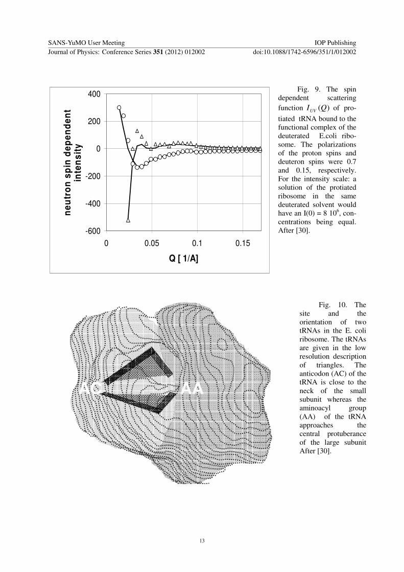
Fig 9 The spin dependent scattering function )(QIUV of pro-
tiated tRNA bound to the functional complex of the deuterated Ecoli ribo-some The polarizations of the proton spins and deuteron spins were 07 and 015 respectively For the intensity scale a solution of the protiated ribosome in the same deuterated solvent would have an I(0) = 8 106 con-centrations being equal After [30]
AC AA
Fig 10 The site and the orientation of two tRNAs in the E coli ribosome The tRNAs are given in the low resolution description of triangles The anticodon (AC) of the tRNA is close to the neck of the small subunit whereas the aminoacyl group (AA) of the tRNA approaches the central protuberance of the large subunit After [30]
SANS-YuMO User Meeting IOP PublishingJournal of Physics Conference Series 351 (2012) 012002 doi1010881742-65963511012002
13
Nevertheless polarized neutron scattering from the nuclear spin polarized sample clearly revealed the basic scattering functions of proton and deuteron spin contrast variation [30] The change of the neutron scattering intensity with the polarization direction of the incident neutron beam is shown in Fig 9 for both the proton spin polarized and the deuteron spin polarized sample
The cross terms shown in Fig 9 differ in the sign at very small scattering angles and so do the contrasts induced by the polarization of protons and deuterons respectively As the density of deuterons in the solvent was higher than in the solute the deuteron spin contrast was negative Hence the proton spin contrast was positive and so was U(0) At Q gt 005 the intensity is mainly due to the amplitude of the protiated tRNAs
The determination of the in situ structure of the two tRNAs was done in two steps In a first step the centre of gravity of the (tRNA)2-mRNA complex was determined In a next step the orientation of this complex with respect to the ribosome had to be found Two facts were helpful (i) the structure of the tRNA is known to atomic resolution (ii) the extremes of the structure of tRNA must be rather close to each other There is also an estimation of the angle between the planes of the tRNAs which may be as large as 90deg [31] The orientation of the tRNA complex was varied over the whole range of Eulerian angles while the center was kept The best fit of the experimental data was obtained with orientation of the tRNAs shown in Fig 10
8 Conclusion Contrast variation techniques have found many applications in neutron small-angle scattering both in life sciences and in condensed matter research in general As the structural aspect prevails in biological applications we have developed the concept and the mathematical formalism of contrast variation in this frame Isotopic substitution is still the mostly used technique It discriminates between chemically different components of complex structures Nuclear spin contrast variation remains an excellent amplifier of an existing contrast
References
[1] HB Stuhrmann RG Kirste Z f physik Chemie Neue Folge 46 247-250 (1965) [2] RG Kirste HB Stuhrmann Z f physik Chemie Neue Folge 56 338-341 (1967) [3] HB Stuhrmann Zf physik Chemie Neue Folge 72 177-184 (1970) [4] Y Zheng PC Doerschuk JE Johnson Biophysical Journal 69 619-639 (1995) [5] HB Stuhrmann Acta Cryst 72 177-184 (1970) [6] DI Svergun VV Volkov MB Kozin HB Stuhrmann Acta Cryst A52 419-426 (1996) [7] HB Stuhrmann Zf physik Chemie Neue Folge 72 185-198 (1970) [8] G Bodo HM Dintzis JC Kendrew WH Wyckoff Proc Royal Soc A 253 70 (1959) [9] P Vachette DI Svergun Structure and Dynamics of Macromolecules HERCULES Vol IV
edited by E Fanchon E Geissler J-L Hodeau J-R Regnard PA Timmins pp 199-237 Oxford University Press (2000)
[10] K Ibel HB Stuhrmann J Mol Biol 93 255-265 (1975) [11] L Mateu A Tardieu V Luzzati L Aggerbeck AM Scanu J Mol Biol 70 105-116 (1972) [12] IN Serdyuk AK Grenader FEBS Letters 59 133 (1975) [13] HB Stuhrmann J Haas K Ibel MHJ Koch B de Wolf R Parfait RR Crichton Proc
Nat Acad Sci USA 73 2379-2383 (1976) [14] RR Crichton DM Engelman J Haas MH Koch PB Moore R Parfait HB Stuhrmann
Proc Nat Acad Sci USA 12 5547-5550 (1977) [15] HB Stuhrmann MHJ Koch R Parfait J Haas K Ibel RR Crichton Proc Nat Acad Sci
USA 74 2316-2320 (1977) [16] C Chauvin Thesis Etude de la structure interne de virus spheacuteriques par diffusion neutronique
University of Grenoble (1979)
SANS-YuMO User Meeting IOP PublishingJournal of Physics Conference Series 351 (2012) 012002 doi1010881742-65963511012002
14
[17] RG Kirste WA Kruse J Schelten Die Makromolekulare Chemie 162 299-303 (1973) [18] JP Cotton B Farnoux G Jannink J Chem Phys 57 290-294 (1972) [19] IN Serdyuk M Yu Pavlov Makromol Chem Macromol Symp 15 167-184 (1988) [20] M Yu Pavlov IN Rublevskaya IN Serdyuk G Zaccai R Leberman Yu M Ostanievich J
Appl Cryst 24 243-254 (1991) [21] DM Engelman PB Moore Proc Nat Acad Sci USA 69 1997 (1972) [22] V Ramakrishnan M Capel M Kjeldgaard DM Engelman PB Moore J Mol Biol 174
265 (1984) [23] Capel MS et al Science 238 1403 (1988) [24] A Abragam Phys Rev 98 1729 [25] A Abragam M Goldman Nuclear Magnetism Order and Disorder Oxford Clarendon
((1982) [26] HB Stuhrmann Rep Prog Phys 67 1073-1115 (2004) [27] RP May HB Stuhrmann KH Nierhaus Neutrons in Biology edited by BP Schoenborn
Basic Life Sciences Vol 27 A Hollaender General Editor pp 25-45 Plenum Press (1984)
[28] J Frank P Penczek R Grasucci S Srivastave J Cell Biol 115 597-605 (1991) [29] R Willumeit et al Biochim Biophys Acta 1520 7-20 (2001) [30] HB Stuhrmann KH Nierhaus Neutrons in Biology edited by Schoenborn and Knott pp
397-413 Plenum Press (1996) [31] V Lim C Venclovas A Spirin R Brimacombe P Mitchell F Muumlller Nucleic Acids
Research 20 2627-2637 (1992)
SANS-YuMO User Meeting IOP PublishingJournal of Physics Conference Series 351 (2012) 012002 doi1010881742-65963511012002
15
Contrast Variation Application in Small-Angle Neutron
Scattering Experiments
H B Stuhrmann
Helmholtz-Forschungszentrum Geesthacht Germany and Institut de Biologie Structurale Jean-Pierre Ebel CEACNRSUJF Grenoble France
heinrichstuhrmannorangefr
Abstract The mathematical formalism of contrast variation is presented in terms of an expansion of spherical harmonics Early attempts of contrast variation in X-ray small-angle scattering are compared with the corresponding more versatile techniques of neutron small-angle scattering Some applications in life sciences illustrate the power of nuclear spin contrast variation
1 Introduction In the early times contrast variation aimed at the elimination of intra-particle background scattering in X-ray small-angle scattering [1] X-ray small-angle scattering from solutions of myoglobin in solvents of different electron density allowed the determination of three basic scattering functions one of them being due to the scattering from the shape of the protein molecule [2] As the shape of a molecule is described by much less parameters than its total structure the probability to find a unique molecular shape from its scattering function is greatly enhanced [3]
2 The spherical harmonics as natural co-ordinates in small-angle scattering
Spherical harmonics can be considered as the natural co-ordinates for the description of the structure of spherical viruses [4] Another reason for the use of spherical harmonics is the question of uniqueness of structure determination from small-angle scattering (SAS) In order to discuss this point a reminder of some basic equations is mandatory
We start from an expansion of )r()( ωρ=ρ rrrr and of its Fourier transform the scattering
amplitude )Q(A)(A Ω=QQQQ as a series of spherical harmonics mlY
)(Y)Q(A)(A)(Y)r()( ml0l
ml0l
mlml Ω=hArrωρ=ρ sumsuminfin
=
infin
=
QQQQrrrr ( 1 )
Q is a vector in the Fourier space and Q is its modulus ω and Ω are unit vectors in the real space and in the Fourier space respectively The intensity of small-angle scattering )(QI then is
obtained as the sum of absolute squares of the coefficients )( QA ml
SANS-YuMO User Meeting IOP PublishingJournal of Physics Conference Series 351 (2012) 012002 doi1010881742-65963511012002
Published under licence by IOP Publishing Ltd 1
sumsumsuminfin
=
infin
= minus=
equiv=0
2
0
2
2 )(2)(2)(
l
l
l
l
lm
ml QIQAQI ππ ( 2 )
The coefficients )( QA ml are the Hankel transforms of )( rmlρ They may be calculated from
the polar co-ordinates ( nnnr ϕθ ) of the N atoms of a molecule
Fig 1 Various density maps giving rise to the scattering function of a cube
SANS-YuMO User Meeting IOP PublishingJournal of Physics Conference Series 351 (2012) 012002 doi1010881742-65963511012002
2
)()(2
)()(2
)(
10
2 nnmln
N
n
l
r
l
lml
l
ml YQrjidrrQrjriQA ϕθπ
ρπ sumint
=
infin
=
== ( 3 )
lj are the spherical Bessel functions ml
m
ml YY minusminus= )1( is the conjugate complex of mlY Each
partial structure )()( ϕθρ mlml Yr gives rise to a partial scattering function 2
)(QA ml
The rotation of a single set of partial structures summinus=
l
lm
mlml Yr )()( ϕθρ by an arbitrary angle
with respect to the rest of the structure has no influence on )(QI l defined in (2) while )(rrrrρ may
change considerably [5] Some density maps of )(rrrrρ giving rise to the scattering function of a cube are shown in Fig 1 The rotation of the partial structures of the cube by l-dependent angles blurs the sharp contours of the original structure Hence the guess may be allowed that there is only one shape which may be associated with the scattering function of a cube
Let us turn to the molecular shape determination as it has been proposed 40 years ago [3] It was assumed that the surface of the shape could be described by a unique function
sumsum= minus=
=L
l
l
lm
mlml YfF0
)()( ωω The shape scattering function is developed as a power series of 2Q [3]
suminfin
==
0
222)(k
k
kC QbQI π where
( )intsum sum sum =+minusminus++
==
minus
= minus=
+minusminus++minus
ω
ωωω dYFfplkpl
ffddb ml
qq
ml
k
l
lk
p
l
lm
plk
ml
pl
mlpklpl
k )()()32()3(
)(
0 0
)32(
)3(
(4)
( 7 )
Computer simulations with model bodies indicate that the low resolution shape determination from error-free data is unique even when very limited ranges are used in the simulated curves [6]
3 Solvent contrast variation Introduction In a first paper the idea of contrast variation has been illustrated by a simulated model consisting
of a cube with a non-uniform density [1] The structure of the model is described by two terms the shape )(rrrrCρ multiplied by the contrast ρ and the internal structure )(rrrrSρ
)()()( rrrrrrrrrrrr SC ρρρρ += (5)
The contrast ρ is the difference between the average scattering density of the dissolved particle
and that of the solvent The shape )(rrrrCρ has the value 1 inside the volume excluded to the solvent
The intensity diffracted by )(rrrrρ is the absolute square of the corresponding amplitude 22
)()()( QQQQQQQQQQQQ SC AAA += ρ For randomly oriented particles we obtain from (2)
SANS-YuMO User Meeting IOP PublishingJournal of Physics Conference Series 351 (2012) 012002 doi1010881742-65963511012002
3
)()()()()()( 22
0
)(
)( QIQIQIQAQAQI SCSC
l
l
lm
S
ml
C
ml ++equiv+=sumsuminfin
= minus=
ρρρ (6 )
Among the three basic scattering functions of solvent contrast variation )(QI S is the only one
which in favorable cases can be measured directly at zero contrast 0=ρ The shape scattering
function )(QIC is dominant at high contrast The cross term )(QICS reflects the convolution
between the shape with the internal structure As int ==v
S
SdVA 0)()0()(
00 rrrrρ both )0(CSI and )0(SI
are zero
Sperm whale myoglobin was the first protein studied by X-ray small-angle scattering using solvent contrast variation The electron density of the solvent was changed by adding glycerol to water The seven small-angle scattering curves shown in Fig2 were analyzed in term of the basic scattering functions of (6)
0 04 08 12 16
Q [ A-1]
002
004
006
008
010
012
I(Q)
Fig 2 X-ray small-angle scattering of myoglobin in glycerolwater mixtures The weight concentration of glycerol varies between 0 (open circles) and 94 (full circles) At large Q small-angle scattering is mainly due to the internal structure )(rrrrSρ
The shape scattering function )(QIC of myoglobin is developed as a power series of Q2 Up to
9 coefficients of the power series were used for the determination of the shape of the myoglobin molecule The result from small-angle scattering [5] shown in Fig 3 is in good agreement with the model from crystallographic studies [8] More recent examples of protein shape determination have been reviewed by Vachette and Svergun [9]
SANS-YuMO User Meeting IOP PublishingJournal of Physics Conference Series 351 (2012) 012002 doi1010881742-65963511012002
4
1
0
-1
0 180 360
cos θ
ϕ
Fig 3 The shape function )(cos)( ϕθω FF = of myoglobin determined from the shape scattering
function )(QIC The coefficients mlf up to l=3 of )(ωF have been determined The distances of
the isohypse lines are given in Aring
The internal structure is more complicated than the shape Its elucidation is often restricted to the analysis of the variation of the apparent radius of gyration with the contrast ρ [10]
222
ρβ
ρα minus+= CRR (7)
The coefficients α and β provide an unmistakable feature of the low-resolution of complex particles in solution Particles with a high density core (eg ribosomes ferritin) will give rise to a negative α whereas particle with a low density core (eg nucleosome core particle low density lipoprotein) give rise to positive α in a spherical approximation A non-vanishing β will be due to a dipolar structure ie the centers of mass of different components do not coincide
Having said this we find ourselves in the field of neutron scattering In fact there are only very few examples of contrast variation using X-ray small-angle scattering [211] But there are considerably more applications of contrast variation in neutron scattering
4 Neutron scattering from hydrogen
Nearly all applications of contrast variation in neutron scattering use the extraordinary properties of the interaction of neutrons with the hydrogen nuclei 1H (=H) and 2H (=Deuterium) More recent applications use dependence of neutron scattering length cohb of these hydrogen isotopes on their
nuclear polarization P(H) and P(D) respectively
SANS-YuMO User Meeting IOP PublishingJournal of Physics Conference Series 351 (2012) 012002 doi1010881742-65963511012002
5
[ ][ ] cmDpPDb
cmHpPHb
coh
coh
12
12
10)(2706670)(
10)(45613740)(minus
minus
++=
+minus= (8)
This equation holds if a completely polarized neutron beam is used The polarization p of the neutron beam then will be +1 or -1 The nuclear polarization may assume values between -1 and +1 The methods for achieving neutron and nuclear polarization will be presented in the other paper
Similarly there is a strong variation of the cross section of incoherent scattering with polarization
( )[ ] 2242
2242
10201)(
1041
21
43
105)(
cmPpPD
cmPpPH
inc
inc
minus
minus
minusminus=
minusminus=
σ
σ (9)
There is no incoherent scattering for P=1
5 Solvent contrast variation by isotopic substitution
Most of the applications of neutron small-angle scattering rely on the difference between the lengths of coherent scattering of H and D This difference is large compared with scattering lengths of other elements Another fact is also important hydrogen is abundant in organic matter The variation of the scattering density of organic solvents due to isotopic substitution in neutron scattering is much larger than equivalent methods in X-ray scattering Solvent contrast variation by isotopic substitution is almost exclusively used in neutron scattering
Neutron small-angle scattering in H20D20 distinguishes clearly between lipids material protein and RNADNA (Fig 4) Most of the applications aim at the internal structure of complex macromolecules As an example we cite the studies on the large subunit of Ecoli ribosomes which consists of rRNA by two third of its mass and more than thirty different ribosomal proteins The variation of the apparent radius of gyration with the D2O content of H20D20 mixtures provides a low resolution model with a high RNA content close to the centre and proteins preferring the surface region [1213] Deuteration of the rRNA confirmed this model [14] (Fig 5) The shape of this ribosomal subunit being asymmetric [15] the elucidation of a more detailed internal structure had to be left to labeling techniques using specific deuteration (see below)
The analysis of the internal structure is greatly simplified with spherical particles because of the absence of spherical harmonics with l gt 0 in (2)
2
2
0
00
2
0
2000
sin)()()()( drr
Qr
QrrdrrQrjrQI
rr
intintinfin
=
infin
=
=asymp ρρ (10)
SANS-YuMO User Meeting IOP PublishingJournal of Physics Conference Series 351 (2012) 012002 doi1010881742-65963511012002
6
-2
0
2
4
6
8
0 02 04 06 08 1
D2O in H20D2O solvent
sc
att
eri
ng
de
ns
ity
[1
010 c
m-2
]
-CD2- deuterated protein
deuterated RNA
RNA
DNA
protein
waterphospholipid
-CH2-
0
2000
4000
6000
8000
-12 -08 -04 0 04 08 12
ρ-1 [ 10
-10 cm
2]
Rg
2
Fig4 The scattering density of water and of some components of biological macromolecules The scattering density of H20D20 mixtures varies between -056 1010 (H20) to 64 1010 cm-2 (D20) covering the scattering densities of the main components of living cells
Fig 5 The variation of the radius of gyration Rg of the large subunit of E coli ribosomes with the contrast ρ The deuteration of the ribosomal proteins has been varied from 0 (∆) through 40 () to about 80 (O) After [14]
1
001
01
0001
I(Q)
0 002 004 006 008
Q
0
01
02
03
04
05
06
07
0 100 200 300
radius [A]
de
nsity
Fig 6 Angular distribution of neutrons scattered by cauliflower mosaic virus Solution in water () solution with 65 D2O in the solvent () solution with 42 D2O in the solvent (+) The insert shows the radial distribution of DNA (broken line) and of protein (line) [16]
SANS-YuMO User Meeting IOP PublishingJournal of Physics Conference Series 351 (2012) 012002 doi1010881742-65963511012002
7
As an example we shall cite the studies on spherical virus the cauliflower mosaic virus [16] The virus has a diameter of 50 nm with a molecular weight of 228 106 dalton Its double stranded DNA has a molecular weight of about 4 106 dalton Fig 6 shows the intensity of neutron small-angle scattering from the virus in H20 42 D20 (protein masked) and 65 D20 (DNA masked) The very high intensity of the first side maximum in all buffers is due to the scattering from a hollow sphere indicating that both the protein and the DNA are essentially distributed on shells on the outer part of the virus [16] A more quantitative analysis is based on a concentric spherical shell model shown in the insert of Fig6 The solid curves in Fig 6 are calculated from this model [16]
6 Specific isotopic labeling
Specific deuteration has led to unique applications of neutron scattering With this technique single polymer chains can be studied amongst others which are identical except for their hydrogen isotope content The determination of the radius of gyration of polymethylmethacrylate in a glassy matrix elucidated the conformation of a polymer in solid state [17] Similar studies were done with dilute and semidilute solutions of polystyrene [18]
Biological macromolecules often are complexes of several unique components (subunits) of identical or different chemical composition Sometimes their dissociation and reconstitution offers en elegant way of selective deuteration Let )(rrrrOρ be the structure of non-spherical particle A region
)(rrrrLρ inside this particle will have an isotopic composition that is different from that of the rest of
the particle The contrast of the label due to the different isotopic composition is ρ The intensity of neutron scattering is written as
)()()()()()( 22
0
)(
)( QIQIQIQAQAQI LOLO
l
l
lm
L
ml
O
ml ρρρ ++equiv+=sumsuminfin
= minus=
(11)
The scattering intensity of the unlabelled particle being known from a separate measurement we are left with the sum of a cross term )(0 QI L and the scattering function )(QI L of the label The
determination of the latter function requires the preparation of a third sample with an intermediate deuteration of )(rrrrLρ The set of three scattering functions is a basis for internal or label contrast variation
)(4)(2)()(
)()()()(
)()(
2
3
22
1
QIQIQIQI
QIQIQIQI
labelingnoQIQI
LOLO
LOLO
O
ρρρρ
++=
++=
=
(12)
The sum of )()( 21 QIQI + diminished by )(2 3 QI yields the scattering function )(2
2
QI L
ρ
of the label In practice the intensity of neutron scattering )(21 QI from a mixture of the solutes
giving rise to )(1 QI and )(2 QI is measured The difference )(2)( 321 QIQI minus provides the
scattering function of the label [19] The latter procedure largely eliminates interparticle scattering and scattering from contamination and aggregates provided these effects are equal for each sample [20]
SANS-YuMO User Meeting IOP PublishingJournal of Physics Conference Series 351 (2012) 012002 doi1010881742-65963511012002
8
The determination of the in situ structure of components of a complex is one of the numerous applications of the method of triple isotopic substitution (TIS) [20]
Biological macromolecules are often composed several well-defined constituents An important approach to determine their architecture starts with the measurement of the spatial correlation function of two selected components The number of different distinct pair correlation functions is increased to a point where the total structure emerges The pair of selected regions )(1 rrrrLρ and )(2 rrrrLρ is
embedded in the total structure )(rrrrOρ The corresponding amplitudes are )()( 21 QQQQQQQQ AA and
)(QQQQOA respectively Each of the two regions may be labeled ie it may have its isotope 1H
substituted by 2H Each of the labeled region may have an excess spatial scattering length distribution
described by [ ]sum minusminusn
nHbDb )()()( rrrrrrrrδ In fact four different solutions have to be prepared in order
to obtain the inter label scattering function from neutron scattering 1 the unlabelled particle 2 )(1 rrrrLρ is labeled 3 )(2 rrrrLρ is labeled 4 )(1 rrrrLρ and )(2 rrrrLρ are labeled Each of these samples
gives rise to the following scattering functions
21201022
21
222104
222
22203
211
22102
21
222)(
2)(
2)(
AAAAAAAAAAAAI
AAAAAAI
AAAAAAI
AI
O
OO
OO
O
+++++=++=
++=+=
++=+=
=
(13 )
The samples giving rise 1I and 4I are mixed and so are the samples giving rise to 2I and 3I
From the difference between 41 II + and 32 II + one obtains the cross term 212 AA This method of
mixing the samples has the great advantage that it eliminates interparticle scattering Using (3) and the
development of
sin
rr
rrQ
minusminus
as a series of spherical harmonics we obtain
sumsumsumsum sumsum
sumsum
infin
= minus= = = = =
infin
= minus=
minus
minusasympasymp
=
0 1
1 1
1)2(
)1(
)2(
)1(
)2(
)1(
)2(
)1(
0
)2(
)1(21
sin)()()()(
)()(
l
l
lm
N
n
N
n
N
n
N
n nn
nn
nmlnmlnlnl
l
l
lm
mlml
Q
QYYQrjQrj
QAQAAA
rrrrrrrr
rrrrrrrrωω
(14)
The super scripts (1) and (2) refer to the hydrogen atoms of the labels )(1 rrrrLρ and )(2 rrrrLρ respectively Representing the labels by their center of gravity one obtains
[ ] [ ]Qd
QdQIQIQIQIQI X
sin)()()()(()( 3241 asymp+minus+= (15 )
)(QI X images the spatial relationship of the pair of components in question [21] The Fourier
transform of )(QI X is the distribution of lengths of all possible vectors connecting the non-exchangeable hydrogen sites in the two labeled regions The second moment of the length distribution related to a pair of labels is equal to the sum of the squares of their radii of gyration in situ plus the
SANS-YuMO User Meeting IOP PublishingJournal of Physics Conference Series 351 (2012) 012002 doi1010881742-65963511012002
9
square of the separation between their centers [22] A table of such distances yields the spatial arrangement of the components In total 2)1( minusnn distances exist between n components In order
to reconstruct their spatial arrangement by triangulation at least 104 minusn distance values must be known for 4gen [23] The three-dimensional model of the 21 proteins of the small ribosomal subunit is shown in Fig 7 It has been constructed by triangulation from 105 distances appreciably more than the minimum of 78 required for a 21 protein structure [23]
Fig 7 Two views of the neutron map of the small ribosomal subunit from Escherichia coli Each protein is represented by a sphere whose volume is the same as that of the protein The maximum linear dimension of the array is about 190 Aring The two views are related to each other by a 180deg rotation about an axis oriented vertically in the plane of the figure After [23]
7 Nuclear spin contrast variation
In contrast to the methods of isotopic substitution which require the preparation of several samples a single sample will sufficient for contrast variation by nuclear spin polarization Using a completely polarized neutron beam the variation of the length of coherent scattering and of the intensity of coherent scattering with the polarization of the hydrogen isotopes 1H and 2H is given by (11) and (12) respectively
A high polarization of protons or deuterons is obtained by the method of dynamic nuclear spin polarization (DNP) Without entering into details of this method we mention that we use the method proposed by Abragam which works in insulators [24][25] In this case the nuclear spins are polarized in the presence of a small amount of paramagnetic centers The temperature is kept between 01 and 1 K and a high magnetic field is used Microwave irradiation at a frequency slightly below or above the paramagnetic resonance will polarize the nuclear spins in positive or negative direction with respect to the external magnetic field direction respectively With solutions of biomolecules in a mixture of deuterated glycerol and heavy water the final values of nuclear polarization will be around 70 (or -70) for protons and 20 (or -20) for deuterons after several hours of microwave irradiation At a temperature of 01 K the polarization of the protons and deuterons remains nearly constant for days
SANS-YuMO User Meeting IOP PublishingJournal of Physics Conference Series 351 (2012) 012002 doi1010881742-65963511012002
10
A proton spin polarized sample is obtained by destroying the deuteron polarization A deuteron spin sample is obtained by destroying the proton polarization At a temperature of 01 K differently polarized nuclear spin systems will coexist for many hours without significant change of polarization
A typical experiment of polarized neutron scattering takes 5 days [26] Loading the frozen sample and cooling the refrigerator to 1 K takes 6 hours After calibration of the proton NMR signal and neutron scattering from the sample at P=0 microwave irradiation will polarize the nuclear spins of the sample at a temperature well below 1 K The microwaves are switched off and the temperature of the sample will drop to 01 K The deuterons are depolarized Neutron scattering from the proton spin polarized sample is measured for two days The direction of the neutron spin polarization is changed each ten minutes Then the same procedure is repeated for the deuteron spin polarized sample for another two days The sample is unloaded on a Friday afternoon The data set consists of five spectra of neutron small-angle scattering The spectra of the sample at P=0 and the spectra of the proton polarized target and deuteron polarized target at two neutron beam polarizations in direction and opposite to the external field [26]
0
05
1
15
2
25
3
-1 -05 0 05 1
proton spin polarisation
sq
uare
ro
ot
of
forw
ard
scatt
eri
ng
Fig 8 The variation of zero-angle of a protein in a mixture of deuterated solvent and heavy water (11) with proton polarization
Nuclear spin contrast variation is mainly used as a method of internal contrast variation
sumsuminfin
=
=
minus=plusmn ++equiv+=
0
22
)( )()()()()()(l
lm
lm
VUVUmlml QIPQIPpQIQVPpQUQI (16 )
)()( QI + and )()( QI minus are obtained with positive and negative polarization p of the neutron
beam respectively The nuclear polarization P stands for the proton polarization P(H) or deuteron polarization P(D) )(QQQQU is the amplitude of the sample at P=0 With deuterated solvents )0(U will be negative With increasing proton spin polarization P(H) the amplitude P(H)V(0) will decrease the intensity of forward scattering (Fig 8) At P(H) = 065 the nuclear spin contrast matches the native
SANS-YuMO User Meeting IOP PublishingJournal of Physics Conference Series 351 (2012) 012002 doi1010881742-65963511012002
11
contrast of the protein The matching point changes hardly with RNA or DNA as solute This means that nuclear spin contrast is an excellent amplifier of an existing contrast but unlike the method of isotopic substitution it is not suitable for the distinction between chemically different components
Assuming that the extreme polarizations P are equal the basic scattering functions are easily obtained from )()( QI + and )()( QI minus
[ ][ ]
2
)()(
)()(
2
2)()()(
)()(21
)(
P
IQIQIQI
QIQIP
QI
U
V
UV
minus+=
minus=
minus+
minus+
( 17 )
The gain in contrast due to nuclear polarization considerably extends the application of contrast variation The neutron scattering studies on ribosomes at the end of the last century may serve as an illustration While the small 30S subunit of the Ecoli ribosome has been studied in great detail (Fig 7) a similar approach for the large 50S subunit of the ribosome seemed to be prohibitively difficult Attempts were made to reduce the incoherent scattering by massive deuteration of the solvent and of the solute The idea of the contrast free lsquoglassy ribosomersquo should facilitate the in situ structure determination of the labeled (protiated) component [27] However the interference of any residual internal contrast between deuterated rRNA and the deuterated proteins with the strong contrast of the label could not be entirely excluded It was at that time when first experiments of nuclear spin contrast variation were started It happened that the optimal composition of samples for this kind of study were those developed for the lsquoglassy ribosomersquo In fact the scattering density of a completely deuterated ribosome is almost equal to that of a mixture of deuterated glycerol and D20 The first experiments of proton spin contrast variation on the deuterated 50S subunit with one of its proteins left protiated showed that the forward scattering from the high contrast of the label and the weak contrast of the deuterated ribosome were comparable in size Thus the analysis of the data had to take into account the shape and internal structure in order to determine the site (and if possible) the shape the labeled ribosomal component [26]
The four additional basic scattering functions from nuclear spin contrast variation of (20) increased considerably the structural information Clearly the basic scattering functions from proton spin contrast variation and in particular the cross term )(QIUV were most important Valuable
additional structural information came from electron microscopy [28] A number of ribosomal proteins of the large subunit have been studied by this method We point out to the fact that the results concerning the co-ordinates of the label with respect to the large ribosomal subunit depend on the knowledge of the low resolution structure of the ribosome known at that time [29]
The study of labeled components in the functional E coli ribosome with its two protiated tRNAs and a protiated mRNA fragment in a completely deuterated environment was a still larger challenge for two reasons
1 The contrast of RNA in a deuterated solvent is lower by a factor 15 with respect to proteins due to the lower hydrogen content The same holds for the proton spin added contrast
2 the occupation density of the ribosomal binding sites by tRNAs does not exceed 40
SANS-YuMO User Meeting IOP PublishingJournal of Physics Conference Series 351 (2012) 012002 doi1010881742-65963511012002
12
-600
-400
-200
0
200
400
0 005 01 015
Q [ 1A]
ne
utr
on
sp
in d
ep
en
de
nt
inte
ns
ity
Fig 9 The spin dependent scattering function )(QIUV of pro-
tiated tRNA bound to the functional complex of the deuterated Ecoli ribo-some The polarizations of the proton spins and deuteron spins were 07 and 015 respectively For the intensity scale a solution of the protiated ribosome in the same deuterated solvent would have an I(0) = 8 106 con-centrations being equal After [30]
AC AA
Fig 10 The site and the orientation of two tRNAs in the E coli ribosome The tRNAs are given in the low resolution description of triangles The anticodon (AC) of the tRNA is close to the neck of the small subunit whereas the aminoacyl group (AA) of the tRNA approaches the central protuberance of the large subunit After [30]
SANS-YuMO User Meeting IOP PublishingJournal of Physics Conference Series 351 (2012) 012002 doi1010881742-65963511012002
13
Nevertheless polarized neutron scattering from the nuclear spin polarized sample clearly revealed the basic scattering functions of proton and deuteron spin contrast variation [30] The change of the neutron scattering intensity with the polarization direction of the incident neutron beam is shown in Fig 9 for both the proton spin polarized and the deuteron spin polarized sample
The cross terms shown in Fig 9 differ in the sign at very small scattering angles and so do the contrasts induced by the polarization of protons and deuterons respectively As the density of deuterons in the solvent was higher than in the solute the deuteron spin contrast was negative Hence the proton spin contrast was positive and so was U(0) At Q gt 005 the intensity is mainly due to the amplitude of the protiated tRNAs
The determination of the in situ structure of the two tRNAs was done in two steps In a first step the centre of gravity of the (tRNA)2-mRNA complex was determined In a next step the orientation of this complex with respect to the ribosome had to be found Two facts were helpful (i) the structure of the tRNA is known to atomic resolution (ii) the extremes of the structure of tRNA must be rather close to each other There is also an estimation of the angle between the planes of the tRNAs which may be as large as 90deg [31] The orientation of the tRNA complex was varied over the whole range of Eulerian angles while the center was kept The best fit of the experimental data was obtained with orientation of the tRNAs shown in Fig 10
8 Conclusion Contrast variation techniques have found many applications in neutron small-angle scattering both in life sciences and in condensed matter research in general As the structural aspect prevails in biological applications we have developed the concept and the mathematical formalism of contrast variation in this frame Isotopic substitution is still the mostly used technique It discriminates between chemically different components of complex structures Nuclear spin contrast variation remains an excellent amplifier of an existing contrast
References
[1] HB Stuhrmann RG Kirste Z f physik Chemie Neue Folge 46 247-250 (1965) [2] RG Kirste HB Stuhrmann Z f physik Chemie Neue Folge 56 338-341 (1967) [3] HB Stuhrmann Zf physik Chemie Neue Folge 72 177-184 (1970) [4] Y Zheng PC Doerschuk JE Johnson Biophysical Journal 69 619-639 (1995) [5] HB Stuhrmann Acta Cryst 72 177-184 (1970) [6] DI Svergun VV Volkov MB Kozin HB Stuhrmann Acta Cryst A52 419-426 (1996) [7] HB Stuhrmann Zf physik Chemie Neue Folge 72 185-198 (1970) [8] G Bodo HM Dintzis JC Kendrew WH Wyckoff Proc Royal Soc A 253 70 (1959) [9] P Vachette DI Svergun Structure and Dynamics of Macromolecules HERCULES Vol IV
edited by E Fanchon E Geissler J-L Hodeau J-R Regnard PA Timmins pp 199-237 Oxford University Press (2000)
[10] K Ibel HB Stuhrmann J Mol Biol 93 255-265 (1975) [11] L Mateu A Tardieu V Luzzati L Aggerbeck AM Scanu J Mol Biol 70 105-116 (1972) [12] IN Serdyuk AK Grenader FEBS Letters 59 133 (1975) [13] HB Stuhrmann J Haas K Ibel MHJ Koch B de Wolf R Parfait RR Crichton Proc
Nat Acad Sci USA 73 2379-2383 (1976) [14] RR Crichton DM Engelman J Haas MH Koch PB Moore R Parfait HB Stuhrmann
Proc Nat Acad Sci USA 12 5547-5550 (1977) [15] HB Stuhrmann MHJ Koch R Parfait J Haas K Ibel RR Crichton Proc Nat Acad Sci
USA 74 2316-2320 (1977) [16] C Chauvin Thesis Etude de la structure interne de virus spheacuteriques par diffusion neutronique
University of Grenoble (1979)
SANS-YuMO User Meeting IOP PublishingJournal of Physics Conference Series 351 (2012) 012002 doi1010881742-65963511012002
14
[17] RG Kirste WA Kruse J Schelten Die Makromolekulare Chemie 162 299-303 (1973) [18] JP Cotton B Farnoux G Jannink J Chem Phys 57 290-294 (1972) [19] IN Serdyuk M Yu Pavlov Makromol Chem Macromol Symp 15 167-184 (1988) [20] M Yu Pavlov IN Rublevskaya IN Serdyuk G Zaccai R Leberman Yu M Ostanievich J
Appl Cryst 24 243-254 (1991) [21] DM Engelman PB Moore Proc Nat Acad Sci USA 69 1997 (1972) [22] V Ramakrishnan M Capel M Kjeldgaard DM Engelman PB Moore J Mol Biol 174
265 (1984) [23] Capel MS et al Science 238 1403 (1988) [24] A Abragam Phys Rev 98 1729 [25] A Abragam M Goldman Nuclear Magnetism Order and Disorder Oxford Clarendon
((1982) [26] HB Stuhrmann Rep Prog Phys 67 1073-1115 (2004) [27] RP May HB Stuhrmann KH Nierhaus Neutrons in Biology edited by BP Schoenborn
Basic Life Sciences Vol 27 A Hollaender General Editor pp 25-45 Plenum Press (1984)
[28] J Frank P Penczek R Grasucci S Srivastave J Cell Biol 115 597-605 (1991) [29] R Willumeit et al Biochim Biophys Acta 1520 7-20 (2001) [30] HB Stuhrmann KH Nierhaus Neutrons in Biology edited by Schoenborn and Knott pp
397-413 Plenum Press (1996) [31] V Lim C Venclovas A Spirin R Brimacombe P Mitchell F Muumlller Nucleic Acids
Research 20 2627-2637 (1992)
SANS-YuMO User Meeting IOP PublishingJournal of Physics Conference Series 351 (2012) 012002 doi1010881742-65963511012002
15
)()(2
)()(2
)(
10
2 nnmln
N
n
l
r
l
lml
l
ml YQrjidrrQrjriQA ϕθπ
ρπ sumint
=
infin
=
== ( 3 )
lj are the spherical Bessel functions ml
m
ml YY minusminus= )1( is the conjugate complex of mlY Each
partial structure )()( ϕθρ mlml Yr gives rise to a partial scattering function 2
)(QA ml
The rotation of a single set of partial structures summinus=
l
lm
mlml Yr )()( ϕθρ by an arbitrary angle
with respect to the rest of the structure has no influence on )(QI l defined in (2) while )(rrrrρ may
change considerably [5] Some density maps of )(rrrrρ giving rise to the scattering function of a cube are shown in Fig 1 The rotation of the partial structures of the cube by l-dependent angles blurs the sharp contours of the original structure Hence the guess may be allowed that there is only one shape which may be associated with the scattering function of a cube
Let us turn to the molecular shape determination as it has been proposed 40 years ago [3] It was assumed that the surface of the shape could be described by a unique function
sumsum= minus=
=L
l
l
lm
mlml YfF0
)()( ωω The shape scattering function is developed as a power series of 2Q [3]
suminfin
==
0
222)(k
k
kC QbQI π where
( )intsum sum sum =+minusminus++
==
minus
= minus=
+minusminus++minus
ω
ωωω dYFfplkpl
ffddb ml
qq
ml
k
l
lk
p
l
lm
plk
ml
pl
mlpklpl
k )()()32()3(
)(
0 0
)32(
)3(
(4)
( 7 )
Computer simulations with model bodies indicate that the low resolution shape determination from error-free data is unique even when very limited ranges are used in the simulated curves [6]
3 Solvent contrast variation Introduction In a first paper the idea of contrast variation has been illustrated by a simulated model consisting
of a cube with a non-uniform density [1] The structure of the model is described by two terms the shape )(rrrrCρ multiplied by the contrast ρ and the internal structure )(rrrrSρ
)()()( rrrrrrrrrrrr SC ρρρρ += (5)
The contrast ρ is the difference between the average scattering density of the dissolved particle
and that of the solvent The shape )(rrrrCρ has the value 1 inside the volume excluded to the solvent
The intensity diffracted by )(rrrrρ is the absolute square of the corresponding amplitude 22
)()()( QQQQQQQQQQQQ SC AAA += ρ For randomly oriented particles we obtain from (2)
SANS-YuMO User Meeting IOP PublishingJournal of Physics Conference Series 351 (2012) 012002 doi1010881742-65963511012002
3
)()()()()()( 22
0
)(
)( QIQIQIQAQAQI SCSC
l
l
lm
S
ml
C
ml ++equiv+=sumsuminfin
= minus=
ρρρ (6 )
Among the three basic scattering functions of solvent contrast variation )(QI S is the only one
which in favorable cases can be measured directly at zero contrast 0=ρ The shape scattering
function )(QIC is dominant at high contrast The cross term )(QICS reflects the convolution
between the shape with the internal structure As int ==v
S
SdVA 0)()0()(
00 rrrrρ both )0(CSI and )0(SI
are zero
Sperm whale myoglobin was the first protein studied by X-ray small-angle scattering using solvent contrast variation The electron density of the solvent was changed by adding glycerol to water The seven small-angle scattering curves shown in Fig2 were analyzed in term of the basic scattering functions of (6)
0 04 08 12 16
Q [ A-1]
002
004
006
008
010
012
I(Q)
Fig 2 X-ray small-angle scattering of myoglobin in glycerolwater mixtures The weight concentration of glycerol varies between 0 (open circles) and 94 (full circles) At large Q small-angle scattering is mainly due to the internal structure )(rrrrSρ
The shape scattering function )(QIC of myoglobin is developed as a power series of Q2 Up to
9 coefficients of the power series were used for the determination of the shape of the myoglobin molecule The result from small-angle scattering [5] shown in Fig 3 is in good agreement with the model from crystallographic studies [8] More recent examples of protein shape determination have been reviewed by Vachette and Svergun [9]
SANS-YuMO User Meeting IOP PublishingJournal of Physics Conference Series 351 (2012) 012002 doi1010881742-65963511012002
4
1
0
-1
0 180 360
cos θ
ϕ
Fig 3 The shape function )(cos)( ϕθω FF = of myoglobin determined from the shape scattering
function )(QIC The coefficients mlf up to l=3 of )(ωF have been determined The distances of
the isohypse lines are given in Aring
The internal structure is more complicated than the shape Its elucidation is often restricted to the analysis of the variation of the apparent radius of gyration with the contrast ρ [10]
222
ρβ
ρα minus+= CRR (7)
The coefficients α and β provide an unmistakable feature of the low-resolution of complex particles in solution Particles with a high density core (eg ribosomes ferritin) will give rise to a negative α whereas particle with a low density core (eg nucleosome core particle low density lipoprotein) give rise to positive α in a spherical approximation A non-vanishing β will be due to a dipolar structure ie the centers of mass of different components do not coincide
Having said this we find ourselves in the field of neutron scattering In fact there are only very few examples of contrast variation using X-ray small-angle scattering [211] But there are considerably more applications of contrast variation in neutron scattering
4 Neutron scattering from hydrogen
Nearly all applications of contrast variation in neutron scattering use the extraordinary properties of the interaction of neutrons with the hydrogen nuclei 1H (=H) and 2H (=Deuterium) More recent applications use dependence of neutron scattering length cohb of these hydrogen isotopes on their
nuclear polarization P(H) and P(D) respectively
SANS-YuMO User Meeting IOP PublishingJournal of Physics Conference Series 351 (2012) 012002 doi1010881742-65963511012002
5
[ ][ ] cmDpPDb
cmHpPHb
coh
coh
12
12
10)(2706670)(
10)(45613740)(minus
minus
++=
+minus= (8)
This equation holds if a completely polarized neutron beam is used The polarization p of the neutron beam then will be +1 or -1 The nuclear polarization may assume values between -1 and +1 The methods for achieving neutron and nuclear polarization will be presented in the other paper
Similarly there is a strong variation of the cross section of incoherent scattering with polarization
( )[ ] 2242
2242
10201)(
1041
21
43
105)(
cmPpPD
cmPpPH
inc
inc
minus
minus
minusminus=
minusminus=
σ
σ (9)
There is no incoherent scattering for P=1
5 Solvent contrast variation by isotopic substitution
Most of the applications of neutron small-angle scattering rely on the difference between the lengths of coherent scattering of H and D This difference is large compared with scattering lengths of other elements Another fact is also important hydrogen is abundant in organic matter The variation of the scattering density of organic solvents due to isotopic substitution in neutron scattering is much larger than equivalent methods in X-ray scattering Solvent contrast variation by isotopic substitution is almost exclusively used in neutron scattering
Neutron small-angle scattering in H20D20 distinguishes clearly between lipids material protein and RNADNA (Fig 4) Most of the applications aim at the internal structure of complex macromolecules As an example we cite the studies on the large subunit of Ecoli ribosomes which consists of rRNA by two third of its mass and more than thirty different ribosomal proteins The variation of the apparent radius of gyration with the D2O content of H20D20 mixtures provides a low resolution model with a high RNA content close to the centre and proteins preferring the surface region [1213] Deuteration of the rRNA confirmed this model [14] (Fig 5) The shape of this ribosomal subunit being asymmetric [15] the elucidation of a more detailed internal structure had to be left to labeling techniques using specific deuteration (see below)
The analysis of the internal structure is greatly simplified with spherical particles because of the absence of spherical harmonics with l gt 0 in (2)
2
2
0
00
2
0
2000
sin)()()()( drr
Qr
QrrdrrQrjrQI
rr
intintinfin
=
infin
=
=asymp ρρ (10)
SANS-YuMO User Meeting IOP PublishingJournal of Physics Conference Series 351 (2012) 012002 doi1010881742-65963511012002
6
-2
0
2
4
6
8
0 02 04 06 08 1
D2O in H20D2O solvent
sc
att
eri
ng
de
ns
ity
[1
010 c
m-2
]
-CD2- deuterated protein
deuterated RNA
RNA
DNA
protein
waterphospholipid
-CH2-
0
2000
4000
6000
8000
-12 -08 -04 0 04 08 12
ρ-1 [ 10
-10 cm
2]
Rg
2
Fig4 The scattering density of water and of some components of biological macromolecules The scattering density of H20D20 mixtures varies between -056 1010 (H20) to 64 1010 cm-2 (D20) covering the scattering densities of the main components of living cells
Fig 5 The variation of the radius of gyration Rg of the large subunit of E coli ribosomes with the contrast ρ The deuteration of the ribosomal proteins has been varied from 0 (∆) through 40 () to about 80 (O) After [14]
1
001
01
0001
I(Q)
0 002 004 006 008
Q
0
01
02
03
04
05
06
07
0 100 200 300
radius [A]
de
nsity
Fig 6 Angular distribution of neutrons scattered by cauliflower mosaic virus Solution in water () solution with 65 D2O in the solvent () solution with 42 D2O in the solvent (+) The insert shows the radial distribution of DNA (broken line) and of protein (line) [16]
SANS-YuMO User Meeting IOP PublishingJournal of Physics Conference Series 351 (2012) 012002 doi1010881742-65963511012002
7
As an example we shall cite the studies on spherical virus the cauliflower mosaic virus [16] The virus has a diameter of 50 nm with a molecular weight of 228 106 dalton Its double stranded DNA has a molecular weight of about 4 106 dalton Fig 6 shows the intensity of neutron small-angle scattering from the virus in H20 42 D20 (protein masked) and 65 D20 (DNA masked) The very high intensity of the first side maximum in all buffers is due to the scattering from a hollow sphere indicating that both the protein and the DNA are essentially distributed on shells on the outer part of the virus [16] A more quantitative analysis is based on a concentric spherical shell model shown in the insert of Fig6 The solid curves in Fig 6 are calculated from this model [16]
6 Specific isotopic labeling
Specific deuteration has led to unique applications of neutron scattering With this technique single polymer chains can be studied amongst others which are identical except for their hydrogen isotope content The determination of the radius of gyration of polymethylmethacrylate in a glassy matrix elucidated the conformation of a polymer in solid state [17] Similar studies were done with dilute and semidilute solutions of polystyrene [18]
Biological macromolecules often are complexes of several unique components (subunits) of identical or different chemical composition Sometimes their dissociation and reconstitution offers en elegant way of selective deuteration Let )(rrrrOρ be the structure of non-spherical particle A region
)(rrrrLρ inside this particle will have an isotopic composition that is different from that of the rest of
the particle The contrast of the label due to the different isotopic composition is ρ The intensity of neutron scattering is written as
)()()()()()( 22
0
)(
)( QIQIQIQAQAQI LOLO
l
l
lm
L
ml
O
ml ρρρ ++equiv+=sumsuminfin
= minus=
(11)
The scattering intensity of the unlabelled particle being known from a separate measurement we are left with the sum of a cross term )(0 QI L and the scattering function )(QI L of the label The
determination of the latter function requires the preparation of a third sample with an intermediate deuteration of )(rrrrLρ The set of three scattering functions is a basis for internal or label contrast variation
)(4)(2)()(
)()()()(
)()(
2
3
22
1
QIQIQIQI
QIQIQIQI
labelingnoQIQI
LOLO
LOLO
O
ρρρρ
++=
++=
=
(12)
The sum of )()( 21 QIQI + diminished by )(2 3 QI yields the scattering function )(2
2
QI L
ρ
of the label In practice the intensity of neutron scattering )(21 QI from a mixture of the solutes
giving rise to )(1 QI and )(2 QI is measured The difference )(2)( 321 QIQI minus provides the
scattering function of the label [19] The latter procedure largely eliminates interparticle scattering and scattering from contamination and aggregates provided these effects are equal for each sample [20]
SANS-YuMO User Meeting IOP PublishingJournal of Physics Conference Series 351 (2012) 012002 doi1010881742-65963511012002
8
The determination of the in situ structure of components of a complex is one of the numerous applications of the method of triple isotopic substitution (TIS) [20]
Biological macromolecules are often composed several well-defined constituents An important approach to determine their architecture starts with the measurement of the spatial correlation function of two selected components The number of different distinct pair correlation functions is increased to a point where the total structure emerges The pair of selected regions )(1 rrrrLρ and )(2 rrrrLρ is
embedded in the total structure )(rrrrOρ The corresponding amplitudes are )()( 21 QQQQQQQQ AA and
)(QQQQOA respectively Each of the two regions may be labeled ie it may have its isotope 1H
substituted by 2H Each of the labeled region may have an excess spatial scattering length distribution
described by [ ]sum minusminusn
nHbDb )()()( rrrrrrrrδ In fact four different solutions have to be prepared in order
to obtain the inter label scattering function from neutron scattering 1 the unlabelled particle 2 )(1 rrrrLρ is labeled 3 )(2 rrrrLρ is labeled 4 )(1 rrrrLρ and )(2 rrrrLρ are labeled Each of these samples
gives rise to the following scattering functions
21201022
21
222104
222
22203
211
22102
21
222)(
2)(
2)(
AAAAAAAAAAAAI
AAAAAAI
AAAAAAI
AI
O
OO
OO
O
+++++=++=
++=+=
++=+=
=
(13 )
The samples giving rise 1I and 4I are mixed and so are the samples giving rise to 2I and 3I
From the difference between 41 II + and 32 II + one obtains the cross term 212 AA This method of
mixing the samples has the great advantage that it eliminates interparticle scattering Using (3) and the
development of
sin
rr
rrQ
minusminus
as a series of spherical harmonics we obtain
sumsumsumsum sumsum
sumsum
infin
= minus= = = = =
infin
= minus=
minus
minusasympasymp
=
0 1
1 1
1)2(
)1(
)2(
)1(
)2(
)1(
)2(
)1(
0
)2(
)1(21
sin)()()()(
)()(
l
l
lm
N
n
N
n
N
n
N
n nn
nn
nmlnmlnlnl
l
l
lm
mlml
Q
QYYQrjQrj
QAQAAA
rrrrrrrr
rrrrrrrrωω
(14)
The super scripts (1) and (2) refer to the hydrogen atoms of the labels )(1 rrrrLρ and )(2 rrrrLρ respectively Representing the labels by their center of gravity one obtains
[ ] [ ]Qd
QdQIQIQIQIQI X
sin)()()()(()( 3241 asymp+minus+= (15 )
)(QI X images the spatial relationship of the pair of components in question [21] The Fourier
transform of )(QI X is the distribution of lengths of all possible vectors connecting the non-exchangeable hydrogen sites in the two labeled regions The second moment of the length distribution related to a pair of labels is equal to the sum of the squares of their radii of gyration in situ plus the
SANS-YuMO User Meeting IOP PublishingJournal of Physics Conference Series 351 (2012) 012002 doi1010881742-65963511012002
9
square of the separation between their centers [22] A table of such distances yields the spatial arrangement of the components In total 2)1( minusnn distances exist between n components In order
to reconstruct their spatial arrangement by triangulation at least 104 minusn distance values must be known for 4gen [23] The three-dimensional model of the 21 proteins of the small ribosomal subunit is shown in Fig 7 It has been constructed by triangulation from 105 distances appreciably more than the minimum of 78 required for a 21 protein structure [23]
Fig 7 Two views of the neutron map of the small ribosomal subunit from Escherichia coli Each protein is represented by a sphere whose volume is the same as that of the protein The maximum linear dimension of the array is about 190 Aring The two views are related to each other by a 180deg rotation about an axis oriented vertically in the plane of the figure After [23]
7 Nuclear spin contrast variation
In contrast to the methods of isotopic substitution which require the preparation of several samples a single sample will sufficient for contrast variation by nuclear spin polarization Using a completely polarized neutron beam the variation of the length of coherent scattering and of the intensity of coherent scattering with the polarization of the hydrogen isotopes 1H and 2H is given by (11) and (12) respectively
A high polarization of protons or deuterons is obtained by the method of dynamic nuclear spin polarization (DNP) Without entering into details of this method we mention that we use the method proposed by Abragam which works in insulators [24][25] In this case the nuclear spins are polarized in the presence of a small amount of paramagnetic centers The temperature is kept between 01 and 1 K and a high magnetic field is used Microwave irradiation at a frequency slightly below or above the paramagnetic resonance will polarize the nuclear spins in positive or negative direction with respect to the external magnetic field direction respectively With solutions of biomolecules in a mixture of deuterated glycerol and heavy water the final values of nuclear polarization will be around 70 (or -70) for protons and 20 (or -20) for deuterons after several hours of microwave irradiation At a temperature of 01 K the polarization of the protons and deuterons remains nearly constant for days
SANS-YuMO User Meeting IOP PublishingJournal of Physics Conference Series 351 (2012) 012002 doi1010881742-65963511012002
10
A proton spin polarized sample is obtained by destroying the deuteron polarization A deuteron spin sample is obtained by destroying the proton polarization At a temperature of 01 K differently polarized nuclear spin systems will coexist for many hours without significant change of polarization
A typical experiment of polarized neutron scattering takes 5 days [26] Loading the frozen sample and cooling the refrigerator to 1 K takes 6 hours After calibration of the proton NMR signal and neutron scattering from the sample at P=0 microwave irradiation will polarize the nuclear spins of the sample at a temperature well below 1 K The microwaves are switched off and the temperature of the sample will drop to 01 K The deuterons are depolarized Neutron scattering from the proton spin polarized sample is measured for two days The direction of the neutron spin polarization is changed each ten minutes Then the same procedure is repeated for the deuteron spin polarized sample for another two days The sample is unloaded on a Friday afternoon The data set consists of five spectra of neutron small-angle scattering The spectra of the sample at P=0 and the spectra of the proton polarized target and deuteron polarized target at two neutron beam polarizations in direction and opposite to the external field [26]
0
05
1
15
2
25
3
-1 -05 0 05 1
proton spin polarisation
sq
uare
ro
ot
of
forw
ard
scatt
eri
ng
Fig 8 The variation of zero-angle of a protein in a mixture of deuterated solvent and heavy water (11) with proton polarization
Nuclear spin contrast variation is mainly used as a method of internal contrast variation
sumsuminfin
=
=
minus=plusmn ++equiv+=
0
22
)( )()()()()()(l
lm
lm
VUVUmlml QIPQIPpQIQVPpQUQI (16 )
)()( QI + and )()( QI minus are obtained with positive and negative polarization p of the neutron
beam respectively The nuclear polarization P stands for the proton polarization P(H) or deuteron polarization P(D) )(QQQQU is the amplitude of the sample at P=0 With deuterated solvents )0(U will be negative With increasing proton spin polarization P(H) the amplitude P(H)V(0) will decrease the intensity of forward scattering (Fig 8) At P(H) = 065 the nuclear spin contrast matches the native
SANS-YuMO User Meeting IOP PublishingJournal of Physics Conference Series 351 (2012) 012002 doi1010881742-65963511012002
11
contrast of the protein The matching point changes hardly with RNA or DNA as solute This means that nuclear spin contrast is an excellent amplifier of an existing contrast but unlike the method of isotopic substitution it is not suitable for the distinction between chemically different components
Assuming that the extreme polarizations P are equal the basic scattering functions are easily obtained from )()( QI + and )()( QI minus
[ ][ ]
2
)()(
)()(
2
2)()()(
)()(21
)(
P
IQIQIQI
QIQIP
QI
U
V
UV
minus+=
minus=
minus+
minus+
( 17 )
The gain in contrast due to nuclear polarization considerably extends the application of contrast variation The neutron scattering studies on ribosomes at the end of the last century may serve as an illustration While the small 30S subunit of the Ecoli ribosome has been studied in great detail (Fig 7) a similar approach for the large 50S subunit of the ribosome seemed to be prohibitively difficult Attempts were made to reduce the incoherent scattering by massive deuteration of the solvent and of the solute The idea of the contrast free lsquoglassy ribosomersquo should facilitate the in situ structure determination of the labeled (protiated) component [27] However the interference of any residual internal contrast between deuterated rRNA and the deuterated proteins with the strong contrast of the label could not be entirely excluded It was at that time when first experiments of nuclear spin contrast variation were started It happened that the optimal composition of samples for this kind of study were those developed for the lsquoglassy ribosomersquo In fact the scattering density of a completely deuterated ribosome is almost equal to that of a mixture of deuterated glycerol and D20 The first experiments of proton spin contrast variation on the deuterated 50S subunit with one of its proteins left protiated showed that the forward scattering from the high contrast of the label and the weak contrast of the deuterated ribosome were comparable in size Thus the analysis of the data had to take into account the shape and internal structure in order to determine the site (and if possible) the shape the labeled ribosomal component [26]
The four additional basic scattering functions from nuclear spin contrast variation of (20) increased considerably the structural information Clearly the basic scattering functions from proton spin contrast variation and in particular the cross term )(QIUV were most important Valuable
additional structural information came from electron microscopy [28] A number of ribosomal proteins of the large subunit have been studied by this method We point out to the fact that the results concerning the co-ordinates of the label with respect to the large ribosomal subunit depend on the knowledge of the low resolution structure of the ribosome known at that time [29]
The study of labeled components in the functional E coli ribosome with its two protiated tRNAs and a protiated mRNA fragment in a completely deuterated environment was a still larger challenge for two reasons
1 The contrast of RNA in a deuterated solvent is lower by a factor 15 with respect to proteins due to the lower hydrogen content The same holds for the proton spin added contrast
2 the occupation density of the ribosomal binding sites by tRNAs does not exceed 40
SANS-YuMO User Meeting IOP PublishingJournal of Physics Conference Series 351 (2012) 012002 doi1010881742-65963511012002
12
-600
-400
-200
0
200
400
0 005 01 015
Q [ 1A]
ne
utr
on
sp
in d
ep
en
de
nt
inte
ns
ity
Fig 9 The spin dependent scattering function )(QIUV of pro-
tiated tRNA bound to the functional complex of the deuterated Ecoli ribo-some The polarizations of the proton spins and deuteron spins were 07 and 015 respectively For the intensity scale a solution of the protiated ribosome in the same deuterated solvent would have an I(0) = 8 106 con-centrations being equal After [30]
AC AA
Fig 10 The site and the orientation of two tRNAs in the E coli ribosome The tRNAs are given in the low resolution description of triangles The anticodon (AC) of the tRNA is close to the neck of the small subunit whereas the aminoacyl group (AA) of the tRNA approaches the central protuberance of the large subunit After [30]
SANS-YuMO User Meeting IOP PublishingJournal of Physics Conference Series 351 (2012) 012002 doi1010881742-65963511012002
13
Nevertheless polarized neutron scattering from the nuclear spin polarized sample clearly revealed the basic scattering functions of proton and deuteron spin contrast variation [30] The change of the neutron scattering intensity with the polarization direction of the incident neutron beam is shown in Fig 9 for both the proton spin polarized and the deuteron spin polarized sample
The cross terms shown in Fig 9 differ in the sign at very small scattering angles and so do the contrasts induced by the polarization of protons and deuterons respectively As the density of deuterons in the solvent was higher than in the solute the deuteron spin contrast was negative Hence the proton spin contrast was positive and so was U(0) At Q gt 005 the intensity is mainly due to the amplitude of the protiated tRNAs
The determination of the in situ structure of the two tRNAs was done in two steps In a first step the centre of gravity of the (tRNA)2-mRNA complex was determined In a next step the orientation of this complex with respect to the ribosome had to be found Two facts were helpful (i) the structure of the tRNA is known to atomic resolution (ii) the extremes of the structure of tRNA must be rather close to each other There is also an estimation of the angle between the planes of the tRNAs which may be as large as 90deg [31] The orientation of the tRNA complex was varied over the whole range of Eulerian angles while the center was kept The best fit of the experimental data was obtained with orientation of the tRNAs shown in Fig 10
8 Conclusion Contrast variation techniques have found many applications in neutron small-angle scattering both in life sciences and in condensed matter research in general As the structural aspect prevails in biological applications we have developed the concept and the mathematical formalism of contrast variation in this frame Isotopic substitution is still the mostly used technique It discriminates between chemically different components of complex structures Nuclear spin contrast variation remains an excellent amplifier of an existing contrast
References
[1] HB Stuhrmann RG Kirste Z f physik Chemie Neue Folge 46 247-250 (1965) [2] RG Kirste HB Stuhrmann Z f physik Chemie Neue Folge 56 338-341 (1967) [3] HB Stuhrmann Zf physik Chemie Neue Folge 72 177-184 (1970) [4] Y Zheng PC Doerschuk JE Johnson Biophysical Journal 69 619-639 (1995) [5] HB Stuhrmann Acta Cryst 72 177-184 (1970) [6] DI Svergun VV Volkov MB Kozin HB Stuhrmann Acta Cryst A52 419-426 (1996) [7] HB Stuhrmann Zf physik Chemie Neue Folge 72 185-198 (1970) [8] G Bodo HM Dintzis JC Kendrew WH Wyckoff Proc Royal Soc A 253 70 (1959) [9] P Vachette DI Svergun Structure and Dynamics of Macromolecules HERCULES Vol IV
edited by E Fanchon E Geissler J-L Hodeau J-R Regnard PA Timmins pp 199-237 Oxford University Press (2000)
[10] K Ibel HB Stuhrmann J Mol Biol 93 255-265 (1975) [11] L Mateu A Tardieu V Luzzati L Aggerbeck AM Scanu J Mol Biol 70 105-116 (1972) [12] IN Serdyuk AK Grenader FEBS Letters 59 133 (1975) [13] HB Stuhrmann J Haas K Ibel MHJ Koch B de Wolf R Parfait RR Crichton Proc
Nat Acad Sci USA 73 2379-2383 (1976) [14] RR Crichton DM Engelman J Haas MH Koch PB Moore R Parfait HB Stuhrmann
Proc Nat Acad Sci USA 12 5547-5550 (1977) [15] HB Stuhrmann MHJ Koch R Parfait J Haas K Ibel RR Crichton Proc Nat Acad Sci
USA 74 2316-2320 (1977) [16] C Chauvin Thesis Etude de la structure interne de virus spheacuteriques par diffusion neutronique
University of Grenoble (1979)
SANS-YuMO User Meeting IOP PublishingJournal of Physics Conference Series 351 (2012) 012002 doi1010881742-65963511012002
14
[17] RG Kirste WA Kruse J Schelten Die Makromolekulare Chemie 162 299-303 (1973) [18] JP Cotton B Farnoux G Jannink J Chem Phys 57 290-294 (1972) [19] IN Serdyuk M Yu Pavlov Makromol Chem Macromol Symp 15 167-184 (1988) [20] M Yu Pavlov IN Rublevskaya IN Serdyuk G Zaccai R Leberman Yu M Ostanievich J
Appl Cryst 24 243-254 (1991) [21] DM Engelman PB Moore Proc Nat Acad Sci USA 69 1997 (1972) [22] V Ramakrishnan M Capel M Kjeldgaard DM Engelman PB Moore J Mol Biol 174
265 (1984) [23] Capel MS et al Science 238 1403 (1988) [24] A Abragam Phys Rev 98 1729 [25] A Abragam M Goldman Nuclear Magnetism Order and Disorder Oxford Clarendon
((1982) [26] HB Stuhrmann Rep Prog Phys 67 1073-1115 (2004) [27] RP May HB Stuhrmann KH Nierhaus Neutrons in Biology edited by BP Schoenborn
Basic Life Sciences Vol 27 A Hollaender General Editor pp 25-45 Plenum Press (1984)
[28] J Frank P Penczek R Grasucci S Srivastave J Cell Biol 115 597-605 (1991) [29] R Willumeit et al Biochim Biophys Acta 1520 7-20 (2001) [30] HB Stuhrmann KH Nierhaus Neutrons in Biology edited by Schoenborn and Knott pp
397-413 Plenum Press (1996) [31] V Lim C Venclovas A Spirin R Brimacombe P Mitchell F Muumlller Nucleic Acids
Research 20 2627-2637 (1992)
SANS-YuMO User Meeting IOP PublishingJournal of Physics Conference Series 351 (2012) 012002 doi1010881742-65963511012002
15
)()()()()()( 22
0
)(
)( QIQIQIQAQAQI SCSC
l
l
lm
S
ml
C
ml ++equiv+=sumsuminfin
= minus=
ρρρ (6 )
Among the three basic scattering functions of solvent contrast variation )(QI S is the only one
which in favorable cases can be measured directly at zero contrast 0=ρ The shape scattering
function )(QIC is dominant at high contrast The cross term )(QICS reflects the convolution
between the shape with the internal structure As int ==v
S
SdVA 0)()0()(
00 rrrrρ both )0(CSI and )0(SI
are zero
Sperm whale myoglobin was the first protein studied by X-ray small-angle scattering using solvent contrast variation The electron density of the solvent was changed by adding glycerol to water The seven small-angle scattering curves shown in Fig2 were analyzed in term of the basic scattering functions of (6)
0 04 08 12 16
Q [ A-1]
002
004
006
008
010
012
I(Q)
Fig 2 X-ray small-angle scattering of myoglobin in glycerolwater mixtures The weight concentration of glycerol varies between 0 (open circles) and 94 (full circles) At large Q small-angle scattering is mainly due to the internal structure )(rrrrSρ
The shape scattering function )(QIC of myoglobin is developed as a power series of Q2 Up to
9 coefficients of the power series were used for the determination of the shape of the myoglobin molecule The result from small-angle scattering [5] shown in Fig 3 is in good agreement with the model from crystallographic studies [8] More recent examples of protein shape determination have been reviewed by Vachette and Svergun [9]
SANS-YuMO User Meeting IOP PublishingJournal of Physics Conference Series 351 (2012) 012002 doi1010881742-65963511012002
4
1
0
-1
0 180 360
cos θ
ϕ
Fig 3 The shape function )(cos)( ϕθω FF = of myoglobin determined from the shape scattering
function )(QIC The coefficients mlf up to l=3 of )(ωF have been determined The distances of
the isohypse lines are given in Aring
The internal structure is more complicated than the shape Its elucidation is often restricted to the analysis of the variation of the apparent radius of gyration with the contrast ρ [10]
222
ρβ
ρα minus+= CRR (7)
The coefficients α and β provide an unmistakable feature of the low-resolution of complex particles in solution Particles with a high density core (eg ribosomes ferritin) will give rise to a negative α whereas particle with a low density core (eg nucleosome core particle low density lipoprotein) give rise to positive α in a spherical approximation A non-vanishing β will be due to a dipolar structure ie the centers of mass of different components do not coincide
Having said this we find ourselves in the field of neutron scattering In fact there are only very few examples of contrast variation using X-ray small-angle scattering [211] But there are considerably more applications of contrast variation in neutron scattering
4 Neutron scattering from hydrogen
Nearly all applications of contrast variation in neutron scattering use the extraordinary properties of the interaction of neutrons with the hydrogen nuclei 1H (=H) and 2H (=Deuterium) More recent applications use dependence of neutron scattering length cohb of these hydrogen isotopes on their
nuclear polarization P(H) and P(D) respectively
SANS-YuMO User Meeting IOP PublishingJournal of Physics Conference Series 351 (2012) 012002 doi1010881742-65963511012002
5
[ ][ ] cmDpPDb
cmHpPHb
coh
coh
12
12
10)(2706670)(
10)(45613740)(minus
minus
++=
+minus= (8)
This equation holds if a completely polarized neutron beam is used The polarization p of the neutron beam then will be +1 or -1 The nuclear polarization may assume values between -1 and +1 The methods for achieving neutron and nuclear polarization will be presented in the other paper
Similarly there is a strong variation of the cross section of incoherent scattering with polarization
( )[ ] 2242
2242
10201)(
1041
21
43
105)(
cmPpPD
cmPpPH
inc
inc
minus
minus
minusminus=
minusminus=
σ
σ (9)
There is no incoherent scattering for P=1
5 Solvent contrast variation by isotopic substitution
Most of the applications of neutron small-angle scattering rely on the difference between the lengths of coherent scattering of H and D This difference is large compared with scattering lengths of other elements Another fact is also important hydrogen is abundant in organic matter The variation of the scattering density of organic solvents due to isotopic substitution in neutron scattering is much larger than equivalent methods in X-ray scattering Solvent contrast variation by isotopic substitution is almost exclusively used in neutron scattering
Neutron small-angle scattering in H20D20 distinguishes clearly between lipids material protein and RNADNA (Fig 4) Most of the applications aim at the internal structure of complex macromolecules As an example we cite the studies on the large subunit of Ecoli ribosomes which consists of rRNA by two third of its mass and more than thirty different ribosomal proteins The variation of the apparent radius of gyration with the D2O content of H20D20 mixtures provides a low resolution model with a high RNA content close to the centre and proteins preferring the surface region [1213] Deuteration of the rRNA confirmed this model [14] (Fig 5) The shape of this ribosomal subunit being asymmetric [15] the elucidation of a more detailed internal structure had to be left to labeling techniques using specific deuteration (see below)
The analysis of the internal structure is greatly simplified with spherical particles because of the absence of spherical harmonics with l gt 0 in (2)
2
2
0
00
2
0
2000
sin)()()()( drr
Qr
QrrdrrQrjrQI
rr
intintinfin
=
infin
=
=asymp ρρ (10)
SANS-YuMO User Meeting IOP PublishingJournal of Physics Conference Series 351 (2012) 012002 doi1010881742-65963511012002
6
-2
0
2
4
6
8
0 02 04 06 08 1
D2O in H20D2O solvent
sc
att
eri
ng
de
ns
ity
[1
010 c
m-2
]
-CD2- deuterated protein
deuterated RNA
RNA
DNA
protein
waterphospholipid
-CH2-
0
2000
4000
6000
8000
-12 -08 -04 0 04 08 12
ρ-1 [ 10
-10 cm
2]
Rg
2
Fig4 The scattering density of water and of some components of biological macromolecules The scattering density of H20D20 mixtures varies between -056 1010 (H20) to 64 1010 cm-2 (D20) covering the scattering densities of the main components of living cells
Fig 5 The variation of the radius of gyration Rg of the large subunit of E coli ribosomes with the contrast ρ The deuteration of the ribosomal proteins has been varied from 0 (∆) through 40 () to about 80 (O) After [14]
1
001
01
0001
I(Q)
0 002 004 006 008
Q
0
01
02
03
04
05
06
07
0 100 200 300
radius [A]
de
nsity
Fig 6 Angular distribution of neutrons scattered by cauliflower mosaic virus Solution in water () solution with 65 D2O in the solvent () solution with 42 D2O in the solvent (+) The insert shows the radial distribution of DNA (broken line) and of protein (line) [16]
SANS-YuMO User Meeting IOP PublishingJournal of Physics Conference Series 351 (2012) 012002 doi1010881742-65963511012002
7
As an example we shall cite the studies on spherical virus the cauliflower mosaic virus [16] The virus has a diameter of 50 nm with a molecular weight of 228 106 dalton Its double stranded DNA has a molecular weight of about 4 106 dalton Fig 6 shows the intensity of neutron small-angle scattering from the virus in H20 42 D20 (protein masked) and 65 D20 (DNA masked) The very high intensity of the first side maximum in all buffers is due to the scattering from a hollow sphere indicating that both the protein and the DNA are essentially distributed on shells on the outer part of the virus [16] A more quantitative analysis is based on a concentric spherical shell model shown in the insert of Fig6 The solid curves in Fig 6 are calculated from this model [16]
6 Specific isotopic labeling
Specific deuteration has led to unique applications of neutron scattering With this technique single polymer chains can be studied amongst others which are identical except for their hydrogen isotope content The determination of the radius of gyration of polymethylmethacrylate in a glassy matrix elucidated the conformation of a polymer in solid state [17] Similar studies were done with dilute and semidilute solutions of polystyrene [18]
Biological macromolecules often are complexes of several unique components (subunits) of identical or different chemical composition Sometimes their dissociation and reconstitution offers en elegant way of selective deuteration Let )(rrrrOρ be the structure of non-spherical particle A region
)(rrrrLρ inside this particle will have an isotopic composition that is different from that of the rest of
the particle The contrast of the label due to the different isotopic composition is ρ The intensity of neutron scattering is written as
)()()()()()( 22
0
)(
)( QIQIQIQAQAQI LOLO
l
l
lm
L
ml
O
ml ρρρ ++equiv+=sumsuminfin
= minus=
(11)
The scattering intensity of the unlabelled particle being known from a separate measurement we are left with the sum of a cross term )(0 QI L and the scattering function )(QI L of the label The
determination of the latter function requires the preparation of a third sample with an intermediate deuteration of )(rrrrLρ The set of three scattering functions is a basis for internal or label contrast variation
)(4)(2)()(
)()()()(
)()(
2
3
22
1
QIQIQIQI
QIQIQIQI
labelingnoQIQI
LOLO
LOLO
O
ρρρρ
++=
++=
=
(12)
The sum of )()( 21 QIQI + diminished by )(2 3 QI yields the scattering function )(2
2
QI L
ρ
of the label In practice the intensity of neutron scattering )(21 QI from a mixture of the solutes
giving rise to )(1 QI and )(2 QI is measured The difference )(2)( 321 QIQI minus provides the
scattering function of the label [19] The latter procedure largely eliminates interparticle scattering and scattering from contamination and aggregates provided these effects are equal for each sample [20]
SANS-YuMO User Meeting IOP PublishingJournal of Physics Conference Series 351 (2012) 012002 doi1010881742-65963511012002
8
The determination of the in situ structure of components of a complex is one of the numerous applications of the method of triple isotopic substitution (TIS) [20]
Biological macromolecules are often composed several well-defined constituents An important approach to determine their architecture starts with the measurement of the spatial correlation function of two selected components The number of different distinct pair correlation functions is increased to a point where the total structure emerges The pair of selected regions )(1 rrrrLρ and )(2 rrrrLρ is
embedded in the total structure )(rrrrOρ The corresponding amplitudes are )()( 21 QQQQQQQQ AA and
)(QQQQOA respectively Each of the two regions may be labeled ie it may have its isotope 1H
substituted by 2H Each of the labeled region may have an excess spatial scattering length distribution
described by [ ]sum minusminusn
nHbDb )()()( rrrrrrrrδ In fact four different solutions have to be prepared in order
to obtain the inter label scattering function from neutron scattering 1 the unlabelled particle 2 )(1 rrrrLρ is labeled 3 )(2 rrrrLρ is labeled 4 )(1 rrrrLρ and )(2 rrrrLρ are labeled Each of these samples
gives rise to the following scattering functions
21201022
21
222104
222
22203
211
22102
21
222)(
2)(
2)(
AAAAAAAAAAAAI
AAAAAAI
AAAAAAI
AI
O
OO
OO
O
+++++=++=
++=+=
++=+=
=
(13 )
The samples giving rise 1I and 4I are mixed and so are the samples giving rise to 2I and 3I
From the difference between 41 II + and 32 II + one obtains the cross term 212 AA This method of
mixing the samples has the great advantage that it eliminates interparticle scattering Using (3) and the
development of
sin
rr
rrQ
minusminus
as a series of spherical harmonics we obtain
sumsumsumsum sumsum
sumsum
infin
= minus= = = = =
infin
= minus=
minus
minusasympasymp
=
0 1
1 1
1)2(
)1(
)2(
)1(
)2(
)1(
)2(
)1(
0
)2(
)1(21
sin)()()()(
)()(
l
l
lm
N
n
N
n
N
n
N
n nn
nn
nmlnmlnlnl
l
l
lm
mlml
Q
QYYQrjQrj
QAQAAA
rrrrrrrr
rrrrrrrrωω
(14)
The super scripts (1) and (2) refer to the hydrogen atoms of the labels )(1 rrrrLρ and )(2 rrrrLρ respectively Representing the labels by their center of gravity one obtains
[ ] [ ]Qd
QdQIQIQIQIQI X
sin)()()()(()( 3241 asymp+minus+= (15 )
)(QI X images the spatial relationship of the pair of components in question [21] The Fourier
transform of )(QI X is the distribution of lengths of all possible vectors connecting the non-exchangeable hydrogen sites in the two labeled regions The second moment of the length distribution related to a pair of labels is equal to the sum of the squares of their radii of gyration in situ plus the
SANS-YuMO User Meeting IOP PublishingJournal of Physics Conference Series 351 (2012) 012002 doi1010881742-65963511012002
9
square of the separation between their centers [22] A table of such distances yields the spatial arrangement of the components In total 2)1( minusnn distances exist between n components In order
to reconstruct their spatial arrangement by triangulation at least 104 minusn distance values must be known for 4gen [23] The three-dimensional model of the 21 proteins of the small ribosomal subunit is shown in Fig 7 It has been constructed by triangulation from 105 distances appreciably more than the minimum of 78 required for a 21 protein structure [23]
Fig 7 Two views of the neutron map of the small ribosomal subunit from Escherichia coli Each protein is represented by a sphere whose volume is the same as that of the protein The maximum linear dimension of the array is about 190 Aring The two views are related to each other by a 180deg rotation about an axis oriented vertically in the plane of the figure After [23]
7 Nuclear spin contrast variation
In contrast to the methods of isotopic substitution which require the preparation of several samples a single sample will sufficient for contrast variation by nuclear spin polarization Using a completely polarized neutron beam the variation of the length of coherent scattering and of the intensity of coherent scattering with the polarization of the hydrogen isotopes 1H and 2H is given by (11) and (12) respectively
A high polarization of protons or deuterons is obtained by the method of dynamic nuclear spin polarization (DNP) Without entering into details of this method we mention that we use the method proposed by Abragam which works in insulators [24][25] In this case the nuclear spins are polarized in the presence of a small amount of paramagnetic centers The temperature is kept between 01 and 1 K and a high magnetic field is used Microwave irradiation at a frequency slightly below or above the paramagnetic resonance will polarize the nuclear spins in positive or negative direction with respect to the external magnetic field direction respectively With solutions of biomolecules in a mixture of deuterated glycerol and heavy water the final values of nuclear polarization will be around 70 (or -70) for protons and 20 (or -20) for deuterons after several hours of microwave irradiation At a temperature of 01 K the polarization of the protons and deuterons remains nearly constant for days
SANS-YuMO User Meeting IOP PublishingJournal of Physics Conference Series 351 (2012) 012002 doi1010881742-65963511012002
10
A proton spin polarized sample is obtained by destroying the deuteron polarization A deuteron spin sample is obtained by destroying the proton polarization At a temperature of 01 K differently polarized nuclear spin systems will coexist for many hours without significant change of polarization
A typical experiment of polarized neutron scattering takes 5 days [26] Loading the frozen sample and cooling the refrigerator to 1 K takes 6 hours After calibration of the proton NMR signal and neutron scattering from the sample at P=0 microwave irradiation will polarize the nuclear spins of the sample at a temperature well below 1 K The microwaves are switched off and the temperature of the sample will drop to 01 K The deuterons are depolarized Neutron scattering from the proton spin polarized sample is measured for two days The direction of the neutron spin polarization is changed each ten minutes Then the same procedure is repeated for the deuteron spin polarized sample for another two days The sample is unloaded on a Friday afternoon The data set consists of five spectra of neutron small-angle scattering The spectra of the sample at P=0 and the spectra of the proton polarized target and deuteron polarized target at two neutron beam polarizations in direction and opposite to the external field [26]
0
05
1
15
2
25
3
-1 -05 0 05 1
proton spin polarisation
sq
uare
ro
ot
of
forw
ard
scatt
eri
ng
Fig 8 The variation of zero-angle of a protein in a mixture of deuterated solvent and heavy water (11) with proton polarization
Nuclear spin contrast variation is mainly used as a method of internal contrast variation
sumsuminfin
=
=
minus=plusmn ++equiv+=
0
22
)( )()()()()()(l
lm
lm
VUVUmlml QIPQIPpQIQVPpQUQI (16 )
)()( QI + and )()( QI minus are obtained with positive and negative polarization p of the neutron
beam respectively The nuclear polarization P stands for the proton polarization P(H) or deuteron polarization P(D) )(QQQQU is the amplitude of the sample at P=0 With deuterated solvents )0(U will be negative With increasing proton spin polarization P(H) the amplitude P(H)V(0) will decrease the intensity of forward scattering (Fig 8) At P(H) = 065 the nuclear spin contrast matches the native
SANS-YuMO User Meeting IOP PublishingJournal of Physics Conference Series 351 (2012) 012002 doi1010881742-65963511012002
11
contrast of the protein The matching point changes hardly with RNA or DNA as solute This means that nuclear spin contrast is an excellent amplifier of an existing contrast but unlike the method of isotopic substitution it is not suitable for the distinction between chemically different components
Assuming that the extreme polarizations P are equal the basic scattering functions are easily obtained from )()( QI + and )()( QI minus
[ ][ ]
2
)()(
)()(
2
2)()()(
)()(21
)(
P
IQIQIQI
QIQIP
QI
U
V
UV
minus+=
minus=
minus+
minus+
( 17 )
The gain in contrast due to nuclear polarization considerably extends the application of contrast variation The neutron scattering studies on ribosomes at the end of the last century may serve as an illustration While the small 30S subunit of the Ecoli ribosome has been studied in great detail (Fig 7) a similar approach for the large 50S subunit of the ribosome seemed to be prohibitively difficult Attempts were made to reduce the incoherent scattering by massive deuteration of the solvent and of the solute The idea of the contrast free lsquoglassy ribosomersquo should facilitate the in situ structure determination of the labeled (protiated) component [27] However the interference of any residual internal contrast between deuterated rRNA and the deuterated proteins with the strong contrast of the label could not be entirely excluded It was at that time when first experiments of nuclear spin contrast variation were started It happened that the optimal composition of samples for this kind of study were those developed for the lsquoglassy ribosomersquo In fact the scattering density of a completely deuterated ribosome is almost equal to that of a mixture of deuterated glycerol and D20 The first experiments of proton spin contrast variation on the deuterated 50S subunit with one of its proteins left protiated showed that the forward scattering from the high contrast of the label and the weak contrast of the deuterated ribosome were comparable in size Thus the analysis of the data had to take into account the shape and internal structure in order to determine the site (and if possible) the shape the labeled ribosomal component [26]
The four additional basic scattering functions from nuclear spin contrast variation of (20) increased considerably the structural information Clearly the basic scattering functions from proton spin contrast variation and in particular the cross term )(QIUV were most important Valuable
additional structural information came from electron microscopy [28] A number of ribosomal proteins of the large subunit have been studied by this method We point out to the fact that the results concerning the co-ordinates of the label with respect to the large ribosomal subunit depend on the knowledge of the low resolution structure of the ribosome known at that time [29]
The study of labeled components in the functional E coli ribosome with its two protiated tRNAs and a protiated mRNA fragment in a completely deuterated environment was a still larger challenge for two reasons
1 The contrast of RNA in a deuterated solvent is lower by a factor 15 with respect to proteins due to the lower hydrogen content The same holds for the proton spin added contrast
2 the occupation density of the ribosomal binding sites by tRNAs does not exceed 40
SANS-YuMO User Meeting IOP PublishingJournal of Physics Conference Series 351 (2012) 012002 doi1010881742-65963511012002
12
-600
-400
-200
0
200
400
0 005 01 015
Q [ 1A]
ne
utr
on
sp
in d
ep
en
de
nt
inte
ns
ity
Fig 9 The spin dependent scattering function )(QIUV of pro-
tiated tRNA bound to the functional complex of the deuterated Ecoli ribo-some The polarizations of the proton spins and deuteron spins were 07 and 015 respectively For the intensity scale a solution of the protiated ribosome in the same deuterated solvent would have an I(0) = 8 106 con-centrations being equal After [30]
AC AA
Fig 10 The site and the orientation of two tRNAs in the E coli ribosome The tRNAs are given in the low resolution description of triangles The anticodon (AC) of the tRNA is close to the neck of the small subunit whereas the aminoacyl group (AA) of the tRNA approaches the central protuberance of the large subunit After [30]
SANS-YuMO User Meeting IOP PublishingJournal of Physics Conference Series 351 (2012) 012002 doi1010881742-65963511012002
13
Nevertheless polarized neutron scattering from the nuclear spin polarized sample clearly revealed the basic scattering functions of proton and deuteron spin contrast variation [30] The change of the neutron scattering intensity with the polarization direction of the incident neutron beam is shown in Fig 9 for both the proton spin polarized and the deuteron spin polarized sample
The cross terms shown in Fig 9 differ in the sign at very small scattering angles and so do the contrasts induced by the polarization of protons and deuterons respectively As the density of deuterons in the solvent was higher than in the solute the deuteron spin contrast was negative Hence the proton spin contrast was positive and so was U(0) At Q gt 005 the intensity is mainly due to the amplitude of the protiated tRNAs
The determination of the in situ structure of the two tRNAs was done in two steps In a first step the centre of gravity of the (tRNA)2-mRNA complex was determined In a next step the orientation of this complex with respect to the ribosome had to be found Two facts were helpful (i) the structure of the tRNA is known to atomic resolution (ii) the extremes of the structure of tRNA must be rather close to each other There is also an estimation of the angle between the planes of the tRNAs which may be as large as 90deg [31] The orientation of the tRNA complex was varied over the whole range of Eulerian angles while the center was kept The best fit of the experimental data was obtained with orientation of the tRNAs shown in Fig 10
8 Conclusion Contrast variation techniques have found many applications in neutron small-angle scattering both in life sciences and in condensed matter research in general As the structural aspect prevails in biological applications we have developed the concept and the mathematical formalism of contrast variation in this frame Isotopic substitution is still the mostly used technique It discriminates between chemically different components of complex structures Nuclear spin contrast variation remains an excellent amplifier of an existing contrast
References
[1] HB Stuhrmann RG Kirste Z f physik Chemie Neue Folge 46 247-250 (1965) [2] RG Kirste HB Stuhrmann Z f physik Chemie Neue Folge 56 338-341 (1967) [3] HB Stuhrmann Zf physik Chemie Neue Folge 72 177-184 (1970) [4] Y Zheng PC Doerschuk JE Johnson Biophysical Journal 69 619-639 (1995) [5] HB Stuhrmann Acta Cryst 72 177-184 (1970) [6] DI Svergun VV Volkov MB Kozin HB Stuhrmann Acta Cryst A52 419-426 (1996) [7] HB Stuhrmann Zf physik Chemie Neue Folge 72 185-198 (1970) [8] G Bodo HM Dintzis JC Kendrew WH Wyckoff Proc Royal Soc A 253 70 (1959) [9] P Vachette DI Svergun Structure and Dynamics of Macromolecules HERCULES Vol IV
edited by E Fanchon E Geissler J-L Hodeau J-R Regnard PA Timmins pp 199-237 Oxford University Press (2000)
[10] K Ibel HB Stuhrmann J Mol Biol 93 255-265 (1975) [11] L Mateu A Tardieu V Luzzati L Aggerbeck AM Scanu J Mol Biol 70 105-116 (1972) [12] IN Serdyuk AK Grenader FEBS Letters 59 133 (1975) [13] HB Stuhrmann J Haas K Ibel MHJ Koch B de Wolf R Parfait RR Crichton Proc
Nat Acad Sci USA 73 2379-2383 (1976) [14] RR Crichton DM Engelman J Haas MH Koch PB Moore R Parfait HB Stuhrmann
Proc Nat Acad Sci USA 12 5547-5550 (1977) [15] HB Stuhrmann MHJ Koch R Parfait J Haas K Ibel RR Crichton Proc Nat Acad Sci
USA 74 2316-2320 (1977) [16] C Chauvin Thesis Etude de la structure interne de virus spheacuteriques par diffusion neutronique
University of Grenoble (1979)
SANS-YuMO User Meeting IOP PublishingJournal of Physics Conference Series 351 (2012) 012002 doi1010881742-65963511012002
14
[17] RG Kirste WA Kruse J Schelten Die Makromolekulare Chemie 162 299-303 (1973) [18] JP Cotton B Farnoux G Jannink J Chem Phys 57 290-294 (1972) [19] IN Serdyuk M Yu Pavlov Makromol Chem Macromol Symp 15 167-184 (1988) [20] M Yu Pavlov IN Rublevskaya IN Serdyuk G Zaccai R Leberman Yu M Ostanievich J
Appl Cryst 24 243-254 (1991) [21] DM Engelman PB Moore Proc Nat Acad Sci USA 69 1997 (1972) [22] V Ramakrishnan M Capel M Kjeldgaard DM Engelman PB Moore J Mol Biol 174
265 (1984) [23] Capel MS et al Science 238 1403 (1988) [24] A Abragam Phys Rev 98 1729 [25] A Abragam M Goldman Nuclear Magnetism Order and Disorder Oxford Clarendon
((1982) [26] HB Stuhrmann Rep Prog Phys 67 1073-1115 (2004) [27] RP May HB Stuhrmann KH Nierhaus Neutrons in Biology edited by BP Schoenborn
Basic Life Sciences Vol 27 A Hollaender General Editor pp 25-45 Plenum Press (1984)
[28] J Frank P Penczek R Grasucci S Srivastave J Cell Biol 115 597-605 (1991) [29] R Willumeit et al Biochim Biophys Acta 1520 7-20 (2001) [30] HB Stuhrmann KH Nierhaus Neutrons in Biology edited by Schoenborn and Knott pp
397-413 Plenum Press (1996) [31] V Lim C Venclovas A Spirin R Brimacombe P Mitchell F Muumlller Nucleic Acids
Research 20 2627-2637 (1992)
SANS-YuMO User Meeting IOP PublishingJournal of Physics Conference Series 351 (2012) 012002 doi1010881742-65963511012002
15
1
0
-1
0 180 360
cos θ
ϕ
Fig 3 The shape function )(cos)( ϕθω FF = of myoglobin determined from the shape scattering
function )(QIC The coefficients mlf up to l=3 of )(ωF have been determined The distances of
the isohypse lines are given in Aring
The internal structure is more complicated than the shape Its elucidation is often restricted to the analysis of the variation of the apparent radius of gyration with the contrast ρ [10]
222
ρβ
ρα minus+= CRR (7)
The coefficients α and β provide an unmistakable feature of the low-resolution of complex particles in solution Particles with a high density core (eg ribosomes ferritin) will give rise to a negative α whereas particle with a low density core (eg nucleosome core particle low density lipoprotein) give rise to positive α in a spherical approximation A non-vanishing β will be due to a dipolar structure ie the centers of mass of different components do not coincide
Having said this we find ourselves in the field of neutron scattering In fact there are only very few examples of contrast variation using X-ray small-angle scattering [211] But there are considerably more applications of contrast variation in neutron scattering
4 Neutron scattering from hydrogen
Nearly all applications of contrast variation in neutron scattering use the extraordinary properties of the interaction of neutrons with the hydrogen nuclei 1H (=H) and 2H (=Deuterium) More recent applications use dependence of neutron scattering length cohb of these hydrogen isotopes on their
nuclear polarization P(H) and P(D) respectively
SANS-YuMO User Meeting IOP PublishingJournal of Physics Conference Series 351 (2012) 012002 doi1010881742-65963511012002
5
[ ][ ] cmDpPDb
cmHpPHb
coh
coh
12
12
10)(2706670)(
10)(45613740)(minus
minus
++=
+minus= (8)
This equation holds if a completely polarized neutron beam is used The polarization p of the neutron beam then will be +1 or -1 The nuclear polarization may assume values between -1 and +1 The methods for achieving neutron and nuclear polarization will be presented in the other paper
Similarly there is a strong variation of the cross section of incoherent scattering with polarization
( )[ ] 2242
2242
10201)(
1041
21
43
105)(
cmPpPD
cmPpPH
inc
inc
minus
minus
minusminus=
minusminus=
σ
σ (9)
There is no incoherent scattering for P=1
5 Solvent contrast variation by isotopic substitution
Most of the applications of neutron small-angle scattering rely on the difference between the lengths of coherent scattering of H and D This difference is large compared with scattering lengths of other elements Another fact is also important hydrogen is abundant in organic matter The variation of the scattering density of organic solvents due to isotopic substitution in neutron scattering is much larger than equivalent methods in X-ray scattering Solvent contrast variation by isotopic substitution is almost exclusively used in neutron scattering
Neutron small-angle scattering in H20D20 distinguishes clearly between lipids material protein and RNADNA (Fig 4) Most of the applications aim at the internal structure of complex macromolecules As an example we cite the studies on the large subunit of Ecoli ribosomes which consists of rRNA by two third of its mass and more than thirty different ribosomal proteins The variation of the apparent radius of gyration with the D2O content of H20D20 mixtures provides a low resolution model with a high RNA content close to the centre and proteins preferring the surface region [1213] Deuteration of the rRNA confirmed this model [14] (Fig 5) The shape of this ribosomal subunit being asymmetric [15] the elucidation of a more detailed internal structure had to be left to labeling techniques using specific deuteration (see below)
The analysis of the internal structure is greatly simplified with spherical particles because of the absence of spherical harmonics with l gt 0 in (2)
2
2
0
00
2
0
2000
sin)()()()( drr
Qr
QrrdrrQrjrQI
rr
intintinfin
=
infin
=
=asymp ρρ (10)
SANS-YuMO User Meeting IOP PublishingJournal of Physics Conference Series 351 (2012) 012002 doi1010881742-65963511012002
6
-2
0
2
4
6
8
0 02 04 06 08 1
D2O in H20D2O solvent
sc
att
eri
ng
de
ns
ity
[1
010 c
m-2
]
-CD2- deuterated protein
deuterated RNA
RNA
DNA
protein
waterphospholipid
-CH2-
0
2000
4000
6000
8000
-12 -08 -04 0 04 08 12
ρ-1 [ 10
-10 cm
2]
Rg
2
Fig4 The scattering density of water and of some components of biological macromolecules The scattering density of H20D20 mixtures varies between -056 1010 (H20) to 64 1010 cm-2 (D20) covering the scattering densities of the main components of living cells
Fig 5 The variation of the radius of gyration Rg of the large subunit of E coli ribosomes with the contrast ρ The deuteration of the ribosomal proteins has been varied from 0 (∆) through 40 () to about 80 (O) After [14]
1
001
01
0001
I(Q)
0 002 004 006 008
Q
0
01
02
03
04
05
06
07
0 100 200 300
radius [A]
de
nsity
Fig 6 Angular distribution of neutrons scattered by cauliflower mosaic virus Solution in water () solution with 65 D2O in the solvent () solution with 42 D2O in the solvent (+) The insert shows the radial distribution of DNA (broken line) and of protein (line) [16]
SANS-YuMO User Meeting IOP PublishingJournal of Physics Conference Series 351 (2012) 012002 doi1010881742-65963511012002
7
As an example we shall cite the studies on spherical virus the cauliflower mosaic virus [16] The virus has a diameter of 50 nm with a molecular weight of 228 106 dalton Its double stranded DNA has a molecular weight of about 4 106 dalton Fig 6 shows the intensity of neutron small-angle scattering from the virus in H20 42 D20 (protein masked) and 65 D20 (DNA masked) The very high intensity of the first side maximum in all buffers is due to the scattering from a hollow sphere indicating that both the protein and the DNA are essentially distributed on shells on the outer part of the virus [16] A more quantitative analysis is based on a concentric spherical shell model shown in the insert of Fig6 The solid curves in Fig 6 are calculated from this model [16]
6 Specific isotopic labeling
Specific deuteration has led to unique applications of neutron scattering With this technique single polymer chains can be studied amongst others which are identical except for their hydrogen isotope content The determination of the radius of gyration of polymethylmethacrylate in a glassy matrix elucidated the conformation of a polymer in solid state [17] Similar studies were done with dilute and semidilute solutions of polystyrene [18]
Biological macromolecules often are complexes of several unique components (subunits) of identical or different chemical composition Sometimes their dissociation and reconstitution offers en elegant way of selective deuteration Let )(rrrrOρ be the structure of non-spherical particle A region
)(rrrrLρ inside this particle will have an isotopic composition that is different from that of the rest of
the particle The contrast of the label due to the different isotopic composition is ρ The intensity of neutron scattering is written as
)()()()()()( 22
0
)(
)( QIQIQIQAQAQI LOLO
l
l
lm
L
ml
O
ml ρρρ ++equiv+=sumsuminfin
= minus=
(11)
The scattering intensity of the unlabelled particle being known from a separate measurement we are left with the sum of a cross term )(0 QI L and the scattering function )(QI L of the label The
determination of the latter function requires the preparation of a third sample with an intermediate deuteration of )(rrrrLρ The set of three scattering functions is a basis for internal or label contrast variation
)(4)(2)()(
)()()()(
)()(
2
3
22
1
QIQIQIQI
QIQIQIQI
labelingnoQIQI
LOLO
LOLO
O
ρρρρ
++=
++=
=
(12)
The sum of )()( 21 QIQI + diminished by )(2 3 QI yields the scattering function )(2
2
QI L
ρ
of the label In practice the intensity of neutron scattering )(21 QI from a mixture of the solutes
giving rise to )(1 QI and )(2 QI is measured The difference )(2)( 321 QIQI minus provides the
scattering function of the label [19] The latter procedure largely eliminates interparticle scattering and scattering from contamination and aggregates provided these effects are equal for each sample [20]
SANS-YuMO User Meeting IOP PublishingJournal of Physics Conference Series 351 (2012) 012002 doi1010881742-65963511012002
8
The determination of the in situ structure of components of a complex is one of the numerous applications of the method of triple isotopic substitution (TIS) [20]
Biological macromolecules are often composed several well-defined constituents An important approach to determine their architecture starts with the measurement of the spatial correlation function of two selected components The number of different distinct pair correlation functions is increased to a point where the total structure emerges The pair of selected regions )(1 rrrrLρ and )(2 rrrrLρ is
embedded in the total structure )(rrrrOρ The corresponding amplitudes are )()( 21 QQQQQQQQ AA and
)(QQQQOA respectively Each of the two regions may be labeled ie it may have its isotope 1H
substituted by 2H Each of the labeled region may have an excess spatial scattering length distribution
described by [ ]sum minusminusn
nHbDb )()()( rrrrrrrrδ In fact four different solutions have to be prepared in order
to obtain the inter label scattering function from neutron scattering 1 the unlabelled particle 2 )(1 rrrrLρ is labeled 3 )(2 rrrrLρ is labeled 4 )(1 rrrrLρ and )(2 rrrrLρ are labeled Each of these samples
gives rise to the following scattering functions
21201022
21
222104
222
22203
211
22102
21
222)(
2)(
2)(
AAAAAAAAAAAAI
AAAAAAI
AAAAAAI
AI
O
OO
OO
O
+++++=++=
++=+=
++=+=
=
(13 )
The samples giving rise 1I and 4I are mixed and so are the samples giving rise to 2I and 3I
From the difference between 41 II + and 32 II + one obtains the cross term 212 AA This method of
mixing the samples has the great advantage that it eliminates interparticle scattering Using (3) and the
development of
sin
rr
rrQ
minusminus
as a series of spherical harmonics we obtain
sumsumsumsum sumsum
sumsum
infin
= minus= = = = =
infin
= minus=
minus
minusasympasymp
=
0 1
1 1
1)2(
)1(
)2(
)1(
)2(
)1(
)2(
)1(
0
)2(
)1(21
sin)()()()(
)()(
l
l
lm
N
n
N
n
N
n
N
n nn
nn
nmlnmlnlnl
l
l
lm
mlml
Q
QYYQrjQrj
QAQAAA
rrrrrrrr
rrrrrrrrωω
(14)
The super scripts (1) and (2) refer to the hydrogen atoms of the labels )(1 rrrrLρ and )(2 rrrrLρ respectively Representing the labels by their center of gravity one obtains
[ ] [ ]Qd
QdQIQIQIQIQI X
sin)()()()(()( 3241 asymp+minus+= (15 )
)(QI X images the spatial relationship of the pair of components in question [21] The Fourier
transform of )(QI X is the distribution of lengths of all possible vectors connecting the non-exchangeable hydrogen sites in the two labeled regions The second moment of the length distribution related to a pair of labels is equal to the sum of the squares of their radii of gyration in situ plus the
SANS-YuMO User Meeting IOP PublishingJournal of Physics Conference Series 351 (2012) 012002 doi1010881742-65963511012002
9
square of the separation between their centers [22] A table of such distances yields the spatial arrangement of the components In total 2)1( minusnn distances exist between n components In order
to reconstruct their spatial arrangement by triangulation at least 104 minusn distance values must be known for 4gen [23] The three-dimensional model of the 21 proteins of the small ribosomal subunit is shown in Fig 7 It has been constructed by triangulation from 105 distances appreciably more than the minimum of 78 required for a 21 protein structure [23]
Fig 7 Two views of the neutron map of the small ribosomal subunit from Escherichia coli Each protein is represented by a sphere whose volume is the same as that of the protein The maximum linear dimension of the array is about 190 Aring The two views are related to each other by a 180deg rotation about an axis oriented vertically in the plane of the figure After [23]
7 Nuclear spin contrast variation
In contrast to the methods of isotopic substitution which require the preparation of several samples a single sample will sufficient for contrast variation by nuclear spin polarization Using a completely polarized neutron beam the variation of the length of coherent scattering and of the intensity of coherent scattering with the polarization of the hydrogen isotopes 1H and 2H is given by (11) and (12) respectively
A high polarization of protons or deuterons is obtained by the method of dynamic nuclear spin polarization (DNP) Without entering into details of this method we mention that we use the method proposed by Abragam which works in insulators [24][25] In this case the nuclear spins are polarized in the presence of a small amount of paramagnetic centers The temperature is kept between 01 and 1 K and a high magnetic field is used Microwave irradiation at a frequency slightly below or above the paramagnetic resonance will polarize the nuclear spins in positive or negative direction with respect to the external magnetic field direction respectively With solutions of biomolecules in a mixture of deuterated glycerol and heavy water the final values of nuclear polarization will be around 70 (or -70) for protons and 20 (or -20) for deuterons after several hours of microwave irradiation At a temperature of 01 K the polarization of the protons and deuterons remains nearly constant for days
SANS-YuMO User Meeting IOP PublishingJournal of Physics Conference Series 351 (2012) 012002 doi1010881742-65963511012002
10
A proton spin polarized sample is obtained by destroying the deuteron polarization A deuteron spin sample is obtained by destroying the proton polarization At a temperature of 01 K differently polarized nuclear spin systems will coexist for many hours without significant change of polarization
A typical experiment of polarized neutron scattering takes 5 days [26] Loading the frozen sample and cooling the refrigerator to 1 K takes 6 hours After calibration of the proton NMR signal and neutron scattering from the sample at P=0 microwave irradiation will polarize the nuclear spins of the sample at a temperature well below 1 K The microwaves are switched off and the temperature of the sample will drop to 01 K The deuterons are depolarized Neutron scattering from the proton spin polarized sample is measured for two days The direction of the neutron spin polarization is changed each ten minutes Then the same procedure is repeated for the deuteron spin polarized sample for another two days The sample is unloaded on a Friday afternoon The data set consists of five spectra of neutron small-angle scattering The spectra of the sample at P=0 and the spectra of the proton polarized target and deuteron polarized target at two neutron beam polarizations in direction and opposite to the external field [26]
0
05
1
15
2
25
3
-1 -05 0 05 1
proton spin polarisation
sq
uare
ro
ot
of
forw
ard
scatt
eri
ng
Fig 8 The variation of zero-angle of a protein in a mixture of deuterated solvent and heavy water (11) with proton polarization
Nuclear spin contrast variation is mainly used as a method of internal contrast variation
sumsuminfin
=
=
minus=plusmn ++equiv+=
0
22
)( )()()()()()(l
lm
lm
VUVUmlml QIPQIPpQIQVPpQUQI (16 )
)()( QI + and )()( QI minus are obtained with positive and negative polarization p of the neutron
beam respectively The nuclear polarization P stands for the proton polarization P(H) or deuteron polarization P(D) )(QQQQU is the amplitude of the sample at P=0 With deuterated solvents )0(U will be negative With increasing proton spin polarization P(H) the amplitude P(H)V(0) will decrease the intensity of forward scattering (Fig 8) At P(H) = 065 the nuclear spin contrast matches the native
SANS-YuMO User Meeting IOP PublishingJournal of Physics Conference Series 351 (2012) 012002 doi1010881742-65963511012002
11
contrast of the protein The matching point changes hardly with RNA or DNA as solute This means that nuclear spin contrast is an excellent amplifier of an existing contrast but unlike the method of isotopic substitution it is not suitable for the distinction between chemically different components
Assuming that the extreme polarizations P are equal the basic scattering functions are easily obtained from )()( QI + and )()( QI minus
[ ][ ]
2
)()(
)()(
2
2)()()(
)()(21
)(
P
IQIQIQI
QIQIP
QI
U
V
UV
minus+=
minus=
minus+
minus+
( 17 )
The gain in contrast due to nuclear polarization considerably extends the application of contrast variation The neutron scattering studies on ribosomes at the end of the last century may serve as an illustration While the small 30S subunit of the Ecoli ribosome has been studied in great detail (Fig 7) a similar approach for the large 50S subunit of the ribosome seemed to be prohibitively difficult Attempts were made to reduce the incoherent scattering by massive deuteration of the solvent and of the solute The idea of the contrast free lsquoglassy ribosomersquo should facilitate the in situ structure determination of the labeled (protiated) component [27] However the interference of any residual internal contrast between deuterated rRNA and the deuterated proteins with the strong contrast of the label could not be entirely excluded It was at that time when first experiments of nuclear spin contrast variation were started It happened that the optimal composition of samples for this kind of study were those developed for the lsquoglassy ribosomersquo In fact the scattering density of a completely deuterated ribosome is almost equal to that of a mixture of deuterated glycerol and D20 The first experiments of proton spin contrast variation on the deuterated 50S subunit with one of its proteins left protiated showed that the forward scattering from the high contrast of the label and the weak contrast of the deuterated ribosome were comparable in size Thus the analysis of the data had to take into account the shape and internal structure in order to determine the site (and if possible) the shape the labeled ribosomal component [26]
The four additional basic scattering functions from nuclear spin contrast variation of (20) increased considerably the structural information Clearly the basic scattering functions from proton spin contrast variation and in particular the cross term )(QIUV were most important Valuable
additional structural information came from electron microscopy [28] A number of ribosomal proteins of the large subunit have been studied by this method We point out to the fact that the results concerning the co-ordinates of the label with respect to the large ribosomal subunit depend on the knowledge of the low resolution structure of the ribosome known at that time [29]
The study of labeled components in the functional E coli ribosome with its two protiated tRNAs and a protiated mRNA fragment in a completely deuterated environment was a still larger challenge for two reasons
1 The contrast of RNA in a deuterated solvent is lower by a factor 15 with respect to proteins due to the lower hydrogen content The same holds for the proton spin added contrast
2 the occupation density of the ribosomal binding sites by tRNAs does not exceed 40
SANS-YuMO User Meeting IOP PublishingJournal of Physics Conference Series 351 (2012) 012002 doi1010881742-65963511012002
12
-600
-400
-200
0
200
400
0 005 01 015
Q [ 1A]
ne
utr
on
sp
in d
ep
en
de
nt
inte
ns
ity
Fig 9 The spin dependent scattering function )(QIUV of pro-
tiated tRNA bound to the functional complex of the deuterated Ecoli ribo-some The polarizations of the proton spins and deuteron spins were 07 and 015 respectively For the intensity scale a solution of the protiated ribosome in the same deuterated solvent would have an I(0) = 8 106 con-centrations being equal After [30]
AC AA
Fig 10 The site and the orientation of two tRNAs in the E coli ribosome The tRNAs are given in the low resolution description of triangles The anticodon (AC) of the tRNA is close to the neck of the small subunit whereas the aminoacyl group (AA) of the tRNA approaches the central protuberance of the large subunit After [30]
SANS-YuMO User Meeting IOP PublishingJournal of Physics Conference Series 351 (2012) 012002 doi1010881742-65963511012002
13
Nevertheless polarized neutron scattering from the nuclear spin polarized sample clearly revealed the basic scattering functions of proton and deuteron spin contrast variation [30] The change of the neutron scattering intensity with the polarization direction of the incident neutron beam is shown in Fig 9 for both the proton spin polarized and the deuteron spin polarized sample
The cross terms shown in Fig 9 differ in the sign at very small scattering angles and so do the contrasts induced by the polarization of protons and deuterons respectively As the density of deuterons in the solvent was higher than in the solute the deuteron spin contrast was negative Hence the proton spin contrast was positive and so was U(0) At Q gt 005 the intensity is mainly due to the amplitude of the protiated tRNAs
The determination of the in situ structure of the two tRNAs was done in two steps In a first step the centre of gravity of the (tRNA)2-mRNA complex was determined In a next step the orientation of this complex with respect to the ribosome had to be found Two facts were helpful (i) the structure of the tRNA is known to atomic resolution (ii) the extremes of the structure of tRNA must be rather close to each other There is also an estimation of the angle between the planes of the tRNAs which may be as large as 90deg [31] The orientation of the tRNA complex was varied over the whole range of Eulerian angles while the center was kept The best fit of the experimental data was obtained with orientation of the tRNAs shown in Fig 10
8 Conclusion Contrast variation techniques have found many applications in neutron small-angle scattering both in life sciences and in condensed matter research in general As the structural aspect prevails in biological applications we have developed the concept and the mathematical formalism of contrast variation in this frame Isotopic substitution is still the mostly used technique It discriminates between chemically different components of complex structures Nuclear spin contrast variation remains an excellent amplifier of an existing contrast
References
[1] HB Stuhrmann RG Kirste Z f physik Chemie Neue Folge 46 247-250 (1965) [2] RG Kirste HB Stuhrmann Z f physik Chemie Neue Folge 56 338-341 (1967) [3] HB Stuhrmann Zf physik Chemie Neue Folge 72 177-184 (1970) [4] Y Zheng PC Doerschuk JE Johnson Biophysical Journal 69 619-639 (1995) [5] HB Stuhrmann Acta Cryst 72 177-184 (1970) [6] DI Svergun VV Volkov MB Kozin HB Stuhrmann Acta Cryst A52 419-426 (1996) [7] HB Stuhrmann Zf physik Chemie Neue Folge 72 185-198 (1970) [8] G Bodo HM Dintzis JC Kendrew WH Wyckoff Proc Royal Soc A 253 70 (1959) [9] P Vachette DI Svergun Structure and Dynamics of Macromolecules HERCULES Vol IV
edited by E Fanchon E Geissler J-L Hodeau J-R Regnard PA Timmins pp 199-237 Oxford University Press (2000)
[10] K Ibel HB Stuhrmann J Mol Biol 93 255-265 (1975) [11] L Mateu A Tardieu V Luzzati L Aggerbeck AM Scanu J Mol Biol 70 105-116 (1972) [12] IN Serdyuk AK Grenader FEBS Letters 59 133 (1975) [13] HB Stuhrmann J Haas K Ibel MHJ Koch B de Wolf R Parfait RR Crichton Proc
Nat Acad Sci USA 73 2379-2383 (1976) [14] RR Crichton DM Engelman J Haas MH Koch PB Moore R Parfait HB Stuhrmann
Proc Nat Acad Sci USA 12 5547-5550 (1977) [15] HB Stuhrmann MHJ Koch R Parfait J Haas K Ibel RR Crichton Proc Nat Acad Sci
USA 74 2316-2320 (1977) [16] C Chauvin Thesis Etude de la structure interne de virus spheacuteriques par diffusion neutronique
University of Grenoble (1979)
SANS-YuMO User Meeting IOP PublishingJournal of Physics Conference Series 351 (2012) 012002 doi1010881742-65963511012002
14
[17] RG Kirste WA Kruse J Schelten Die Makromolekulare Chemie 162 299-303 (1973) [18] JP Cotton B Farnoux G Jannink J Chem Phys 57 290-294 (1972) [19] IN Serdyuk M Yu Pavlov Makromol Chem Macromol Symp 15 167-184 (1988) [20] M Yu Pavlov IN Rublevskaya IN Serdyuk G Zaccai R Leberman Yu M Ostanievich J
Appl Cryst 24 243-254 (1991) [21] DM Engelman PB Moore Proc Nat Acad Sci USA 69 1997 (1972) [22] V Ramakrishnan M Capel M Kjeldgaard DM Engelman PB Moore J Mol Biol 174
265 (1984) [23] Capel MS et al Science 238 1403 (1988) [24] A Abragam Phys Rev 98 1729 [25] A Abragam M Goldman Nuclear Magnetism Order and Disorder Oxford Clarendon
((1982) [26] HB Stuhrmann Rep Prog Phys 67 1073-1115 (2004) [27] RP May HB Stuhrmann KH Nierhaus Neutrons in Biology edited by BP Schoenborn
Basic Life Sciences Vol 27 A Hollaender General Editor pp 25-45 Plenum Press (1984)
[28] J Frank P Penczek R Grasucci S Srivastave J Cell Biol 115 597-605 (1991) [29] R Willumeit et al Biochim Biophys Acta 1520 7-20 (2001) [30] HB Stuhrmann KH Nierhaus Neutrons in Biology edited by Schoenborn and Knott pp
397-413 Plenum Press (1996) [31] V Lim C Venclovas A Spirin R Brimacombe P Mitchell F Muumlller Nucleic Acids
Research 20 2627-2637 (1992)
SANS-YuMO User Meeting IOP PublishingJournal of Physics Conference Series 351 (2012) 012002 doi1010881742-65963511012002
15
[17] RG Kirste WA Kruse J Schelten Die Makromolekulare Chemie 162 299-303 (1973) [18] JP Cotton B Farnoux G Jannink J Chem Phys 57 290-294 (1972) [19] IN Serdyuk M Yu Pavlov Makromol Chem Macromol Symp 15 167-184 (1988) [20] M Yu Pavlov IN Rublevskaya IN Serdyuk G Zaccai R Leberman Yu M Ostanievich J
Appl Cryst 24 243-254 (1991) [21] DM Engelman PB Moore Proc Nat Acad Sci USA 69 1997 (1972) [22] V Ramakrishnan M Capel M Kjeldgaard DM Engelman PB Moore J Mol Biol 174
265 (1984) [23] Capel MS et al Science 238 1403 (1988) [24] A Abragam Phys Rev 98 1729 [25] A Abragam M Goldman Nuclear Magnetism Order and Disorder Oxford Clarendon
((1982) [26] HB Stuhrmann Rep Prog Phys 67 1073-1115 (2004) [27] RP May HB Stuhrmann KH Nierhaus Neutrons in Biology edited by BP Schoenborn
Basic Life Sciences Vol 27 A Hollaender General Editor pp 25-45 Plenum Press (1984)
[28] J Frank P Penczek R Grasucci S Srivastave J Cell Biol 115 597-605 (1991) [29] R Willumeit et al Biochim Biophys Acta 1520 7-20 (2001) [30] HB Stuhrmann KH Nierhaus Neutrons in Biology edited by Schoenborn and Knott pp
397-413 Plenum Press (1996) [31] V Lim C Venclovas A Spirin R Brimacombe P Mitchell F Muumlller Nucleic Acids
Research 20 2627-2637 (1992)
SANS-YuMO User Meeting IOP PublishingJournal of Physics Conference Series 351 (2012) 012002 doi1010881742-65963511012002