The FASEB Journal express article 10.1096/fj.01-0320fje. Published online October 29, 2001. Control by the endogenous cannabinoid system of ras oncogene-dependent tumor growth Maurizio Bifulco* , , Chiara Laezza*, Giuseppe Portella*, Mario Vitale*, Pierangelo Orlando , Luciano De Petrocellis § , and Vincenzo Di Marzo ║ Endocannabinoid Research Group, *Centro di Endocrinologia ed Oncologia Sperimentale, Consiglio Nazionale delle Ricerche, and Dipartimento di Biologia e Patologia Cellulare e Molecolare L. Califano, Universit di Napoli Federico II; Dipartimento di Medicina Sperimentale e Clinica "G. Salvatore", Universit di Catanzaro; Istituto di Biochimica delle Proteine ed Enzimologia, § Istituto di Cibernetica and ║ Istituto di Chimica Biomolecolare, Consiglio Nazionale delle Ricerche, Via Campi Flegrei 34, Comprensorio Olivetti, Fabbr. 70, 80078, Pozzuoli, Napoli, Italy Corresponding authors: Maurizio Bifulco, Endocannabinoid Research Group, *Centro di Endocrinologia ed Oncologia Sperimentale, Consiglio Nazionale delle Ricerche, and Dipartimento di Biologia e Patologia Cellulare e Molecolare L. Califano, Universit di Napoli Federico II; Dipartimento di Medicina Sperimentale e Clinica "G. Salvatore", Universit di Catanzaro, Pozzuoli, Napoli, Italy. E-mail: [email protected]; and Vincenzo Di Marzo, Istituto di Chimica Biomolecolare, Consiglio Nazionale delle Ricerche, Via Campi Flegrei 34, Comprensorio Olivetti, Fabbr. 70, 80078, Pozzuoli, Napoli, Italy. E-mail: [email protected]ABSTRACT We investigated the effect of 2-methyl-arachidonyl-2-fluoro-ethylamide (Met-F-AEA), a stable analog of the endocannabinoid anandamide, on a rat thyroid epithelial cell line (FRTL-5) transformed by the K-ras oncogene, and on epithelial tumors derived from these cells. Met-F- AEA effect in vivo was evaluated in a nude mouse xenograft model, where K-ras-transformed (KiMol) cells were implanted subcutaneously. Met-F-AEA (0.5 mg/kg/dose) induced a drastic reduction in tumor volume. This effect was inhibited by the CB 1 receptor antagonist SR141716A (0.7 mg/kg/dose) and was accompanied by a strong reduction of K-ras activity. Accordingly, KiMol cells and tumors express CB 1 receptors. Met-F-AEA inhibited (IC 50 ∼5 µM) the proliferation in vitro and the transition to the S phase of KiMol cells and it reduced K-ras activity; these effects were antagonized by SR141716A. Met-F-AEA cytostatic action was significantly smaller in nontransformed FRTL-5 cells than in KiMol cells. Met-F-AEA treatment exerted opposite effects on the expression of CB 1 receptors in KiMol and FRTL-5 cells, with a strong up-regulation in the former case and a suppression in nontransformed cells. The data suggest that: 1) Met-F-AEA inhibits ras oncogene-dependent tumor growth in vivo through CB 1 cannabinoid receptors; and 2) responsiveness of FRTL-5 cells to endocannabinoids depends on whether or not they are transformed by K-ras. Key words: anandamide • CB 1 receptor • K-ras oncogene • thyroid cell • cancer
Transcript
The FASEB Journal express article 10.1096/fj.01-0320fje. Published online October 29, 2001.
Control by the endogenous cannabinoid system of ras oncogene-dependent tumor growth
Maurizio Bifulco*,�, Chiara Laezza*, Giuseppe Portella*, Mario Vitale*, Pierangelo Orlando�, Luciano De Petrocellis§, and Vincenzo Di Marzo║
Endocannabinoid Research Group, *Centro di Endocrinologia ed Oncologia Sperimentale, Consiglio Nazionale delle Ricerche, and Dipartimento di Biologia e Patologia Cellulare e Molecolare �L. Califano�, Università di Napoli �Federico II; �Dipartimento di Medicina Sperimentale e Clinica "G. Salvatore", Università di Catanzaro; �Istituto di Biochimica delle Proteine ed Enzimologia, §Istituto di Cibernetica and ║Istituto di Chimica Biomolecolare, Consiglio Nazionale delle Ricerche, Via Campi Flegrei 34, Comprensorio Olivetti, Fabbr. 70, 80078, Pozzuoli, Napoli, Italy Corresponding authors: Maurizio Bifulco, Endocannabinoid Research Group, *Centro di Endocrinologia ed Oncologia Sperimentale, Consiglio Nazionale delle Ricerche, and Dipartimento di Biologia e Patologia Cellulare e Molecolare �L. Califano�, Università di Napoli �Federico II; �Dipartimento di Medicina Sperimentale e Clinica "G. Salvatore", Università di Catanzaro, Pozzuoli, Napoli, Italy. E-mail: [email protected]; and Vincenzo Di Marzo, Istituto di Chimica Biomolecolare, Consiglio Nazionale delle Ricerche, Via Campi Flegrei 34, Comprensorio Olivetti, Fabbr. 70, 80078, Pozzuoli, Napoli, Italy. E-mail: [email protected] ABSTRACT We investigated the effect of 2-methyl-arachidonyl-2�-fluoro-ethylamide (Met-F-AEA), a stable analog of the endocannabinoid anandamide, on a rat thyroid epithelial cell line (FRTL-5) transformed by the K-ras oncogene, and on epithelial tumors derived from these cells. Met-F-AEA effect in vivo was evaluated in a nude mouse xenograft model, where K-ras-transformed (KiMol) cells were implanted subcutaneously. Met-F-AEA (0.5 mg/kg/dose) induced a drastic reduction in tumor volume. This effect was inhibited by the CB1 receptor antagonist SR141716A (0.7 mg/kg/dose) and was accompanied by a strong reduction of K-ras activity. Accordingly, KiMol cells and tumors express CB1 receptors. Met-F-AEA inhibited (IC50 ∼5 µM) the proliferation in vitro and the transition to the S phase of KiMol cells and it reduced K-ras activity; these effects were antagonized by SR141716A. Met-F-AEA cytostatic action was significantly smaller in nontransformed FRTL-5 cells than in KiMol cells. Met-F-AEA treatment exerted opposite effects on the expression of CB1 receptors in KiMol and FRTL-5 cells, with a strong up-regulation in the former case and a suppression in nontransformed cells. The data suggest that: 1) Met-F-AEA inhibits ras oncogene-dependent tumor growth in vivo through CB1 cannabinoid receptors; and 2) responsiveness of FRTL-5 cells to endocannabinoids depends on whether or not they are transformed by K-ras. Key words: anandamide • CB1 receptor • K-ras oncogene • thyroid cell • cancer
R
as proteins (H-Ras, N-Ras, K-Ras4A, and K-Ras4B) are generally considered key molecular targets in the signal transduction pathways leading to cell proliferation, differentiation, or death. Ras functions as a molecular switch that cycles between an
inactive guanosine 5�-diphosphate (GDP)-bound form and an active guanosine 5�-triphosphate (GTP)-bound state (1). The involvement of ras in uncontrolled growth is witnessed by the observation of increased expression of normal or mutated ras in 30%�40% of human cancers, including colon cancer, pancreatic carcinoma, and anaplastic and follicular thyroid carcinoma (2). As to the ras genes identified so far, mutation of K-ras is most commonly found in human tumors, whereas N-ras mutations are encountered less often and H-ras mutations quite rarely (3). The high incidence of K-ras mutations in human tumors has motivated much research on the development of inhibitors that block the growth of tumors that harbor this mutated oncogene, by inhibiting the activity of its product, the p21ras protein. New therapeutic strategies interfering with p21ras action, and thereby impeding oncogenic ras function in vivo, include the blockade of ras gene expression by complementary oligodeoxynucleotides (4) and the inhibition of p21ras farnesylation by farnesyl transferase inhibitors (5). Endocannabinoids are the endogenous agonists for the cannabinoid receptors, the sites of action of marijuana�s psychotropic principle, (-)∆9-tetrahydrocannabinol (THC) (6). These substances, and particularly anandamide (7), seem to be ubiquitous in mammals (8), and it has been suggested that they control cell survival or death (9). This control is not necessarily exerted via the two-cannabinoid-receptor subtypes characterized so far. Of these receptors, the CB1 and CB2 receptors, however, anandamide efficaciously activates only the former (10).Anandamide inhibits the proliferation of human breast cancer cells by blocking the G0/G1-S phase transition of the cell cycle through CB1 receptor-coupled signal transducing events (11�13), but no evidence exists for an antiproliferative action of this substance in vivo. Conversely, THC inhibits the growth of glioma tumors in vivo by inducing cell apoptosis, but the involvement of cannabinoid receptors in this effect has not been clarified fully (14). No direct evidence exists for the interference of cannabimimetic agents with p21ras activity. In this study we present evidence that anandamide and CB1 receptors in concert are part of an endogenous signaling system that can be targeted pharmacologically for the inhibition of K-ras oncogene-dependent cancer growth. We induced tumors in nude mice by injection of K-ras-transformed (KiMol) thyroid FRTL-5 cells and found that an anandamide analog, by blocking p21ras activity via CB1 receptors, inhibits the growth of these tumors. We also report that the expression of CB1 receptors in healthy and transformed FRTL-5 cells is regulated in opposite ways and determines the extent of the responsiveness of these cells to endocannabinoids. Our data support the use of anandamide-based drugs as antitumor drugs. MATERIALS AND METHODS Cells and culture We cultured FRTL-5 cells as described previously (15). KiMol cells, derived from FRTL-5 cells on infection and transformation with a wild-type strain of KiMSV-MolMuLV (16, 17), were kindly provided by G. Vecchio and A. Fusco (Napoli, Italy). We grew KiMol cells at 37°C in Coon�s modified Ham�s F-12 medium, supplemented with 5% calf serum.
Cell proliferation assays Cell proliferation assays were carried out in six-well dishes containing subconfluent cells at a density of ~60,000 cells/well. This was according to the method previously described (11). Tumorigenicity assay We performed all experiments in 6-week-old male athymic mice. Animals were injected (day 0) on the dorsal right side with a suspension of 0.3 ml containing 1 × 10⁶ KiMol cells, and the site of injection was marked. At day 3 animals were divided in three groups, and 2-methyl-arachidonyl-2�-fluoro-ethylamide (Met-F-AEA, Calbiochem, Darnstadt, Germany) or Met-F-AEA + SR141716A (a kind gift from Sanofi Recherche, Montpellier, France) were dissolved in 0.2 ml of sterile saline solution (0.9% NaCl) and injected subcutaneously (s.c.) at the previous injection site. SR141716A was introduced from a stock solution in dimethylsulphoxyde (DMSO), whose final concentration in the injected saline solution was <0.1%. Before injection, the solutions were sonicated in order to facilitate the solution of lipophilic components. Saline solution was used for the injection of the control group. Treatment was repeated at 72-h intervals. Tumor diameters were measured with calipers every other day until the animals were killed. Tumor volumes (V) were calculated by the formula of rotational ellipsoid: V= A × B2/2 (A = axial diameter, B = rotational diameter). After 20 days in the control group, tumor burden was exceeding 10% of the host weight and, therefore, according to the regulation for animal welfare, the experiment was stopped, the animals were killed, and the tumor weight was evaluated. During the treatment, none of mice showed signs of wasting or other visible indications of toxicity. Furthermore, the dose of Met-F-AEA showed no detectable reduction of the spontaneous activity, as we observed unimpaired locomotion of the treated mice. All mice were maintained at the Dipartimento di Biologia e Patologia animal facility, and all animal studies were conducted in accordance with the Italian regulation for the welfare of animals in experimental neoplasia. Western immunoblot We prepared cell extracts from subconfluent cells grown in 100-mm petri dishes. Cells were washed twice in phosphate buffered saline (PBS), scraped in PBS, and pelleted by centrifugation. Cell pellets were resuspended in lysis buffer (10 mM Tris-HCl, pH 7.2; 1 mM phenylmethylsulphonylfluoride; 2 µM aprotinin; and 10 µM leupeptin) disrupted by sonication and centrifuged at 3,000 rpm for 10 min at 4°C. Protein (50 µg) from liver homogenates and cell supernatants were electrophoresed on 12% sodium dodecylsulfate polyacrylamide gel electrophoresis (SDS-PAGE), transferred to nitrocellulose membrane, and blocked with 7.5% milk in tris-buffered saline-Tween 20 for 1 h at room temperature. The filters were then probed with CB1 receptor polyclonal antibodies (Cayman, East Lansing, MI) at a 1:500 dilution. Immunoreactive proteins were detected by incubation with horseradish peroxidase-conjugated donkey anti rabbit IgG (BioRad, Hercules, CA), by using the enhanced chemiluminescence system (ECL, Amersham, Buckinghamshire, U.K.). The coefficient of analytical variations of Western blot experiments was 4%.
Immunofluorescence microscopy KiMol cells grown on coverslip glasses were incubated for 24 h in culture medium containing 5% calf serum in the presence or in the absence of 10 µM Met-F-AEA. The cells were then fixed with 3.7% paraformaldehyde and were permeabilized or not with 0.05% Triton X-100. The primary antibody anti CB1 receptor was the same used for Western blot experiments. The secondary antibodies were tetramethyl rhodamine isothiocyanate-conjugated anti rabbit IgG (Sigma, St. Louis, MO). We determined nonspecific labeling similarly, except that we substituted nonimmune rabbit antiserum for anti-CB1 receptor antibodies. Coverslips were mounted on 50% glycerol in PBS and examined by fluorescence microscopy (18). p21ras activity assay Ras activity was assayed by affinity precipitation by using a Ras Activation Assay Kit (Upstate Biotechnology, Lake Placid, NY). Briefly, 4 × 10⁶ KiMol cells and tumors were treated with Met-F-AEA or vehicle as previously described. Cells were then lysed with lysis buffer (125 mM Hepes, pH 7.5; 750 mM NaCl; 5% Igepal CA630; 50 mM MgCl2; 5 mM ethylenediamine-tetraacticacid (EDTA); and 10% glycerol) and incubated with 5 µl of a 50% slurry of Raf-1 RBD (ras binding domain peptide) for 30 min at 4°C. The beads were then boiled in reducing sample buffer, adsorbed proteins were resolved by electrophoresis transferred to nitrocellulose and probed with a monoclonal anti-Ras (MLB, Raf-1 RBD, and anti-Ras were included in a Ras Activation Assay Kit, Upstate Biotechnology). We visualized proteins by using a horseradish peroxidase-conjugated secondary antibody and ECL. RT-PCR amplification of CB1 mRNA Total RNA was prepared from FRTL-5 cells and rat thyroids by Trizol® method (Life Technologies, Rockville, MD), according to manufacturer information. To remove contaminant DNA, we followed DNA-free (Ambion) protocol to DNase digest 12 µg of RNA samples. Retro-transcription of mRNA into cDNA was performed in a 20 µl reaction mixture containing 75 mM KCl; 3 mM MgCl2; 10 mM dithiothreitol; 1 mM dNTPs; 50 mM Tris-HCl, pH 8.3; 5 µg total RNA; 20 units of RNAse inhibitor (Boehringer-Roche, Mannheim, Germany); 0.125 A260 units of hexanucleotide mixture (Boehringer-Roche) for random-priming; and 200 units of MoMuLV reverse transcriptase (Superscript, GIBCO, Rockville, MD). The reaction mixture was incubated at room temperature for 10 min and at 37°C for 90 min. The incubation was stopped by heating at 98°C for 5 min, and the mixture was cooled in ice and stored to �20°C. Control samples (no-RT) were prepared by omitting MoMuLV reverse transcriptase in the retrotrascription mixture. DNA amplification was performed in a 50 µl polymerase chain reaction (PCR) mixture containing: 0.5�2 µl of the retrotrascription mixture, 1× PCR buffer (supplied as component of the DNA polymerase kit), 3 mM MgCl2, 250 µM dNTPs, 0.5 µM each of 5' and 3' primers, and 2.5 units of Platinum® Taq DNA polymerase (Life Technologies). We amplified the mixture in a PE Gene Amp PCR System 2400 thermocycler (Perkin Elmer, Foster City, CA). The primers used were CB1 sense primer, 5'- GGAGAACATCCAGTGTGGGG-3'; CB1 antisense primer 5'-CATTGGGGCTGTCTTTACGG-3' (*) β-beta-actin sense primer 5'-GGCATCCTGACCCTGAAGTACCCC -3'; β-beta-actin antisense primer 5'-AGACGCAGGATGGCATGAGGGAGC-3'. The amplification profile for CB1 consisted of an initial denaturation of 2 min at 95°C; 15 cycles of pre-PCR: 30 s at 95°C, annealing
for 30 s at 60°C (0.2°C decrease for each successive cycle), elongation for 2 min at 72°C; 10�20 cycles of PCR, 30 s at 95°C, annealing for 30 s at 57°C, elongation for 60�155 s at 72°C (5 s of time elongation for each successive cycle). The amplification profile for β-actin consisted of an initial denaturation of 2 min at 95°C and 20�35 cycles of 30 s at 95°C, annealing for 1 min at 60°C and elongation for 2 min at 72°C. A final extension of 10 min was carried out at 72°C. The expected sizes of the amplicons were 284 bp for CB1 and 348 bp for β-actin. The β-actin housekeeping gene expression was used in order to evaluate variations in the mRNA quality and content and to monitor cDNA synthesis in the different preparations. Furthermore the PCR primers β-actin were selected by including an intron sequence; consequently, in the presence of contaminant genomic DNA, the expected size of the β-actin amplicon would be 811 bp. Quantification of expression levels was performed in the exponential phase of amplification determined, for a fixed quantity of retrotrascription mixture, by analyzing the amount of amplicon synthesized at different numbers of amplification cycles. PCR products (10�20 µl) were electrophoresed on 2% agarose gel (MS agarose, Boehringer-Roche) in 1× TAE buffer at 4 V/cm for 4 h. We included ethidium bromide (0.1 µg/ml) in both the gel and the electrophoresis buffer, and PCR products were detected by UV visualization. Flow cytometry analysis We performed flow cytometry by collecting floating cells and adherent cells obtained by trypsin/EDTA, washing them in cold PBS, and fixing them in 70% cold ethanol for 30 min. Ethanol was removed by PBS wash, and cells were incubated in PBS, 50 µg/ml propidium iodide, 10 µg/ml RNase A DNase-free overnight at 4°C. Cells were then analyzed by flow cytometry by using a FACScan (Becton Dickinson and Co., Mountain View, CA). RESULTS AND DISCUSSION Because anandamide is metabolized rapidly in vivo (6), we used a metabolically stable analog, Met-F-AEA (19). The effect of Met-F-AEA in vivo was evaluated in a nude mouse xenograft model, where KiMol cells were implanted s.c. KiMol cells can induce the growth of undifferentiated carcinomas when injected s.c. into syngenic animals or athymic mice (20). In order to evaluate the efficacy of Met-F-AEA treatment in vivo on the growth of thyroid KiMol cells, we inoculated s.c. 30 athymic mice with 1 × 10⁶ KiMol cells. After two days, animals were injected with saline solution containing Met-F-AEA. Saline solution was injected in the control group. Met-F-AEA was injected s.c. in the peri-tumoral area on days 2 and 5 of a 7-day cycle, for 3 cycles. Met-F-AEA treatment (0.5 mg/kg/dose) induced a drastic reduction in tumor weight (~80%) with respect to the vehicle-control treated mice, with no detectable toxic or hypolocomotor effects on the treated animals (Fig. 1). This effect was inhibited significantly by the CB1 receptor antagonist SR141716A (21) (0.7 mg/kg/dose, s.c. intra-tumor) (Fig. 1), thus suggesting the involvement of CB1 receptors in the tumor growth inhibitory effect of Met-F-AEA. Although no statistically significant difference was observed at any time point between tumor size in control animals or in animals treated with Met-F-AEA plus SR141716A, blockade of the antitumor effect of the anandamide analog by the CB1 receptor antagonist appeared to be incomplete at the dose used. Because cannabinoid CB2 receptors have been suggested recently to mediate the inhibitory effect of cannabinoids on glioma growth in vivo (22), we cannot rule out the participation of this receptor subtype to the antitumor effect of Met-F-AEA. However, this
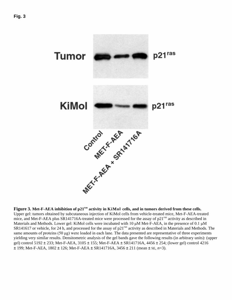
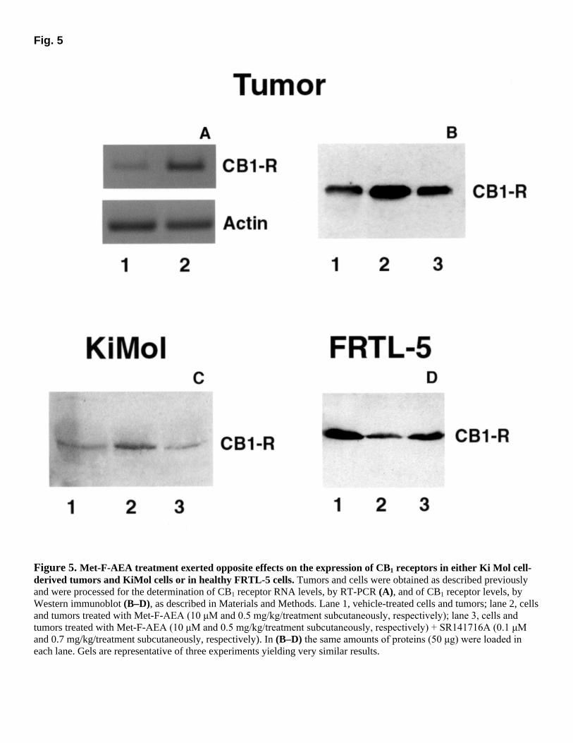
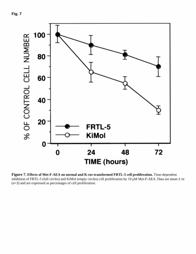
anandamide analog was shown previously to have much lower affinity for CB2 than CB1 cannabinoid receptors (19). The participation of CB1 receptors to the tumor-growth inhibitory effect of Met-F-AEA was also supported by the finding, in tumor tissue, of a CB1 mRNA transcript, as detected by RT-PCR technique, and of a CB1-immunoreactive receptor protein as detected by Western immunoblotting (Fig. 2a, b). The sizes of both the mRNA transcript and the immuno-reactive protein were compatible to what expected for CB1 receptors. Furthermore the immuno-reactive band disappeared if the gel was co-incubated with the immunizing peptide (not shown). The antitumor effect of Met-F-AEA was accompanied by a strong decrease in of p21ras activity in tumors, which was attenuated significantly by SR 141716A (Fig. 3, upper panel). We also looked at the effect of Met-F-AEA on KiMol cells in vitro and found that the anandamide analog (10 µM) inhibits their proliferation. After a 24-h treatment, cells had proliferated ~36% less than vehicle-treated cells (P<0.05 by ANOVA). By contrast, the proliferation of nontransformed FRTL-5 cells after a 24-h treatment was not affected by 10 µM Met-F-AEA (9% inhibition, not statistically significant by ANOVA, see below). The effect of Met-F-AEA was not due to toxicity or to apoptosis of cells, but instead to the dose-dependent (IC50 = 5 µM) arrest of the cell cycle at the G0/G1 phase, associated with a significant reduction of cells in the S and G2/M phase, as shown by cytofluorimetric analysis (Fig. 4). The antiproliferative effect was accompanied by a striking reduction of p21ras activity (Fig. 3, lower panel). All these effects were attenuated significantly by SR141716A (0.1 µM) (Fig. 3, lower panel, and Fig. 4). In fact, also KiMol cells expressed a CB1 mRNA transcript and a CB1-immunoreactive protein (Fig. 2b). These findings indicate that two fundamental components of the endogenous cannabinoid system; that is, the endocannabinoids and the cannabinoid CB1 receptor represent a potentially useful target for the development of therapeutic agents controlling ras oncogene-dependent tumor growth. Finally, we assessed whether cannabinoid CB1 receptors are regulated during endocannabinoid inhibition of tumor development. We were surprised to find that the decrease in tumor volume induced by Met-F-AEA was accompanied by a strong up-regulation of CB1 receptor mRNA and protein compared with vehicle-treated tumors (Fig. 5a, b). Similarly, KiMol cells treated with Met-F-AEA expressed significantly more CB1 receptors, and this effect was abolished by SR141716A (0.1 µM) (Fig. 5c). Cell immunofluorescence studies with both permeabilized and nonpermeabilized cells showed that Met-F-AEA increased the levels of CB1 receptors both on the cell membrane and in the cytosol (Fig. 6). By contrast, nontransformed FRTL-5 cells treated with Met-F-AEA exhibited less CB1 receptors than vehicle-treated cells in both the cell membrane and cytosol, as assessed by Western immunoblot (Fig. 5d) and immunofluorescence analysis (Fig. 6). This finding agrees with the down-regulation of the expression of CB1 receptors generally observed in several healthy cell types and tissues after chronic exposure to CB1 receptor agonists (10). In accordance with this opposite regulation of CB1 receptor expression in transformed vs. healthy cells, we found that after we treated cells with 10 µM Met-F-AEA, the proliferation of Ki Mol cells was significantly more strongly inhibited by the cannabimimetic substance (up to 70% inhibition) than the response of FRTL-5 cells, which barely reached statistical significance after 3 days (up to 28% inhibition, Fig. 7).
Previous observations have shown that THC can effectively reduce the growth of glioma tumors in mice by inducing apoptosis of glioma cancer cells (14). This effect was blocked by a combination of CB1 and CB2 cannabinoid receptor antagonists but not by each antagonist alone, thus leaving the role of each cannabinoid receptor subtype in this effect still to be fully clarified. Furthermore, the apoptotic effect of THC in glioma cells appeared to be mediated by sphingomyelin hydrolysis and ceramide formation (14, 23). More recent studies have confirmed the role of this signaling pathway in cannabinoid-induced inhibition of glioma cell growth (24). By contrast, we have reported previously that the cytostatic effect of anandamide on human breast and prostate cancer cells was due to activation of CB1 receptors (11, 13), inhibition of cAMP-mediated signaling and/or activation of p42/p44 extracellular regulated kinases (ERK) (12), and inhibition of the expression of the receptors for prolactin and nerve growth factor, which act as mitogenic factors for these cells (13). Therefore, we believe that the data presented here are important inasmuch as they indicate for the first time that: 1) anandamide analogs, which have a lower potential for physical dependence than THC and synthetic cannabinoids (25), inhibit tumor growth also in vivo, and at nonpsychotropic doses; 2) the antitumor effects of these substances in vivo can be exerted through CB1 cannabinoid receptors and via inhibition of p21ras activity; and 3) anandamide analogs inhibit the growth of epithelial tumors and, in particular, those derived from KiMol thyroid cells. Indeed, it should be emphasized that epithelial cell-derived tumor models may have particular importance because the large majority of human neoplasias are epithelial in origin (26). Further work will be necessary to establish the exact molecular mechanism of Met-F-AEA-induced suppression of p21ras activity. It has been shown recently that endocannabinoids stimulate ERK, c-Jun amino-terminal kinases (JNK), and p38 mitogen activated protein kinases (MAPKs) in several cell lines via CB1/CB2 receptor-dependent and independent mechanisms (12, 27�29), whereas an inhibitor of p21ras farnesyltransferase was shown to attenuate the CB1-mediated activation of JNK (28). Hence, on the basis of data reported here and previously, we speculate that, by modulating the activity of both p21ras and MAPKs, the CB1 receptor may regulate the fate of cancer cells (e.g., cessation of proliferation and/or apoptosis). Another important finding described here is that CB1 receptors may be regulated by endocannabinoids in KiMol cells in a manner entirely different from that observed with nontransformed cells, thus allowing cancer cells, but not healthy cells, to respond ever more efficaciously to the antiproliferative effect of CB1 agonists. Indeed, we found that transformed cells in culture become ever more responsive to Met-F-AEA, whereas nontransformed cells do not. We speculate that this phenomenon may contribute to the putative tumor-suppressing role proposed for endocannabinoids in previous studies (9, 11, 30). Furthermore, this finding indicates that Met-F-AEA can inhibit the growth of cancer thyroid cells much more effectively than healthy, nontransformed cells can, and strengthens our proposal that endocannabinoid-based drugs should be kept into account as novel antitumor agents. CONCLUSIONS In conclusion, we have shown here that anandamide-based drugs may be efficacious drugs for the inhibition of K-ras-induced epithelial cancer cell growth in vivo through the activation of CB1 receptors, inhibition of p21ras activity, and blockade of the cell cycle. Because multiple pathways are important for the proliferation of tumor cells and because combination therapies
are often more effective than single-drug administration, cannabimimetic substances may complement other anticancer agents that may or may not affect K-ras-mediated pathways. Because THC-like compounds are used to inhibit nausea and induce appetite in cancer patients, and anandamide appears to be an endogenous orexigenic mediator (31), the finding of possible antitumor effect for these substances might have a tremendous potential for therapeutic intervention in preventing the progression of cancer and, at the same time, in alleviating its symptoms. ACKNOWLEDGMENTS This work was supported by the Associazione Italiana per la Ricerca sul Cancro (AIRC) and the Associazione Partenopea Ricerche Oncologiche (APRO) (to M. B.) and by the Ministero dell�Università e Ricerca Scientifica e Tecnologica (MURST) (3933 to V.D.M.). We thank T. Bisogno, G. Mazziotti , S. Stingo, L. Fiorentino, and R. D�Elia for technical assistance, and M. Berardone for the art work. REFERENCES 1. Barbacid, M. (1987) Ras genes. Annu. Rev. Biochem. 56, 779�827 2. Suarez, H. G., Du Villard, J. A., Caillou, B., Schlumberger, M., Tubiana, M., Parmentier, C., and Monier, R. (1988) Detection of activated ras oncogenes in human thyroid carcinomas. Oncogene 2, 403�406 3. Bos, J. L. (1989) Ras oncogenes in human cancer: a review. Cancer Res. 49, 4682�4689 4. Schwab, G., Chavany, C., Duroux, I., Goubin, G., Lebeau, J., Helene, C., and Saison-Behmoaras, T. (1994) Antisense oligonucleotides adsorbed to polyalkylcyanoacrylate nanoparticles specifically inhibit mutated Ha-ras-mediated cell proliferation and tumorigenicity in nude mice. Proc. Natl. Acad. Sci. USA 91, 10460�10464
5. Kohl, N. E., Wilson, F. R., Mosser, S. D., Giuliani, E., deSolms, S. J., Conner, M. W., Anthony, N. J., Holtz, W. J., Gomez, R. P., Lee, T. J., et al. (1994) Protein farnesyltransferase inhibitors block the growth of ras-dependent tumors in nude mice. Proc. Natl. Acad. Sci. USA 91, 9141�9145
6. Mechoulam, R., Fride, E., Di Marzo, V. (1998) Endocannabinoids. Eur J Pharmacol. 359, 1�18
7. Devane, W. A., Hanus, L., Breuer, A., Pertwee, R. G., Stevenson, L. A., Griffin, G., Gibson, D., and Mechoulam, R. (1992) Isolation and structure of a brain constituent that binds to the cannabinoid receptor. Science. 258, 1946�1949
8. Di Marzo V. �Endocannabinoids� and other fatty acid derivatives with cannabimimetic properties: biochemistry and possible physiopathological relevance. (1998) Biochim. Biophys. Acta. 1392, 153�175
9. Guzman, M., Sanchez, C., and Galve-Roperh, I. (2001) Control of the cell survival/death decision by cannabinoids. J. Mol. Med. 78, 613�625
10. Pertwee, R. G. (1997) Pharmacology of cannabinoid CB1 and CB2 receptors. Pharmacol. Ther. 74, 129�180
11. De Petrocellis, L., Melck, D., Palmisano, A., Bisogno, T., Laezza, C., Bifulco, M., and Di Marzo V. (1998) The endogenous cannabinoid anandamide inhibits human breast cancer cell proliferation. Proc. Natl. Acad. Sci. USA 95, 8375�8380
12. Melck, D., Rueda, D., Galve-Roperh, I., De Petrocellis, L., Guzman, M., and Di Marzo, V. (1999) Involvement of the cAMP/protein kinase A pathway and of mitogen-activated protein kinase in the anti-proliferative effects of anandamide in human breast cancer cells. FEBS Lett. 463, 235�240
13. Melck, D., De Petrocellis, L., Orlando, P., Bisogno, T., Laezza, C., Bifulco, M., and Di Marzo V. (2000) Suppression of nerve growth factor Trk receptors and prolactin receptors by endocannabinoids leads to inhibition of human breast and prostate cancer cell proliferation. Endocrinology 141, 118�126
14. Galve-Roperh, I., Sanchez, C., Cortes, M. L., del Pulgar, T. G., Izquierdo, M., and Guzman, M. (2000) Anti-tumoral action of cannabinoids: involvement of sustained ceramide accumulation and extracellular signal-regulated kinase activation. Nat. Med. 6, 313�319
15. Ambesi-Impiombato, F. S., Parks, L.A.M., and Coon, H. G. (1980) Culture of hormone-dependent functional epithelial cells from rat thyroids. Proc. Natl. Acad. Sci. USA 77, 3455�3459
16. Fusco, A., Pinto, A., Ambesi-Impiombato, F. S., Vecchio, G., and Tsuchida, N. (1981) Transformation of rat thyroid epithelial cells by Kirsten murine sarcoma virus. Int. J. Cancer 28, 655�662
17. Laezza, C., Di Marzo, V., and Bifulco, M. (1998) v-K-ras leads to preferential farnesylation of p21(ras) in FRTL-5 cells: multiple interference with the isoprenoid pathway. Proc. Natl. Acad. Sci. USA 95, 13646�13651
18. Bifulco, M., Laezza, C., Aloj, S. M., and Garbi, C. (1993) Mevalonate controls cytoskeleton organization and cell morphology in thyroid epithelial cells. J. Cell Physiol. 155, 340�348
19. Martin, B. R., Mechoulam, R., and Razdan, R. K. (1999) Discovery and characterization of endogenous cannabinoids. Life Sci. 65, 573�95
20. Fusco, A., Pinto, A., Tramontano, D., Tajana, G., Vecchio, G., and Tsuchida, N. (1982) Block in the expression of differentiation markers of rat thyroid epithelial cells by transformation with Kirsten murine sarcoma virus. Cancer Res. 42, 618�626
21. Rinaldi-Carmona, M., Barth, F., Heaulme, M., Shire, D., Calandra, B., Congy, C., Martinez, S., Maruani, J., Neliat, G., Caput, D., Ferrara, P., Soubrié, P., Breliere, J. C., and Le Fur, G. (1994) SR141716A, a potent and selective antagonist of the brain cannabinoid receptor. FEBS Lett. 350, 240�244
22. Sanchez, C., de Caballos, M. L., del Pulgar, T. G., Rueda, D., Corbacho, C., Velasco, G., Galve-Roperh, I., Huffman, J.W., Ramon y Cajal, S., and Guzman, M. (2001) Inhibition of glioma growth in vivo by selective activation of the CB2 cannabinoid receptor. Cancer Res. 61, 5784�5789
23. Sanchez, C., Galve-Roperh, I., Canova, C., Brachet, P., and Guzman, M. (1998) Delta9-tetrahydrocannabinol induces apoptosis in C6 glioma cells. FEBS Lett. 436, 6�10
24. Guzman, M., Galve-Roperh, I., and Sanchez, C. (2001) Ceramide: a new second messenger of cannabinoid action. Trends Pharmacol. Sci. 22, 19�22
25. Aceto, M. D., Scates, S. M., Razdan, R. K., Martin, B. R. (1998) Anandamide, an endogenous cannabinoid, has a very low physical dependence potential. J. Pharmacol. Exp. Ther. 287, 598�605
26. Foulds, L. (1969) Neoplastic Development, vol. 1. London: Academic Press. 27. Kobayashi, Y., Arai, S., Waku, K., and Sugiura, T. (2001) Activation by 2-arachidonoylglycerol, an endogenous cannabinoid receptor ligand, of p42/44 mitogen-activated protein kinase in HL-60 cells. J. Biochem. 129, 665�669
28. Rueda, D., Galve-Roperh, I., Haro, A., and Guzman, M. (2000) The CB(1) cannabinoid receptor is coupled to the activation of c-Jun N-terminal kinase. Mol. Pharmacol. 58, 814�820
29. Derkinderen, P., Ledent, C., Parmentier, M., and Girault, J. A. (2001) Cannabinoids activate p38 mitogen-activated protein kinases through CB1 receptors in hippocampus. J. Neurochem. 77, 957�960
30. Bisogno, T., Katayama, K., Melck, D., Ueda, N, De Petrocellis, L., Yamamoto, S., and Di Marzo, V. (1998) Biosynthesis and degradation of bioactive fatty acid amides in human breast cancer and rat pheochromocytoma cells�implications for cell proliferation and differentiation. Eur. J. Biochem. 254, 634�642
31. Di Marzo, V., Goparaju, S. K., Wang, L., Liu, J., Batkai, S., Jarai, Z., Fezza, F., Miura, G. I., Palmiter, R. D., Sugiura, T., and Kunos, G. (2001) Leptin-regulated endocannabinoids are involved in maintaining food intake. Nature (London) 410, 822�825
Received June 18, 2001; revised August 28, 2001.
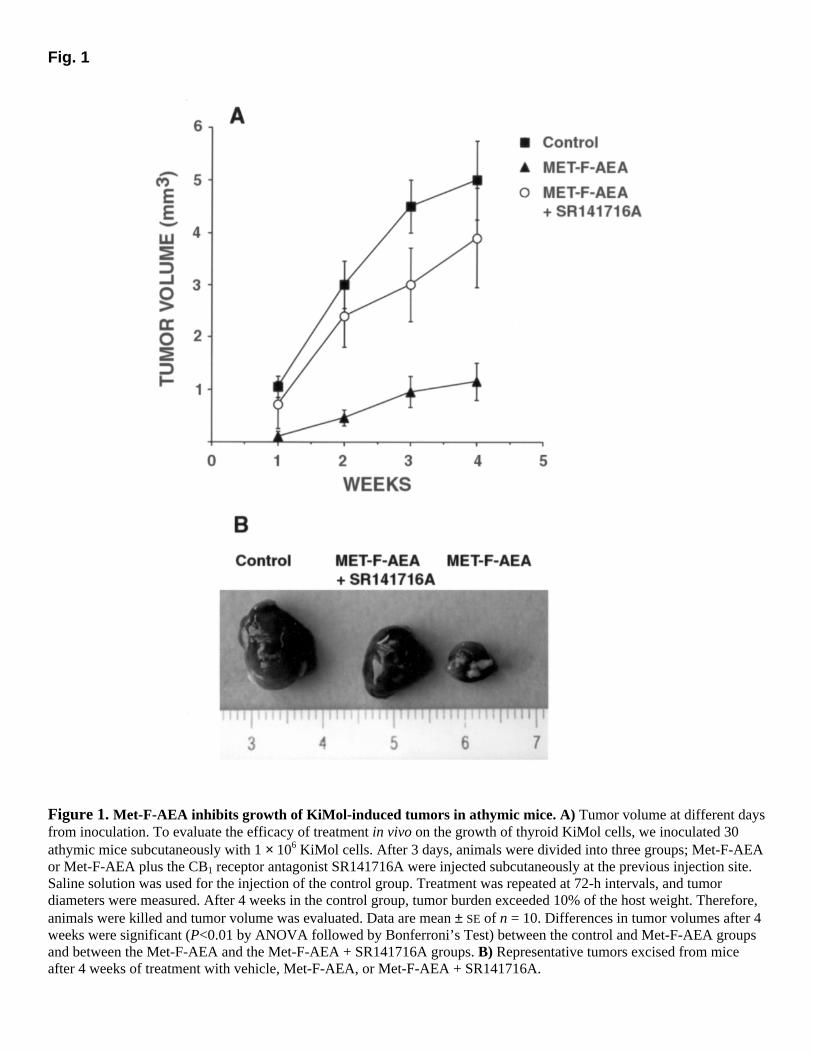
Fig. 1
Figure 1. Met-F-AEA inhibits growth of KiMol-induced tumors in athymic mice. A) Tumor volume at different days from inoculation. To evaluate the efficacy of treatment in vivo on the growth of thyroid KiMol cells, we inoculated 30 athymic mice subcutaneously with 1 × 10⁶ KiMol cells. After 3 days, animals were divided into three groups; Met-F-AEA or Met-F-AEA plus the CB1 receptor antagonist SR141716A were injected subcutaneously at the previous injection site. Saline solution was used for the injection of the control group. Treatment was repeated at 72-h intervals, and tumor diameters were measured. After 4 weeks in the control group, tumor burden exceeded 10% of the host weight. Therefore, animals were killed and tumor volume was evaluated. Data are mean ± SE of n = 10. Differences in tumor volumes after 4 weeks were significant (P<0.01 by ANOVA followed by Bonferroni’s Test) between the control and Met-F-AEA groups and between the Met-F-AEA and the Met-F-AEA + SR141716A groups. B) Representative tumors excised from mice after 4 weeks of treatment with vehicle, Met-F-AEA, or Met-F-AEA + SR141716A.
Fig. 2
Figure 2. Molecular characterization of CB1 receptors in K-ras transformed FRTL-5 cells and in tumors derived from these cells. A) Identification by RT-PCR of a CB1 transcript in the RNA from KiMol cell-derived tumors. The transcript was of the size expected from the use of the oligoprobes described in the Materials and Methods. STD indicates the lane where the base pair ladder was loaded. B) Western immunoblot of proteins from KiMol cells and the derived tumor. Molecular weight markers are shown. The size of the immunoreactive protein was compatible with that reported for cannabinoid CB1 receptors (10). The gel and the blot are representative of three different experiments.
Fig. 3
Figure 3. Met-F-AEA inhibition of p21ras activity in KiMol cells, and in tumors derived from these cells. Upper gel: tumors obtained by subcutaneous injection of KiMol cells from vehicle-treated mice, Met-F-AEA-treated mice, and Met-F-AEA plus SR141716A-treated mice were processed for the assay of p21ras activity as described in Materials and Methods. Lower gel: KiMol cells were incubated with 10 µM Met-F-AEA, in the presence of 0.1 µM
SR141617 or vehicle, for 24 h, and processed for the assay of p21ras activity as described in Materials and Methods. The same amounts of proteins (50 µg) were loaded in each lane. The data presented are representative of three experiments yielding very similar results. Densitometric analysis of the gel bands gave the following results (in arbitrary units): (upper gel) control 5192 ± 233; Met-F-AEA, 3105 ± 155; Met-F-AEA ± SR141716A, 4456 ± 254; (lower gel) control 4216 ± 199; Met-F-AEA, 1802 ± 126; Met-F-AEA ± SR141716A, 3456 ± 211 (mean ± SE, n=3).
Fig. 4
Figure 4. Dose-dependent accumulation of KiMol cells in the Gi/Go phase and reduction in the S phase of the cell cycle by Met-F-AEA. Samples were collected at 24 h, in the presence of different concentrations of Met-F-AEA, and then were analyzed for DNA content by flow cytometry. The effect of the CB1 selective antagonist, SR141617A (0.1 µM)
on Met-F-AEA (20 µM) inhibition of KiMol cell proliferation, is shown (full circles) and was statistically significant
(P<0.05, for the G0/G1 phase, and P<0.01 for the S and G2/M phases, by ANOVA).
Fig. 5
Figure 5. Met-F-AEA treatment exerted opposite effects on the expression of CB1 receptors in either Ki Mol cell-derived tumors and KiMol cells or in healthy FRTL-5 cells. Tumors and cells were obtained as described previously and were processed for the determination of CB1 receptor RNA levels, by RT-PCR (A), and of CB1 receptor levels, by Western immunoblot (B–D), as described in Materials and Methods. Lane 1, vehicle-treated cells and tumors; lane 2, cells and tumors treated with Met-F-AEA (10 µM and 0.5 mg/kg/treatment subcutaneously, respectively); lane 3, cells and
tumors treated with Met-F-AEA (10 µM and 0.5 mg/kg/treatment subcutaneously, respectively) + SR141716A (0.1 µM
and 0.7 mg/kg/treatment subcutaneously, respectively). In (B–D) the same amounts of proteins (50 µg) were loaded in
each lane. Gels are representative of three experiments yielding very similar results.
Fig. 6
Figure 6. Immunofluorescence of CB1-receptors in KiMol and FRTL-5 cells. Cells were pre-incubated for 24 h in the absence (control) and in the presence of 10 µM Met-F-AEA and/or 0.1 µM SR141716A before processing for the immunostaining of CB1-receptors. The figure is representative of three independent experiments. Cells were either permeabilized (A , B , F , G) or not (C, D, E) with 0.05% Triton X-100.
Fig. 7
Figure 7. Effects of Met-F-AEA on normal and K-ras-transformed FRTL-5 cell proliferation. Time-dependent inhibition of FRTL-5 (full circles) and KiMol (empty circles) cell proliferation by 10 µM Met-F-AEA. Data are mean ± SE
(n=3) and are expressed as percentages of cell proliferation.