J Physiol 561.1 (2004) pp 39–51 39 Cooperative effect of S4–S5 loops in domains D3 and D4 on fast inactivation of the Na + channel M. Oana Popa 1 , Alexi K. Alekov 1 , Sigrid Bail 1 , Frank Lehmann-Horn 1 and Holger Lerche 1,2 Departments of 1 Applied Physiology and 2 Neurology, University of Ulm, D-89069 Ulm, Germany Cytoplasmic S4–S5 loops have been shown to be involved in fast inactivation of voltage- gated ion channels. We studied mutations in these loops and their potential cooperative effects in domains D3 (N1151C, A1152C, I1160C/A) and D4 (F1473C, L1482C/A) of the human skeletal muscle Na + channel α-subunit (hNa v 1.4) using expression in tsA201 cells and the whole cell patch-clamp technique. All cysteine mutations were accessible to intra- cellularly applied sulfhydryl reagents which considerably destabilized fast inactivation. For different combinations of corresponding D3/D4 double mutations, fast inactivation could be almost completely removed. Thermodynamic cycle analysis indicated an additive effect for N1151C/F1473C and a significant cooperative effect for I1160/L1482 double mutations. Application of oxidizing reagents such as Cu-phenanthroline to link two cysteines via a disulfide bridge did not reveal evidence for a direct physical interaction of cysteines in D3 and D4. In addition to the pronounced alterations of fast inactivation, mutations of I1160 shifted steady-state activation in the hyperpolarizing direction and slowed the kinetics of both activation and deactivation. Sulfhydryl reagents had charge-dependent effects on I1160C suggesting interaction with negative charges in another protein region. We conclude that fast inactivation of the Na + channel involves both S4–S5 loops in D3 and D4 in a cooperative manner. D3/S4–S5 also plays an important role in activation and deactivation. (Received 5 April 2004; accepted after revision 27 September 2004; first published online 30 September 2004) Corresponding author H. Lerche: Departments of Applied Physiology and Neurology, University of Ulm, Zentrum Klinische Forschung, Helmholtzstr. 8/1, D-89081 Ulm, Germany. Email: [email protected]Voltage-gated Na + and K + channels provide the basis for the generation and conduction of action potentials in nerve and muscle cells. In response to membrane depolarization, they open from the resting, closed state, and then Na + channels and some types of K + channels inactivate. Voltage-gated ion channels have a common tetrameric structure of homologous domains (D1–D4) each with six transmembrane segments (S1–S6). Whereas K + channels are constituted by four identical domains, the about fourfold longer Na + channel α-subunits have four homologous but distinct domains. All four S4 segments contain positively charged residues conferring voltage dependence to the channel protein (Hille, 2001). As first proposed by Armstrong & Bezanilla (1977), fast inactivation of voltage-gated channels is supposed to function in a ball and chain or hinged-lid fashion with a tethered inactivation particle occluding the internal mouth of the pore from the cytoplasmic side (Hoshi et al. 1990; West et al. 1992). S4–S5 loops are short sequences of 15–20 amino acids adjacent to the voltage sensors and exposed to the cytoplasm. They have been shown previously to play an important role in fast inactivation of both K + and Na + channels, and have been proposed to contribute to the formation of a receptor site for the inactivation particle (Isacoff et al. 1991; Holmgren et al. 1996; Smith & Goldin, 1997; Lerche et al. 1997; Filatov et al. 1998; McPhee et al. 1998; Tang et al. 1998; reviewed by Catterall, 2000). A direct interaction between an inactivation peptide and S4–S5 in Shaker K + channels (Holmgren et al. 1996) as well as between the proposed Na + channel inactivation particle IFM and an amino acid in D3/S4–S5 of the rat brain type IIa Na + channel (rNa v 1.2) (Smith & Goldin, 1997) was suggested by experiments using substituted, complementary charges. Structure–function analysis using cysteine mutagenesis of the D4/S4–S5 loop of the rat or human skeletal muscle Na + channel confirmed an important role in fast inactivation, but revealed that most parts of D4/S4–S5 are still accessible when the channel is inactivated (Lerche et al. 1997; Filatov et al. 1998), arguing against a direct function of the proximal part of D4/S4–S5 as a receptor site for the IFM. In a more recent study with K + channels, the receptor site for the inactivation ball was located in the central cavity C The Physiological Society 2004 DOI: 10.1113/jphysiol.2004.065912
Transcript
J Physiol 561.1 (2004) pp 39–51 39
Cooperative effect of S4–S5 loops in domainsD3 and D4 on fast inactivation of the Na+ channel
M. Oana Popa1, Alexi K. Alekov1, Sigrid Bail1, Frank Lehmann-Horn1 and Holger Lerche1,2
Departments of 1Applied Physiology and 2Neurology, University of Ulm, D-89069 Ulm, Germany
Cytoplasmic S4–S5 loops have been shown to be involved in fast inactivation of voltage-gated ion channels. We studied mutations in these loops and their potential cooperativeeffects in domains D3 (N1151C, A1152C, I1160C/A) and D4 (F1473C, L1482C/A) of thehuman skeletal muscle Na+ channel α-subunit (hNav1.4) using expression in tsA201 cellsand the whole cell patch-clamp technique. All cysteine mutations were accessible to intra-cellularly applied sulfhydryl reagents which considerably destabilized fast inactivation. Fordifferent combinations of corresponding D3/D4 double mutations, fast inactivation couldbe almost completely removed. Thermodynamic cycle analysis indicated an additive effectfor N1151C/F1473C and a significant cooperative effect for I1160/L1482 double mutations.Application of oxidizing reagents such as Cu-phenanthroline to link two cysteines via adisulfide bridge did not reveal evidence for a direct physical interaction of cysteines inD3 and D4. In addition to the pronounced alterations of fast inactivation, mutations ofI1160 shifted steady-state activation in the hyperpolarizing direction and slowed the kineticsof both activation and deactivation. Sulfhydryl reagents had charge-dependent effects onI1160C suggesting interaction with negative charges in another protein region. We concludethat fast inactivation of the Na+ channel involves both S4–S5 loops in D3 and D4 in acooperative manner. D3/S4–S5 also plays an important role in activation and deactivation.
(Received 5 April 2004; accepted after revision 27 September 2004; first published online 30 September 2004)Corresponding author H. Lerche: Departments of Applied Physiology and Neurology, University of Ulm,Zentrum Klinische Forschung, Helmholtzstr. 8/1, D-89081 Ulm, Germany. Email: [email protected]
Voltage-gated Na+ and K+ channels provide the basisfor the generation and conduction of action potentialsin nerve and muscle cells. In response to membranedepolarization, they open from the resting, closed state,and then Na+ channels and some types of K+ channelsinactivate. Voltage-gated ion channels have a commontetrameric structure of homologous domains (D1–D4)each with six transmembrane segments (S1–S6). WhereasK+ channels are constituted by four identical domains, theabout fourfold longer Na+ channel α-subunits have fourhomologous but distinct domains. All four S4 segmentscontain positively charged residues conferring voltagedependence to the channel protein (Hille, 2001). Asfirst proposed by Armstrong & Bezanilla (1977), fastinactivation of voltage-gated channels is supposed tofunction in a ball and chain or hinged-lid fashion with atethered inactivation particle occluding the internal mouthof the pore from the cytoplasmic side (Hoshi et al. 1990;West et al. 1992).
S4–S5 loops are short sequences of 15–20 aminoacids adjacent to the voltage sensors and exposed to thecytoplasm. They have been shown previously to play
an important role in fast inactivation of both K+ andNa+ channels, and have been proposed to contributeto the formation of a receptor site for the inactivationparticle (Isacoff et al. 1991; Holmgren et al. 1996; Smith& Goldin, 1997; Lerche et al. 1997; Filatov et al. 1998;McPhee et al. 1998; Tang et al. 1998; reviewed by Catterall,2000). A direct interaction between an inactivationpeptide and S4–S5 in Shaker K+ channels (Holmgrenet al. 1996) as well as between the proposed Na+ channelinactivation particle IFM and an amino acid in D3/S4–S5of the rat brain type IIa Na+ channel (rNav1.2) (Smith& Goldin, 1997) was suggested by experiments usingsubstituted, complementary charges. Structure–functionanalysis using cysteine mutagenesis of the D4/S4–S5 loopof the rat or human skeletal muscle Na+ channel confirmedan important role in fast inactivation, but revealed thatmost parts of D4/S4–S5 are still accessible when thechannel is inactivated (Lerche et al. 1997; Filatov et al.1998), arguing against a direct function of the proximalpart of D4/S4–S5 as a receptor site for the IFM. In amore recent study with K+ channels, the receptor sitefor the inactivation ball was located in the central cavity
of the pore formed by the S6 segments (Zhou et al.2001).
An important question for Na+ channels in contrastto K+ channels is whether the distinct domains playdifferent roles in channel gating. Previous studies revealedthat in particular D4 plays a prominent role in fastinactivation, whereas D1 to D3 are more important forchannel activation (Stuhmer et al. 1989; Chahine et al.1994; Lerche et al. 1996; Chen et al. 1996; Mitrovic et al.1998; Cha et al. 1999). Second, the question for bothNa+ and K+ channels arises as to whether the voltagesensors and other protein structures in different domainsact independently or in a cooperative manner. Severalstudies revealed evidence for cooperative interactions ofthe voltage sensors during activation of K+ channels(Tytgat & Hess, 1992; Schoppa et al. 1992; Bezanilla et al.1994; Zagotta et al. 1994; Schoppa & Sigworth, 1998;Smith-Maxwell et al. 1998a,b; Ledwell & Aldrich, 1999;Mannuzzu & Isacoff, 2000) and recent work also pointsto such cooperative effects in Na+ channels (Chanda et al.2004).
Our paper is focused on cooperative effects of S4–S5loops in domains D3 and D4 with regard to fastinactivation of the Na+ channel. We chose three sites inD3/S4–S5 (N1151, A1152, I1160) and two in D4/S4–S5(F1473, L1482) which either harbour mutations causingion channel disorders (Ptacek et al. 1994; Wang et al. 1995;
Figure 1. Model of the adult human muscle Na+ channel α-subunit (Nav1.4)All mutations within the intracellular S4–S5 loops of domains D3 and D4 are shown. The lower part of the figureshows a putative alignment of the amino acid sequences in S4 and S4–S5 segments of domains D1–D4 of Nav1.4,the Shaker K+ channel (Kv1.1). Positive charges of the voltage sensors (S4 segments) are shown in italic, conservedresidues in S4–S5 are underlined, and the residues mutated in this study are shown in bold.
Mitrovic et al. 1996; Richmond et al. 1997; Fleischhaueret al. 1998) or have otherwise been shown to be importantfor fast inactivation of the channel (Smith & Goldin,1997; Lerche et al. 1997; Filatov et al. 1998; McPhee et al.1998; Alekov et al. 2001). According to an alignmentusing positively charged residues in S4 segments andfive conserved residues in S4–S5 loops, N1151/F1473as well as I1160/L1482 should be correspondingresidues in D3/S4–S5 and D4/S4–S5, respectively(Fig. 1). These two pairs were used for cooperativitystudies.
Methods
Mutagenesis
Site-directed mutagenesis to introduce the differentalanine and cysteine mutations in S4–S5 loops of domainsD3 and D4 of the α-subunit of the human skeletal muscleNa+ channel (Nav1.4, gene: SCN4A) was performed usingthe Altered Sites system employing the plasmid vectorpSELECT (Promega Corporation; Lerche et al. 1997;Alekov et al. 2001) or PCR-based strategies. All mutationswere verified by dideoxynucleotide sequencing. Full lengthwild-type (WT) and mutant constructs of single anddouble mutations were assembled in the expression vectorspRC/CMV for transfection into the mammalian cell linetsA201.
J Physiol 561.1 Cooperative effect of D3/S4–S5 and D4/S4–S5 of the Na+ channel 41
Expression in tsA201 cells
Transfection of plasmids containing the cDNA of WTor mutant Na+ channel α-subunits into tsA201 cellswas performed using a standard calcium phosphatemethod. A CD8-cDNA containing plasmid wascotransfected to recognize transfected cells usinganti-CD8 antibody-coated microbeads (DynabeadsM450, Dynal, Oslo, Norway) (Lerche et al. 1997).
Electrophysiology
Na+ currents from mammalian cells were recordedusing an EPC-7 patch clamp amplifier (List Electronics,Darmstadt, Germany), a Digidata 1200 digitizer andpCLAMP 6 data acquisition software (Axon Instruments,Union City, CA, USA). Leakage and capacitive currentswere automatically subtracted using a pre-pulse protocol(−P/4). Currents were filtered at 3 kHz and digitized at20 kHz, except for tail currents which were filtered at10 kHz and digitized at 50 kHz. All measurements wereperformed at room temperature of 21–23◦C.
The experiments were conducted in tsA201 cells usingthe whole cell patch clamp technique. Na+ currents of2–15 nA were recorded from transfected tsA201 cells, atleast 10 min after establishing the whole cell configuration.Borosilicate glass pipettes were fire polished with a finaltip resistance of 0.8–1.2 M� when filled with internalrecording solution (see below). We carefully checked thatthe maximal voltage error due to residual series resistanceafter up to 90% compensation was always ≤ 5 mV.
The sulfhydryl reagents [2-(trimethylammonium)ethyl]methanethio-sulfonate bromide and sodium(2-sulfonatoethyl)methanethiosulfonate (MTSET andMTSES, Toronto Research Chemicals, North York,Ontario, Canada) were kept in a H2O-stock solution(500 mm) at −20◦C and diluted 1 : 50 (MTSES) or1 : 200 (MTSET) to the pipette solution immediatelybefore the experiments. Reagents with lower watersolubility (MTS-1-MTS) were prepared as stocks indimethylsulfoxide (DMSO) and stored at −20◦C. Analiquot of the stock solution was thawed each day andused as in the case of water soluble reagents.
Cu(II)1,10-phenanthroline3 stock solution wasprepared by dissolving Cu(II)SO4 and 1,10-phenanthroline to reach final concentrations of 150and 500 mm, respectively, in a 4 : 1 water/ethanol solution(Careaga & Falke, 1992). This stock solution was diluted1 : 1000 to the pipette solution at the time of use.
Data analysis
All data were analysed using a combination ofpCLAMP, Microsoft Excel and Origin (OriginLabInc., Northampton, MA, USA) software. For statisticalevaluation, Student’s t test was applied. All data are shownas means ± s.e.m., unless otherwise indicated.
The membrane was depolarized to various testpotentials from a holding potential of −140 mV. Thetime course of inactivation was best fitted to a doubleexponential function yielding two time constants ofinactivation. The weight of the second slower timeconstant was relatively small (0–25%) except for mutationscontaining F1473C-ES for which we found an amplitudeof 50% for the slow component. To describe the slowingof inactivation in the Results section, only the fasttime constant was therefore used, which was termed τ h.Persistent Na+ currents (I ss, for ‘steady-state’ current),were determined at the end of 70 ms-lasting depolarizingpulses to 0 mV and are given relative to the initialpeak current (Ipeak). Recovery from inactivation wasrecorded from a holding potential of −100 mV. Cellswere depolarized to −20 mV for 100 ms to inactivate allNa+ channels and then repolarized to various recoverypotentials for increasing duration. The time course ofrecovery from inactivation was best fitted to a doubleexponential function with an initial delay. For comparisonamong WT, mutants and effects of MTSET/MTSES, onlythe fast time constant was used, since the slow one hada relatively small weight (< 25%) and did not vary muchcompared to the fast one, which was termed τ rec.
Steady-state inactivation was determined using 300 msconditioning pulses to various potentials followed by thetest pulse to −20 mV at which the peak current reflectedthe percentage of non-inactivated channels. Inactivationcurves were fitted to a standard Boltzmann function:
I/Imax(V ) = 1/(1 + exp[(V − V 1/2 )/kV]),
with Imax being the maximal current amplitude, V 1/2
the voltage of half-maximal inactivation, and kV theslope factor. The activation curve (conductance–voltagerelationship) was derived from the current–voltagerelationship by measuring the peak current at various stepdepolarizations from the holding potential of −140 mV,and also fit to a Boltzmann function:
g/gmax(V ) = 1/(1 + exp[(V − V 1/2 )/kV]),
with g = I/(V – V rev) being the conductance, gmax themaximal conductance, V rev the Na+ reversal potential, V 1/2
the voltage of half-maximal activation, and kV the slopefactor.
Na+ current traces were fitted to the Hodgkin-Huxleyequation (m3h, Hodgkin & Huxley, 1952) to quantify the
where τm is the activation time constant, τ h theinactivation time constant, A an amplitude factor, t thetime after onset of the depolarization, and t0 the delay toactivation of the channel.
To characterize deactivation of WT and mutant Na+
channels, a short depolarizing pulse (0.26 ms to 70 mV),which activated all channels without causing significantinactivation, was followed by the test pulse to theindicated potentials. The deactivation time constant, τ d,was obtained by a single exponential fit to the tail currentdecay.
Entry into, recovery from and steady-state slowinactivation were characterized using cumulativeprotocols (Alekov et al. 2001). To measure the entry,cells were held at −100 mV, depolarized to 0 mV forincreasing duration, repolarized for 100 ms to −100 mVto let the channels recover from fast inactivation, andthen depolarized again shortly for 3 ms to determine thefraction of slow inactivated channels without inducingfurther slow inactivation. The time course of slowinactivation was well fitted to a single exponentialfunction. Recovery from slow inactivation was measuredfor different time points at −120 mV after a 30 sconditioning pulse to 0 mV. Curves were fitted to a doubleexponential function. For steady-state slow inactivation30 s conditioning pulses starting at −140 mV andstepping by 10 mV increasingly to 10 mV were used, witheach followed by a 20 ms hyperpolarization to −140 mV(to let the channels recover from fast inactivation) andthe 5 ms test pulse to −20 mV. Curves were fitted to astandard Boltzmann function as for fast inactivation (seeabove).
Double mutant cycle analysis
The mutational effects in Nav1.4 were analysed througha thermodynamic description of site-specific effects. Wecalculated the change in free energy due to inactivation ofthe WT and mutant proteins according to
�G = −RT lnKeq,
where R is the gas constant, T is the absolute temperature inkelvins, and K eq is the equilibrium constant, kon/koff, withkon and koff being the on and off rate constants for fastinactivation. For strong depolarizations, the steady-statelevel of the current relative to the peak current canbe approximated as I ss/Ipeak ≈ koff/(koff + kon). Thus, K eq
was calculated as K eq = kon/koff ≈ (1 − I ss/Ipeak)/(I ss/Ipeak)(McPhee et al. 1995). We used the steady-state currentdetermined at 0 mV, 70 ms after onset of the depolarizationfor these calculations (see above). At this voltage, WT andall mutant channels were fully activated, as can be seen in
Fig. 3. As a measure of the effect on fast inactivation ofthe site-directed mutation, we calculated the difference��G(WT→MUT) = �GMUT − �GWT. In the absenceof coupling between two mutations, the change in thefree energy associated with that transition upon a doublemutation equals the sum of changes in free energy dueto the single mutations. The deviation from additivitygives a measure of the change in the coupling between thetwo mutations due to the specified transition (Horovitz &Fersht, 1990), in our case fast inactivation:
�Gcoupling = ��G(WT→MUT1/MUT2)
−(��G(WT→MUT1) + ��G(WT→MUT2)).
�G for WT and mutations was calculated for eachcell separately. Standard deviations for �Gcoupling werecalculated by error propagation, i.e. the square root of thesum of the variances.
Results
To evaluate a potential cooperative effect of mutations inD3/S4–S5 and D4/S4–S5, we first studied the biophysicalproperties of the single mutations and second those of thedifferent D3/D4 double mutants. We mainly introducedcysteines and modified those by intracellularly appliedsulfhydryl reagents (Stauffer & Karlin, 1994), but we alsointroduced alanines at positions 1160 and 1482 to confirmthe effects of cysteines in an independent experiment.Cooperative effects were analysed using thermodynamicmutant cycles. Third, we tested a direct physicalinteraction of D3/D4 cysteine double mutations usingoxidizing reagents to induce disulfide bridges. In addition,we present interesting data on defects of channel activationand deactivation, and report results on slow inactivation.
Effects of single mutations in D3/S4–S5 and D4/S4–S5
Three cysteine mutations (N1151C, A1152C, and I1160C)and one alanine mutation (I1160A) were investigatedin D3/S4–S5 using transient expression in tsA201 cellsand the whole cell patch clamp technique. We aimed tocharacterize the accessibility of the introduced cysteinesin the resting and inactivated states of the channelusing MTSES. The negatively charged reagent has beenshown to successfully modify cysteine mutants throughoutD4/S4–S5; additional use of the positively charged MTSETdid not yield further information (Lerche et al. 1997).Therefore, we here investigated charge-dependent effects,i.e. using both MTSES and MTSET, only at one site− I1160– at which we observed large alterations in channel gating.Cysteines were modified by intracellularly applied MTSES(10 mm) or MTSET (2.5 mm) (denoted as C-ES or C-ETin the following). The reactions of sulfhydryl reagents withthe different cysteine mutations were generally slow (Fig. 2,
J Physiol 561.1 Cooperative effect of D3/S4–S5 and D4/S4–S5 of the Na+ channel 43
inset). Reaction rates were determined from the increase inpersistent Na+ current or the slowing of inactivation. Theyranged from 0.11 to 0.03 min−1. For L1482C the reactionwas extremely slow with a rate of about 0.008 min−1.
The results in the absence and presence of sulfhydrylreagents revealed profound alterations of both activationand inactivation parameters, most pronounced forI1160C-ES or -ET. Families of raw current traces areshown in Fig. 2. It is noteworthy that I1160C could bemodified by MTSES or MTSET only at a depolarizedmembrane but not when the cells were hyperpolarized(Fig. 2, inset), whereas modification of the other twocysteine mutations was independent of the membranepotential. This indicates a relative conformational changeof D3/S4–S5 exposing C1160 to the cytoplasm uponmembrane depolarization.
Steady-state activation and inactivation curves arepresented in Fig. 3A. The activation curve was largely
Figure 2. Whole-cell currents of D3/S4–S5, D4/S4–S5 and D3/D4 double mutantsDifferent pairs of mutations were investigated combining corresponding mutations at positions 1151 (D3/S4–S5)and 1473 (D4/S4–S5), as well as those at 1160 (D3/S4–S5) and 1482 (D4/S4–S5) (compare alignment in Fig. 1).Shown are representative raw current traces as recorded for each of the mutants expressed in tsA201 cells, inthe absence and presence of MTSES (10 mM) or MTSET (2.5 mM), elicited by depolarizing the membrane between−105 mV and 67.5 mV in 7.5-mV steps from a holding potential of −140 mV. Note that – beside a distinct slowingof the inactivation time course and a persistent Na+ current at the end of the depolarization – the activation timecourse was slowed significantly for I1160C-ES, but not for I1160C-ET. The scale bars represent 1 nA (vertical) and5 ms (horizontal), respectively. The inset shows the reaction of I1160C with MTSES monitored as the change withtime of the persistent current relative to the peak current (Iss/Ipeak) 20 ms after onset of the depolarization, at twodifferent holding potentials. At −20 mV (filled symbols), the reaction rate was 0.06 ± 0.03 min−1, whereas therewas no detectable reaction at −140 mV (open symbols), n = 3–4.
shifted in the hyperpolarizing direction, in particular formutations at I1160, whereas steady-state inactivation wasless affected (Fig. 3B and C, and Table 1).
The time course of fast inactivation was slowed3- to 4-fold for all mutations modified by the negativelycharged MTSES without altering its voltage sensitivity,whereas kinetics were only slowed minimally for thepositively charged I1160C-ET (Fig. 4A and B). The mostprominent alteration of fast inactivation parameterswas an increase of the persistent Na+ current. Seventymilliseconds after onset of the depolarization to 0 mV, weobserved a persistent current of 20 ± 4% of the maximalpeak current (I ss/Ipeak) for I1160C-ET and less pronouncedincreases for the other mutations compared to 0.4 ± 0.1%for wild-type (WT) channels (Fig. 4C). I1160A was theonly mutation with a stabilizing effect on fast inactivation– left shift of the availability curve (Fig. 3C) and slowingof the recovery (Fig. 4D).
The mutations F1473C, L1482C and L1482A inD4/S4–S5 have been already investigated in previousstudies of our group using different solutions (with 2 mmMTSES) and protocols at that time (Lerche et al. 1997;Alekov et al. 2001). Raw currents of these mutants asrecorded in the study presented here are shown in Fig. 2,their gating parameters in Figs 3B and C and 4B–D, andTable 1. The results were very similar to those reportedbefore. Additionally, a hyperpolarizing shift of steadystate activation observed for F1473C-ES now reachedsignificance. Using 10 mm MTSES, we now observed anincrease in persistent sodium current for L1482C-EScompared to L1482C. The reaction of L1482C with MTSESwas very slow (see above) and resulted in small effects(Figs 3B and C and 4B–D). Therefore, it could not beclearly determined if there was any voltage dependenceof the availability of C1482. Also, a native cysteine mighthave become little accessible after mutating L1482inducing these slowly progressing changes. However, as theobserved effect was an increase in persistent Na+ currentoccurring as well for L1482C without reagents, L1482Aand other mutations in that channel region (Lerche et al.1997), we believe that it was specific for L1482C.
Figure 3. Activation and steady-state inactivation parameters of the studied mutantsA, voltage dependence of steady-state activation (open symbols), and steady-state inactivation (filled symbols), forI1160C (upper diagram) and for I1160C/L1482A (lower diagram) before and after modification by MTSES/MTSET.Symbols as indicated in the legend. Lines represent fits to standard Boltzmann functions. Values for V1/2
are given inTable 1. B, V1/2
of activation curve for all mutants relative to the WT. C, V1/2of steady-state inactivation curve for all
mutants relative to the WT. Symbols of B and C as indicated in the legend. All values are shown as means ± S.E.M.,n = 3–15.
Effects on activation and deactivation mutating I1160
The left shift of the activation curve observed for manyof the investigated mutants (Fig. 3B) might have arisenat least in part from the disrupted inactivation whichcan alter the apparent voltage dependence of activation.However, for mutants containing I1160C the activation,as well as deactivation, parameters were strongly affected– independent of the observed changes in inactivation –suggesting a direct effect on activation and deactivation.Beside the strong hyperpolarizing shift of the activationcurve (up to −30 mV for I1160C-ET, Fig. 3A and B, andTable 1), we observed a distinct slowing of the activationtime course for I1160C and I1160C-ES. Interestingly, thisdid not occur with introduction of a positive charge(I1160C-ET) (see raw currents in Fig. 2). The activationtime constants, τm, for these mutations are shown inFig. 5A over the whole voltage range investigated. For themore conservative mutant I1160A, a less pronounced butsignificant slowing of activation was also observed at −60and −70 mV. The time course of activation was normal forall other single mutations in D3/S4–S5 or D4/S4–S5 (Fig. 2,time constants not shown). For the double mutationscontaining I1160C(-ES), we obtained similar results to
Values for V1/2, the voltage of half-maximal activation and inactivation, and the slope factor kv were
derived from Boltzmann fits to activation and inactivation curves (see Methods). n = 3–15. Significancelevels are indicated as follows: ∗P < 0.05, ∗∗P < 0.01, ∗∗∗P < 0.001.
those for I1160C(-ES) alone, but for those with I1160C-ETactivation was again fast (Figs 2 and 5B).
Representative examples of raw current traces of thedeactivation time course are shown in Fig. 5C, and thedeactivation time constants, τ d, in Fig. 5D and E, forall examined single and double mutations, respectively.Deactivation was slowed up to 10-fold for all mutationscontaining I1160C(-ES/-ET) and for I1160A, regardless ofwhether the substituted modified or unmodified residuein position 1160 was neutral, or negatively or positivelycharged. In contrast to the differential effects of theintroduced charges on the time course of activation,MTSET even had a slightly stronger effect on deactivationthan MTSES. Thus, a positive charge in position 1160prevents the slowing of activation kinetics observed forI1160C and I1160C-ES, but all other effects on gatingwere not qualitatively different for a positive charge in thisposition when compared to the polar cysteine residue or anegative charge.
Cooperativity of corresponding mutationsin D3/S4–S5 and D4/S4–S5
Families of raw current traces for D3/D4 doublemutations before and after modification by sulfhydryl
reagents are shown in Fig. 2 in comparison to therespective single mutations. For N1151C/F1473C weobserved similar effects as for F1473C, in particulara strong slowing of fast inactivation. None of thealterations in channel gating indicated a cooperativeaction of both mutations (Figs 3B and C and4B–D).
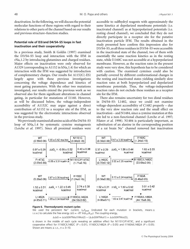
In contrast, for the different combinations of doublemutations in positions 1160/1482, the most obviouschanges were much larger persistent Na+ currents thanexpected for a simple additive effect, suggesting acooperative effect of mutations in those two positions.For the modified double mutations, fast inactivationcould be almost completely removed (Figs 2 and 4C).The coupling energies for each pair of mutations weredetermined by calculating the change in the free energyof the channel state due to inactivation for all singlemutations as well as for the double mutations and thedifferences between them in thermodynamic mutantcycles (see Methods). The coupling energies for all threepairs of mutations in the absence of sulfhydryl reagentswere significantly different from zero (P < 0.05 or < 0.01,respectively, Fig. 6) indicating that these double mutationsact interdependently with regard to their effects on fastinactivation.
The other gating parameters of 1160/1482 doublemutations were altered in a similar way as already observedfor the respective single mutations. Activation curveswere largely shifted in the hyperpolarizing direction asfor I1160C/A mutations, and the inactivation curvestowards more depolarized membrane potentials as seenfor L1482C/A mutations (Fig. 3A–C). The kinetics of fastinactivation were slowed and recovery from inactivationwas accelerated (Fig. 4B and D). These changes point toa considerably destabilized fast inactivated state of thedouble mutant channel.
Exploring a direct interaction of D3/S4–S5and D4/S4–S5 using cysteine double mutations,Cu-phenanthroline and MTS–linkers
The interaction of two protein regions can be examinedby double cysteine mutagenesis, when experimentsare designed to link the two cysteine side chainseither directly via a disulfide bridge using oxidizingreagents such as Cu-phenanthroline, or with sulfhydrylreagents having a reactive MTS group at both ends(MTS-1-MTS). We performed such experiments withboth pairs of combined cysteine mutations in D3/S4–S5and D4/S4–S5 (N1151C/F1473C, I1160C/L1482C) usingCu-phenanthroline (0.15 mm) or H2O2 (up to 1%) asoxidizing reagents, and MTS-1-MTS as a sulfhydryl linker.All substances were applied intracellularly, i.e. added to thepipette solution in whole cell experiments.
Figure 4. Kinetics and steady-state current of D3/S4–S5, D4/S4–S5 and D3/D4 double mutantsA, fast inactivation time constant, τh, as a function of voltage for I1160C (upper diagram) and I1160C/L1482A(lower diagram) before and after modification by thiol reagents. Symbols as indicated in the legend. For comparison,τh at 0 mV (B), the sustained current relative to the peak current 70 ms after onset of the depolarization at 0 mV,Iss/Ipeak (C), and the time constant of recovery from inactivation at −100 mV, τ rec (D), are shown. Symbols asindicated in the legend. All values are shown as means ± S.E.M., n = 3–13.
For N1151C/F1473C, we did not observe significanteffects on channel gating using either of the oxidizingsubstances. Applying the sulfhydryl linker, a slowing of theinactivation time course occurred, similar to that observedafter modification by MTSES. There was no effect onchannel gating pointing to a direct interaction of bothcysteines via the MTS-linker, such as a channel block or anabolished inactivation that might have been expected for asevere conformational change of the protein implicatedby the formation of a new disulfide bond (results notshown).
When MTS-1-MTS was applied to I1160C/L1482Cthere was also no substantial effect on channel gatingother than those already known for MTSES or MTSETmodification. In contrast, when Cu-phenanthroline(150 µm) was applied to this double mutation, weobserved an inhibition of Na+ current at highly positivemembrane potentials which developed in a use-dependentmanner. The Cu-phenanthroline-induced inhibition wasreversible without using reducing reagents, in thepresence of Cu-phenanthroline, contrary to what wouldhave been expected for the formation of a disulfidebond between the two introduced cysteines. Moreover,another oxidizing reagent, H2O2, was not able to inducecurrent inhibition in contrast to Cu-phenanthroline.These results clearly indicate that there is no evidencefor a direct interaction of both cysteine residues andthat the observed current inhibition is due to achannel block by Cu-phenanthroline. However, when we
J Physiol 561.1 Cooperative effect of D3/S4–S5 and D4/S4–S5 of the Na+ channel 47
examined the respective single mutations, I1160C andL1482C, we only observed a very weak block forboth mutations − 14.3 ± 3.9% of blocked channels at+60 mV for I1160C, 18.2 ± 3.2% for L1482C comparedwith 67.9 ± 5.3% for the I1160C/L1482C (results forCu-phenanthroline not shown). Thus, the doublemutation strongly enhanced channel block which under-lines the cooperative action of both residues alreadydescribed for fast inactivation.
Slow inactivation
Since slow inactivation was affected by mutationsL1482C/A in D4/S4–S5 as reported previously byour group (Alekov et al. 2001), we also looked forchanges in slow inactivation of mutation I1160C at thecorresponding site in D3/S4–S5 and the double mutationI1160C/L1482C. All mutations had small but significanteffects on slow inactivation. I1160C shifted the steady-stateavailability curve by −7 mV (and by −10 mV after MTSESapplication) and accelerated the entry (at 0 mV) into slowinactivation by 1.8-fold. L1482C increased the steepnessof the availability curve and slowed recovery from slow
Figure 5. Effects on the kinetics of activation anddeactivationA, activation time constant, τm, as a function of voltagefor I1160C(-ES/-ET), I1160A mutations. Activation wassignificantly slowed for I1160C in the absence andpresence of MTSES, but not MTSET. B, voltagedependence of activation time constants for the doublemutants I1160C(-ES/-ET)/L1482A andI1160C-ES/L1482C-ES. C, deactivation pulse protocoland representative recordings for WT and I1160C-ES.Scale bars represent 1 nA (vertical) and 1 ms (horizontal),respectively. D and E, time constant of deactivation, τd,for the single mutations (D) and the double mutations(E) as a function of voltage. Symbols as indicated in thelegend (inset). Shown are means ± S.E.M., n = 3–6.
inactivation (at −120 mV) by 6.4-fold. Consequently,for the double mutation we observed a negativelyshifted (−4 mV) and steeper availability curve, a 1.8-foldaccelerated entry and a 2.5-fold slowed recovery, thus aconsistent stabilization of slow inactivation. Applicationof MTSES did not substantially alter these results (resultsfor slow inactivation are not shown).
Discussion
Although the four different domains of the Na+ channelcertainly are specialized in their function, and in particularthe voltage sensor D4/S4 is most important for fastinactivation in contrast to D1–D3/S4 (see Introduction),we here present evidence that S4–S5 loops of both D3and D4 play crucial roles in fast inactivation of thechannel and – more importantly – that the distal partsof these regions act in a cooperative manner. Mutations inboth regions also affected voltage-dependent activation.However, most of the observed effects were relativelysmall compared to the alterations of fast inactivation,except for mutations at position 1160 affecting profoundlythe voltage dependence and kinetics of activation and
deactivation. In the following, we will discuss the potentialmolecular functions of these regions with regard to theirrelations to other parts of the channel based on our resultsand previous structure–function studies.
Potential role of D3/and D4/S4–S5 loops in fastinactivation and their cooperativity
In a previous study, Smith & Goldin (1997) examinedthe D3/S4–S5 loop and interaction with the IFM inrNav1.2 by introducing glutamines and charged residues.Major effects on inactivation were only observed forA1329 (corresponding to A1152 in hNav1.4) for which aninteraction with the IFM was suggested by introductionof complementary charges. Our results for A1152C(-ES)largely agree with these previous investigationsconcerning the voltage dependence and kinetics ofmost gating parameters. With the other two mutationsinvestigated, our results extend the previous work as weobserved also for them significant alterations in channelgating, in particular for mutations of I1160. However,as will be discussed below, the voltage-independentaccessibility of A1152C may argue against a directcontribution of A1152 to a receptor site of the IFM, aswas suggested by the electrostatic interactions observedin the previous study.
We previously examined all amino acids of the D4/S4–S5loop of hNav1.4 by systematic cysteine mutagenesis(Lerche et al. 1997). Since all proximal residues were
Figure 6. Thermodynamic mutant cyclesWe used the persistent Na+ current, Iss/Ipeak (indicated for each mutation in brackets, means ±S.E.M.) to calculate the free energy �G = –RT ln(kon/koff). The coupling energy,
is shown in the middle of each cycle revealing an additive effect for N1151C/F1473C, and a significantcooperative effect for I1160C/L1482C (P < 0.01), I1160C/L1482A (P < 0.05) and I1160A/L1482A (P < 0.05).Shown are means ± S.D., n = 3–10.
accessible to sulfhydryl reagents with approximately thesame kinetics at depolarized membrane potentials (i.e.inactivated channel) as at hyperpolarized potentials (i.e.resting closed channel), we concluded that they do notdirectly participate in a receptor site for the putativeinactivation particle IFM. The results obtained in thestudy presented here confirm this impression also forD3/S4–S5, as all three residues in D3/S4–S5 were accessiblein the inactivated state of the channel, two of them withessentially the same reaction kinetics as in the restingstate, while I1160C was not accessible at a hyperpolarizedmembrane. However, as the reaction rates in the presentstudy were very slow, this hypothesis has to be consideredwith caution. The examined cysteines could also bepartially covered by different conformational changes inthe resting and inactivated states yielding similarly slowreaction rates at both hyperpolarized and depolarizedmembrane potentials. Thus, the voltage-independentreaction rates do not exclude these residues as a receptorsite for the IFM.
There also remains uncertainty for two distal residuesin D4/S4–S5: L1482, since we could not examinevoltage-dependent accessibility of C1482 properly – dueto the very slow reaction rate and the small effect oninactivation – and N1484, since a cysteine mutation at thissite led to a non-functional channel (Lerche et al. 1997;Filatov et al. 1998). N1484 is particularly important, assubstitution of an alanine in the corresponding positionof a rat brain Na+ channel removed fast inactivation
J Physiol 561.1 Cooperative effect of D3/S4–S5 and D4/S4–S5 of the Na+ channel 49
almost completely (McPhee et al. 1998). Hence, this distalpart of D4/S4–S5 might directly participate in a receptorsite for the IFM.
Our data presented here demonstrate the importanceof studying gating parameters in Na+ channels not onlyin isolated mutations of one domain but also theircooperativity in different domains. Persistent Na+ currentsfor the single mutations were relatively small and wouldnot have indicated the crucial role of these loops for fastinactivation that was shown by the double mutationsin positions 1160 and 1482. The persistent currentincreased from 1.2 ± 0.2% and 0.7 ± 0.1% for the singlecysteine mutants to 23.7 ± 2% when these mutationswere combined, strongly pointing to a cooperativity of themutations concerning destabilization of the inactivatedstate. We attempted to quantify this cooperativity byapplying a double mutant cycle analysis. Using thisanalysis we got a quantitative estimation of the inter-dependency of the I1160 and L1482 action with regardto inactivation, but no information if this is due to adirect or indirect interaction. Values of coupling energiesreported in the literature for residue pairs which havebeen proven to be in direct contact by revealing theirprotein crystal structure vary between 1.3 and 7 kcal mol−1
(Schreiber & Fersht, 1995; Goldman et al. 1997; Dall’Acquaet al. 1998). For different paired substitutions of I1160and L1482, we obtained coupling energies in the range1.2–1.6 kcal mol−1, similar with what has been found forresidues forming either a hydrogen bond or 2–3 van derWaals contacts (Goldman et al. 1997; Dall’Acqua et al.1998).
Our experiments with Cu-phenanthroline also hint at acooperative action of I1160C and L1482C, since channelblock by Cu-phenanthroline was strongly enhanced inthe double mutant. However, in multiple cross-linkingexperiments we could not find any conclusive evidencefor a direct interaction of S4–S5 loops in D3 and D4.Therefore, the 1.2–1.6 kcal mol−1 coupling energy mostprobably does not result from a direct but rather an indirectinteraction of I1160 with L1482 through other proteinregions. How this interdependency of these two regionsfunctions on a molecular level remains a matter ofspeculation. One possibility could be an interaction ofS4–S5 with S6 segments in each domain and a cooperationof S6 segments in forming the receptor site for theinactivation particle.
Potential role of I1160 in channel activationand deactivation
A striking additional result of this study is the strongalteration of activation and deactivation gating bymutations in position 1160 at the C-terminal end ofD3/S4–S5 indicating an important role of this residuefor those processes. All mutations destabilized the resting
closed relative to the open state by shifting the activationcurve in the hyperpolarizing direction and I1160C(-ES/-ET) also reduced the apparent gating charge, sincethe slopes of the activation curves were significantlydecreased (−1e, P < 0.0001, for cysteine alone, −1.6e,P < 0.00001, for I1160C-ES, and −1.8e, P < 0.0001, forI1160C-ET, respectively). These results indicate that aposition of the voltage sensor or of the activation gatetowards the activated state is favoured by the mutationsand their modification by positively and negativelycharged reagents. The effects on the voltage dependenceof activation and also on deactivation kinetics wereneither related to charge, as I1160C-ET exhibited thestrongest negative shift in steady-state activation andslowed deactivation even slightly more than I1160C-ES,nor related to the size of the introduced residue, asalso I1160A showed the same effects. Thus, the bulkyhydrophobic isoleucine seems to exert a specific functionimportant for these gating parameters.
In contrast, activation kinetics were slowed for allsingle and double mutants containing the polar cysteineor the negatively charged C-ES at position 1160, but notfor the positive I1160C-ET for which activation kineticswere not significantly different from the WT. For theneutral alanine, kinetics were only a little slowed. Thedifferential effects with introduced charges on the voltagedependence and the kinetics of activation indicate adissociation of the two gating processes. The slowedactivation kinetics could be caused either by slowedmovement of the voltage sensors or by a slowed openingof the gate. In the latter case, this might suggest partialuncoupling of voltage sensor movement from the openingof the gate for I1160C(-ES).
The dissociation of effects between MTSES andMTSET furthermore hints to charge-dependentinteractions of the introduced mutations withanother protein region. For Shaker K+ channelsand hyperpolarization-activated pacemaker channelsrecent studies revealed strong evidence for a directinteraction of S4–S5 with S6 segments thereby couplingvoltage sensor movement to the opening of the pore, i.e. toactivation of the channels (Chen et al. 2001; Lu et al. 2002;Decher et al. 2004). An alignment of these interactingregions from the Shaker K+ channel (Lu et al. 2002) andthe corresponding regions of hNav1.4 reveal two negativecharges in D3/S6 (D1296) and D4/S6 (E1601) that maypotentially interact with I1160C-ES/-ET. In this regard wehave to consider that interaction of I1160 with S6 in thenative channel would have to be primarily hydrophobic, asisoleucine is a strongly hydrophobic residue. However, thepolar cysteine and more particularly the negative chargeof I1160C-ES could disrupt this hydrophobic interactionin the WT channel by a charge-dependent repulsioninvolving D1296 or E1601. This could be compensated forby a charge-dependent attraction of I1160C-ET with one
of these residues and might therefore be responsible forthe observed differences in kinetics by the introduction ofdistinct charges. The relatively small effects on activationkinetics upon introduction of the neutral alanine couldbe explained by less intense hydrophobic interactionscompared to the native isoleucine.
In summary, our results reveal a crucial cooperative roleof D3/S4–S5 and D4/S4–S5 for fast inactivation of thevoltage-gated Na+ channel, even if these two loops may notdirectly contribute to a receptor site for the inactivationparticle IFM and not directly interact with each other.I1160 is intimately involved in channel activation anddeactivation and might interact with another proteinregion, which could be the S6 segment.
References
Alekov AK, Peter W, Mitrovic N, Lehmann-Horn F & Lerche H(2001). Two mutations in the IV/S4–S5 segment of thehuman skeletal muscle Na+ channel disrupt fast andenhance slow inactivation. Neurosci Lett 306,173–176.
Armstrong CM & Bezanilla F (1977). Inactivation of thesodium channel. II. Gating current experiments. J GeneralPhysiol 70, 567–590.
Bezanilla F, Perozo E & Stefani E (1994). Gating of Shaker K+channels. II. The components of gating currents and a modelof channel activation. Biophys J 66, 1011–1021.
Careaga CL & Falke JJ (1992). Thermal motions of surfacealpha-helices in the d-galactose chemosensory receptor.Detection by disulfide trapping. J Mol Biol 226,1219–1235.
Catterall WA (2000). From ionic currents to molecularmechanisms: the structure and function of voltage-gatedsodium channels. Neuron 26, 13–25.
Cha A, Ruben PC, George AL Jr, Fujimoto E & Bezanilla F(1999). Voltage sensors in domains III and IV, but not I andII, are immobilized by Na+ channel fast inactivation. Neuron22, 73–87.
Chahine M, George AL Jr, Zhou M, Ji S, Sun W, Barchi RL &Horn R (1994). Sodium channel mutations in paramyotoniacongenita uncouple inactivation from activation. Neuron 12,281–294.
Chanda B, Asamoah OK & Bezanilla F (2004). Couplinginteractions between voltage sensors of the sodium channelas revealed by site-specific measurements. J General Physiol123, 217–230.
Chen J, Mitcheson J, Tristani-Firouzi M, Lin M & SanguinettiM (2001). The S4-S5 linker couples voltage sensing andactivation of pacemaker channels. Proc Natl Acad Sci U S A98, 11277–11282.
Chen LQ, Santarelli V, Horn R & Kallen RG (1996). A uniquerole for the S4 segment of domain 4 in the inactivation ofsodium channels. J General Physiol 108, 549–556.
Dall’Acqua W, Goldman ER, Lin W, Teng C, Tsuchiya D, Li Het al. (1998). A mutational analysis of binding interactions inan antigen-antibody protein-protein complex. Biochemistry37, 7981–7991.
Decher N, Chen J & Sanguinetti MC (2004). Voltage-dependent gating of hyperpolarization-activated, cyclicnucleotide-gated pacemaker channels: molecular couplingbetween the S4–S5 and C-linkers. J Biol Chem 279,13859–13865.
Filatov GN, Nguyen TP, Kraner SD & Barchi RL (1998).Inactivation and secondary structure in the D4/S4–5 regionof the SkM1 sodium channel. J General Physiol 111, 703–715.
Fleischhauer R, Mitrovic N, Deymeer F, Lehmann-Horn F &Lerche H (1998). Effects of temperature and mexiletine onthe F1473S Na+ channel mutation causing paramyotoniacongenita. Pflugers Arch 436, 757–765.
Goldman ER, Dall’Acqua W, Braden BC & Mariuzza RA(1997). Analysis of binding interactions in an idiotope-antiidiotope protein-protein complex by double mutantcycles. Biochemistry 36, 49–56.
Hille B (2001). Ionic Channels in Excitable Membranes, 3rd edn.Sinauer Associates, Inc. Sunderland, MA, USA.
Hodgkin AL & Huxley AF (1952). A quantitative description ofmembrane current and its application to conduction andexcitation in nerve. J Physiol 117, 500–544.
Holmgren M, Jurman ME & Yellen GN (1996). N-typeinactivation and the S4–S5 region of the Shaker K+ channel.J General Physiol 108, 195–206.
Horovitz A & Fersht AR (1990). Strategy for analysing theco-operativity of intramolecular interactions in peptides andproteins. J Mol Biol 214, 613–617.
Isacoff EY, Jan YN & Jan LY (1991). Putative receptor for thecytoplasmic inactivation gate in the Shaker K+ channel.Nature 353, 86–90.
Ledwell JL & Aldrich RW (1999). Mutations in the S4 regionisolate the final voltage-dependent cooperative step inpotassium channel activation. J General Physiol 113,389–414.
Lerche H, Mitrovic N, Dubowitz V & Lehmann-Horn F (1996).Paramyotonia congenita: the R1448P Na+ channel mutationin adult human skeletal muscle. Ann Neurol 39, 599–608.
Lerche H, Peter W, Fleischhauer R, Pika-Hartlaub U, Malina T,Mitrovic N & Lehmann-Horn F (1997). Role in fastinactivation of the IV/S4–S5 loop of the human muscle Na+channel probed by cysteine mutagenesis. J Physiol 505,345–352.
Lu Z, Klem AM & Ramu YC (2002). Coupling between voltagesensors and activation gate in voltage-gated K+ channels.J General Physiol 120, 663–676.
McPhee JC, Ragsdale DS, Scheuer T & Catterall WA (1995).A critical role for transmembrane segment IVS6 of thesodium channel alpha subunit in fast inactivation. J BiolChem 270, 12025–12034.
McPhee JC, Ragsdale DS, Scheuer T & Catterall WA (1998).A critical role for the S4–S5 intracellular loop in domain IVof the sodium channel alpha-subunit in fast inactivation.J Biol Chem 273, 1121–1129.
Mannuzzu LM & Isacoff EY (2000). Independence andcooperativity in rearrangements of a potassium channelvoltage sensor revealed by single subunit fluorescence.J General Physiol 115, 257–268.
J Physiol 561.1 Cooperative effect of D3/S4–S5 and D4/S4–S5 of the Na+ channel 51
Mitrovic N, George AL Jr & Horn R (1998). Independentversus coupled inactivation in sodium channels. Role of thedomain 2, S4 segment. J General Physiol 111, 451–462.
Mitrovic N, Lerche H, Heine R, Fleischhauer R, Pika-HartlaubU, Hartlaub U et al. (1996). Role in fast inactivation ofconserved amino acids in the IV/S4–S5 loop of the humanmuscle Na+ channel. Neurosci Lett 214, 9–12.
Richmond JE, VanDeCarr D, Featherstone DE, George AL Jr &Ruben PC (1997). Defective fast inactivation recovery anddeactivation account for sodium channel myotonia in theI1160V mutant. Biophys J 73, 1896–1903.
Schoppa NE, McCormack K, Tanouye MA & Sigworth FJ(1992). The size of gating charge in wild-type and mutantShaker potassium channels. Science 255, 1712–1715.
Schoppa NE & Sigworth FJ (1998). Activation of Shakerpotassium channels. III. An activation gating model forwild-type and V2 mutant channels. J General Physiol 111,313–342.
Schreiber G & Fersht AR (1995). Energetics of protein–proteininteractions: analysis of the barnase–barstar interface bysingle mutations and double mutant cycles. J Mol Biol 248,478–486.
Smith MR & Goldin AL (1997). Interaction between thesodium channel inactivation linker and domain III, S4–S5.Biophys J 73, 1885–1895.
Smith-Maxwell CJ, Ledwell JL & Aldrich RW (1998a). Role ofthe S4 in cooperativity of voltage-dependent potassiumchannel activation. J General Physiol 111, 399–420.
Smith-Maxwell CJ, Ledwell JL & Aldrich RW (1998b).Uncharged S4 residues and cooperativity in voltage-dependent potassium channel activation. J General Physiol111, 421–439.
Stauffer DA & Karlin A (1994). Electrostatic potential of theacetylcholine binding sites in the nicotinic receptor probedby reactions of binding-site cysteines with chargedmethanethiosulfonates. Biochemistry 33, 6840–6849.
Stuhmer W, Conti F, Suzuki H, Wang XD, Noda M, Yahagi Net al. (1989). Structural parts involved in activation andinactivation of the sodium channel. Nature 339, 597–603.
Tang L, Chehab N, Wieland SJ & Kallen RG (1998). Glutaminesubstitution at alanine1649 in the S4–S5 cytoplasmic loop ofdomain 4 removes the voltage sensitivity of fast inactivationin the human heart sodium channel. J General Physiol 111,639–652.
Tytgat J & Hess P (1992). Evidence for cooperative interactionsin potassium channel gating. Nature 359, 420–423.
Wang Q, Shen J, Li Z, Timothy K, Vincent GM, Priori SG et al.(1995). Cardiac sodium channel mutations in patients withlong QT syndrome, an inherited cardiac arrhythmia. HumMol Genet 4, 1603–1607.
West JW, Patton DE, Scheuer T, Wang Y, Goldin AL & CatterallWA (1992). A cluster of hydrophobic amino acid residuesrequired for fast Na+-channel inactivation. Proc Natl AcadSci U S A 89, 10910–10914.
Zagotta WN, Hoshi T & Aldrich RW (1994). Shaker potassiumchannel gating. III: Evaluation of kinetic models foractivation. J General Physiol 103, 321–362.
Zhou M, Morais-Cabral JH, Mann S & MacKinnon R (2001).Potassium channel receptor site for the inactivation gate andquaternary amine inhibitors. Nature 411, 657–661.
Acknowledgements
We thank A. Bellan-Koch for technical assistance and Drs R.Horn, N. Mitrovic and W. Peter for helpful discussion onthe manuscript. This work was supported by the DeutscheForschungsgemeinschaft (DFG, Le1030/5-1,/5-2). H.L. is aHeisenberg fellow of the DFG.