23
Chronic Obstructive Pulmonary Disease
| Date post: | 24-Dec-2015 |
| Category: |
Documents |
| Upload: | vvijayakanth7656 |
| View: | 226 times |
| Download: | 1 times |

Chronic Obstructive Pulmonary Disease

This term is commonly used to consider two common conditions
1. Chronic bronchitis 2. Emphysema
When a patient with these conditions comes with SOB, there will be considerable airway obstruction which is irreversible
These two conditions usually coexist because of same pathogenesis - smoking however, can occur alone.
Common in men

Emphysema:- Defined pathologicallyPermanent enlargement of airspaces distal
to the terminal bronchioles accompanied by destruction of their walls
Here pathological process confined to Acinus and structure dislat to the terminal bronchioles
What is Acinus- part of lung distal to Terminalbronchiole
Terminal bronchiole Respiratory bronchiole
Alveoli Alveolar duct

Lobule-: 3-5 Acinus together forms lobule
Alveoli
Terminal.bronchioe Alveolar duct Respiratory bronchiole Alveoli originates from terminal bronchiole to alveolar
duct
Types of Emphysema1. Centriacinar2. Pan aciner3. Distal Acinar

Centriacinar EmphysemaThe proximal parts of the acini formed by
respiratory bronchioles are affected with spairing of distal Alveoli.
In severe cases can involve distal acinai also.
Commonly involve apical segment of upper lobe
Commonly seen in smokers without 1
Antitrypsin deficiency

Pan Acinar EnphysemaAll parts of Acini is affectedCommon in lower zonesCommon in patients with 1 Antitrypsin
deficiecy

Distal Acinar typeDistal part of acini is affectedCommon in upper half of lung juxta
pleural parts of lung and margins of lobules
Also occur adjacent to fibrosis, scaring and atelectasis
Characteristically multiple enlarged air spaces forms cystlike structure
Prograssively enlarge and forms bullae

PathogenesisIt occurs due to two critical imbalances1. Protease – antiprotease imbalance2. Oxidant – antioxidant imbalance
Usually both coexist and cause additive pathogenesis

Protease – Antiprotease imbalance1 antitrypsin is protease inhibitor present
in serum, tissue fluid and macrophages It is codes on (Pi) locus of chromosome 14Protease are secreted by neutrophils during
inflammation which leads to tissue damageSo whenever there is inflammation in the
lung with neutrophil infiltration the lung is liable to damage when 1 Antitrypsin is deficient. So emphysema is common in patient with 1 Antitrypsin deficiency

In smokers without alpha 1 antitypsin
deficiency How it causes emphysema?– Nocotine & O2 free radicles in smok
act as chemoaltractant for Neutrophil& macrophage Activate transcription of Nuclear factor kB(NFkB)
Gene for Tumor necrosis factor & IL 8
Activate neutrophil

Activated Neutrophils
Release Granules
Protease Proteinase 3 Cathepsin G Metaloproteins Tissue
damage
*Smoking also elastase activity in macrophages*This elastase cannot be inhibited by 1
Antitrypsin *This elastase can digest the 1 antitrypsin
So relative deficiency of antitrypsin. So proteolytic tissue damage is increased
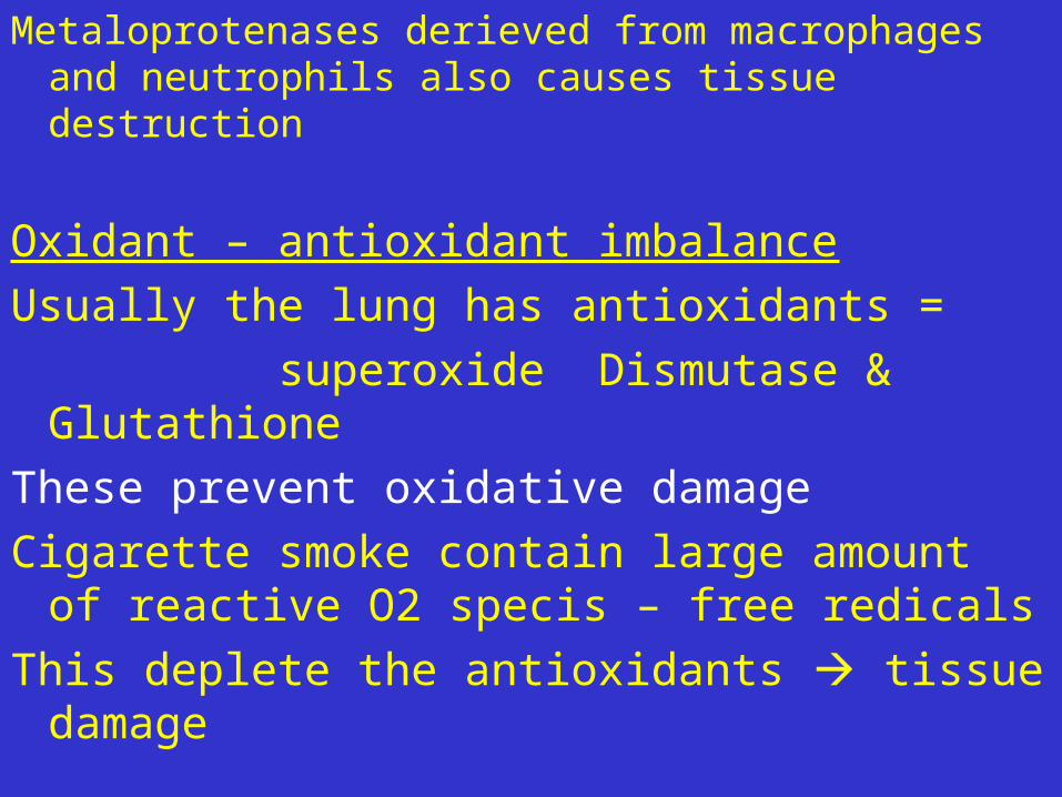
Metaloprotenases derieved from macrophages and neutrophils also causes tissue destruction
Oxidant – antioxidant imbalanceUsually the lung has antioxidants = superoxide Dismutase & GlutathioneThese prevent oxidative damageCigarette smoke contain large amount of
reactive O2 specis – free redicalsThis deplete the antioxidants tissue
damage

Activated neutrophils also produce O2 free radicles Oxidative injury also inactivate antiprotease
functional 1 Antitrypsin
Macroscopic appearance – Pan acinar typeWhen chest wall is opened upLung is pale, hyper inflated, obliterate the
heart
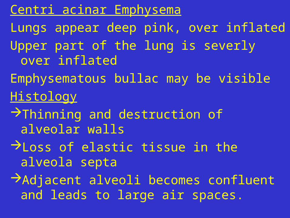
Centri acinar EmphysemaLungs appear deep pink, over inflatedUpper part of the lung is severly over
inflatedEmphysematous bullac may be visibleHistologyThinning and destruction of alveolar
wallsLoss of elastic tissue in the alveola
septaAdjacent alveoli becomes confluent and
leads to large air spaces.

Terminal and respiratory bronchioles deformed
Fibrosis of respiratory bronchiole.
Because of loss of elastic tissue in the alvolar septa
reduced radial traction on small airway
Small airways collapse on expiration
Air trapingAlveolar capillary surface also deminished

Clinical presentationProgressive SOB (hyperventilation)Pink puffer pink but puffing
Secondary pulmonary HT R heart failure
Due to - hypoxia with vascular spasm - pulmonary vascular reduction
So feattures of R heart failure pedal oedema

Chronic Bronchitis – clinical definitionInflammation of all part of bronchial system
Defnition -: consecutive productive cough for at lease 3 consecutive months in last 2 consecutive years
It will affect both small and large airwaysLarge airway – trachea & BronchiMucus hyper secretion coughInflamationSmall airways – bronchiolesPeribronchiolar fibrosis air way
obstruction

4 different types1. Simple chronic bronchits cough & mucoid sputum No obstruction2. Mucopurulent bronchitis cough with purulent sputum3. Chronic asthmatic bronchites Demonstrate hyper-responsiveness
4. Chronic obstructive bronchitis Shows significant airflow obstruction Obstruction mainly occurs at bronchiolar
level

It result from inflammation, fibrosis and narrowing of
bronchioles usually there will be emphysema alsoPathogenesis Epidermal growth factor receptor
Cigarette smoke irritate upper airway Air pollution Nicotin recurint SO2, N2O neutrophils
Inflamation sets in

Inflammation with cellular infiltrate Neutrophil,CD8 Lymphocyte,
Macrophages
Mucus secretion with hypertrophy of mucus gland and increased number of goblet cells, Epithelial damage
Metaplasia Dysplasia and fibrosis
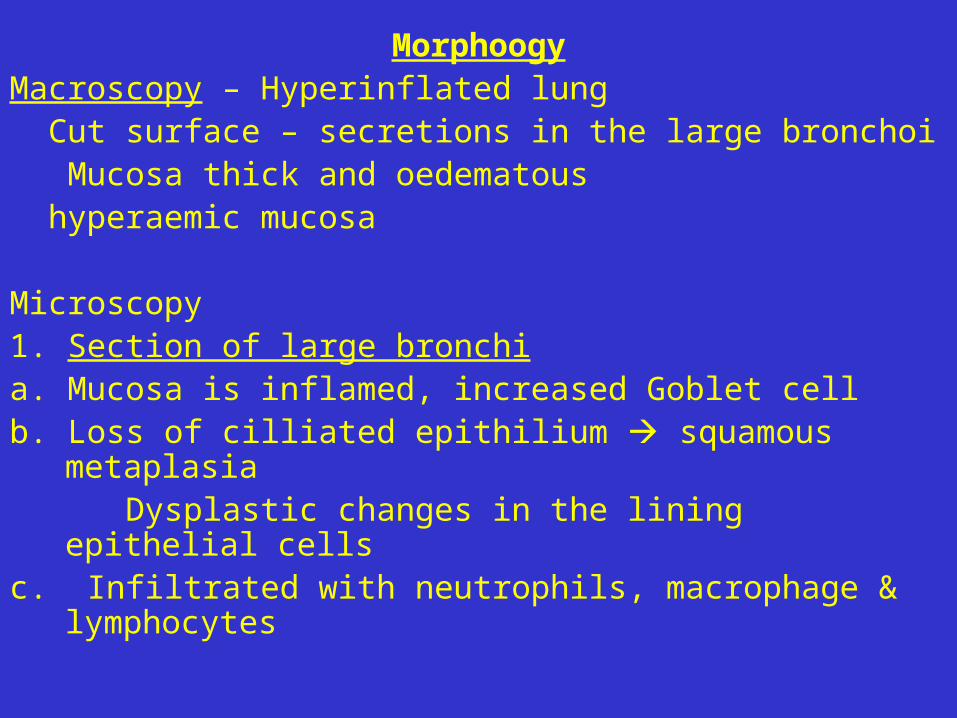
MorphoogyMacroscopy – Hyperinflated lung Cut surface – secretions in the large bronchoi Mucosa thick and oedematous hyperaemic mucosa
Microscopy1. Section of large bronchia. Mucosa is inflamed, increased Goblet cellb. Loss of cilliated epithilium squamous
metaplasia Dysplastic changes in the lining epithelial cellsc. Infiltrated with neutrophils, macrophage &
lymphocytes
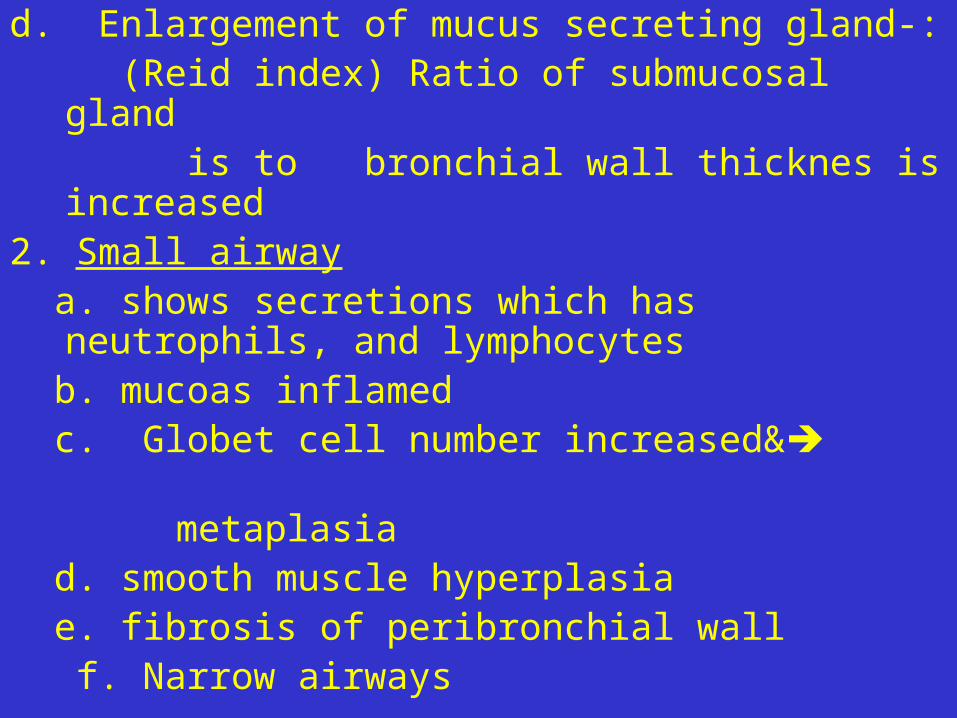
d. Enlargement of mucus secreting gland-: (Reid index) Ratio of submucosal gland is to bronchial wall thicknes is
increased2. Small airway a. shows secretions which has
neutrophils, and lymphocytes b. mucoas inflamed c. Globet cell number increased& metaplasia d. smooth muscle hyperplasia e. fibrosis of peribronchial wall f. Narrow airways

Conditions that mimic emphysema1. Compensatory emphysema – when one
bronchus is blocked and collapsed other lung expands with air
2. Senile emphysema3. Obstructive over inflation when a bronchus is obstructed partially with
each respiratory cycle the is traped in
4. Mediastinal emphysema leakage of air in the mediastinal tissue
in – rib #5. subcutaneous emphysema


