CURRENTOPINION Systolic heart failure: diagnosis and therapy
Copyrig
0952-7907 Copyright � 2016 Wolte
Janek Henes and Peter Rosenberger
Purpose of review
The present review highlights recent findings on perioperative systolic heart failure. It briefly summarizes thepathophysiology of heart failure and provides the reader with new insight in diagnosis and treatment ofsystolic heart failure. In addition, we review new therapeutic strategies with pharmacologic agents andmechanical assist devices to treat systolic heart failure.
Recent findings
Left ventricular systolic heart failure is a high-risk disease for patients undergoing cardiac and noncardiacsurgery and poses a high burden on the anesthesiologist in charge. Perioperative echocardiography is wellestablished for urgent diagnosis in the operating room and is superior to biomarker-based diagnosis.Although cardiovascular disease associated mortality decreases, systolic heart failure related mortalityremains at a high of 50% after 5 years. As a consequence, left ventricular assist device implantation ratesgrow rapidly and include approximately 30–40% patients with desperate clinical situation and destinationtherapy. Extracorporeal life support for acute heart failure needs further investigation to document possibleindications and side-effects.
Summary
Recent advances in the field of cardiovascular anesthesiology comprise advanced use of perioperativeechocardiography, mechanical circulatory assist devices, and customized pharmacologic management.
Department of Anesthesiology and Intensive Care Medicine, TubingenUniversity Hospital, Tubingen, Germany
Correspondence to Janek Henes, MD, Department of Anesthesiologyand Intensive Care Medicine, University Hospital Tubingen, Hoppe-Seyler-Straße 3, D-72076 Tubingen, Germany. Tel: +49 7071 2981110;e-mail: [email protected]
Curr Opin Anesthesiol 2016, 29:55–60
DOI:10.1097/ACO.0000000000000270
INTRODUCTION
Heart failure is an end-stage clinical syndromebased on a broad variety of underlying cardiacconditions. With its high prevalence of over5.7 million Americans older than 20 years of ageand a projected rise of 46% from 2012 to 2030,resulting in more than 8 million people sufferingfrom it in the USA [1,2] and more than 23 millionworldwide, heart failure is of major significance formodern healthcare systems and has significantfinancial implications for healthcare systems. Heartfailure is primarily a disease of the elderly andaffects about 6.6% males and 4.8% females aged60–79 years, with females leading the 80þ group(10.6% male vs. 13.5% female) [1,2]. Among allindividuals (asymptomatic or validated clinicalheart failure) the prevalence of left ventricular sys-tolic dysfunction is 6% [3,4]. Left ventricular sys-tolic dysfunction includes patients suffering fromheart failure and a reduced left ventricular ejectionfraction (HFrEF), compared with patients with dias-tolic heart failure and preserved ejection fraction(HFpEF), which is reviewed in another chapter inthis issue of the journal.
The overall mortality remains high with 50% ofpatientsdying5yearsafterthediagnosisofheartfailure[5], interestingly without significant differences inmortalitybetweenthegroupsHFrEF(EF<40%),HFbEF(borderline ejection fraction 40–50%) and HFpEF(ejection fraction �50%) [6]. Physicians should beaware of the fact that acute decompensation of heartfailureor intensificationofchronicheart failure, is themost common cause of hospital admission amongpatients with heart failure. These patients frequentlyrequire invasive therapy and anesthesia during theirhospital stay. Anesthesiologists are confronted withthese patients in multiple locations including theoperating room (OR) and outside-of-OR areas duringa variety of interventions. Moreover, heart failure is
� Although mortality of cardiovascular disease (CVD) intotal declines [34], 5-year survival of heart failureremains at approximately 50%.
� Undiagnosed and untreated patients with heart failurehave a high risk of acute decompensation in theperioperative setting.
� Diagnosis and therapy must be handled withexperience (inotropic treatment/mechanical assist) toprovide best possible outcome.
� More patients receive continuous-flow left ventricularassist devices as destination therapy for disabling heartfailure. Anesthesia care providers must be familiar withthe management of the technical advances in this field(coagulation, TEE).
Cardiovascular anesthesia
quite frequent in postoperative patients after cardiacsurgery with an incidence of approximately 12%and is also the second most frequent reason forhospital readmission after an infection (approx.16%) [7]. This shows that heart failure of differentcauses poses a huge challenge on healthcare provi-ders, in particular those in acute care situations.We therefore review the latest literature for systolicheart failure (HFrEF) for the cardiovascular anesthesi-ologist with focus on new diagnostic and therapeuticapproaches in the review period.
Pathophysiology and cause of systolic heartfailure
Different pathophysiologic models have been pro-posed in the last decades, altogether describing themechanisms of heart failure. The hemodynamicmodel from 1967 defines heart failure as a patho-logical state in which an abnormality of myocardialfunction is responsible for the failure of the heart topump blood at a rate commensurate with therequirements of the metabolizing tissues duringordinary activity [8]. This was supported by datashowing that intrinsic cardiac muscle contractilitywas reduced during hemodynamic load. The associ-ated ventricular remodeling occurs in both types ofheart failure with a high impact on hemodynamicstability [9]. As a consequence, patients with HFrEFtypically present with dilated left ventricular cavityand a normal or reduced ratio of end-diastolic vol-ume. The extracellular matrix hereby determinesthe ventricular architecture and provides a basisfor efficient pumping. Myocardial injury leads toremodeling of the extracellular matrix throughfibroblast proliferation, which results in ventricular
thinning and impairment of systolic function. Thecardiorenal model refers to the close functionalrelationship between the heart and kidneys. Renalsodium and water retention are then leading to theclinical symptoms of dyspnea and edema [10]. Thisform of heart failure is treated primarily by diuretics and dietary sodium restriction. The associatedneurohumoral activation results in sympatheticnervous activation, increased contractility, vasocon-striction, elevated blood pressure, and long-termmaladaptive remodeling with progressive worsen-ing of myocardial injury. Proof of this neurohumoral model was provided through drug-medi-ated interference with the adrenergic and renin–angiotensin–aldosteron system with significantlyimproved survival in patients with heart failure[11–14]. The role of the Ca2þ metabolism throughthe ryanodin receptor and the SERCA2a pump in thedevelopment of heart failure was also highlighted inseveral studies [15,16]. Other approaches focus oncardiac myocyte cell death through necrosis andapoptosis because of excessive adrenergic activity,inflammation, oxidative stress or toxic substances[17–19]. At least genome wide association studiesaim to identify heart failure syndrome-related genecandidates in order to help understand the mech-anisms by which genetic aberration can affect car-diac function [20,21]. All these approaches describethe complex pathophysiological picture of heartfailure and are reflected in the current treatmentguidelines of the ACCF and AHA [22].
Recent advances in heart failure diagnosis
A prospective trial by O’Meara et al. [23&
] investi-gated the role of renal disease, interleukins, andspecific left ventricular remodeling processes inpatients with HFrEF with/without anemia by com-paring clinical, echocardiographic, and circulatingbiomarker profiles with a control group. This studyfound a strong association between anemia, heartdisease markers, and the level of renal dysfunctionin patients with HFrEF, represented by increasedmyocardial remodeling, inflammation, and volumeoverload. The HFrEF group with anemia showedsignificantly higher NT-proBNP levels and moreoften elevated troponin T levels than the nonane-mic group. Furthermore, patients with anemicHFrEF showed more advanced chronic kidney dis-ease (CKD) than those without anemia, reachingcontrol-group levels of GFR (those with diagnosedCKD). Inflammatory cytokines IL-6 and IL-10 werehigher in the anemia group, and the echocardio-graphic markers left ventricular mass, mitral regur-gitation, and left atrial end-systolic volume index.All these echocardiographic markers were elevated
Health, Inc. All rights reserved.
Volume 29 � Number 1 � February 2016
Systolic heart failure Henes and Rosenberger
in patients with anemic HFrEF representing a greaterleft ventricular remodeling process. Altogether thisstudy demonstrates that patients suffering fromHFrEF and anemia bare a complex pathophysiologyand have a more advanced and active heart diseasethan nonanemic patients. This finding should benoticed by anesthesiologists for preoperative evalu-ation and patient blood management.
Echocardiography in the evaluation ofperioperative pump function
Recent recommendations from the European Associ-ation of Cardiovascular Imaging and the AcuteCardiovascular Care Association summarized the peri-operative use of ultrasound imaging [24]. Transthora-cic and transesophageal echocardiography (TTE, TEE)is a standard tool in the intraoperative and postoper-ative hemodynamic assessment of patients followingcardiac surgery. Postoperatively TTE should be per-formed first but will often be supplemented by TEEbecause of poor quality of the transthoracic acousticwindow in these patients. Postoperative compli-cations of systolic function include pericardialcollection and cardiac tamponade. Suboptimal intra-operative myocardial protection and long bypasstimes during complex heart surgery often cause sig-nificant myocardial depression. A diagnostic approachto assess systolic cardiac function should includeinspection of the first 2–4cm of the coronary arteries,which are accessible to the TEE. Imaging of the rightventricle should also be performed. For that mattertricuspid annular plane systolic excursion (TAPSE) is avalidated parameter of global right ventricular func-tion and should be measured intraoperatively as areference value for postoperative assessment. TAPSEis easily obtainable, correlates well with other stand-ardized parameters of right ventricular function and isfurthermore an independent predictor of poor out-come in patients with acute right heart failure [25].Echocardiography for cannula placement and deviceimplementation is essential and has a vital role inexcluding nonheart failure causes of cardiorespiratoryfailure. Guidelines from the American Society of Anes-thesiologists (Practice Guidelines for PerioperativeTransesophageal Echocardiography; update in 2016and estimated publication 2017) describe the perio-perative use of echocardiography in detail [26].
Perioperative treatment of the patient withsystolic heart failure
Inotropic treatment
In the situation of low cardiac output in a patientwith a failing ventricle and typical clinical signs of
0952-7907 Copyright � 2016 Wolters Kluwer Health, Inc. All rights rese
systolic heart failure, it is of vital interest to improvethe cardiac performance. A recent study from Spon-holz et al. [27
&
] evaluated the use of catecholaminesduring heart surgery in Germany and reported,that 80–100% of all individuals enrolled in thisstudy received vasopressors during the perioperativeperiod. First-line therapy in the case of hypotensioncaused by low cardiac output was dobutamine, whichwas administered by 32% of all care providers, fol-lowed by epinephrine with 30%. Only 8% appliedprimarilya phosphodiesterase inhibitor (PDEI). Vaso-plegia was treated with norepinephrine by 96% of allphysicians reported in this study. Second-line treat-ment was performed by PDEI (50%), epinephrine(42%), and levosimendan (22%).
A propensity score-matched analysis in 2340patients by Nielsen et al. investigated the associationbetween intraoperative and postoperative use ofinotropes, mortality, and postoperative compli-cations in heart surgery patients [28
&&
]. Resultsshowed a strong association between the intraoper-ative and postoperative use of inotropes, increasedmortality, and major postoperative morbidity. Ino-tropic therapy was independently linked to post-operative myocardial infarction (adjusted OR, 2.1;95% CI, 1.4–3.0), stroke (adjusted OR, 2.4; 95% CI,1.4–4.3), and renal replacement therapy (adjustedOR, 7.9; 95% CI, 3.8–16.4). Recent years have seenthe advent of the calcium-sensitizer Levosimendan,which is still under extensive clinical investigation(LICORN, soon reporting, NCT 02184819; LEVO-CTS,recruiting, NCT 02025621; HSR-LEVO, recruiting,NCT00994825; Intracoronary administration oflevosimendan in cardiac surgery patients, recruiting,NCT01500785; prophylactic administration oflevosimendan in patients undergoing coronarysurgery, soon reporting, NCT01318460). The latestEuropean expert opinion on the preoperative andperioperative use of Levosimendan in cardiacsurgery summarizes the growing body of knowledge[29
&&
] and stated that levosimendan effectivelyimproves general and pulmonary hemodynamicsin patients undergoing cardiac surgery. It therebyreduces the need for inotropic and mechanical sup-port with benefit for renal and hepatic function,even though pronounced vasodilation frequentlyraises vasopressor demand. It can effectively reducethe length of stay on both ICU and hospital.Preoperative administration of levosimendan isrecommended in patients who have a generallycompromised myocardial function, including rightventricular dysfunction. Bolus administration oflevosimendan out of the OR is not advisable, insteada continuous infusion of 0.1 mg/kg/min for 24 h wasconsidered the optimal dose. If not possible, bolusdose after induction of anesthesia is feasible.
r Health, Inc. All rights reserved.
rved. www.co-anesthesiology.com 57
Cardiovascular anesthesia
If vasodilation emerges, treatment with norepi-nephrine or vasopressin is recommended, possiblysupplemented by the inotrope dobutamine. At last arecent study from Greco et al. [30
&
] was mentioned,in which commonly used inotropes (dobutamine,phosphodiesterase inhibitors) were compared tolevosimendan. Interestingly, only levosimendanwas associated with a decrease in mortality whencompared with placebo (OR¼0.48; 95% CI, 0.28–0.80). The expert panel and the authors of this studyagreed upon the need for large-scale RCTs to supportthis finding.
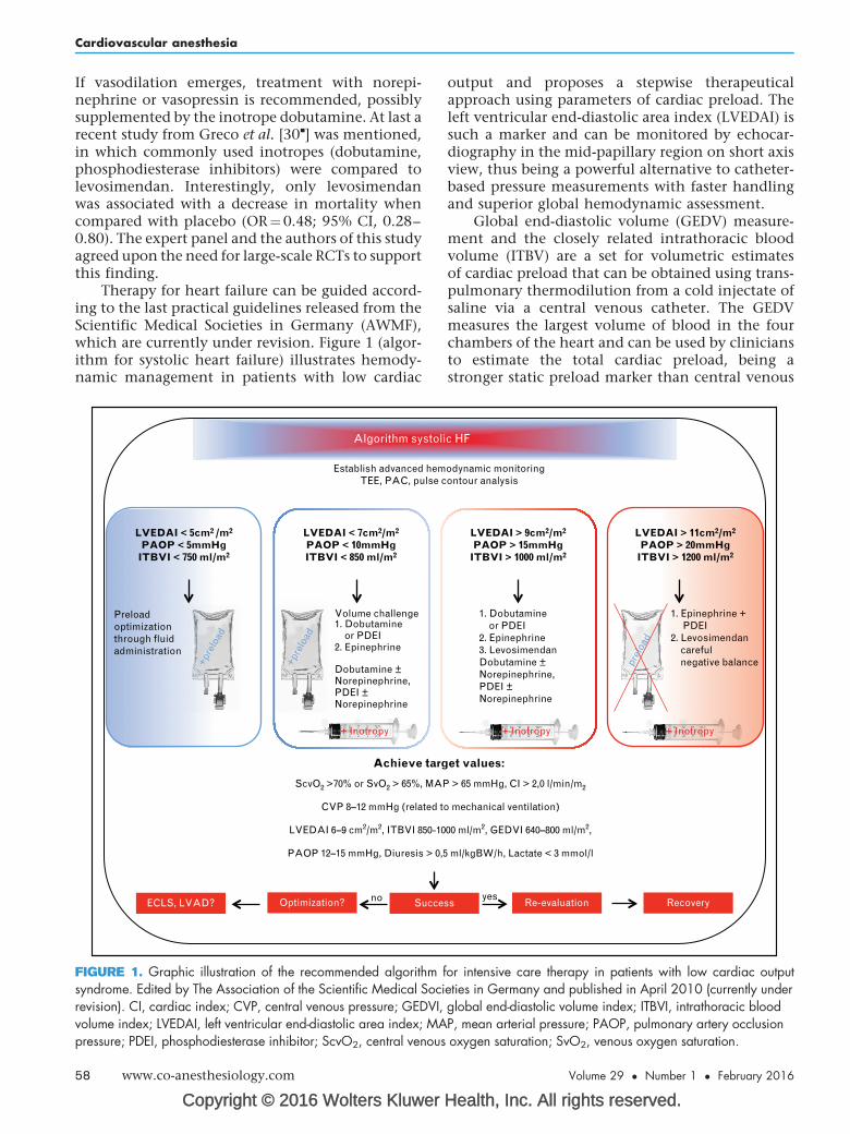
Therapy for heart failure can be guided accord-ing to the last practical guidelines released from theScientific Medical Societies in Germany (AWMF),which are currently under revision. Figure 1 (algor-ithm for systolic heart failure) illustrates hemody-namic management in patients with low cardiac
FIGURE 1. Graphic illustration of the recommended algorithm fsyndrome. Edited by The Association of the Scientific Medical Socirevision). CI, cardiac index; CVP, central venous pressure; GEDVI,volume index; LVEDAI, left ventricular end-diastolic area index; MApressure; PDEI, phosphodiesterase inhibitor; ScvO2, central venous
58 www.co-anesthesiology.com
output and proposes a stepwise therapeuticalapproach using parameters of cardiac preload. Theleft ventricular end-diastolic area index (LVEDAI) issuch a marker and can be monitored by echocar-diography in the mid-papillary region on short axisview, thus being a powerful alternative to catheter-based pressure measurements with faster handlingand superior global hemodynamic assessment.
Global end-diastolic volume (GEDV) measure-ment and the closely related intrathoracic bloodvolume (ITBV) are a set for volumetric estimatesof cardiac preload that can be obtained using trans-pulmonary thermodilution from a cold injectate ofsaline via a central venous catheter. The GEDVmeasures the largest volume of blood in the fourchambers of the heart and can be used by cliniciansto estimate the total cardiac preload, being astronger static preload marker than central venous
Health, Inc. All rights reserved.
odynamic monitoringontour analysis
+ Inotropy + Inotropy
1. Dobutamine or PDEI2. Epinephrine3. LevosimendanDobutamine ±Norepinephrine,PDEI ±Norepinephrine
or intensive care therapy in patients with low cardiac outputeties in Germany and published in April 2010 (currently underglobal end-diastolic volume index; ITBVI, intrathoracic bloodP, mean arterial pressure; PAOP, pulmonary artery occlusionoxygen saturation; SvO2, venous oxygen saturation.
Volume 29 � Number 1 � February 2016
Systolic heart failure Henes and Rosenberger
pressure (CVP) or pulmonary artery occlusion pres-sure (PAOP). Placement of a pulmonary arterycatheter allows intermittent recording of the PAOPas an indirect marker of left ventricular preload. Theintrathoracic blood volume (ITBV) comprises theGEDV and the blood volume from the pulmonaryvasculature and plays a similar role in preload assess-ment, but is more difficult to measure (double-indicator transpulmonary thermodilution) andhas to be corrected by computation. However, ITBVassessment allows further calculation of extravascu-lar lung water as a diagnostic criterion for acute lunginjury and the acute respiratory distress syndrome incritical care patients.
Mechanical assist
Ever since the first LVAD was approved by the USFood and Drug Administration (FDA) in 1994, thefield evolved rapidly and device implantation ratesconstantly grow according to The Interagency Regis-try for Mechanically Assisted Circulatory Support(Intermacs). Intermacs currently lists 158 participat-ing hospitals in the USA and Canada and a total of14 039 patients (21.2% female; 78.6% male) in theirfirst quarterly report from 2015 [31]. Out of thispopulation 8762 individuals (62.2%) received amechanical circulatory assist device as a bridge-to-transplantation and 5084 individuals (36.2%) asdestination therapy. Only 102 patients (0.7%) werebridged to recovery and 78 patients (0.5%) receiveda device for extracorporeal cardiopulmonary resus-citation. LVADs account for 92.5% and BiVADs for5.2% of all registered device implantations withLVADs being the group of devices with the highestexpansion rate. Survival rate from prospectiveimplantation of state-of-the-art continuous flowLVADs between June 2006 to March 2015 is 81%after 1 year and decreases by approximately 10 %/year. LVAD implantation prolongs and improves lifefor patients with heart failure; however, recentreports demonstrate a significant issue concerningthe development of pump thrombosis with arelevant impact on survival of patients having theHeartMate II LVAD [32
&
,33]. Starling et al. describean incidence-increase of pump thrombosis frominitially reported 2.2%, 3-months after implan-tation before March 2011 through 8.4% by January2013 with occurrence peak 1-month after implan-tation. Six-month mortality did not differ betweenpatients who had pump thrombosis and weretreated with device replacement or transplantation,compared with patients without pump thrombosis,yet untreated patients with pump thrombosisshowed an alarming mortality of 48.2% after6 months [32
0952-7907 Copyright � 2016 Wolters Kluwer Health, Inc. All rights rese
technology is capable to improve and prolongpatient life on the one hand, but has to be subjectedto superior monitoring on the other hand in order toscreen for life threatening adverse events. Moreoverconsidering ethical issues, maximum care providersmust be aware that one-third of all patients withLVAD have the device implanted as DT when itcomes to complications (DT 2006–2007: 14.7%vs. DT 2011–2013: 41.6%). More than ever consentin treatment and end-of-life care must be metthrough the patient and all participating specialtiesbefore futile situations occur.
CONCLUSION
We conclude that patients with diminished cardiacpump function demand full attention from theentire perioperative team. Not only must thepatient be well prepared for surgery, includingadequate diagnosis of the underlying cardiac con-dition and pharmacologic pretreatment, but alsocare providers must cooperate best possible toachieve acceptable outcomes. Therefore, currentstandards of care must be met in the use of theTEE device, pharmacologic agents, and knowledgeof the circuit in any hemodynamic situation,especially while implementing mechanical assistdevices. At last levosimendan could favor the out-come of patients in the perioperative course ofcardiac surgery.
Acknowledgements
The authors thank the staff of the CardiothoracicDivision of the Department of Anesthesiology andIntensive Care Medicine, for their outstanding work withpatients and their commitment to teaching youngresidents.
Financial support and sponsorship
Internal funding from the Medical Faculty of the Eber-hard-Karls-University Tubingen to J.H. (fortune ProgramNr. 2228-1-0).
Conflicts of interest
There are no conflicts of interest.
REFERENCES AND RECOMMENDEDREADINGPapers of particular interest, published within the annual period of review, havebeen highlighted as:
& of special interest&& of outstanding interest
1. Mozaffarian D, Benjamin EJ, Go AS, et al. Heart disease and stroke statistics–2015 update: a report from the American Heart Association. Circulation2015; 131:e29–322.
2. Correction. Circulation 2015; 131:e535.
r Health, Inc. All rights reserved.
rved. www.co-anesthesiology.com 59
Cardiovascular anesthesia
3. Lam CS, Lyass A, Kraigher-Krainer E, Massaro JM, et al. Cardiac dysfunctionand noncardiac dysfunction as precursors of heart failure with reduced andpreserved ejection fraction in the community. Circulation 2011; 124:24–30.
4. Yeboah J, Rodriguez CJ, Stacey B, Lima JA, et al. Prognosis of individuals withasymptomatic left ventricular systolic dysfunction in the multiethnic study ofatherosclerosis (MESA). Circulation 2012; 126:2713–2719.
5. National Center for Health Statistics. Mortality Multiple Cause Micro-dataFiles, 2013. Public-use data file and documentation. NHLB1 tabulations;2013. http://wwwcdcgov/nchs/data_access/Vitalstatsonlinehtm - Mortality.[Accessed 24 August 2015]
6. Cheng RK, Cox M, Neely ML, Heidenreich PA, et al. Outcomes in patients withheart failure with preserved, borderline, and reduced ejection fraction in theMedicare population. Am Heart J 2014; 168:721–730.
7. Hannan EL, Zhong Y, Lahey SJ, Culliford AT, et al. 30-day readmissions aftercoronary artery bypass graft surgery in New York State. JACC CardiovascInterv 2011; 4:569–576.
8. Braunwald E, Ross J Jr, Sonnenblick EH. Mechanisms of contraction of thenormal and failing heart. New Engl J Med 1967; 277:794–800.
9. van Heerebeek L, Borbely A, Niessen HW, Bronzwaer JG, et al. Myocardialstructure and function differ in systolic and diastolic heart failure. Circulation2006; 113:1966–1973.
10. Schrier RW, Abraham WT. Hormones and hemodynamics in heart failure.New Engl J Med 1999; 341:577–585.
11. Packer M. The neurohormonal hypothesis: a theory to explain the mechanismof disease progression in heart failure. J Am Coll Cardiol 1992; 20:248–254.
12. Flather MD, Yusuf S, Kober L, Pfeffer M, et al. Long-term ACE-inhibitor therapyin patients with heart failure or left-ventricular dysfunction: a systematicoverview of data from individual patients. ACE-Inhibitor Myocardial InfarctionCollaborative Group. Lancet 2000; 355:1575–1581.
13. Cohn JN, Tognoni G. A randomized trial of the angiotensin-receptor blockervalsartan in chronic heart failure. New Engl J Med 2001; 345:1667–1675.
14. Pfeffer MA, Swedberg K, Granger CB, Held P, et al. Effects of candesartan onmortality and morbidity in patients with chronic heart failure: the CHARM-Overall programme. Lancet 2003; 362:759–766.
15. Respress JL, van Oort RJ, Li N, Rolim N, et al. Role of RyR2 phosphorylation atS2814 during heart failure progression. Circ Res 2012; 110:1474–1483.
17. Konstantinidis K, Whelan RS, Kitsis RN. Mechanisms of cell death in heartdisease. Arteriosclerosis, thrombosis, and vascular biology 2012; 32:1552–1562.
18. Olivetti G, Abbi R, Quaini F, Kajstura J, et al. Apoptosis in the failing humanheart. New Engl J Med 1997; 336:1131–1141.
19. Mudd JO, Kass DA. Tackling heart failure in the twenty-first century. Nature2008; 451:919–928.
20. Morita H, Seidman J, Seidman CE. Genetic causes of human heart failure.J Clin Invest 2005; 115:518–526.
21. Zeller T, Blankenberg S, Diemert P. Genomewide association studies incardiovascular disease – an update 2011. Clin Chem 2012; 58:92–103.
22. Writing Committee M, Yancy CW, Jessup M, Bozkurt B, et al. 2013 ACCF/AHA guideline for the management of heart failure: a report of the AmericanCollege of Cardiology Foundation/American Heart Association Task Force onpractice guidelines. Circulation 2013; 128:e240–327.
O’Meara E, Rouleau JL, White M, Roy K, et al. Heart failure with anemia: novelfindings on the roles of renal disease, interleukins, and specific left ventricularremodeling processes. Circ Heart Fail 2014; 7:773–781.
This prospective trial with 151 patients investigated the association of anemia andheart failure with reduced ejection fraction. They found anemia to be a strongmarker for more advanced and active heart disease.24. Lancellotti P, Price S, Edvardsen T, Cosyns B, et al. The use of echocardio-
graphy in acute cardiovascular care: Recommendations of the EuropeanAssociation of Cardiovascular Imaging and the Acute Cardiovascular CareAssociation. European Heart Journal: Acute Cardiovascular Care 2014. doi:10.1177/2048872614549739.
25. Schmid E, Hilberath JN, Blumenstock G, Shekar PS, et al. Tricuspid annularplane systolic excursion (TAPSE) predicts poor outcome in patients under-going acute pulmonary embolectomy. Heart Lung Vessel 2015; 7:151–158.
26. Practice guidelines for perioperative transesophageal echocardiography. Anupdated report by the American Society of Anesthesiologists and the Societyof Cardiovascular Anesthesiologists Task Force on TransesophagealEchocardiography. Anesthesiology 2010; 112:1084–1096.
27.&
Sponholz C, Schelenz C, Reinhart K, Schirmer U, et al. Catecholamine andvolume therapy for cardiac surgery in Germany – results from a postal survey.PloS one 2014; 9:e103996.
Clinical survey with 81 participating German anesthesia departments investigatingcurrentpractice inhemodynamicmonitoring,catecholamineuse,andvolume therapy.28.&&
Nielsen DV, Hansen MK, Johnsen SP, Hansen M, et al. Health outcomes withand without use of inotropic therapy in cardiac surgery: results of a propensityscore-matched analysis. Anesthesiology 2014; 120:1098–1108.
This study investigated the connection between intraoperative and postoperativeuse of inotropes, mortality and postoperative complications by propensity score-matched analysis of 2340 cardiac surgery patients. Results show increasedmortality and major postoperative morbidity with intraoperative- and postoperativeuse of inotropes.29.&&
Toller W, Heringlake M, Guarracino F, Algotsson L, et al. Preoperative andperioperative use of levosimendan in cardiac surgery: European expertopinion. Int J Cardiol 2015; 184:323–336.
Excellent review of the latest literature on levosimendan use in cardiac surgery.30.&
Greco T, Calabro MG, Covello RD, Greco M, et al. A Bayesian network meta-analysis on the effect of inodilatory agents on mortality. Br J Anaesthesia2015; 114:746–756.
Meta-analysisontheeffectof inodilatorsonmortality incardiacsurgery.They identifiedlevosimendan to be the best inodilator to improve survival in adult cardiac surgery.31. Intermacs Website. Interagency Registry for Mechanically Assisted Circula-
tory Support. National Heart Lung and Blood Institute. Contract AwardHHSN268201100025C. http://www.uab.edu/intermacs. [Accessed 1 Sep-tember 2015]
32.&
StarlingRC,MoazamiN,SilvestrySC,EwaldG,etal.Unexpectedabruptincreasein left ventricular assist device thrombosis. New Engl J Med 2014; 370:33–40.
The authors describe an abrupt increase in left ventricular assist device thrombosisand report management and survival of this serious adverse event.33. Rame JE, Atluri P, Acker MA. Unexpected abrupt increase in left ventricular
assist device thrombosis. New Engl J Med 2014; 370:1466–1467.34. Wilmot KA, O’Flaherty M, Capewell S, Ford ES, et al. Coronary heart disease
mortality declines in the United States from 1979 through 2011: evidence forstagnationinyoungadults,especiallywomen.Circulation2015;137:997–1002.