66
Copyright © 2005 Pearson Education, Inc. publishing as Benjamin Cummings I-1- 1 EXPLORING LIFE & A TOUR OF THE CELL
| Date post: | 20-Dec-2015 |
| Category: |
Documents |
| View: | 213 times |
| Download: | 0 times |

Copyright © 2005 Pearson Education, Inc. publishing as Benjamin Cummings I-1- 1
EXPLORING LIFE & A TOUR OF THE CELL

Copyright © 2005 Pearson Education, Inc. publishing as Benjamin Cummings I-1- 2
Exploring Life• Biology
– Is the scientific study of life
• We recognize life
– By what living things do
Figure 1.1

Copyright © 2005 Pearson Education, Inc. publishing as Benjamin Cummings I-1- 3
Some properties of life
(c) Response to the environment
(a) Order
(d) Regulation
(g) Reproduction (f) Growth and development
(b) Evolutionary adaptation
(e) Energy processing

Copyright © 2005 Pearson Education, Inc. publishing as Benjamin Cummings I-1- 4
From the biosphere to organisms
1 The biosphere

Copyright © 2005 Pearson Education, Inc. publishing as Benjamin Cummings I-1- 5
From cells to molecules
Cell
8 Cells
6 Organs and organ systems
7 Tissues
10 Molecules
9 Organelles
50 µm
10 µm
1 µm
Atoms
Figure 1.3

Copyright © 2005 Pearson Education, Inc. publishing as Benjamin Cummings I-1- 6
A Closer Look at Ecosystems
• Each organism
– Interacts with its environment
• Both organism and environment
– Are affected by the interactions between them

Copyright © 2005 Pearson Education, Inc. publishing as Benjamin Cummings I-1- 7
A Closer Look at Cells
• The cell
– Is the lowest level of organization that can perform all activities required for life
25 µmFigure 1.5

Copyright © 2005 Pearson Education, Inc. publishing as Benjamin Cummings I-1- 8
The Cell’s Heritable Information
• Cells contain chromosomes made partly of DNA, the substance of genes
– Which program the cells’ production of proteins and transmit information from parents to offspring
Egg cell
Sperm cell
NucleicontainingDNA
Fertilized eggwith DNA fromboth parents
Embyro’s cells with copies of inherited DNA Offspring with traits
inherited fromboth parentsFigure 1.6

Copyright © 2005 Pearson Education, Inc. publishing as Benjamin Cummings I-1- 9
The molecular structure of DNA
• Accounts for it information-rich nature
DNA
Cell
Nucleotide
ACTA
T
A
CC
G
G
TA
TA
(b) Single strand of DNA. These geometric shapes and letters are simple symbols for the nucleotides in a small section of one chain of a DNA molecule. Genetic information is encoded in specific sequences
of the four types of nucleotides (their names are abbreviated here as A, T, C, and G).
(a) DNA double helix. This model shows
each atom in a segment of DNA.Made up of two long chains of building blocks called nucleotides, a DNA molecule takes the three-dimensional form of a double helix.
Figure 1.7
Nucleus

Copyright © 2005 Pearson Education, Inc. publishing as Benjamin Cummings I-1- 10
Two Main Forms of Cells
• All cells share certain characteristics
– They are all enclosed by a membrane
– They all use DNA as genetic information
• There are two main forms of cells
– Eukaryotic
– Prokaryotic

Copyright © 2005 Pearson Education, Inc. publishing as Benjamin Cummings I-1- 11
• Eukaryotic cells
– Are subdivided by internal membranes into various membrane-enclosed organelles
• Prokaryotic cells
– Lack the kinds of membrane-enclosed organelles found in eukaryotic cells

Copyright © 2005 Pearson Education, Inc. publishing as Benjamin Cummings I-1- 12
EUKARYOTIC CELL
Membrane
Cytoplasm
Organelles
Nucleus (contains DNA) 1 µm
PROKARYOTIC CELL
DNA
(no nucleus)Membrane
Figure 1.8

Copyright © 2005 Pearson Education, Inc. publishing as Benjamin Cummings I-1- 13
Biologists explore life across its great diversity of species
• Diversity is a hallmark of life
Figure 1.13

Copyright © 2005 Pearson Education, Inc. publishing as Benjamin Cummings I-1- 14
Grouping Species: The Basic Idea
• Taxonomy
– Is the branch of biology that names and classifies species according to a system of broader and broader groups

Copyright © 2005 Pearson Education, Inc. publishing as Benjamin Cummings I-1- 15
Classifying life
Species Genus Family Order Class Phylum Kingdom Domain
Mammalia
Ursusameri-canus(Americanblack bear)
Ursus
Ursidae
Carnivora
Chordata
Animalia
EukaryaFigure 1.14

Copyright © 2005 Pearson Education, Inc. publishing as Benjamin Cummings I-1- 16
The Three Domains of Life
• At the highest level, life is classified into three domains
– Bacteria
– Archaea
– Eukarya
• Domain Bacteria and domain Archaea
– Consist of prokaryotes
• Domain Eukarya, the eukaryotes
– Includes the various protist kingdoms and the kingdoms Plantae, Fungi, and Animalia

Copyright © 2005 Pearson Education, Inc. publishing as Benjamin Cummings I-1- 17
Life’s three domains
Figure 1.15
100 µm
0.5 µm
4 µmBacteria are the most diverse and widespread prokaryotes and are now divided among multiple kingdoms. Each of the rod-shapedstructures in this photo is a bacterial cell.
Protists (multiple kingdoms)are unicellular eukaryotes and their relatively simple multicellular relatives.Pictured here is an assortment of protists inhabiting pond water. Scientists are currently debating how to split the protistsinto several kingdoms that better represent evolution and diversity.
Kingdom Plantae consists of multicellula eukaryotes that carry out photosynthesis, the conversion of light energy to food.
Many of the prokaryotes known as archaea live in Earth‘s extreme environments, such as salty lakes and boiling hot springs. Domain Archaea includes multiple kingdoms. The photoshows a colony composed of many cells.
Kindom Fungi is defined in part by thenutritional mode of its members, suchas this mushroom, which absorb nutrientsafter decomposing organic material.
Kindom Animalia consists of multicellular eukaryotes thatingest other organisms.
DOMAIN ARCHAEA

Copyright © 2005 Pearson Education, Inc. publishing as Benjamin Cummings I-1- 18
Unity in the Diversity of Life
• As diverse as life is
– There is also evidence of remarkable unity
Cilia of Paramecium.The cilia of Parameciumpropel the cell throughpond water.
Cross section of cilium, as viewedwith an electron microscope
15 µm
1.0 µm
5 µm
Cilia of windpipe cells. The cells that line the human windpipe are equipped with cilia that help keep the lungs clean by moving a film of debris-trapping mucus upward.

Copyright © 2005 Pearson Education, Inc. publishing as Benjamin Cummings I-1- 19
A Tour of the CellOverview: The Importance of Cells
• All organisms are made of cells
• The cell is the simplest collection of matter that can live

Copyright © 2005 Pearson Education, Inc. publishing as Benjamin Cummings I-1- 20
Cell structure is correlated to cellular function
10 µm

Copyright © 2005 Pearson Education, Inc. publishing as Benjamin Cummings I-1- 21
To study cells, biologists use microscopes and the tools of biochemistry
• Different types of microscopes
– Can be used to visualize different sized cellular structures
Una
ide
d e
ye
1 m
0.1 nm
10 m
0.1 m
1 cm
1 mm
100 µm
10 µ m
1 µ m
100 nm
10 nm
1 nm
Length of somenerve and muscle cells
Chicken egg
Frog egg
Most plant and Animal cells
Smallest bacteria
Viruses
Ribosomes
Proteins
Lipids
Small molecules
Atoms
NucleusMost bacteriaMitochondrion
Lig
ht m
icro
sco
pe
Ele
ctro
n m
icro
sco
pe
Ele
ctro
n m
icro
sco
pe
Human height
Measurements1 centimeter (cm) = 102 meter (m) = 0.4 inch1 millimeter (mm) = 10–3 m1 micrometer (µm) = 10–3 mm = 10–6 m1 nanometer (nm) = 10–3 mm = 10–9 m

Copyright © 2005 Pearson Education, Inc. publishing as Benjamin Cummings I-1- 22
Use different methods for enhancing visualization of cellular structures
TECHNIQUE RESULT
Brightfield (unstained specimen). Passes light directly through specimen. Unless cell is naturally pigmented or artificially stained, image has little contrast. [Parts (a)–(d) show a human cheek epithelial cell.]
(a)
Brightfield (stained specimen). Staining with various dyes enhances contrast, but most staining procedures require that cells be fixed (preserved).
(b)
Phase-contrast. Enhances contrast in unstained cells by amplifying variations in density within specimen; especially useful for examining living, unpigmented cells.
(c)
50 µm

Copyright © 2005 Pearson Education, Inc. publishing as Benjamin Cummings I-1- 23
Differential-interference-contrast (Nomarski). Like phase-contrast microscopy, it uses optical modifications to exaggerate differences indensity, making the image appear almost 3D.
Fluorescence. Shows the locations of specific molecules in the cell by tagging the molecules with fluorescent dyes or antibodies. These fluorescent substances absorb ultraviolet radiation and emit visible light, as shown here in a cell from an artery.
Confocal. Uses lasers and special optics for “optical sectioning” of fluorescently-stained specimens. Only a single plane of focus is illuminated; out-of-focus fluorescence above and below the plane is subtracted by a computer. A sharp image results, as seen in stained nervous tissue (top), where nerve cells are green, support cells are red, and regions of overlap are yellow. A standard fluorescence micrograph (bottom) of this relatively thick tissue is blurry.
50 µm
50 µm
(d)
(e)
(f)

Copyright © 2005 Pearson Education, Inc. publishing as Benjamin Cummings I-1- 24
Electron microscopes (EMs)
• The scanning electron microscope (SEM)
– Provides for detailed study of the surface of a specimen
TECHNIQUE RESULTS
Scanning electron micro-scopy (SEM). Micrographs takenwith a scanning electron micro-scope show a 3D image of the surface of a specimen. This SEM shows the surface of a cell from a rabbit trachea (windpipe) covered with motile organelles called cilia. Beating of the cilia helps moveinhaled debris upward toward the throat.
(a)
Cilia1 µm

Copyright © 2005 Pearson Education, Inc. publishing as Benjamin Cummings I-1- 25
The transmission electron microscope (TEM)
• Provides for detailed study of the internal ultrastructure of cells
Transmission electron micro-scopy (TEM). A transmission electron microscope profiles a thin section of a specimen. Here we see a section through a tracheal cell, revealing its ultrastructure. In preparing the TEM, some cilia were cut along their lengths, creating longitudinal sections, while other cilia were cut straight across, creating cross sections.
(b)
Longitudinalsection ofcilium
Cross sectionof cilium
1 µm

Copyright © 2005 Pearson Education, Inc. publishing as Benjamin Cummings I-1- 26
Isolating Organelles by Cell Fractionation
Tissuecells
Homogenization
Homogenate1000 g(1000 times theforce of gravity)
10 min Differential centrifugation
Supernatant pouredinto next tube
20,000 g20 min
Pellet rich innuclei andcellular debris
Pellet rich inmitochondria(and chloro-plasts if cellsare from a plant)
Pellet rich in“microsomes”(pieces of plasma mem-branes andcells’ internalmembranes)
Pellet rich inribosomes
150,000 g3 hr
80,000 g60 min

Copyright © 2005 Pearson Education, Inc. publishing as Benjamin Cummings I-1- 27
Comparing Prokaryotic and Eukaryotic Cells
• All cells have several basic features in common
– They are bounded by a plasma membrane
– They contain a semifluid substance called the cytosol
– They contain chromosomes
– They all have ribosomes

Copyright © 2005 Pearson Education, Inc. publishing as Benjamin Cummings I-1- 28
Prokaryotic cells: Do not contain a nucleus; Have their DNA located in a region called the nucleoid.
(b) A thin section through the bacterium Bacillus coagulans (TEM)
Pili: attachment structures onthe surface of some prokaryotes
Nucleoid: region where thecell’s DNA is located (notenclosed by a membrane)
Ribosomes: organelles thatsynthesize proteins
Plasma membrane: membraneenclosing the cytoplasm
Cell wall: rigid structure outsidethe plasma membrane
Capsule: jelly-like outer coatingof many prokaryotes
Flagella: locomotionorganelles ofsome bacteria
(a) A typical rod-shaped bacterium
0.5 µmBacterial
chromosome

Copyright © 2005 Pearson Education, Inc. publishing as Benjamin Cummings I-1- 29
Eukaryotic cells: Contain a true nucleus, bounded by a membranous nuclear envelope; Are generally quite a bit bigger than prokaryotic cells
• The plasma membrane:
Functions as a selective barrier
Allows sufficient passage
of nutrients and waste
Carbohydrate side chain
Outside of cell
Inside of cell
Hydrophilicregion
Hydrophobicregion
Hydrophilicregion
(b) Structure of the plasma membrane
Phospholipid Proteins
TEM of a plasmamembrane. Theplasma membrane,here in a red bloodcell, appears as apair of dark bandsseparated by alight band.
(a)
0.1 µm

Copyright © 2005 Pearson Education, Inc. publishing as Benjamin Cummings I-1- 30
• A animal cell
Rough ER Smooth ER
Centrosome
CYTOSKELETON
Microfilaments
Microtubules
Microvilli
Peroxisome
Lysosome
Golgi apparatus
Ribosomes
In animal cells but not plant cells:LysosomesCentriolesFlagella (in some plant sperm)
Nucleolus
Chromatin
NUCLEUS
Flagelium
Intermediate filaments
ENDOPLASMIC RETICULUM (ER)
Mitochondrion
Nuclear envelope
Plasma membrane
Copyright © 2005 Pearson Education, Inc. publishing as Benjamin Cummings I-1- 31
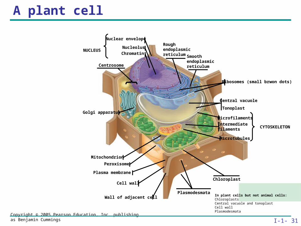
A plant cell
In plant cells but not animal cells:ChloroplastsCentral vacuole and tonoplastCell wallPlasmodesmata
CYTOSKELETON
Ribosomes (small brwon dots)
Central vacuole
Microfilaments
Intermediate filaments
Microtubules
Rough endoplasmic reticulum Smooth
endoplasmic reticulum
ChromatinNUCLEUS
Nuclear envelope
Nucleolus
Chloroplast
PlasmodesmataWall of adjacent cell
Cell wall
Golgi apparatus
Peroxisome
Tonoplast
Centrosome
Plasma membrane
Mitochondrion

Copyright © 2005 Pearson Education, Inc. publishing as Benjamin Cummings I-1- 32
Concept : The eukaryotic cell’s genetic instructions are housed in the nucleus and carried out by the ribosomes
• The Nucleus: Genetic Library of the Cell
• The nucleus
– Contains most of the genes in the eukaryotic cell

Copyright © 2005 Pearson Education, Inc. publishing as Benjamin Cummings I-1- 33
The nuclear envelope
• Encloses the nucleus, separating its contents from the cytoplasm Nucleus
NucleusNucleolus
Chromatin
Nuclear envelope:Inner membrane
Outer membrane
Nuclear pore
Rough ER
Porecomplex
Surface of nuclear envelope.
Pore complexes (TEM). Nuclear lamina (TEM).
Close-up of nuclearenvelope
Ribosome
1 µm
1 µm
0.25 µm

Copyright © 2005 Pearson Education, Inc. publishing as Benjamin Cummings I-1- 34
Ribosomes: Protein Factories in the Cell
– Are particles made of ribosomal RNA and protein; Carry out protein synthesis
ER
Endoplasmic reticulum (ER)
Ribosomes Cytosol
Free ribosomes
Bound ribosomes
Largesubunit
Smallsubunit
TEM showing ER and ribosomes Diagram of a ribosome
0.5 µm

Copyright © 2005 Pearson Education, Inc. publishing as Benjamin Cummings I-1- 35
Concept : The endomembrane system regulates protein traffic and performs metabolic functions in the cell
• The endomembrane system
– Includes many different structures

Copyright © 2005 Pearson Education, Inc. publishing as Benjamin Cummings I-1- 36
The Endoplasmic Reticulum: Biosynthetic Factory
Accounts for more than half the total membrane in many eukaryotic cells
• The ER membrane
Is continuous with the
nuclear envelope
Smooth ER
Rough ER
ER lumenCisternae
RibosomesTransport vesicle
Smooth ER
Transitional ER
Rough ER 200 µm
Nuclearenvelope

Copyright © 2005 Pearson Education, Inc. publishing as Benjamin Cummings I-1- 37
There are two distinct regions of ER
– Smooth ER, which lacks ribosomes
– Rough ER, which contains ribosomes
• The smooth ER: Synthesizes lipids; Metabolizes carbohydrates; Stores calcium; Detoxifies poison
• The rough ER: Has bound ribosomes; Produces proteins and membranes, which are distributed by transport vesicles

Copyright © 2005 Pearson Education, Inc. publishing as Benjamin Cummings I-1- 38
• The Golgi apparatus
– Receives many of the transport vesicles produced in the rough ER
– Consists of flattened membranous sacs called cisternae
• Functions of the Golgi apparatus include
– Modification of the products of the rough ER
– Manufacture of certain macromolecules
The Golgi Apparatus: Shipping and Receiving Center

Copyright © 2005 Pearson Education, Inc. publishing as Benjamin Cummings I-1- 39
Golgiapparatus
TEM of Golgi apparatus
cis face(“receiving” side ofGolgi apparatus)
Vesicles movefrom ER to Golgi Vesicles also
transport certainproteins back to ER
Vesicles coalesce toform new cis Golgi cisternae
Cisternalmaturation:Golgi cisternaemove in a cis-to-transdirection
Vesicles form andleave Golgi, carryingspecific proteins toother locations or tothe plasma mem-brane for secretion
Vesicles transport specificproteins backward to newerGolgi cisternae
Cisternae
trans face(“shipping” side ofGolgi apparatus)
0.1 0 µm16
5
2
3
4
Functions of the Golgi apparatus

Copyright © 2005 Pearson Education, Inc. publishing as Benjamin Cummings I-1- 40
Lysosomes: Digestive Compartments
Membranous sac of hydrolytic enzymes; Can digest all kinds of macromolecules
• Carry out intracellular
• digestion by
• phagocytosis
Figure 6.14 A
(a) Phagocytosis: lysosome digesting food
1 µm
Lysosome containsactive hydrolyticenzymes
Food vacuole fuses with lysosome
Hydrolyticenzymes digestfood particles
Digestion
Food vacuole
Plasma membraneLysosome
Digestiveenzymes
Lysosome
Nucleus

Copyright © 2005 Pearson Education, Inc. publishing as Benjamin Cummings I-1- 41
Vacuoles: Diverse Maintenance Compartments
• A plant or fungal cell
– May have one or several vacuoles
• Food vacuoles
– Are formed by phagocytosis
• Contractile vacuoles
– Pump excess water out of protist cells

Copyright © 2005 Pearson Education, Inc. publishing as Benjamin Cummings I-1- 42
• Central vacuoles
– Are found in plant cells
– Hold reserves of important organic compounds and water
Central vacuole
Cytosol
Tonoplast
Centralvacuole
Nucleus
Cell wall
Chloroplast
5 µm

Copyright © 2005 Pearson Education, Inc. publishing as Benjamin Cummings I-1- 43
Plasma membrane expandsby fusion of vesicles; proteinsare secreted from cell
Transport vesicle carriesproteins to plasma membrane for secretion
Lysosome availablefor fusion with anothervesicle for digestion
4 5 6
Nuclear envelope isconnected to rough ER, which is also continuous
with smooth ER
Nucleus
Rough ER
Smooth ERcis Golgi
trans Golgi
Membranes and proteinsproduced by the ER flow in
the form of transport vesiclesto the Golgi
Nuclear envelop
Golgi pinches off transport Vesicles and other vesicles
that give rise to lysosomes and Vacuoles
1
3
2
Plasmamembrane
The Endomembrane System: A Review
• Relationships among organelles of the endomembrane system

Copyright © 2005 Pearson Education, Inc. publishing as Benjamin Cummings I-1- 44
Concept: Mitochondria and chloroplasts change energy from one form to another
• Mitochondria
– Are the sites of cellular respiration
• Chloroplasts
– Found only in plants, are the sites of photosynthesis

Copyright © 2005 Pearson Education, Inc. publishing as Benjamin Cummings I-1- 45
Mitochondria: Chemical Energy Conversion
• Are found in nearly all eukaryotic cells
• Mitochondria are enclosed by two membranes
– A smooth outer membrane; An inner membrane folded into cristae
Mitochondrion
Intermembrane space
Outermembrane
Freeribosomesin the mitochondrialmatrix
MitochondrialDNA
Innermembrane
Cristae
Matrix
100 µm

Copyright © 2005 Pearson Education, Inc. publishing as Benjamin Cummings I-1- 46
Chloroplasts: Capture of Light Energy
• The chloroplast is a specialized member of a family of closely related plant organelles called plastids
– Contains chlorophyll; Are found in leaves and other green organs of plants and in algae
Chloroplast
ChloroplastDNA
RibosomesStroma
Inner and outermembranes
Thylakoid
1 µm
Granum

Copyright © 2005 Pearson Education, Inc. publishing as Benjamin Cummings I-1- 47
Peroxisomes: Oxidation
– Produce hydrogen peroxide and convert it to water
ChloroplastPeroxisome
Mitochondrion
1 µm

Copyright © 2005 Pearson Education, Inc. publishing as Benjamin Cummings I-1- 48
Concept: The cytoskeleton is a network of fibers that organizes structures and activities in the cell
• Cytoskeleton is a network of fibers extending throughout the cytoplasm
Microtubule
0.25 µm Microfilaments

Copyright © 2005 Pearson Education, Inc. publishing as Benjamin Cummings I-1- 49
Roles of the Cytoskeleton: Support, Motility, and Regulation
– Is involved in cell motility, which utilizes motor proteins
VesicleATP
Receptor formotor protein
Motor protein(ATP powered)
Microtubuleof cytoskeleton
(a) Motor proteins that attach to receptors on organelles can “walk”the organelles along microtubules or, in some cases, microfilaments.
Microtubule Vesicles 0.25 µm
(b) Vesicles containing neurotransmitters migrate to the tips of nerve cell axons via the mechanism in (a). In this SEM of a squid giant axon, two vesicles can be seen moving along a microtubule. (A separate part of the experiment provided the evidence that they were in fact moving.)

Copyright © 2005 Pearson Education, Inc. publishing as Benjamin Cummings I-1- 50
Components of the Cytoskeleton
• There are three main types of fibers that make up the cytoskeleton

Copyright © 2005 Pearson Education, Inc. publishing as Benjamin Cummings I-1- 51
Microtubules
– Shape the cell
– Guide movement of organelles
– Help separate the chromosome copies in dividing cells

Copyright © 2005 Pearson Education, Inc. publishing as Benjamin Cummings I-1- 52
Centrosomes and Centrioles
• The centrosome is considered to be a “microtubule-organizing center”; Contains a pair of centrioles
Centrosome
Microtubule
Centrioles
0.25 µm
Longitudinal sectionof one centriole
Microtubules Cross sectionof the other centrioleFigure 6.22

Copyright © 2005 Pearson Education, Inc. publishing as Benjamin Cummings I-1- 53
Cilia and Flagella
– Contain specialized arrangements of microtubules
– Are locomotor appendages of some cells

Copyright © 2005 Pearson Education, Inc. publishing as Benjamin Cummings I-1- 54
Flagella beating pattern
(a) Motion of flagella. A flagellum usually undulates, its snakelike motion driving a cell in the same direction as the axis of the flagellum. Propulsion of a human sperm cell is an example of flagellatelocomotion (LM).
1 µm
Direction of swimming

Copyright © 2005 Pearson Education, Inc. publishing as Benjamin Cummings I-1- 55
Ciliary motion
(b) Motion of cilia. Cilia have a back- and-forth motion that moves the cell in a direction perpendicular to the axis of the cilium. A dense nap of cilia, beating at a rate of about 40 to 60 strokes a second, covers this Colpidium, a freshwater protozoan (SEM).
15 µm

Copyright © 2005 Pearson Education, Inc. publishing as Benjamin Cummings I-1- 56
Cilia and flagella share a common ultrastructure
(a)
(c)
(b)
Outer microtubuledoublet
Dynein arms
Centralmicrotubule
Outer doublets cross-linkingproteins inside
Radialspoke
Plasmamembrane
Microtubules
Plasmamembrane
Basal body
0.5 µm
0.1 µm
0.1 µm
Cross section of basal body
Triplet

Copyright © 2005 Pearson Education, Inc. publishing as Benjamin Cummings I-1- 57
Microfilaments (Actin Filaments)
– Are built from molecules of the protein actin
– Are found in microvilli
0.25 µm
Microvillus
Plasma membrane
Microfilaments (actinfilaments)
Intermediate filaments

Copyright © 2005 Pearson Education, Inc. publishing as Benjamin Cummings I-1- 58
Microfilaments that function in cellular motility
• Contain the protein myosin in addition to actin
Actin filament
Myosin filament
Myosin motors in muscle cell contraction. (a)
Muscle cell
Myosin arm

Copyright © 2005 Pearson Education, Inc. publishing as Benjamin Cummings I-1- 59
Amoeboid movement
• Involves the contraction of actin and myosin filaments
Cortex (outer cytoplasm):gel with actin network
Inner cytoplasm: sol with actin subunits
Extendingpseudopodium
(b) Amoeboid movement

Copyright © 2005 Pearson Education, Inc. publishing as Benjamin Cummings I-1- 60
Cell Walls of Plants
– Is an extracellular structure of plant cells that distinguishes them from animal cells
– Are made of cellulose fibers embedded in other polysaccharides and protein; May have multiple layers
Central vacuoleof cell
Plasmamembrane
Secondarycell wall
Primarycell wall
Middlelamella
1 µm
Centralvacuoleof cell
Central vacuole Cytosol
Plasma membrane
Plant cell walls
Plasmodesmata

Copyright © 2005 Pearson Education, Inc. publishing as Benjamin Cummings I-1- 61
The Extracellular Matrix (ECM) of Animal Cells
• Animal cells lack cell walls and covered by an elaborate matrix, the ECM.
– Is made up of glycoproteins and other macromolecules
Collagen
Fibronectin
Plasmamembrane
EXTRACELLULAR FLUID
Micro-filaments
CYTOPLASM
Integrins
Polysaccharidemolecule
Carbo-hydrates
Proteoglycanmolecule
Coreprotein
Integrin
A proteoglycan complex

Copyright © 2005 Pearson Education, Inc. publishing as Benjamin Cummings I-1- 62
Functions of the ECM include
– Support
– Adhesion
– Movement
– Regulation

Copyright © 2005 Pearson Education, Inc. publishing as Benjamin Cummings I-1- 63
Intercellular Junctions
– Plants: Plasmodesmata
– Are channels that perforate plant cell walls
Interiorof cell
Interiorof cell
0.5 µm Plasmodesmata Plasma membranes
Cell walls

Copyright © 2005 Pearson Education, Inc. publishing as Benjamin Cummings I-1- 64
Animals: Tight Junctions, Desmosomes, and Gap Junctions
• In animals, there are three types of intercellular junctions
– Tight junctions
– Desmosomes
– Gap junctions

Copyright © 2005 Pearson Education, Inc. publishing as Benjamin Cummings I-1- 65
• Types of intercellular junctions in animals
Tight junctions prevent fluid from moving across a layer of cells
Tight junction
0.5 µm
1 µm
Spacebetweencells
Plasma membranesof adjacent cells
Extracellularmatrix
Gap junction
Tight junctions
0.1 µm
Intermediatefilaments
Desmosome
Gapjunctions
At tight junctions, the membranes ofneighboring cells are very tightly pressedagainst each other, bound together byspecific proteins (purple). Forming continu-ous seals around the cells, tight junctionsprevent leakage of extracellular fluid acrossA layer of epithelial cells.
Desmosomes (also called anchoringjunctions) function like rivets, fastening cellsTogether into strong sheets. IntermediateFilaments made of sturdy keratin proteinsAnchor desmosomes in the cytoplasm.
Gap junctions (also called communicatingjunctions) provide cytoplasmic channels fromone cell to an adjacent cell. Gap junctions consist of special membrane proteins that surround a pore through which ions, sugars,amino acids, and other small molecules maypass. Gap junctions are necessary for commu-nication between cells in many types of tissues,including heart muscle and animal embryos.
TIGHT JUNCTIONS
DESMOSOMES
GAP JUNCTIONS

Copyright © 2005 Pearson Education, Inc. publishing as Benjamin Cummings I-1- 66
The Cell: A Living Unit Greater Than the Sum of Its Parts
• Cells rely on the integration of structures and organelles in order to function
5 µ
m


