+ All Categories
Home > Documents > Copyright © 2005 Pearson Education, Inc. publishing as Benjamin Cummings Osmoregulation and...

Copyright © 2005 Pearson Education, Inc. publishing as Benjamin Cummings Osmoregulation and...

Date post: 18-Dec-2015
Category:
Upload: emily-riley
View: 214 times
Download: 1 times
Share this document with a friend
Popular Tags:
52
Copyright © 2005 Pearson Education, Inc. publishing as Benjamin Cummings Osmoregulation and excretion The physiological systems of animals operate in a fluid environment The relative concentrations of water and solutes in this environment must be maintained within fairly narrow limits
Transcript

Copyright © 2005 Pearson Education, Inc. publishing as Benjamin Cummings

Osmoregulation and excretion

• The physiological systems of animals operate in a fluid environment

• The relative concentrations of water and solutes in this environment must be maintained within fairly narrow limits

Copyright © 2005 Pearson Education, Inc. publishing as Benjamin Cummings

• Freshwater animals show adaptations that reduce water uptake and conserve solutes

• Desert and marine animals face desiccating environments with the potential to quickly deplete the body water

Figure 44.1

Copyright © 2005 Pearson Education, Inc. publishing as Benjamin Cummings

• Osmoregulation regulates solute concentrations and balances the gain and loss of water

• Excretion gets rid of metabolic wastes

Copyright © 2005 Pearson Education, Inc. publishing as Benjamin Cummings

Osmoregulation

• Osmoregulation is based largely on controlled movement of solutes between internal fluids and the external environment.

• Cells require a balance between osmotic gain and loss of water.

• Water uptake and loss are balanced by various mechanisms of osmoregulation in different environments.

Copyright © 2005 Pearson Education, Inc. publishing as Benjamin Cummings

Osmotic Challenges

• Osmoconformers, which are only marine animals are isoosmotic with [at the same concentration as] their surroundings and do not regulate their osmolarity.

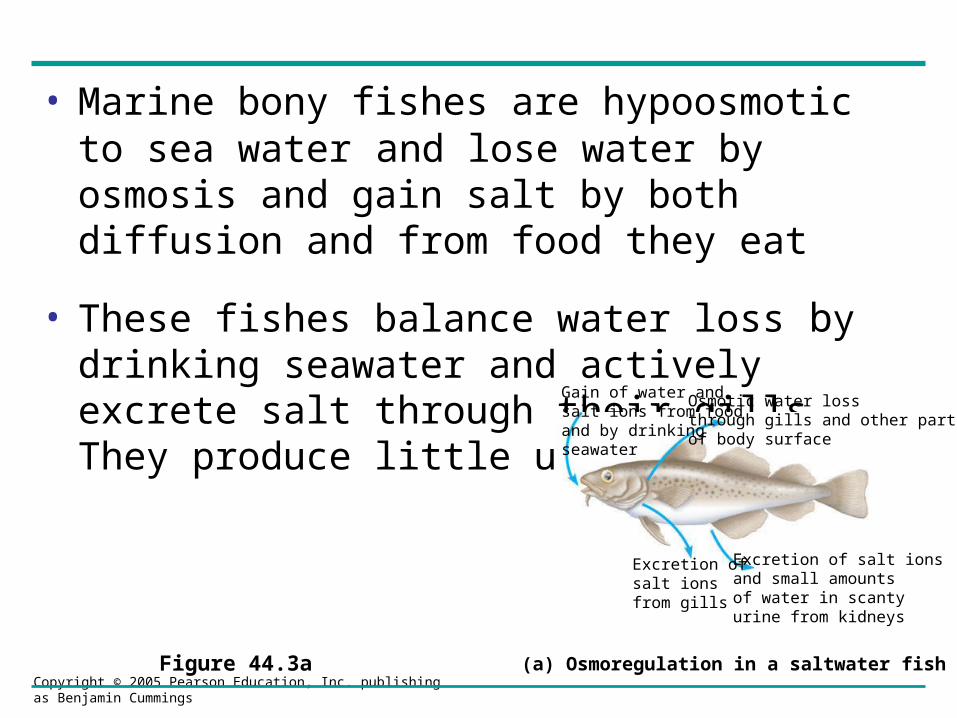
• Osmoregulators expend energy to control water uptake and loss in a hyperosmotic [more concentrated] or hypoosmotic [less concentrated] environment.

Copyright © 2005 Pearson Education, Inc. publishing as Benjamin Cummings

Marine Animals

• Most marine invertebrates are osmoconformers

• Most marine vertebrates and some invertebrates are osmoregulators

Copyright © 2005 Pearson Education, Inc. publishing as Benjamin Cummings

• Marine bony fishes are hypoosmotic to sea water and lose water by osmosis and gain salt by both diffusion and from food they eat

• These fishes balance water loss by drinking seawater and actively excrete salt through their gills. They produce little urine.

Figure 44.3a

Gain of water andsalt ions from foodand by drinkingseawater

Osmotic water lossthrough gills and other partsof body surface

Excretion ofsalt ionsfrom gills

Excretion of salt ionsand small amountsof water in scantyurine from kidneys

(a) Osmoregulation in a saltwater fish

Copyright © 2005 Pearson Education, Inc. publishing as Benjamin Cummings

Freshwater Animals

• Freshwater animals constantly take in water from their hypoosmotic environment

• They lose salts by diffusion

Copyright © 2005 Pearson Education, Inc. publishing as Benjamin Cummings

• Freshwater animals maintain water balance by excreting large amounts of dilute urine

• Salts lost by diffusion are replaced by foods and uptake across the gills

Figure 44.3b

Uptake ofwater and someions in food

Osmotic water gainthrough gills and other partsof body surface

Uptake ofsalt ions by gills

Excretion oflarge amounts ofwater in dilute urine from kidneys

(b) Osmoregulation in a freshwater fish

Copyright © 2005 Pearson Education, Inc. publishing as Benjamin Cummings

Land animals

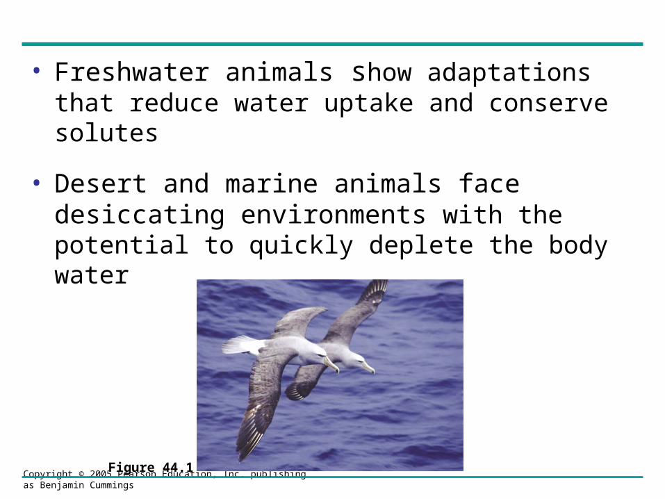
• Land animals manage their water budgets by drinking and eating moist foods and by using metabolic water.

• Animals adapted to deserts produce highly concentrated urine and use a wide range of behavioral adaptations to avoid the heat (burrowing, nocturnal activity)

Copyright © 2005 Pearson Education, Inc. publishing as Benjamin Cummings

Figure 44.5

Waterbalance in a human

(2,500 mL/day= 100%)

Waterbalance in akangaroo rat

(2 mL/day= 100%)

Ingested in food (0.2)

Ingested in food (750)

Ingested in liquid(1,500)

Derived from metabolism (250)Derived from

metabolism (1.8)

Water gain

Feces (0.9)

Urine(0.45)

Evaporation (1.46)

Feces (100)

Urine(1,500)

Evaporation (900)

Water loss

Copyright © 2005 Pearson Education, Inc. publishing as Benjamin Cummings

Benefits of insulation• Desert animals also get major water savings from simple

anatomical features

• Knut and Bodil Schmidt-Nielsen et al. observed that the fur of camels exposed to full sun in the Sahara Desert could reach temperatures of over 70°C, while the animals’ skin remained more than 30°C cooler.

• The Schmidt-Nielsens reasoned that insulation of the skin by fur may substantially reduce the need for evaporative cooling by sweating. To test this hypothesis, they compared the water loss rates of unclipped and clipped camels.

• Removing the fur of a camel increased the rateof water loss through sweating by up to 50%.

Copyright © 2005 Pearson Education, Inc. publishing as Benjamin Cummings

Control group(Unclipped fur)

Experimental group(Clipped fur)

4

3

2

1

0

Wat

er lo

st p

er d

ay(L

/100

kg

body

ma

ss)

Copyright © 2005 Pearson Education, Inc. publishing as Benjamin Cummings

Transport Epithelia

• Transport epithelia are specialized cells that regulate solute movement. The epithelia form a layer of cells across whose selectively permeable membranes solutes must flow.

• These epithelia are essential components of osmotic regulation and metabolic waste disposal and usually are arranged into complex tubular networks.

Copyright © 2005 Pearson Education, Inc. publishing as Benjamin Cummings

• An example of transport epithelia is found in the salt glands of marine birds which remove excess sodium chloride from the blood.

• In birds such as petrels, gulls and albatrosses transport epithelia arranged in tubes in a salt gland actively secrete salt from the blood into collecting ducts [a countercurrent system is used to maximize salt concentration] that drain to the nostrils where the salt solution is shed.

Copyright © 2005 Pearson Education, Inc. publishing as Benjamin Cummings

Nasal salt gland

Nostrilwith saltsecretions

Lumen ofsecretory tubule

NaCl

Bloodflow

Secretory cellof transportepithelium

Centralduct

Directionof saltmovement

Transportepithelium

Secretorytubule

Capillary

Vein

Artery

(a) An albatross’s salt glands empty via a duct into thenostrils, and the salty solution either drips off the tip of the beak or is exhaled in a fine mist.

(b) One of several thousand secretory tubules in a salt-excreting gland. Each tubule is lined by a transportepithelium surrounded by capillaries, and drains intoa central duct.

(c) The secretory cells actively transport salt from theblood into the tubules. Blood flows counter to the flow of salt secretion. By maintaining a concentrationgradient of salt in the tubule (aqua), this countercurrentsystem enhances salt transfer from the blood to the lumen of the tubule.

Copyright © 2005 Pearson Education, Inc. publishing as Benjamin Cummings

Nitrogenous wastes

• An animal’s nitrogenous wastes reflect its phylogeny and habitat

• The type and quantity of an animal’s waste products may have a large impact on its water balance

• Among the most important wastes are the nitrogenous breakdown products of proteins and nucleic acids.

• Different animals excrete nitrogenous wastes in different forms

Copyright © 2005 Pearson Education, Inc. publishing as Benjamin Cummings

Proteins Nucleic acids

Amino acids Nitrogenous bases

–NH2

Amino groups

Most aquaticanimals, includingmost bony fishes

Mammals, mostamphibians, sharks,some bony fishes

Many reptiles(includingbirds), insects,land snails

Ammonia Urea Uric acid

NH3 NH2

NH2

O C

C

CN

CO N

H H

C O

NC

HN

O

H

Copyright © 2005 Pearson Education, Inc. publishing as Benjamin Cummings

Ammonia

• Animals such as fish that excrete nitrogenous wastes as ammonia need access to lots of water.

• Ammonia is released across the whole body surface or through the gills.

Copyright © 2005 Pearson Education, Inc. publishing as Benjamin Cummings

Urea

• The liver of mammals and most adult amphibians converts ammonia to less toxic urea

• Urea is carried to the kidneys, concentrated and excreted with a minimal loss of water

Copyright © 2005 Pearson Education, Inc. publishing as Benjamin Cummings

Uric Acid

• Insects, land snails, and many reptiles, including birds excrete uric acid as their major nitrogenous waste

• Uric acid is largely insoluble in water and can be secreted as a paste with little water loss.

Copyright © 2005 Pearson Education, Inc. publishing as Benjamin Cummings

Excretory Systems

• Diverse excretory systems are variations on a tubular theme

• Most excretory systems produce urine by refining a filtrate derived from body fluids

Copyright © 2005 Pearson Education, Inc. publishing as Benjamin Cummings

• Key functions of most excretory systems are

– Filtration, pressure-filtering of body fluids producing a filtrate

– Reabsorption, reclaiming valuable solutes from the filtrate

– Secretion, addition of toxins and other solutes from the body fluids to the filtrate

– Excretion, the filtrate leaves the system

Copyright © 2005 Pearson Education, Inc. publishing as Benjamin Cummings

Figure 44.9

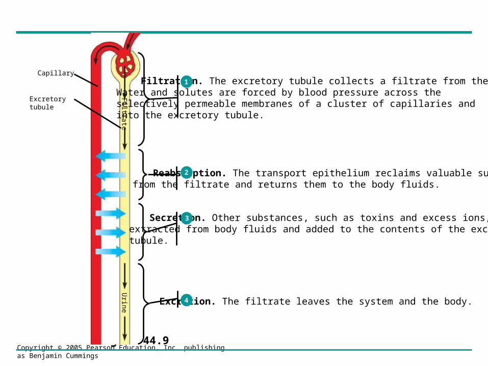
Filtration. The excretory tubule collects a filtrate from the blood.Water and solutes are forced by blood pressure across the selectively permeable membranes of a cluster of capillaries and into the excretory tubule.

Reabsorption. The transport epithelium reclaims valuable substances from the filtrate and returns them to the body fluids.

Secretion. Other substances, such as toxins and excess ions, are extracted from body fluids and added to the contents of the excretory tubule.

Excretion. The filtrate leaves the system and the body.

Capillary

Excretorytubule

Filtrate

Urine

1

2

3

4

Copyright © 2005 Pearson Education, Inc. publishing as Benjamin Cummings

Vertebrate Kidneys

• Kidneys, the excretory organs of vertebrates, function in both excretion and osmoregulation.

• The mammalian excretory system centers on paired kidneys which are also the principal site of water balance and salt regulation.

Copyright © 2005 Pearson Education, Inc. publishing as Benjamin Cummings

• Each kidney is supplied with blood by a renal artery and drained by a renal vein.

Posterior vena cava

Renal artery and vein

Aorta

Ureter

Urinary bladder

Urethra

(a) Excretory organs and major associated blood vessels

Kidney

Copyright © 2005 Pearson Education, Inc. publishing as Benjamin Cummings

• Urine exits each kidney through a duct called the ureter.

• Both ureters drain into a common urinary bladder.

• Urine is voided from the bladder via a duct called the urethra.

Copyright © 2005 Pearson Education, Inc. publishing as Benjamin Cummings

(b) Kidney structure

UreterSection of kidney from a rat

Renalmedulla

Renalcortex

Renalpelvis

Figure 44.13b

Structure and Function of the Nephron and Associated Structures

• The mammalian kidney has two distinct regions:an outer renal cortex and an inner renal medulla

Copyright © 2005 Pearson Education, Inc. publishing as Benjamin Cummings

• The nephron is the functional unit of the vertebrate kidney.

• It consists of a single long tubule and a ball of capillaries called the glomerulus.

Copyright © 2005 Pearson Education, Inc. publishing as Benjamin Cummings

Figure 44.13c, d

Juxta-medullarynephron

Corticalnephron

Collectingduct

To renalpelvis

Renalcortex

Renalmedulla

20 µm

Afferentarteriolefrom renalartery

Glomerulus

Bowman’s capsule

Proximal tubule

Peritubularcapillaries

SEM

Efferentarteriole fromglomerulus

Branch ofrenal vein

DescendinglimbAscendinglimb

Loopof

Henle

Distal tubule

Collectingduct

(c) Nephron

Vasarecta

(d) Filtrate and blood flow

Copyright © 2005 Pearson Education, Inc. publishing as Benjamin Cummings

Filtration of the Blood

• Filtration occurs as blood pressure forces fluid from the blood in the glomerulus into the lumen of the Bowman’s capsule.

• Filtration of small molecules is nonselective and the filtrate in Bowman’s capsule is a mixture that mirrors the concentration of various solutes in the blood plasma.

• The filtrate contains water, urea, salts and other small molecules.

Copyright © 2005 Pearson Education, Inc. publishing as Benjamin Cummings

Pathway of the Filtrate

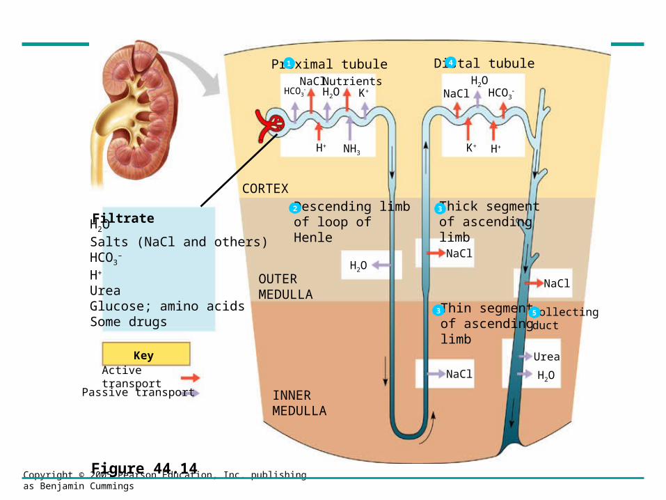
• From the Bowman’s capsule, the filtrate passes through three regions of the nephron the proximal tubule, the loop of Henle, and the distal tubule. [proximal means closest to and distal furthest from in the case the Bowman’s capsule].

• Fluid from several nephrons flows into a collecting duct.

Copyright © 2005 Pearson Education, Inc. publishing as Benjamin Cummings

Proximal tubule

FiltrateH2OSalts (NaCl and others)HCO3

H+

UreaGlucose; amino acidsSome drugs

KeyActive transport

Passive transport

CORTEX

OUTERMEDULLA

INNERMEDULLA

Descending limbof loop ofHenle

Thick segmentof ascendinglimb

Thin segmentof ascendinglimb

Collectingduct

NaCl

NaCl

NaCl

Distal tubuleNaCl Nutrients

Urea

H2O

NaClH2O

H2OHCO3 K+

H+ NH3

HCO3

K+ H+

H2O

1 4

32

3 5

Figure 44.14

Copyright © 2005 Pearson Education, Inc. publishing as Benjamin Cummings

Proximal tubule

• Selective secretion and reabsorption in the proximal tubule substantially alter the volume and composition of filtrate from what is initially produced in the Bowman’s capsule.

• In the proximal tubule hydrogen ions (H+) and ammonia (NH3) are secreted into the tubule as are drugs and toxins.

• Nutrients (e.g. glucose and amino acids) are reabsorbed as are potassium (K+), NaCl and water.

Copyright © 2005 Pearson Education, Inc. publishing as Benjamin Cummings

Descending limb of loop of Henle

• Reabsorption of water out of the filtrate continues as the filtrate moves into the descending limb of the loop of Henle, which is very permeable to water, but not very permeable to salts.

• NaCl concentration in the urine thus increases.

Copyright © 2005 Pearson Education, Inc. publishing as Benjamin Cummings

Ascending Limb of Loop of Henle

• The ascending limb of the Loop of Henle in contrast to the descending loop is permeable to salt, but not to water.

• Thus, as filtrate travels through the ascending limb of the loop of Henle, salt diffuses out of the permeable tubule into the interstitial fluid.

• First the movement of salt is passive, but in the upper section of the ascending loop salt is actively pumped out into the interstitial fluid.

Copyright © 2005 Pearson Education, Inc. publishing as Benjamin Cummings

Ascending Limb of Loop of Henle

• The movement of salt out of the ascending loop of Henle contributes to the development of an osmolarity gradient that is important to the kidney’s ability to produce a concentrated urine as we will see later.

Copyright © 2005 Pearson Education, Inc. publishing as Benjamin Cummings

Distal tubule

• The distal tubule plays a key role in regulating the K+ and NaCl concentration of body fluids by regulating the K+ secreted and NaCl reabsorbed.

Copyright © 2005 Pearson Education, Inc. publishing as Benjamin Cummings

Collecting Duct

• The collecting duct carries the filtrate through the medulla to the renal pelvis and reabsorbs NaCl

Copyright © 2005 Pearson Education, Inc. publishing as Benjamin Cummings

Water conservation ability of the kidney

• The mammalian kidney’s ability to conserve water is a key terrestrial adaptation

• The mammalian kidney can produce urine much more concentrated than body fluids, thus conserving water

Copyright © 2005 Pearson Education, Inc. publishing as Benjamin Cummings

Solute Gradients and Water Conservation

• In a mammalian kidney, the cooperative action and arrangement of the loops of Henle and the collecting ducts produce an osmotic gradient along the length of the loop of Henle in the kidney.

• Two solutes, NaCl and urea, contribute to the osmolarity of the interstitial fluid which causes the reabsorption of water in the kidney and concentrates the urine

Copyright © 2005 Pearson Education, Inc. publishing as Benjamin Cummings

Figure 44.15

H2O

H2O

H2O

H2O

H2O

H2O

H2O

NaCl

NaCl

NaCl

NaCl

NaCl

NaCl

NaCl

300

300 100

400

600

900

1200

700

400

200

100

Activetransport

Passivetransport

OUTERMEDULLA

INNERMEDULLA

CORTEX

H2O

Urea

H2OUrea

H2O

Urea

H2O

H2O

H2O

H2O

1200

1200

900

600

400

300

600

400

300

Osmolarity of interstitial

fluid(mosm/L)

300

Copyright © 2005 Pearson Education, Inc. publishing as Benjamin Cummings

• Water passing down the descending loop of Henle meets more and more concentrated interstitial fluid and so water passively moves out of the tubule into the kidney.

• Because the ascending loop of Henle is not permeable to water, water does not flow back into the filtrate as it ascends. However, salts are actively pumped out to maintain the concentration gradient.

Copyright © 2005 Pearson Education, Inc. publishing as Benjamin Cummings

Collecting duct

• The collecting duct, is also permeable to water but not salt.

• The collecting duct carries the filtrate through the kidney’s osmolarity gradient, and more water exits the filtrate by osmosis

Copyright © 2005 Pearson Education, Inc. publishing as Benjamin Cummings

Collecting duct

• Urea also diffuses out of the collecting duct as it traverses the inner medulla

• Urea and NaCl together form the osmotic gradient that enables the kidney to produce urine that is hyperosmotic to the blood

Copyright © 2005 Pearson Education, Inc. publishing as Benjamin Cummings

• Because of the countercurrent arrangement of the loop of Henle, the longer the loop the more urine can be concentrated.

• Thus, desert rodents have very long loops of Henle, which enable them to produce urine much more concentrated than humans can produce.

Copyright © 2005 Pearson Education, Inc. publishing as Benjamin Cummings

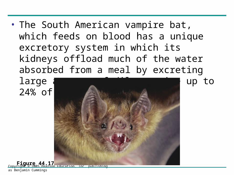
• The South American vampire bat, which feeds on blood has a unique excretory system in which its kidneys offload much of the water absorbed from a meal by excreting large amounts of dilute urine up to 24% of bodyweight per hour.

Figure 44.17

Copyright © 2005 Pearson Education, Inc. publishing as Benjamin Cummings

Regulation of Kidney Function

• The osmolarity of the urine

– Is regulated by nervous and hormonal control of water and salt reabsorption in the kidneys

• Antidiuretic hormone (ADH)

– Increases water reabsorption in the distal tubules and collecting ducts of the kidney

Copyright © 2005 Pearson Education, Inc. publishing as Benjamin Cummings

Osmoreceptorsin hypothalamus

Drinking reducesblood osmolarity

to set point

H2O reab-sorption helpsprevent further

osmolarity increase

STIMULUS:The release of ADH istriggered when osmo-receptor cells in the

hypothalamus detect anincrease in the osmolarity

of the blood

Homeostasis:Blood osmolarity

Hypothalamus

ADH

Pituitarygland

Increasedpermeability

Thirst

Collecting duct

Distaltubule

(a) Antidiuretic hormone (ADH) enhances fluid retention by makingthe kidneys reclaim more water.

Copyright © 2005 Pearson Education, Inc. publishing as Benjamin Cummings

RAAS system

• The renin-angiotensin-aldosterone system (RAAS) is part of a complex feedback circuit that functions in homeostasis.

• The juxtoglomerular apparatus located around arterioles supplying glomeruli detect drop in blood pressure. Releases enzyme renin.

• Renin stimulates production of angiotensin II which decreases blood flow to kidney and stimulates proximal tubules to reabsorb more water na dNaCl.

Copyright © 2005 Pearson Education, Inc. publishing as Benjamin Cummings

Increased Na+

and H2O reab-sorption in

distal tubules

Homeostasis:Blood pressure,

volume

STIMULUS:The juxtaglomerular

apparatus (JGA) respondsto low blood volume or

blood pressure (such as dueto dehydration or loss of

blood)

Aldosterone

Adrenal gland

Angiotensin II

Angiotensinogen

Reninproduction

Renin

Arterioleconstriction

Distal tubule

JGA

(b) The renin-angiotensin-aldosterone system (RAAS) leads to an increasein blood volume and pressure.

Copyright © 2005 Pearson Education, Inc. publishing as Benjamin Cummings

ANF

• Another hormone, atrial natriuretic factor (ANF) opposes the RAAS.

• Walls of the atria of the heart release ANF in response to increase in blood volume and pressure. It inhibits release of renin.


Recommended