+ All Categories
Home > Documents > Copyright © 2006 by Mosby, Inc. Slide 1 Chapter 14 Bronchiectasis Figure 14–1. Bronchiectasis. A,...

Copyright © 2006 by Mosby, Inc. Slide 1 Chapter 14 Bronchiectasis Figure 14–1. Bronchiectasis. A,...

Date post: 27-Dec-2015
Category:
Upload: allyson-walsh
View: 233 times
Download: 2 times
Share this document with a friend
Popular Tags:
45
Copyright © 2006 by Mosby, Inc. Slide 1 Chapter 14 Chapter 14 Bronchiectasis Bronchiectasis Figure 14–1. Bronchiectasis. A, Varicose bronchiectasis. B, Figure 14–1. Bronchiectasis. A, Varicose bronchiectasis. B, Cylindrical bronchiectasis. C, Saccular bronchiectasis. Also Cylindrical bronchiectasis. C, Saccular bronchiectasis. Also illustrated are excessive bronchial secretions (D) and atelectasis illustrated are excessive bronchial secretions (D) and atelectasis (E), which are both common anatomic alterations of the lungs in this (E), which are both common anatomic alterations of the lungs in this disease. disease. D E A B C
Transcript

Copyright © 2006 by Mosby, Inc.Slide 1

Chapter 14Chapter 14 Bronchiectasis Bronchiectasis


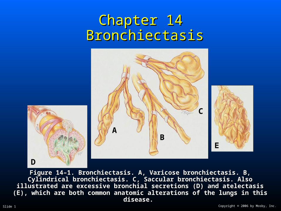
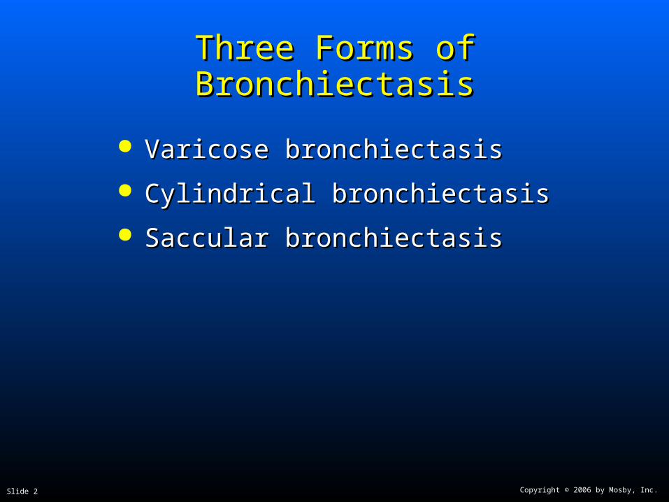
Figure 14–1. Bronchiectasis. A, Varicose bronchiectasis. B, Cylindrical bronchiectasis. C, Figure 14–1. Bronchiectasis. A, Varicose bronchiectasis. B, Cylindrical bronchiectasis. C, Saccular bronchiectasis. Also illustrated are excessive bronchial secretions (D) and Saccular bronchiectasis. Also illustrated are excessive bronchial secretions (D) and

atelectasis (E), which are both common anatomic alterations of the lungs in this disease.atelectasis (E), which are both common anatomic alterations of the lungs in this disease.

D

E

AB

C

Copyright © 2006 by Mosby, Inc.Slide 2

Three Forms of BronchiectasisThree Forms of Bronchiectasis

Varicose bronchiectasisVaricose bronchiectasis

Cylindrical bronchiectasisCylindrical bronchiectasis

Saccular bronchiectasisSaccular bronchiectasis

Copyright © 2006 by Mosby, Inc.Slide 3

Anatomic Alterations of the LungsAnatomic Alterations of the Lungs

Chronic dilation and distortion of bronchial airwaysChronic dilation and distortion of bronchial airways

Excessive production of often foul-smelling sputumExcessive production of often foul-smelling sputum

Smooth muscle constriction of bronchial airwaysSmooth muscle constriction of bronchial airways

Hyperinflation of alveoli (air-trapping)Hyperinflation of alveoli (air-trapping)

Atelectasis, consolidation, and parenchymal fibrosisAtelectasis, consolidation, and parenchymal fibrosis

Hemorrhage secondary to bronchial arterial erosionHemorrhage secondary to bronchial arterial erosion

Copyright © 2006 by Mosby, Inc.Slide 4

EtiologyEtiology

Acquired bronchiectasisAcquired bronchiectasis Recurrent pulmonary infectionRecurrent pulmonary infection

Bronchial obstructionBronchial obstruction

Congenital bronchiectasisCongenital bronchiectasis Kartagener’s syndromeKartagener’s syndrome

HypogammaglobulinemiaHypogammaglobulinemia

Cystic fibrosisCystic fibrosis

Copyright © 2006 by Mosby, Inc.Slide 5

Overview of the Cardiopulmonary Overview of the Cardiopulmonary Clinical Manifestations Associated Clinical Manifestations Associated

with BRONCHIECTASISwith BRONCHIECTASIS

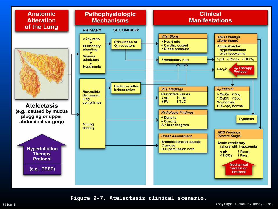
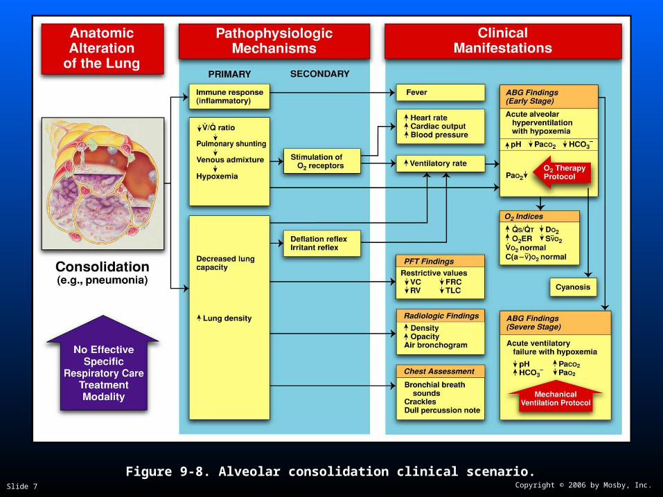
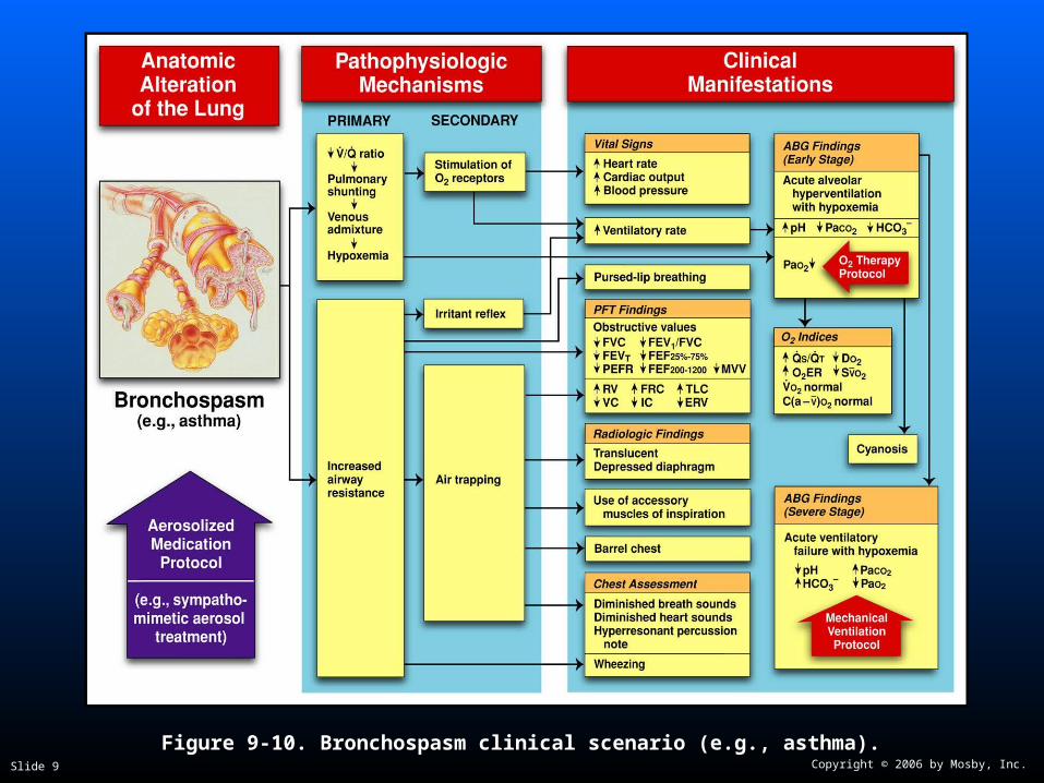
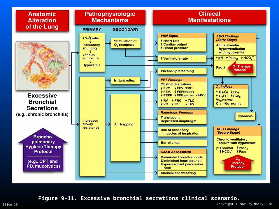
The following clinical manifestations result from the The following clinical manifestations result from the pathophysiologic mechanisms caused (or activated) pathophysiologic mechanisms caused (or activated) by by AtelectasisAtelectasis (see Figure 9-12), (see Figure 9-12), Consolidation Consolidation (see Figure 9-8), (see Figure 9-8), Bronchospasm Bronchospasm (see Figure 9-10), (see Figure 9-10), and and Excessive BronchialExcessive Bronchial Secretions Secretions (see Figure (see Figure 9-11)—the major anatomic alterations of the lungs 9-11)—the major anatomic alterations of the lungs associated with bronchiectasis (see Figure 14-1).associated with bronchiectasis (see Figure 14-1).

Copyright © 2006 by Mosby, Inc.Slide 6


Figure 9-7. Atelectasis clinical scenario.Figure 9-7. Atelectasis clinical scenario.

Copyright © 2006 by Mosby, Inc.Slide 7

Figure 9-8. Alveolar consolidation clinical scenario.Figure 9-8. Alveolar consolidation clinical scenario.

Copyright © 2006 by Mosby, Inc.Slide 8

Figure 9-9. Increased alveolar-capillary membrane thickness clinical scenario.Figure 9-9. Increased alveolar-capillary membrane thickness clinical scenario.

Copyright © 2006 by Mosby, Inc.Slide 9


Figure 9-10. Bronchospasm clinical scenario (e.g., asthma).Figure 9-10. Bronchospasm clinical scenario (e.g., asthma).

Copyright © 2006 by Mosby, Inc.Slide 10


Figure 9-11. Excessive bronchial secretions clinical scenario.Figure 9-11. Excessive bronchial secretions clinical scenario.

Copyright © 2006 by Mosby, Inc.Slide 11

Clinical Data Obtained at the Clinical Data Obtained at the Patient’s BedsidePatient’s Bedside

Vital signsVital signs

Increased respiratory rateIncreased respiratory rate

Increased heart rate, cardiac output, Increased heart rate, cardiac output, blood pressureblood pressure

Copyright © 2006 by Mosby, Inc.Slide 12

Clinical Data Obtained at the Clinical Data Obtained at the Patient’s BedsidePatient’s Bedside

Use of accessory muscles of inspirationUse of accessory muscles of inspiration

Use of accessory muscles of expirationUse of accessory muscles of expiration

Pursed-lip breathingPursed-lip breathing

Increased anteroposterior chest diameter Increased anteroposterior chest diameter (barrel chest)(barrel chest)

CyanosisCyanosis

Digital clubbingDigital clubbing

Copyright © 2006 by Mosby, Inc.Slide 13

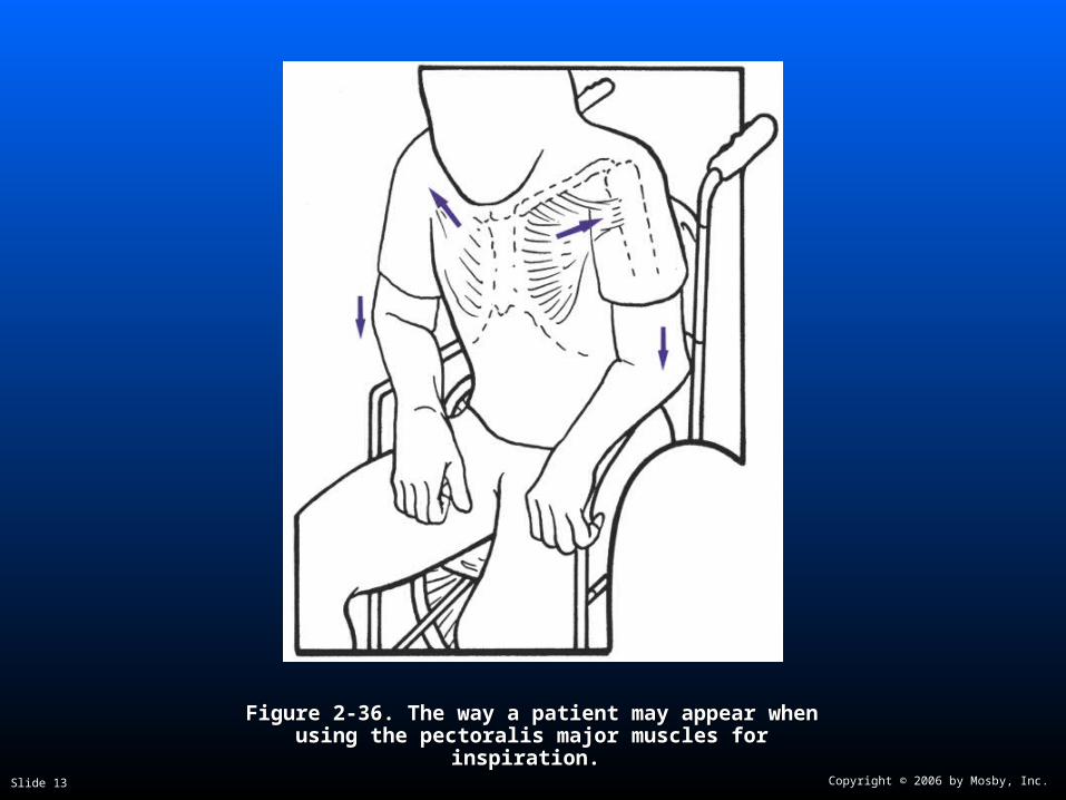
Figure 2-36. Figure 2-36. The way a patient may appear when using the The way a patient may appear when using the pectoralis major muscles for inspiration.pectoralis major muscles for inspiration.

Copyright © 2006 by Mosby, Inc.Slide 14

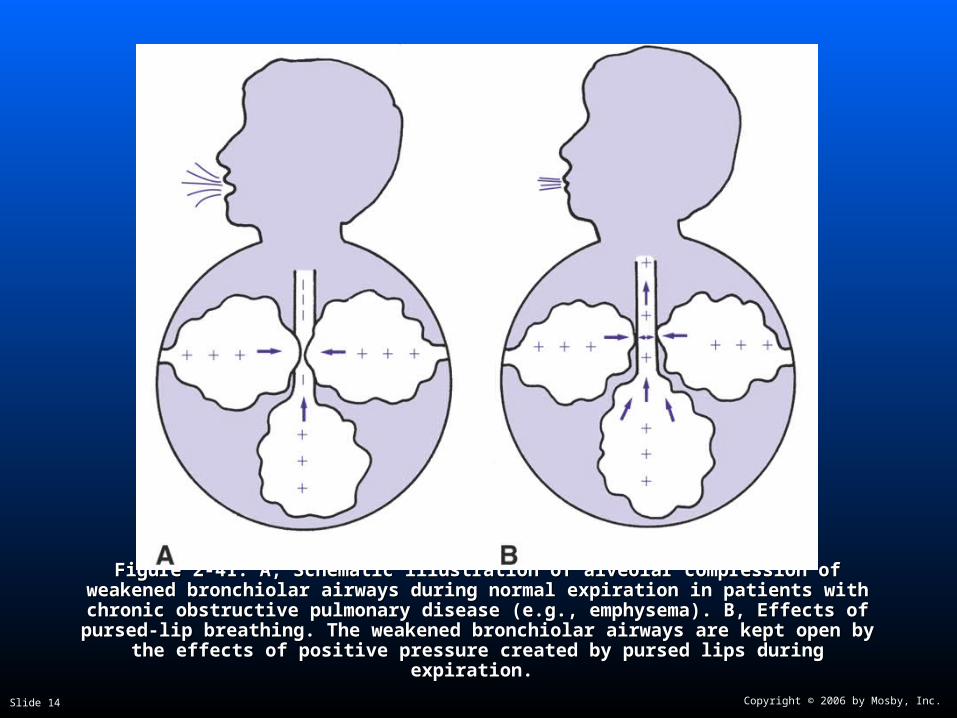
Figure 2-41. Figure 2-41. A, Schematic illustration of alveolar compression of weakened bronchiolar A, Schematic illustration of alveolar compression of weakened bronchiolar airways during normal expiration in patients with chronic obstructive pulmonary disease airways during normal expiration in patients with chronic obstructive pulmonary disease (e.g., emphysema). B, Effects of pursed-lip breathing. The weakened bronchiolar airways (e.g., emphysema). B, Effects of pursed-lip breathing. The weakened bronchiolar airways

are kept open by the effects of positive pressure created by pursed lips during expiration.are kept open by the effects of positive pressure created by pursed lips during expiration.

Copyright © 2006 by Mosby, Inc.Slide 15

Digital Clubbing

Figure 2-46. Digital clubbing.Figure 2-46. Digital clubbing.

Copyright © 2006 by Mosby, Inc.Slide 16

Clinical Data Obtained at the Clinical Data Obtained at the Patient’s BedsidePatient’s Bedside

Peripheral edema and venous distentionPeripheral edema and venous distention Distended neck veinsDistended neck veins

Pitting edemaPitting edema

Enlarged and tender liverEnlarged and tender liver

Copyright © 2006 by Mosby, Inc.Slide 17

DistendedDistendedNeck VeinsNeck Veins

Figure 2-48. Distended neck veins (Figure 2-48. Distended neck veins (arrowsarrows).).

Copyright © 2006 by Mosby, Inc.Slide 18

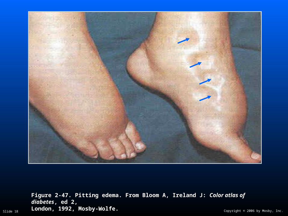
Figure 2-47. Pitting edema. From Bloom A, Ireland J: Figure 2-47. Pitting edema. From Bloom A, Ireland J: Color atlas of diabetesColor atlas of diabetes, ed 2,, ed 2,London, 1992, Mosby-Wolfe.London, 1992, Mosby-Wolfe.

Copyright © 2006 by Mosby, Inc.Slide 19

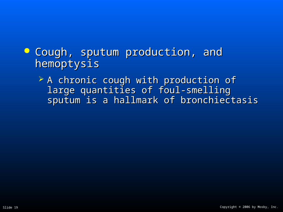
Cough, sputum production, and hemoptysisCough, sputum production, and hemoptysis A chronic cough with production of large quantities A chronic cough with production of large quantities

of foul-smelling sputum is a hallmark of of foul-smelling sputum is a hallmark of bronchiectasisbronchiectasis

Copyright © 2006 by Mosby, Inc.Slide 20

Clinical Data Obtained at the Clinical Data Obtained at the Patient’s BedsidePatient’s Bedside

Chest assessment findings (primarily obstructive)Chest assessment findings (primarily obstructive)

Decreased tactile and vocal fremitusDecreased tactile and vocal fremitus

Hyperresonant percussion noteHyperresonant percussion note

Diminished breath soundsDiminished breath sounds

Rhonchi and WheezingRhonchi and Wheezing

Copyright © 2006 by Mosby, Inc.Slide 21

Clinical Data Obtained at the Clinical Data Obtained at the Patient’s BedsidePatient’s Bedside

Chest assessment findings (primarily restrictive)Chest assessment findings (primarily restrictive)

Increased tactile and vocal fremitusIncreased tactile and vocal fremitus

Bronchial breath soundsBronchial breath sounds

CracklesCrackles

Whispered pectoriloquyWhispered pectoriloquy

Dull percussion noteDull percussion note

Copyright © 2006 by Mosby, Inc.Slide 22

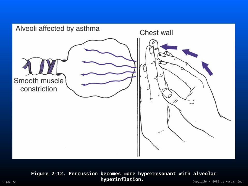
Figure 2-12. Figure 2-12. Percussion becomes more hyperresonant with alveolar hyperinflation.Percussion becomes more hyperresonant with alveolar hyperinflation.

Copyright © 2006 by Mosby, Inc.Slide 23

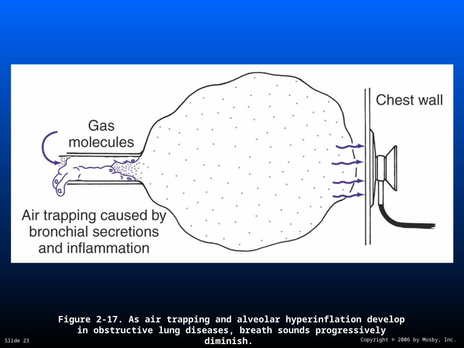
Figure 2-17. Figure 2-17. As air trapping and alveolar hyperinflation develop in obstructive As air trapping and alveolar hyperinflation develop in obstructive lung diseases, breath sounds progressively diminish.lung diseases, breath sounds progressively diminish.

Copyright © 2006 by Mosby, Inc.Slide 24

Clinical Data Obtained from Clinical Data Obtained from Laboratory Tests and Special Laboratory Tests and Special

ProceduresProcedures

Copyright © 2006 by Mosby, Inc.Slide 25

Pulmonary Function Study: Pulmonary Function Study: Expiratory Maneuver FindingsExpiratory Maneuver Findings

Primarily ObstructivePrimarily Obstructive

FVC FEVFVC FEVTT FEF FEF25%-75%25%-75% FEF FEF200-1200200-1200

PEFRPEFR MVV FEFMVV FEF50% 50% FEVFEV1%1%

Copyright © 2006 by Mosby, Inc.Slide 26

Pulmonary Function Study: Pulmonary Function Study: Lung Volume and Capacity FindingsLung Volume and Capacity Findings

Primarily ObstructivePrimarily Obstructive

VVTT RV FRC TLC RV FRC TLC

N or N or N or N or

VCVC IC ERV RV/TLC ratio IC ERV RV/TLC ratio

N or N or

N or N or

Copyright © 2006 by Mosby, Inc.Slide 27

Pulmonary Function Study: Pulmonary Function Study: Expiratory Maneuver Findings Expiratory Maneuver Findings

Primarily RestrictivePrimarily Restrictive

FVC FEVFVC FEVTT FEF FEF25%-75%25%-75% FEF FEF200-1200200-1200

N or N or N or N or N N

PEFRPEFR MVV FEFMVV FEF50% 50% FEVFEV1%1%

N N or N N or N N N or N or

Copyright © 2006 by Mosby, Inc.Slide 28

Pulmonary Function Study: Pulmonary Function Study: Lung Volume and Capacity FindingsLung Volume and Capacity Findings

Primarily RestrictivePrimarily Restrictive

VVTT RV FRC TLC RV FRC TLC

N or N or

VCVC IC ERV RV/TLC ratio IC ERV RV/TLC ratio

N N

Copyright © 2006 by Mosby, Inc.Slide 29

Arterial Blood GasesArterial Blood Gases

Mild to Moderate BronchiectasisMild to Moderate Bronchiectasis

Acute alveolar hyperventilation with Acute alveolar hyperventilation with hypoxemiahypoxemia

pH PaCO2 HCO3- PaO2

(Slightly)

Copyright © 2006 by Mosby, Inc.Slide 30

Time and Progression of Disease Time and Progression of Disease

100100

5050

3030

8080

00

PaCO2

1010

2020

4040

Alveolar HyperventilationAlveolar Hyperventilation

6060

7070

9090 Point at which PaO2 declines enough to stimulate peripheral oxygen receptors

Point at which PaO2 declines enough to stimulate peripheral oxygen receptors

PaO2

Disease OnsetDisease OnsetP

aO2

or

PaC

O2

PaO

2 o

r P

aCO

2

Figure 4-2. PaO2 and PaC02 trends during acute alveolar hyperventilation.

Copyright © 2006 by Mosby, Inc.Slide 31

Arterial Blood GasesArterial Blood Gases

Severe BronchiectasisSevere Bronchiectasis

Chronic ventilatory failure with hypoxemiaChronic ventilatory failure with hypoxemia

pH PaCO2 HCO3- PaO2

Normal (Significantly)

Copyright © 2006 by Mosby, Inc.Slide 32

Time and Progression of DiseaseTime and Progression of Disease

100100

5050

3030

80

0

PaO2

1010

2020

4040

Alveolar HyperventilationAlveolar Hyperventilation

6060

7070

9090Point at which PaO2 declines enough to stimulate peripheral oxygen receptors

Point at which PaO2 declines enough to stimulate peripheral oxygen receptors

PaCO 2

Chronic Ventilatory Failure Chronic Ventilatory FailureDisease OnsetDisease Onset

Point at which disease becomes severe and patient begins to become fatigued

Point at which disease becomes severe and patient begins to become fatigued

Pa0

2 o

r P

aC0 2

Pa0

2 o

r P

aC0 2

Figure 4-7. PaO2 and PaCO2 trends during acute or chronic ventilatory failure.

Copyright © 2006 by Mosby, Inc.Slide 33

Acute Ventilatory Changes on Acute Ventilatory Changes on Chronic Ventilatory FailureChronic Ventilatory Failure

Acute alveolar hyperventilation on chronic Acute alveolar hyperventilation on chronic ventilatory failureventilatory failure

Acute ventilatory failure on chronic ventilatory Acute ventilatory failure on chronic ventilatory failure failure

Copyright © 2006 by Mosby, Inc.Slide 34

Oxygenation IndicesOxygenation Indices

QQSS/Q/QTT D DOO22 V VOO22 C(a-v) C(a-v)OO22

Normal NormalNormal Normal

OO22ER SvER SvOO2 2

Copyright © 2006 by Mosby, Inc.Slide 35

Hemodynamic Indices Hemodynamic Indices (Severe Chronic Bronchiectasis)(Severe Chronic Bronchiectasis)

CVP CVP RAPRAP PAPA PCWPPCWP

NormalNormal

COCO SVSV SVISVI CICI

NormalNormal NormalNormal NormalNormal Normal Normal

RVSWIRVSWI LVSWILVSWI PVRPVR SVRSVR

NormalNormal NormalNormal

Copyright © 2006 by Mosby, Inc.Slide 36

Abnormal Laboratory TestsAbnormal Laboratory Testsand Proceduresand Procedures

Hematology Hematology (Increased hematocrit and hemoglobin) (Increased hematocrit and hemoglobin)

Sputum examinationSputum examination Streptococcus pneumoniaeStreptococcus pneumoniae

Haemophilus influenzaeHaemophilus influenzae

Pseudomonas aeruginosaPseudomonas aeruginosa

Anaerobic organismsAnaerobic organisms

Copyright © 2006 by Mosby, Inc.Slide 37

Radiologic FindingsRadiologic Findings

Chest radiographChest radiograph Translucent (dark) lung fieldsTranslucent (dark) lung fields

Depressed or flattened diaphragmDepressed or flattened diaphragm

Long and narrow heartLong and narrow heart

Enlarged heartEnlarged heart

BronchogramBronchogram

CT scanCT scan

Copyright © 2006 by Mosby, Inc.Slide 38

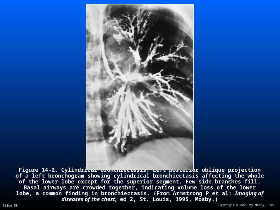
Figure 14-2. Figure 14-2. Cylindrical bronchiectasis. Left posterior oblique projection of a left bronchogram Cylindrical bronchiectasis. Left posterior oblique projection of a left bronchogram showing cylindrical bronchiectasis affecting the whole of the lower lobe except for the superior showing cylindrical bronchiectasis affecting the whole of the lower lobe except for the superior segment. Few side branches fill. Basal airways are crowded together, indicating volume loss of segment. Few side branches fill. Basal airways are crowded together, indicating volume loss of

the lower lobe, a common finding in bronchiectasis. (From Armstrong P et al: the lower lobe, a common finding in bronchiectasis. (From Armstrong P et al: Imaging of Imaging of diseases of the chest,diseases of the chest, ed 2, St. Louis, 1995, Mosby.) ed 2, St. Louis, 1995, Mosby.)

Copyright © 2006 by Mosby, Inc.Slide 39

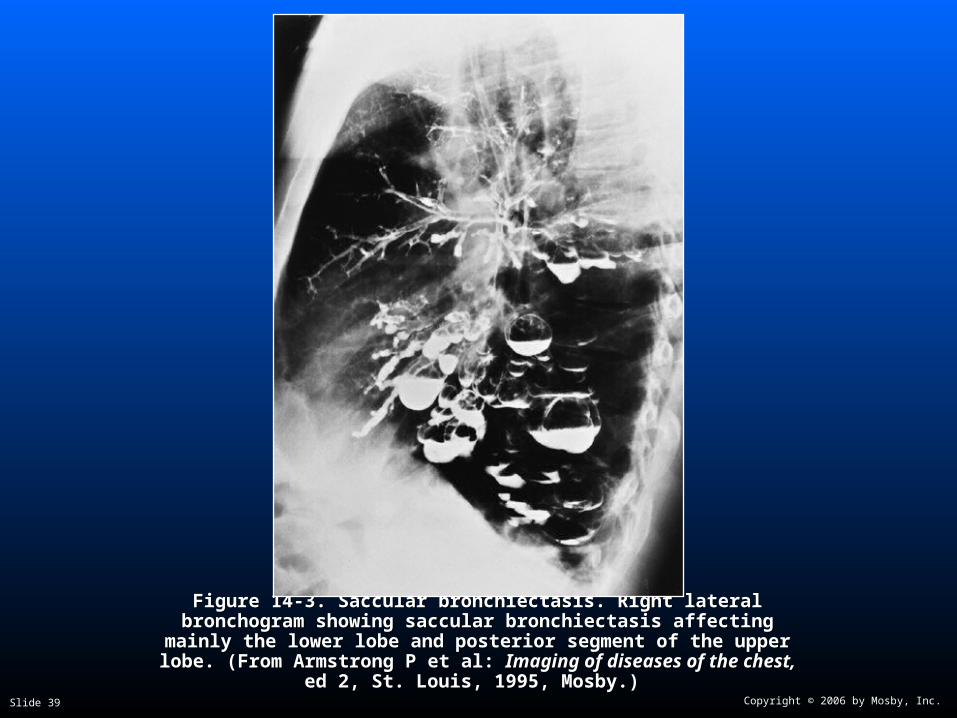
Figure 14-3. Figure 14-3. Saccular bronchiectasis. Right lateral bronchogram showing Saccular bronchiectasis. Right lateral bronchogram showing saccular bronchiectasis affecting mainly the lower lobe and posterior saccular bronchiectasis affecting mainly the lower lobe and posterior

segment of the upper lobe. (From Armstrong P et al: segment of the upper lobe. (From Armstrong P et al: Imaging of diseases Imaging of diseases of the chest,of the chest, ed 2, St. Louis, 1995, Mosby.) ed 2, St. Louis, 1995, Mosby.)

Copyright © 2006 by Mosby, Inc.Slide 40

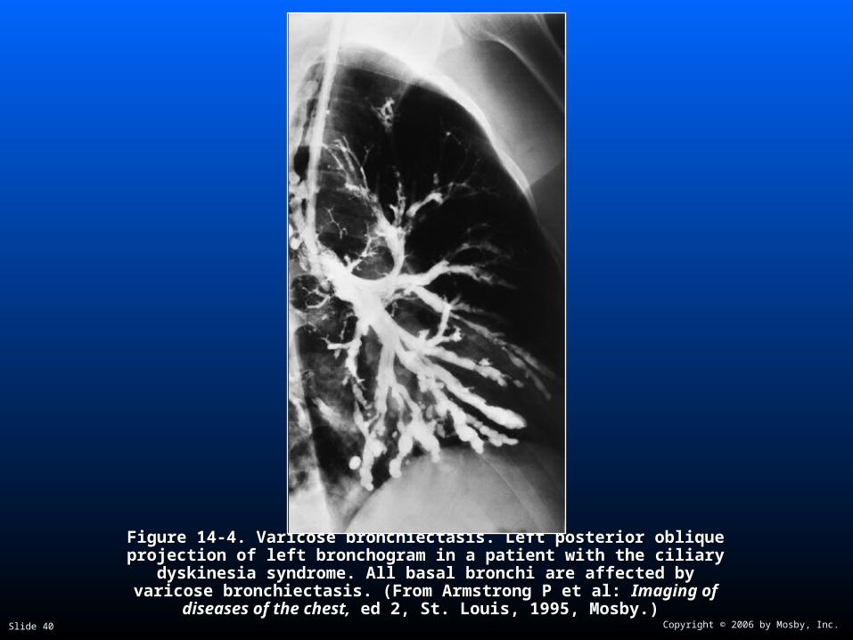
Figure 14-4. Figure 14-4. Varicose bronchiectasis. Left posterior oblique projection of left Varicose bronchiectasis. Left posterior oblique projection of left bronchogram in a patient with the ciliary dyskinesia syndrome. All basal bronchogram in a patient with the ciliary dyskinesia syndrome. All basal

bronchi are affected by varicose bronchiectasis. (From Armstrong P et al: bronchi are affected by varicose bronchiectasis. (From Armstrong P et al: Imaging of diseases of the chest,Imaging of diseases of the chest, ed 2, St. Louis, 1995, Mosby.) ed 2, St. Louis, 1995, Mosby.)

Copyright © 2006 by Mosby, Inc.Slide 41

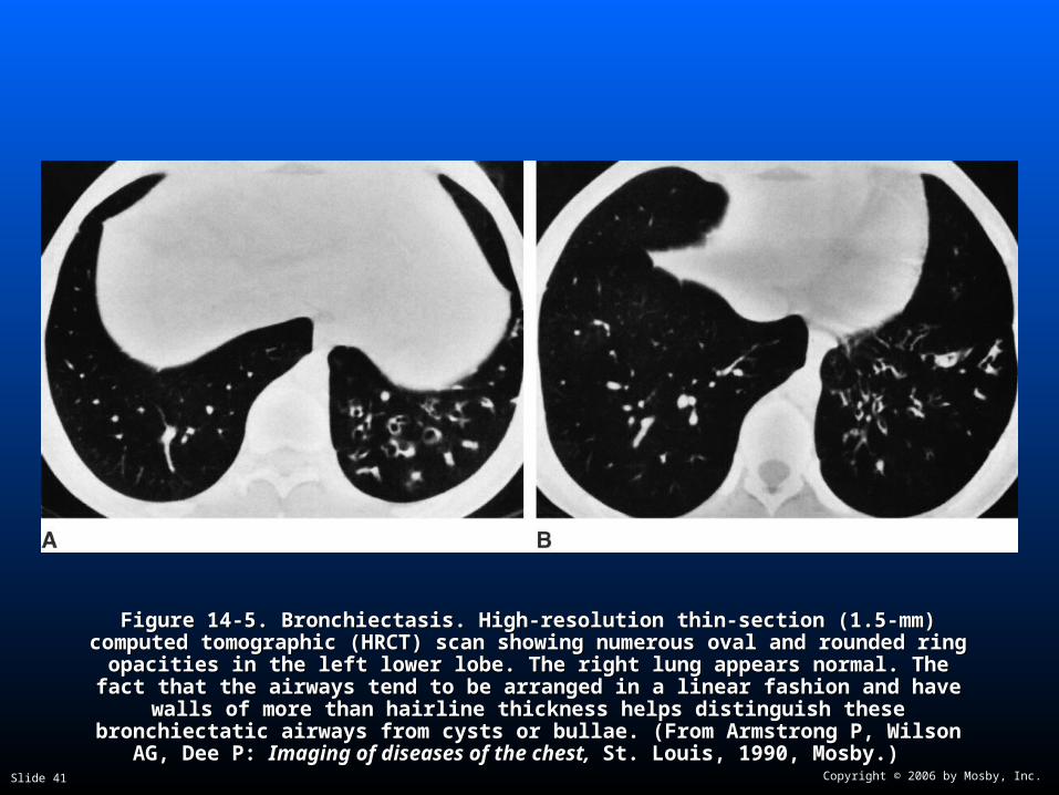
Figure 14-5. Figure 14-5. Bronchiectasis. High-resolution thin-section (1.5-mm) computed tomographic Bronchiectasis. High-resolution thin-section (1.5-mm) computed tomographic (HRCT) scan showing numerous oval and rounded ring opacities in the left lower lobe. (HRCT) scan showing numerous oval and rounded ring opacities in the left lower lobe. The right lung appears normal. The fact that the airways tend to be arranged in a linear The right lung appears normal. The fact that the airways tend to be arranged in a linear

fashion and have walls of more than hairline thickness helps distinguish these fashion and have walls of more than hairline thickness helps distinguish these bronchiectatic airways from cysts or bullae. (From Armstrong P, Wilson AG, Dee P: bronchiectatic airways from cysts or bullae. (From Armstrong P, Wilson AG, Dee P:

Imaging of diseases of the chest,Imaging of diseases of the chest, St. Louis, 1990, Mosby.) St. Louis, 1990, Mosby.)

Copyright © 2006 by Mosby, Inc.Slide 42

General Management of General Management of BronchiectasisBronchiectasis

General treatment includes:General treatment includes:

Controlling pulmonary infectionsControlling pulmonary infections

Controlling airway secretionsControlling airway secretions

Preventing complicationsPreventing complications

Copyright © 2006 by Mosby, Inc.Slide 43

General Management of General Management of BronchiectasisBronchiectasis

Respiratory care treatment protocolsRespiratory care treatment protocols

Oxygen therapy protocolOxygen therapy protocol

Bronchopulmonary hygiene therapy protocolBronchopulmonary hygiene therapy protocol

Hyperinflation therapy protocolHyperinflation therapy protocol

Aerosolized medication protocolAerosolized medication protocol

Mechanical ventilation protocolMechanical ventilation protocol

Copyright © 2006 by Mosby, Inc.Slide 44

General Management of General Management of BronchiectasisBronchiectasis

Other medications commonly prescribedOther medications commonly prescribedby the physicianby the physician

XanthinesXanthines

ExpectorantsExpectorants

AntibioticsAntibiotics

Copyright © 2006 by Mosby, Inc.Slide 45

Classroom DiscussionClassroom DiscussionCase Study: BronchiectasisCase Study: Bronchiectasis


Recommended