+ All Categories
Home > Documents > Copyright 2010, John Wiley & Sons, Inc. Bone Function Support Protection Assist in movements Mineral...

Copyright 2010, John Wiley & Sons, Inc. Bone Function Support Protection Assist in movements Mineral...

Date post: 25-Dec-2015
Category:
Upload: loren-golden
View: 214 times
Download: 0 times
Share this document with a friend
Popular Tags:
71
Copyright 2010, John Wiley & Sons, Inc. Bone Function Support Protection Assist in movements Mineral homeostasis Blood cell production Hemopoiesis - red bone marrow Triglyceride storage
Transcript

Copyright 2010, John Wiley & Sons, Inc.

Bone Function Support Protection Assist in movements Mineral homeostasis Blood cell production

Hemopoiesis - red bone marrow Triglyceride storage

Copyright 2010, John Wiley & Sons, Inc.

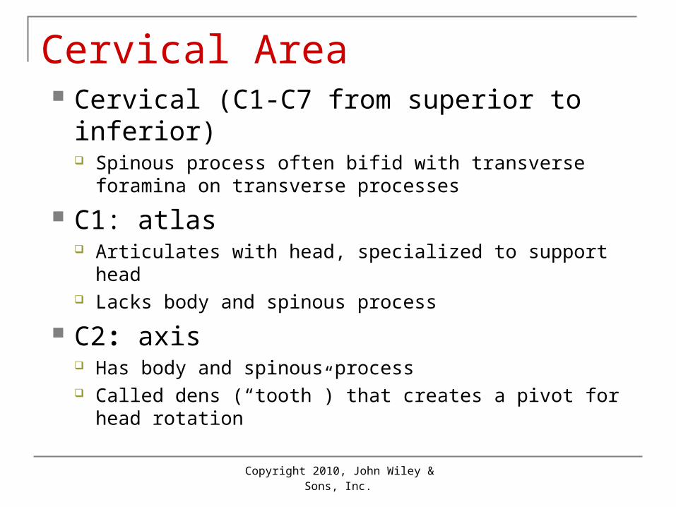
Types of Bones Long bones: longer than wide

Such as thigh, leg, arm, forearm, fingers and toes

Short bones: almost cube shaped Most wrist and ankle bones

Flat bones: thin and extensive surface Such as cranial bones sternum, ribs and scapulas

Irregular bones: Such as vertebrae and some facial bones

• Sesamoid Bones • develop within a tendon

Copyright 2010, John Wiley & Sons, Inc.

Macroscopic Structure Parts of a long bone

Diaphysis: shaft of long bone; made up mostly of compact bone

Epiphysis: broad end of long bone; mostly spongy bone

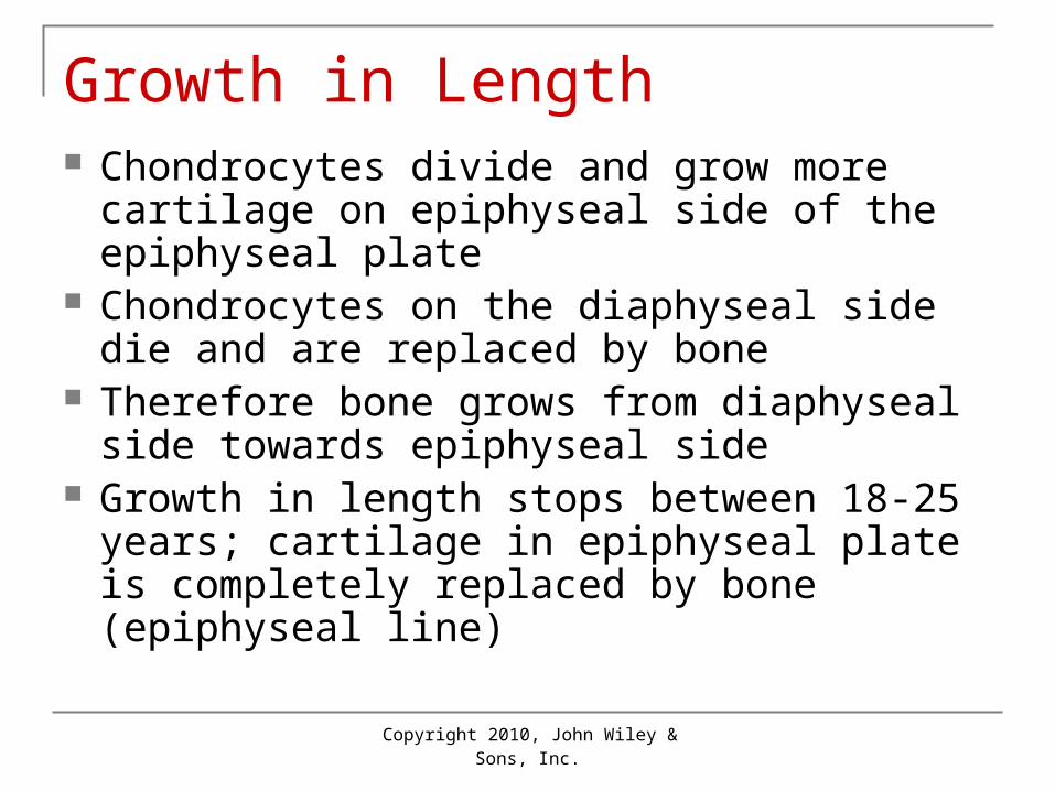
Metaphysis: growth area between diaphysis and epiphysis

Epiphyseal line = remnant of epiphyseal disk/plate Cartilage at the junction of the diaphysis and epiphyses

(growth plate)

Copyright 2010, John Wiley & Sons, Inc.

Parts of a Long Bone• Periosteum: fibrous covering over most of

bone• Endosteum: membrane lining medullary cavity

Contains layer of osteoblasts (bone-forming cells) & osteoclasts (bone-destroying cells)

• Medullary cavity: (yellow marrow) with fat and blood cells

• Nutrient Foramen = perforating canal allowing blood vessels to enter and leave bone

• Articular cartilage = pad of hyaline cartilage on the epiphyses where long bones articulate or join

Copyright 2010, John Wiley & Sons, Inc.

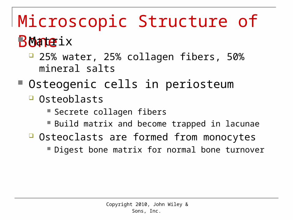
Microscopic Structure of Bone Matrix

25% water, 25% collagen fibers, 50% mineral salts Osteogenic cells in periosteum

Osteoblasts Secrete collagen fibers Build matrix and become trapped in lacunae

Osteoclasts are formed from monocytes Digest bone matrix for normal bone turnover

Copyright 2010, John Wiley & Sons, Inc.

Compact Bone Structure Arranged in osteons (haversian systems)

Cylinders running parallel to long axis of bone

Central canal through center of osteon Contains blood vessels, nerves, lymphatics

Concentric lamellae: layers of matrix Lacunae:

Contain osteocytes (bone cells)

Copyright 2010, John Wiley & Sons, Inc.

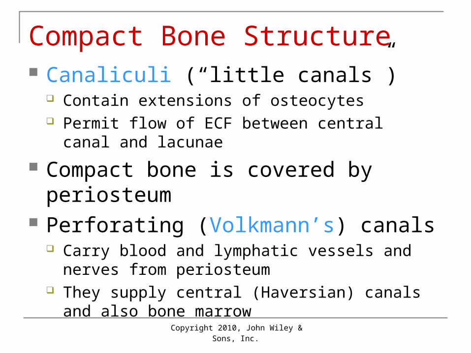
Compact Bone Structure Canaliculi (“little canals”)

Contain extensions of osteocytes Permit flow of ECF between central canal and

lacunae

Compact bone is covered by periosteum Perforating (Volkmann’s) canals

Carry blood and lymphatic vessels and nerves from periosteum

They supply central (Haversian) canals and also bone marrow

Copyright 2010, John Wiley & Sons, Inc.

Spongy Bone Not arranged in osteons Irregular latticework of trabeculae

These contain lacunae with osteocytes and canaliculi

Spaces between trabeculae may contain red bone marrow

Spongy bone is lighter than compact bone, so reduces weight of skeleton

Copyright 2010, John Wiley & Sons, Inc.

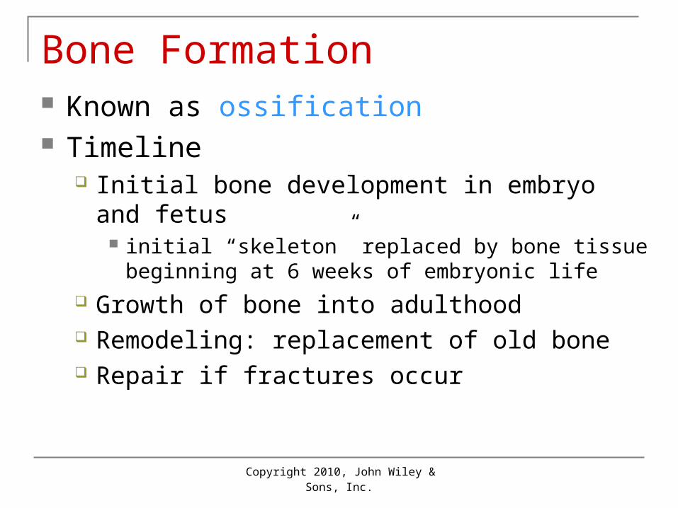
Bone Formation Known as ossification Timeline

Initial bone development in embryo and fetus initial “skeleton” replaced by bone tissue beginning at 6

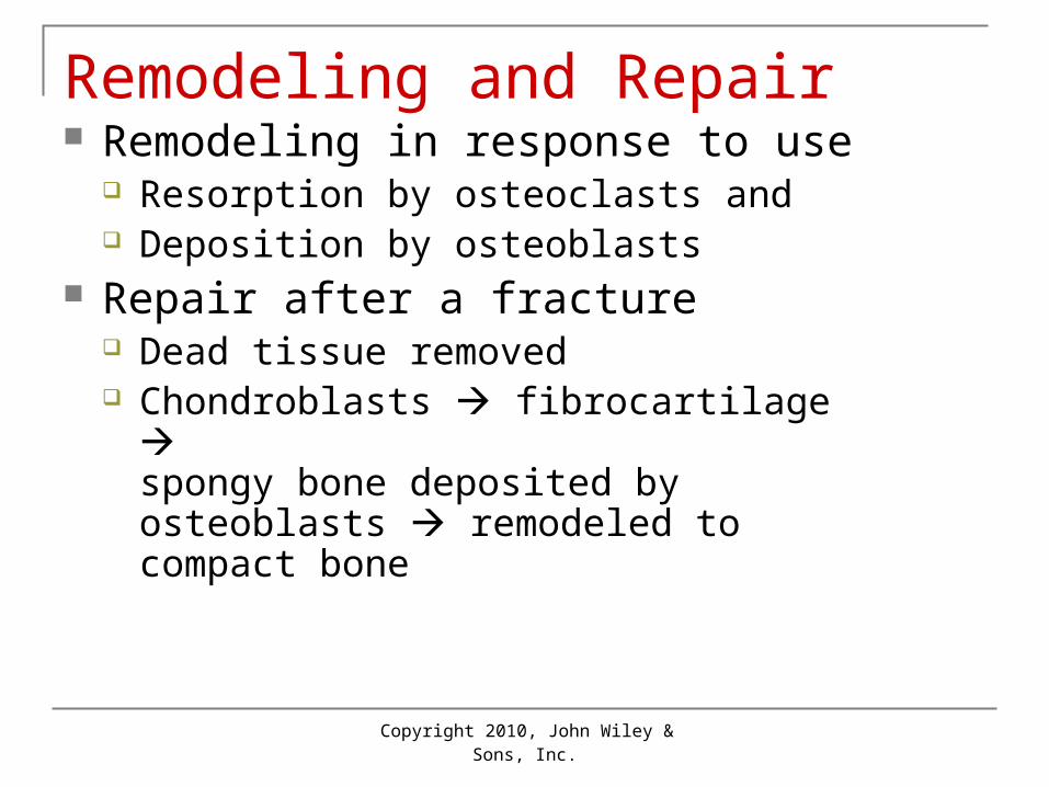
weeks of embryonic life Growth of bone into adulthood Remodeling: replacement of old bone Repair if fractures occur

Copyright 2010, John Wiley & Sons, Inc.

Bone Formation Two different methods of ossification each

result in similar bone tissue1. Intramembranous: bone forms in sheets

Only a few bones form by this process: flat bones of the skull, lower jawbone (mandible), and part of clavicle (collarbone)

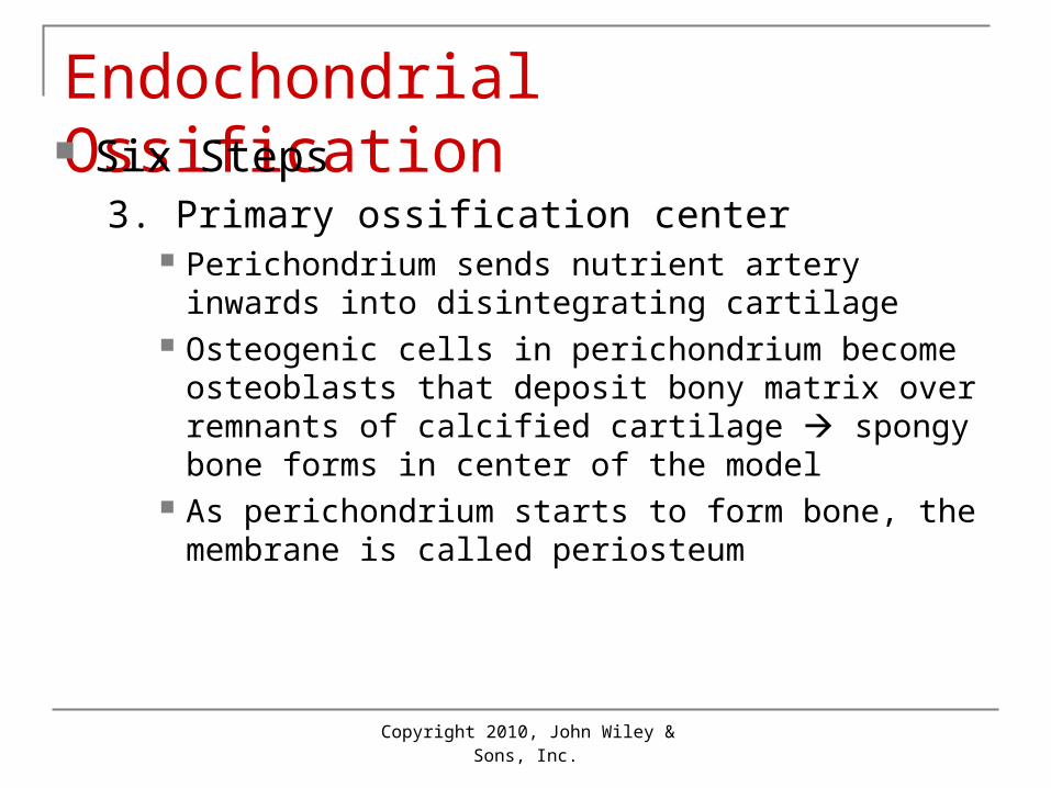
2. Endochondrial: forms hyaline cartilage which then develops into bone All other bones form by this process

Copyright 2010, John Wiley & Sons, Inc.

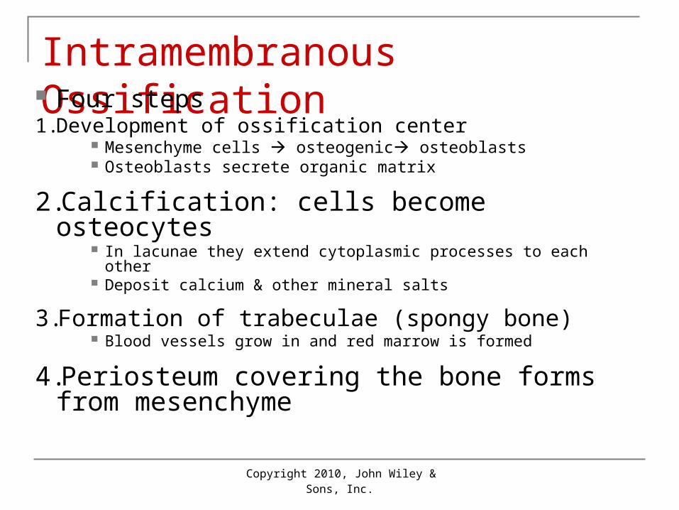
Intramembranous Ossification Four steps1. Development of ossification center

Mesenchyme cells osteogenic osteoblasts Osteoblasts secrete organic matrix

2.Calcification: cells become osteocytes In lacunae they extend cytoplasmic processes to each other Deposit calcium & other mineral salts

3.Formation of trabeculae (spongy bone) Blood vessels grow in and red marrow is formed

4.Periosteum covering the bone forms from mesenchyme

Copyright 2010, John Wiley & Sons, Inc.

Endochondrial Ossification Six Steps

1. Formation of cartilage model of the “bone” As mesenchyme cells develop into chondroblasts

2. Growth of cartilage model Cartilage “bone” grows as chondroblasts secrete

cartilage matrix Chondrocytes increase in size, matrix around them

calcifies Chondrocytes die as they are cut off from nutrients,

leaving small spaces (lacunae)

Copyright 2010, John Wiley & Sons, Inc.

Endochondrial Ossification Six Steps

3. Primary ossification center Perichondrium sends nutrient artery inwards into

disintegrating cartilage Osteogenic cells in perichondrium become osteoblasts

that deposit bony matrix over remnants of calcified cartilage spongy bone forms in center of the model

As perichondrium starts to form bone, the membrane is called periosteum

Copyright 2010, John Wiley & Sons, Inc.

Endochondrial Ossification Six Steps

4. Medullary (marrow) cavity Spongy bone in center of the model grows towards

ends of model Octeoclasts break down some of new spongy bone

forming a cavity (marrow) through most of diaphysis Most of the wall of the diaphysis is replaced by a collar

of compact bone

Copyright 2010, John Wiley & Sons, Inc.

Endochondrial Ossification Six Steps

5. Secondary ossification center Similar to step 3 except that nutrient arteries enter ends

(epiphyses) of bones and osteoblasts deposit bony matrix spongy bone forms in epiphyses from center outwards

Occurs about time of birth

6. Articular cartilage and epiphyseal cartilage Ends of epiphyses becomes articular cartilage Epiphyseal (growth) plate of cartilage remains between

epiphysis and diaphysis until bone growth ceases

Copyright 2010, John Wiley & Sons, Inc.

Growth in Length Chondrocytes divide and grow more cartilage

on epiphyseal side of the epiphyseal plate Chondrocytes on the diaphyseal side die and

are replaced by bone Therefore bone grows from diaphyseal side

towards epiphyseal side Growth in length stops between 18-25 years;

cartilage in epiphyseal plate is completely replaced by bone (epiphyseal line)

Copyright 2010, John Wiley & Sons, Inc.

Growth in Thickness As bones grow in length, they must

also grow in thickness (width) Perichondrial osteoblasts lay down

additional lamellae of compact bone Simultaneously, osteoclasts in the

endosteum destroy interior bone to increase width of the marrow

Copyright 2010, John Wiley & Sons, Inc.

Remodeling and Repair Remodeling in response to use

Resorption by osteoclasts and Deposition by osteoblasts

Repair after a fracture Dead tissue removed Chondroblasts fibrocartilage

spongy bone deposited by osteoblasts remodeled to compact bone

Copyright 2010, John Wiley & Sons, Inc.

Types of Fractures Partial: incomplete break (crack) Complete: bone broken into two or more

pieces Closed (simple): not through skin Open (compound): broken ends break skin

Copyright 2010, John Wiley & Sons, Inc.

Types of Fractures• green stick• fissured• comminuted• transverse• oblique• spiral

7-62

Copyright 2010, John Wiley & Sons, Inc.

Factors Affecting Growth Adequate minerals (Ca, P, Mg) Vitamins A, C, D Hormones

Before puberty: hGH + insulin-like growth factors Thyroid hormone and insulin also required Sex hormones contribute to adolescent growth

spurt Weight-bearing activity

Copyright 2010, John Wiley & Sons, Inc.

• Deficiency of Vitamin A – retards bone development• Deficiency of Vitamin C – results in fragile bones, scurvy• Deficiency of Vitamin D – rickets (children), osteomalacia (adults)• Insufficient Growth Hormone – dwarfism• Excessive Growth Hormone – gigantism, acromegaly • Insufficient Thyroid Hormone – delays bone growth•Physical Stress – stimulates bone growth

7-11

Copyright 2010, John Wiley & Sons, Inc.

Calcium Homeostasis Blood levels of Ca2+ controlled Negative feedback loops Parathyroid hormone (PTH) increases

osteoclast activity + decreases loss of Ca2+ in urine

Calcitonin decreases osteoclast activity

Copyright 2010, John Wiley & Sons, Inc.

Exercise & Bone Tissue Bone strengthened in response to use Bone reabsorbed during disuse; examples:

During prolonged bed rest Fracture with cast/immobilizer Astronauts without gravity

Copyright 2010, John Wiley & Sons, Inc.

Divisions of Skeletal System Two divisions: axial and appendicular

Axial: bones around body axis Examples: skull bones, hyoid, ribs, sternum, vertebrae

Appendicular: bones of upper and lower limbs plus shoulder and hip bones that connect them Examples: collar bone (clavicle), arm (humerus),

forearm (radius and ulna), thigh bone (femur)

Copyright 2010, John Wiley & Sons, Inc.

Skull & Hyoid Bone Eight Cranial bones

Frontal, 2 parietal, 2 temporal, occipital, sphenoid, and ethmoid

Fourteen Facial bones 2 nasal, 2 maxilla, 2 zygomatic, 2 lacrimal 2 palatine, 2 inferior nasal conchae, 1 mandible,

1 vomer

Copyright 2010, John Wiley & Sons, Inc.

Skull

Frontal (1)• forehead•frontal sinuses• supraorbital foramen• coronal suture

7-17

Copyright 2010, John Wiley & Sons, Inc.

Skull

Parietal (2)• side walls of cranium• roof of cranium• sagittal suture

7-18

Copyright 2010, John Wiley & Sons, Inc.

SkullTemporal (2)•squamosal suture• external acoustic meatus• mandibular fossa• mastoid process• styloid process• zygomatic process

7-19

Copyright 2010, John Wiley & Sons, Inc.

Skull

Occipital (1)• back of skull• foramen magnum• occipital condyles• lambdoidal suture

7-20

Copyright 2010, John Wiley & Sons, Inc.

Skull

Sphenoid (1)• sella turcica• sphenoidal sinuses

7-21

Copyright 2010, John Wiley & Sons, Inc.

Skull

Ethmoid (1)•cribiform plates• perpendicular plate• superior and middle nasal conchae• ethmoidal sinuses• crista gallis

7-22

Copyright 2010, John Wiley & Sons, Inc.

Facial Skeleton

Maxillary (2)• upper jaw•alveolar processes• maxillary sinuses• palatine process

7-23

Copyright 2010, John Wiley & Sons, Inc.

Facial Skeleton

Palatine (2)• posterior roof of mouth

7-24

Copyright 2010, John Wiley & Sons, Inc.

Facial Skeleton

Zygomatic (2) • prominences of cheeks• temporal process

7-25

Copyright 2010, John Wiley & Sons, Inc.

Facial Skeleton

Lacrimal (2)• medial walls of orbits

Nasal (2)• bridge of nose

7-26

Copyright 2010, John Wiley & Sons, Inc.

Facial Skeleton

Vomer (1)• inferior portion of nasal septum

7-27

Copyright 2010, John Wiley & Sons, Inc.

Facial Skeleton

Inferior Nasal Conchae (2)• extend from lateral walls of nasal cavity

7-28

Copyright 2010, John Wiley & Sons, Inc.

Facial Skeleton

Mandible (1)• lower jaw• body• ramus• mandibular condyle• coronoid process• alveolar process• mandibular foramen• mental foramen

7-29

Copyright 2010, John Wiley & Sons, Inc.

hyoid

Copyright 2010, John Wiley & Sons, Inc.

Unique Features of Skull Sutures: immovable joint between skull

bones Coronal, sagittal, lambdoidal, squamous

Paranasal sinuses: cavities Located in bones near nasal cavity

Fontanels: soft spot in fetal skull Allow deformation at birth Calcify to form sutures

Copyright 2010, John Wiley & Sons, Inc.

Vertebrae Functions

Encloses spinal cord Supports head Point of attachment for muscles of back, ribs

and pelvic girdle Regions (from superior to inferior)

7 cervical 12 thoracic 5 lumbar 1 sacrum and 1 coccyx

Copyright 2010, John Wiley & Sons, Inc.

Normal Curves in Column Four normal curves

Cervical and lumbar curves are convex (bulge anteriorly)

Thoracic and sacral curves are concave (bulge posteriorly)

Curves increase strength, help in balance and absorb shocks

Copyright 2010, John Wiley & Sons, Inc.

Structure of Vertebra Body: disc-shaped anterior portion Vertebral arch: posteriorly back from body

With the body, creates a hole called vertebral foramen Seven processes from this arch

Transverse process extending laterally on each side Spinous process extending dorsally Two each of superior and inferior articular processes

that form joints with vertebrae

Copyright 2010, John Wiley & Sons, Inc.

Vertebral Column

•rib facets•intervertebral discs• intervertebral foramina

7-32

Copyright 2010, John Wiley & Sons, Inc.

Cervical Area Cervical (C1-C7 from superior to inferior)

Spinous process often bifid with transverse foramina on transverse processes

C1: atlas Articulates with head, specialized to support head Lacks body and spinous process

C2: axis Has body and spinous process Called dens (“tooth”) that creates a pivot for head

rotation

Copyright 2010, John Wiley & Sons, Inc.

Other Vertebrae Thoracic (T1-T12 )

Larger than cervical Have facets for articulations with ribs

Lumbar (L1-L5) Largest and strongest; spinous processes short and

thick Sacrum (S1-S5 fused into one unit)

Foundation for pelvic girdle Contain sacral foramina for nerves and blood vessels

Coccyx: 4 coccygeal vertebrae fused into 1

Copyright 2010, John Wiley & Sons, Inc.

Thorax Thoracic cage: sternum, costal cartilages,

ribs and bodies of T1-T12 Sternum: 3 portions fused by about age 25

Manubrium, body, xiphoid process True ribs #1-7: articulate with sternum by

costal cartilages False ribs #8-12: articulate with coastal

cartilage of true ribs 2 - Floating Ribs (no attachment)

Copyright 2010, John Wiley & Sons, Inc.

Rib Structure• Shaft• Head – articulates with vertebrae• Tubercle • Costal cartilage

7-40

Copyright 2010, John Wiley & Sons, Inc.

Pectoral Girdle Function: attach bones of upper limbs to axial

skeleton Clavicles and scapulas: bilateral

Copyright 2010, John Wiley & Sons, Inc.

Clavicles

• articulate with manubrium• articulate with scapulae (acromion process)

7-43

Copyright 2010, John Wiley & Sons, Inc.

Scapula• spine• supraspinous fossa• infraspinous fossa

• acromion process• coracoid process• glenoid cavity

7-44

Copyright 2010, John Wiley & Sons, Inc.

Upper Limb Humerus: arm bone

Articulates with scapula (glenoid cavity) Articulates with radius and ulna at elbow

Ulna: medial bone Radius: lateral bone (thumb side)

Copyright 2010, John Wiley & Sons, Inc.

Humerus• head• greater tubercle• lesser tubercle• anatomical neck• surgical neck• deltoid tuberosity• capitulum• trochlea• coronoid fossa• olecranon fossa•Lateral epicondyle•Medial epicondyle

7-46

Copyright 2010, John Wiley & Sons, Inc.

Radius• lateral forearm bone• head• radial tuberosity• styloid process

7-47

Copyright 2010, John Wiley & Sons, Inc.

Ulna• medial forearm bone• trochlear notch• olecranon process• coronoid process• styloid process

7-48

Copyright 2010, John Wiley & Sons, Inc.

Wrist and Hand Carpals (wrist): 8 bones Metacarpals: 5 bones of palm of hand

Number 1-5 starting with thumb Phalanges: 14 bones of fingers

Each finger except thumb has proximal, middle and distal phalanges; thumb lacks middle phalanx

Copyright 2010, John Wiley & Sons, Inc.

Wrist and Hand• Carpals • trapezium• trapezoid• capitate• scaphoid• pisiform• triquetrum• hamate• lunate

7-49

Copyright 2010, John Wiley & Sons, Inc.

Pelvic (Hip) Girdle Pelvic girdle includes two hip (coxal) bones

Joined anteriorly at pubic symphysis + sacrum and coccyx False (greater) pelvis: broad region superior

to pelvic brim; contains abdominal organs True (lesser) pelvis: small region inferior to

pelvic brim; contains urinary bladder + internal reproductive organs

Copyright 2010, John Wiley & Sons, Inc.

Parts of Each Hip (Coxal) Bone 3 separate bones fuse by age 23 to form a

hip bone Ilium: largest and most superior Ischium: lower posterior part Pubis: lower anterior part

acetabulum - socket for head of femur

Copyright 2010, John Wiley & Sons, Inc.

Lower Limb Femur : largest bone in the body

Articulates with hip proximally and with the tibia and patella distally

Patella: kneecap in anterior of knee joint Tibia: shin bone

Large medial, weight-bearing bone of leg

Fibula: longest, thinnest bone in body Lateral to tibia and smaller Does not articulate with femur

Copyright 2010, John Wiley & Sons, Inc.

Femur

• head• fovea capitis• neck• greater trochanter• lesser trochanter• linea aspera• condyles (medial/lateral)• epicondyles (medial/lateral)

7-55

Copyright 2010, John Wiley & Sons, Inc.

Patella

• flat sesamoid bone located in a tendon

7-56

Copyright 2010, John Wiley & Sons, Inc.

Tibia• medial to fibula• condyles• tibial tuberosity• anterior crest• medial malleolus

7-57

Copyright 2010, John Wiley & Sons, Inc.

Fibula

Insert figure 7.54

• lateral to tibia• head• lateral malleolus• does not bear any body weight

7-58

Copyright 2010, John Wiley & Sons, Inc.

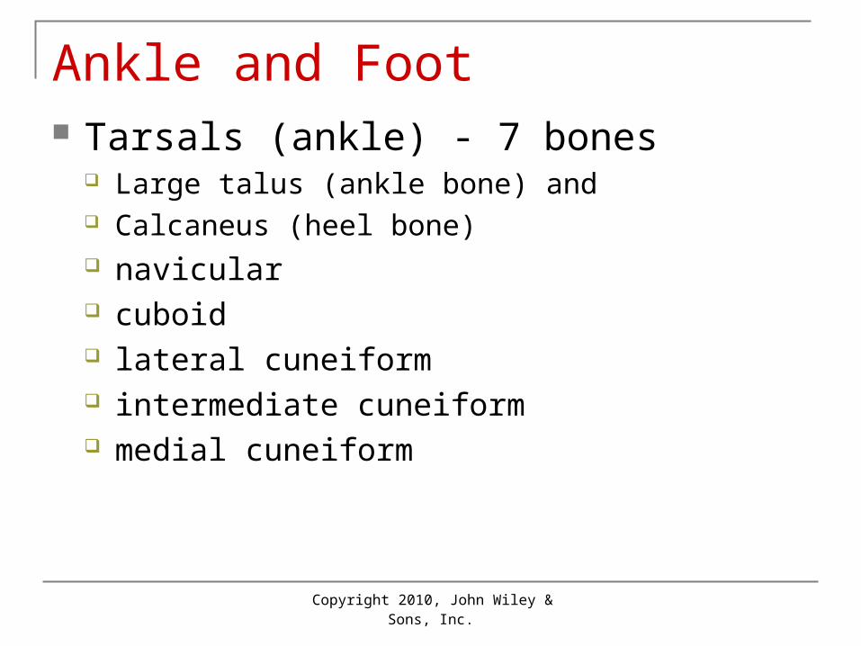
Ankle and Foot Tarsals (ankle) - 7 bones

Large talus (ankle bone) and Calcaneus (heel bone) navicular cuboid lateral cuneiform intermediate cuneiform medial cuneiform

Copyright 2010, John Wiley & Sons, Inc.

Ankle and FootMetatarsals (foot bones)

• Numbered 1 to 5 from medial to lateralPhalanges (toe bones)

• Big toe has proximal and distal phalanges while others have proximal, middle and distal phalanges.

7-59

Copyright 2010, John Wiley & Sons, Inc.

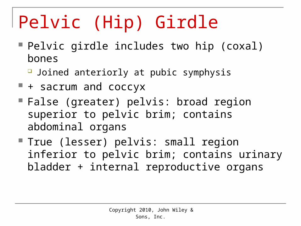
Male and Female Differences Males usually have heavier bones Related to muscle size and strength Female pelvis is wider and shallower than

male pelvis: allows for birth

Copyright 2010, John Wiley & Sons, Inc.

Aging and Skeletal System Birth through adolescence: more bone

formed than lost Young adults: gain and loss about equal As levels of sex steroids decline with age:

bone reabsorption > bone formation Bones become brittle and lose calcium

Copyright 2010, John Wiley & Sons, Inc.

Life-Span Changes• decrease in height at about age 30• osteoclasts outnumber osteoblasts• spongy bone weakens before compact bone• hip fractures common• bone loss rapid in menopausal women•After age 70 bone loss between sexes is similar•vertebral compression fractures common

7-61

Copyright 2010, John Wiley & Sons, Inc.

Homeostatic DisordersSickle Cell DiseaseOsteoporosisCleft PalatePolydactyly


Recommended