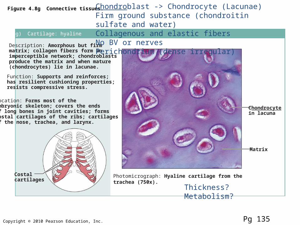
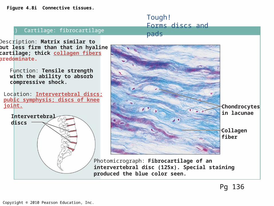
Description: Amorphous but firmmatrix; collagen fibers form animperceptible network; chondroblastsproduce the matrix and when mature(chondrocytes) lie in lacunae.
Function: Supports and reinforces;has resilient cushioning properties;resists compressive stress.
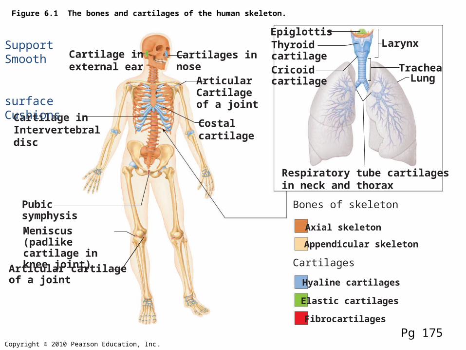
Location: Forms most of theembryonic skeleton; covers the endsof long bones in joint cavities; formscostal cartilages of the ribs; cartilagesof the nose, trachea, and larynx.
Photomicrograph: Hyaline cartilage from thetrachea (750x).
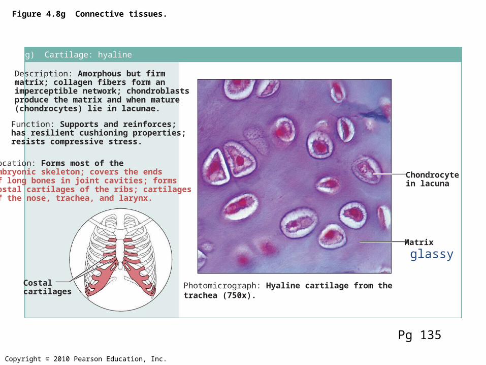
Costalcartilages
Chondrocytein lacuna
Matrix
Pg 135
Chondroblast -> Chondrocyte (Lacunae)Firm ground substance (chondroitin sulfate and water)Collagenous and elastic fibersNo BV or nervesPerichondrium (dense irregular)
Description: Amorphous but firmmatrix; collagen fibers form animperceptible network; chondroblastsproduce the matrix and when mature(chondrocytes) lie in lacunae.
Function: Supports and reinforces;has resilient cushioning properties;resists compressive stress.
Location: Forms most of theembryonic skeleton; covers the endsof long bones in joint cavities; formscostal cartilages of the ribs; cartilagesof the nose, trachea, and larynx.
Photomicrograph: Hyaline cartilage from thetrachea (750x).
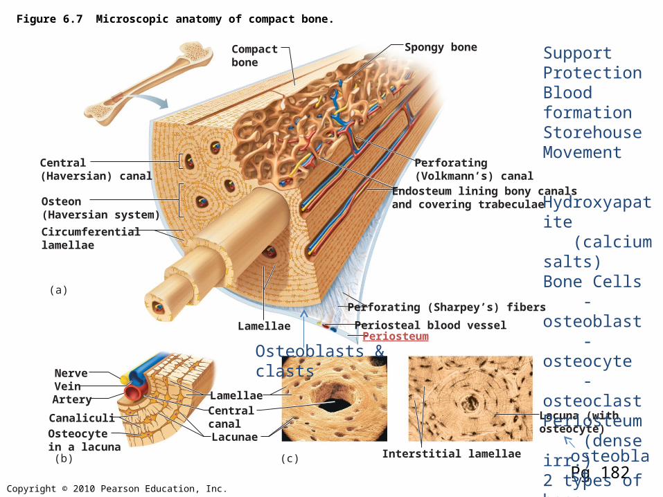
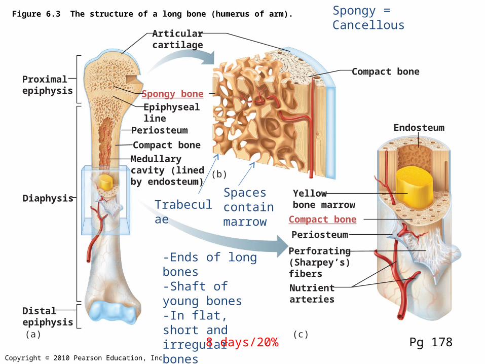
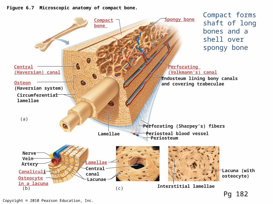
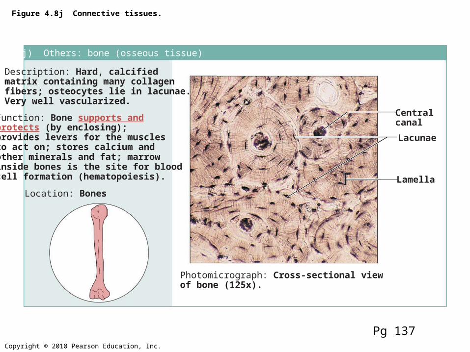
Description: Hard, calcifiedmatrix containing many collagenfibers; osteocytes lie in lacunae.Very well vascularized.
Function: Bone supports andprotects (by enclosing);provides levers for the musclesto act on; stores calcium andother minerals and fat; marrowinside bones is the site for bloodcell formation (hematopoiesis).
Location: Bones
Photomicrograph: Cross-sectional viewof bone (125x).
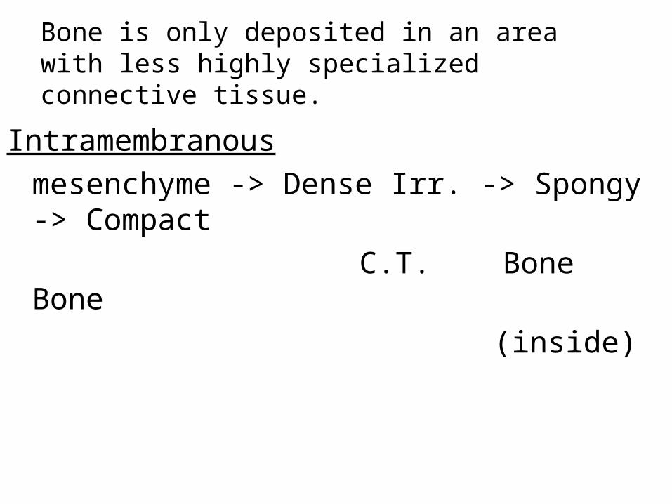
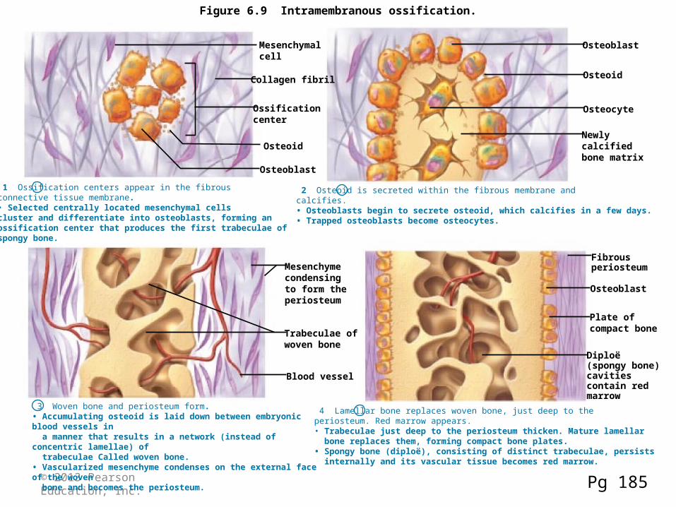
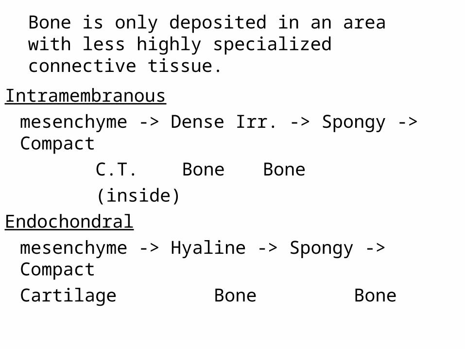
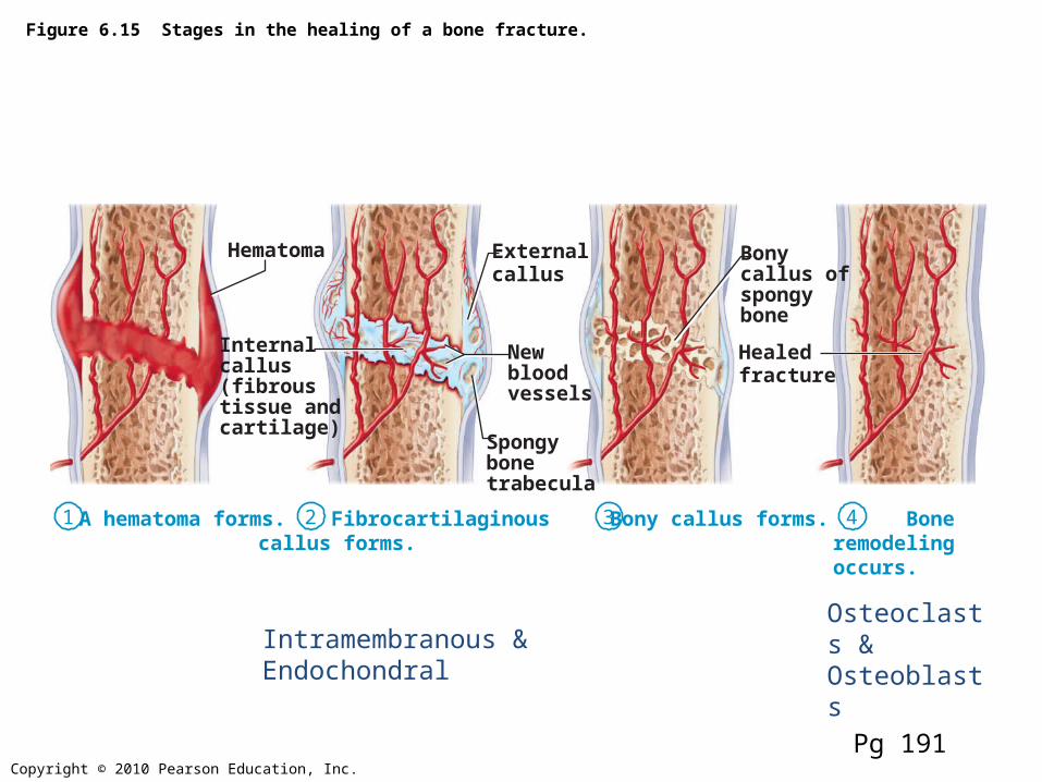
1 Ossification centers appear in the fibrous connective tissue membrane.• Selected centrally located mesenchymal cells cluster and differentiate into osteoblasts, forming an ossification center that produces the first trabeculae ofspongy bone.
Mesenchymal cell
Collagen fibril
Ossification center
Osteoid
Osteoblast
Osteoblast
Osteoid
Osteocyte
Newly calcifiedbone matrix
Mesenchymecondensingto form the periosteum
Trabeculae ofwoven bone
Blood vessel
2 Osteoid is secreted within the fibrous membrane and calcifies.• Osteoblasts begin to secrete osteoid, which calcifies in a few days.• Trapped osteoblasts become osteocytes.
3 Woven bone and periosteum form.• Accumulating osteoid is laid down between embryonic blood vessels in a manner that results in a network (instead of concentric lamellae) of trabeculae Called woven bone.• Vascularized mesenchyme condenses on the external face of the woven bone and becomes the periosteum.
Fibrous periosteum
Osteoblast
Plate ofcompact bone
Diploë (spongy bone)cavities contain red marrow
4 Lamellar bone replaces woven bone, just deep to the periosteum. Red marrow appears. • Trabeculae just deep to the periosteum thicken. Mature lamellar bone replaces them, forming compact bone plates.• Spongy bone (diploë), consisting of distinct trabeculae, persists internally and its vascular tissue becomes red marrow.
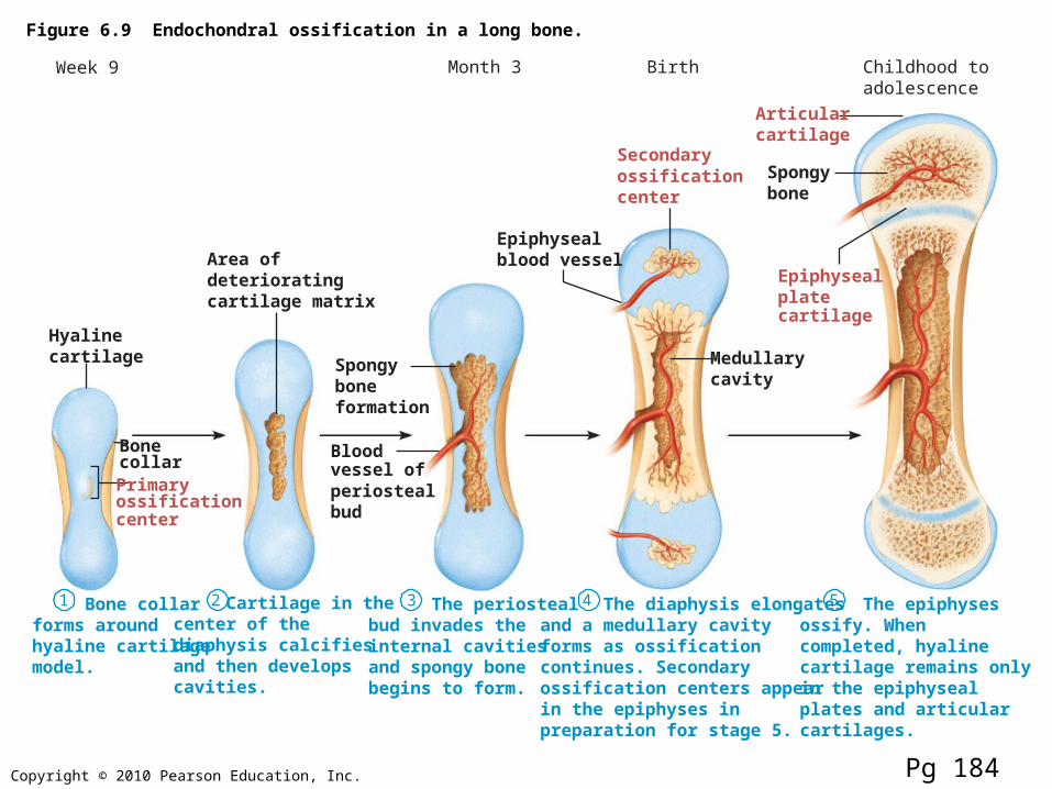
Figure 6.9 Endochondral ossification in a long bone.
1 2 3 4 5 Bone collarforms aroundhyaline cartilagemodel.
Cartilage in thecenter of thediaphysis calcifiesand then developscavities.
The periostealbud invades theinternal cavitiesand spongy bonebegins to form.
The diaphysis elongatesand a medullary cavityforms as ossificationcontinues. Secondaryossification centers appearin the epiphyses inpreparation for stage 5.
The epiphysesossify. Whencompleted, hyalinecartilage remains onlyin the epiphysealplates and articularcartilages.


![Cartilage - Shahid Beheshti Universityfacultymembers.sbu.ac.ir/rajabi/ppt toPDF/Cartilage [Compatibility Mode].pdf · tissue and hyaline cartilage. Chondrocytes may lie singly or](https://static.documents.pub/doc/80x56/5e11522693c7ac3efa2277cb/cartilage-shahid-beheshti-univ-topdfcartilage-compatibility-modepdf-tissue.jpg)


