65
pyright © 2010 Pearson Education, Inc. THE MUSCULAR SYSTEM (PHYSIOLOGY) CHAPTER # 9(a)
| Date post: | 17-Dec-2015 |
| Category: |
Documents |
| Upload: | jodie-campbell |
| View: | 225 times |
| Download: | 0 times |
Copyright © 2010 Pearson Education, Inc.
THE MUSCULAR SYSTEM(PHYSIOLOGY)
CHAPTER # 9(a)
Copyright © 2010 Pearson Education, Inc.
Three Types of Muscle Tissue
1. Skeletal muscle tissue:
• Attached to bones and skin
• Striated
• Voluntary (i.e., conscious control)
• Powerful
• Primary topic of this chapter

Copyright © 2010 Pearson Education, Inc.
Three Types of Muscle Tissue
2. Cardiac muscle tissue:
• Only in the heart
• Striated
• Involuntary
• More details in Chapter 18

Copyright © 2010 Pearson Education, Inc.
Three Types of Muscle Tissue
3. Smooth muscle tissue:
• In the walls of hollow organs, e.g., stomach, urinary bladder, and airways
• Not striated
• Involuntary
• More details later in this chapter

Copyright © 2010 Pearson Education, Inc. Table 9.3

Copyright © 2010 Pearson Education, Inc.
Special Characteristics of Muscle Tissue
• Excitability (responsiveness or irritability): ability to receive and respond to stimuli
• Contractility: ability to shorten when stimulated
• Extensibility: ability to be stretched
• Elasticity: ability to recoil to resting length
Copyright © 2010 Pearson Education, Inc.
Muscle Functions
1. Movement of bones or fluids (e.g., blood)
2. Maintaining posture and body position
3. Stabilizing joints
4. Heat generation (especially skeletal muscle)

Copyright © 2010 Pearson Education, Inc.
Skeletal Muscle
• Each muscle is served by one artery, one nerve, and one or more veins
Copyright © 2010 Pearson Education, Inc.
Skeletal Muscle
• Connective tissue sheaths of skeletal muscle:
• Epimysium: dense regular connective tissue surrounding entire muscle
• Perimysium: fibrous connective tissue surrounding fascicles (groups of muscle fibers)
• Endomysium: fine areolar connective tissue surrounding each muscle fiber
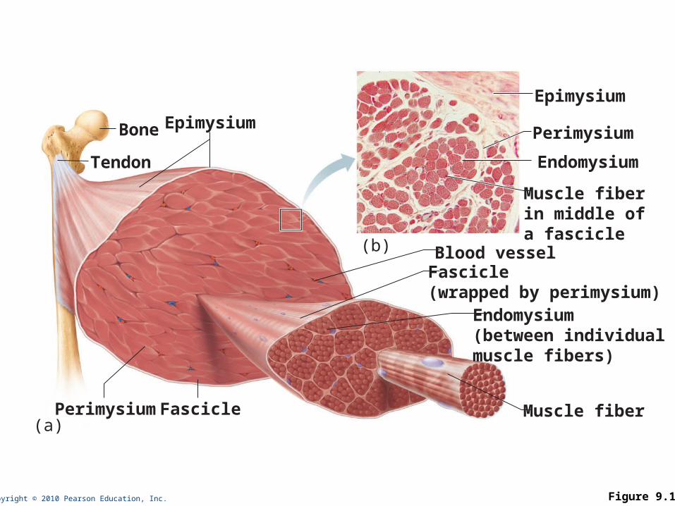
Copyright © 2010 Pearson Education, Inc. Figure 9.1
Bone
Perimysium
Endomysium(between individualmuscle fibers)
Muscle fiber
Fascicle(wrapped by perimysium)
Epimysium
Tendon
Epimysium
Muscle fiberin middle ofa fascicle
Blood vessel
Perimysium
Endomysium
Fascicle(a)
(b)

Copyright © 2010 Pearson Education, Inc.
Skeletal Muscle: Attachments
• Muscles attach:
• Directly—epimysium of muscle is fused to the periosteum of bone or perichondrium of cartilage
• Indirectly—connective tissue wrappings extend beyond the muscle as a ropelike tendon or sheetlike aponeurosis
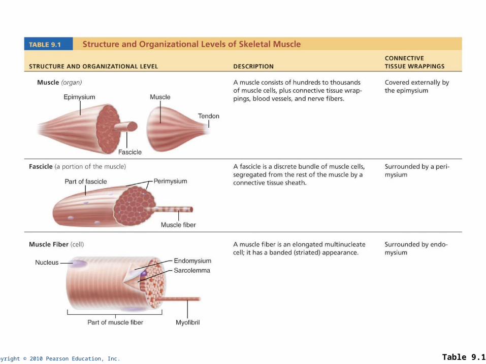
Copyright © 2010 Pearson Education, Inc. Table 9.1

Copyright © 2010 Pearson Education, Inc.
Microscopic Anatomy of a Skeletal Muscle Fiber
• Cylindrical cell 10 to 100 m in diameter, up to 30 cm long
• Multiple peripheral nuclei
• Many mitochondria
• Glycosomes for glycogen storage, myoglobin for O2 storage
• Also contain myofibrils, sarcoplasmic reticulum, and T tubules
Copyright © 2010 Pearson Education, Inc.
Myofibrils
• Densely packed, rodlike elements
• ~80% of cell volume
• Exhibit striations: perfectly aligned repeating series of dark A bands and light I bands

Copyright © 2010 Pearson Education, Inc.
NucleusLight I bandDark A band
Sarcolemma
Mitochondrion
(b) Diagram of part of a muscle fiber showing the myofibrils. Onemyofibril is extended afrom the cut end of the fiber.
Myofibril
Copyright © 2010 Pearson Education, Inc.
Sarcomere
• Smallest contractile unit (functional unit) of a muscle fiber
• The region of a myofibril between two successive Z discs
• Composed of thick and thin myofilaments made of contractile proteins
Copyright © 2010 Pearson Education, Inc.
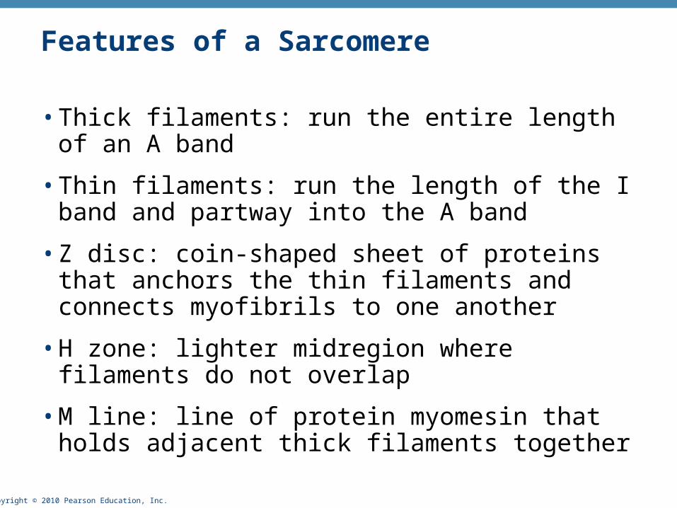
Features of a Sarcomere
• Thick filaments: run the entire length of an A band
• Thin filaments: run the length of the I band and partway into the A band
• Z disc: coin-shaped sheet of proteins that anchors the thin filaments and connects myofibrils to one another
• H zone: lighter midregion where filaments do not overlap
• M line: line of protein myomesin that holds adjacent thick filaments together

Copyright © 2010 Pearson Education, Inc. Figure 9.2c, d
I band I bandA bandSarcomere
H zoneThin (actin)filament
Thick (myosin)filament
Z disc Z disc
M line
(c) Small part of one myofibril enlarged to show the myofilamentsresponsible for the banding pattern. Each sarcomere extends fromone Z disc to the next.
Z disc Z discM line
Sarcomere
Thin (actin)filament
Thick(myosin)filament
Elastic (titin)filaments
(d) Enlargement of one sarcomere (sectioned lengthwise). Notice the myosin heads on the thick filaments.
Copyright © 2010 Pearson Education, Inc.
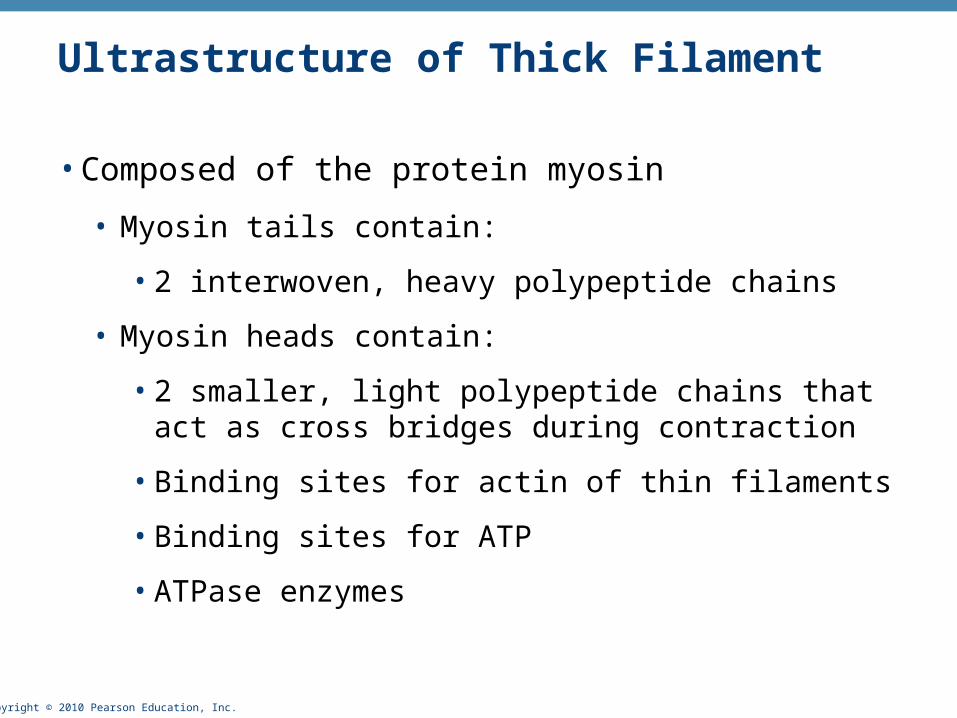
Ultrastructure of Thick Filament
• Composed of the protein myosin
• Myosin tails contain:
• 2 interwoven, heavy polypeptide chains
• Myosin heads contain:
• 2 smaller, light polypeptide chains that act as cross bridges during contraction
• Binding sites for actin of thin filaments
• Binding sites for ATP
• ATPase enzymes

Copyright © 2010 Pearson Education, Inc.
Ultrastructure of Thin Filament
• Twisted double strand of fibrous protein F actin
• F actin consists of G (globular) actin subunits
• G actin bears active sites for myosin head attachment during contraction
• Tropomyosin and troponin: regulatory proteins bound to actin
Copyright © 2010 Pearson Education, Inc. Figure 9.3
Flexible hinge region
Tail
Tropomyosin Troponin Actin
Myosin head
ATP-bindingsite
Heads Active sitesfor myosinattachment
Actinsubunits
Actin-binding sites
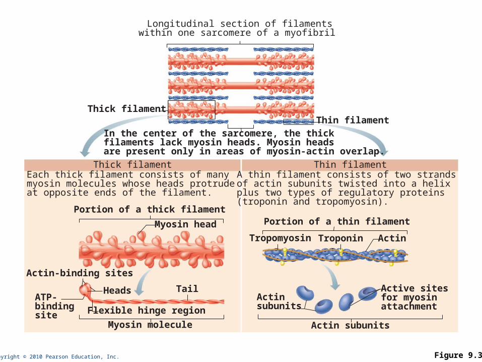
Thick filamentEach thick filament consists of manymyosin molecules whose heads protrude at opposite ends of the filament.
Thin filamentA thin filament consists of two strandsof actin subunits twisted into a helix plus two types of regulatory proteins(troponin and tropomyosin).
Thin filamentThick filament
In the center of the sarcomere, the thickfilaments lack myosin heads. Myosin heads are present only in areas of myosin-actin overlap.
Longitudinal section of filamentswithin one sarcomere of a myofibril
Portion of a thick filamentPortion of a thin filament
Myosin molecule Actin subunits
Copyright © 2010 Pearson Education, Inc.
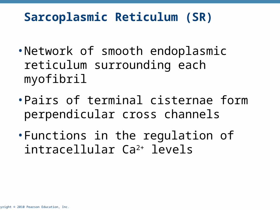
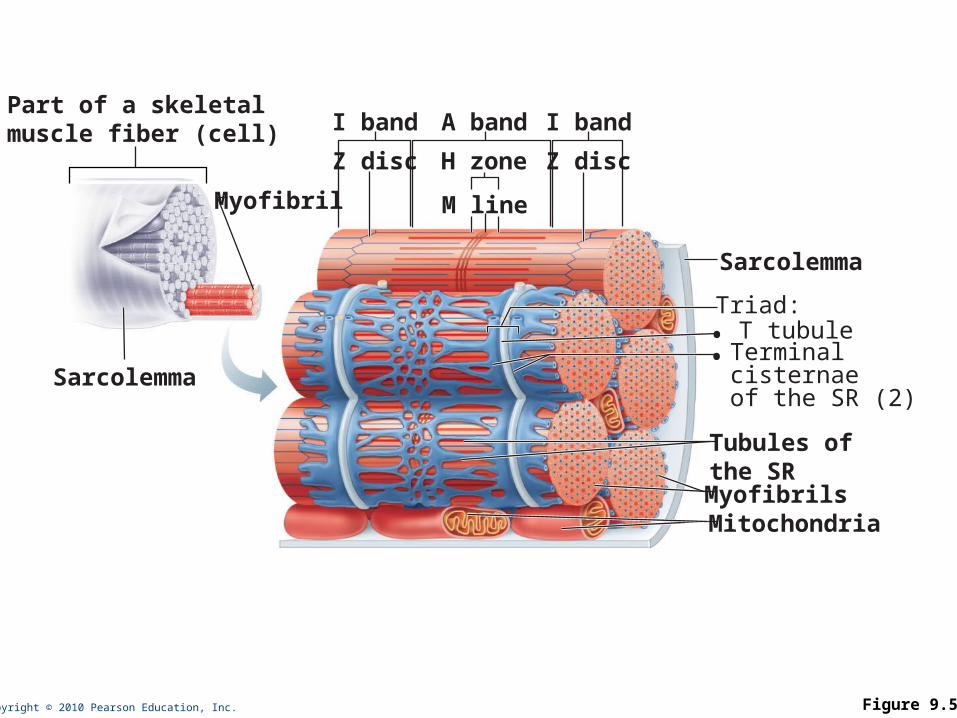
Sarcoplasmic Reticulum (SR)
• Network of smooth endoplasmic reticulum surrounding each myofibril
• Pairs of terminal cisternae form perpendicular cross channels
• Functions in the regulation of intracellular Ca2+ levels
Copyright © 2010 Pearson Education, Inc.
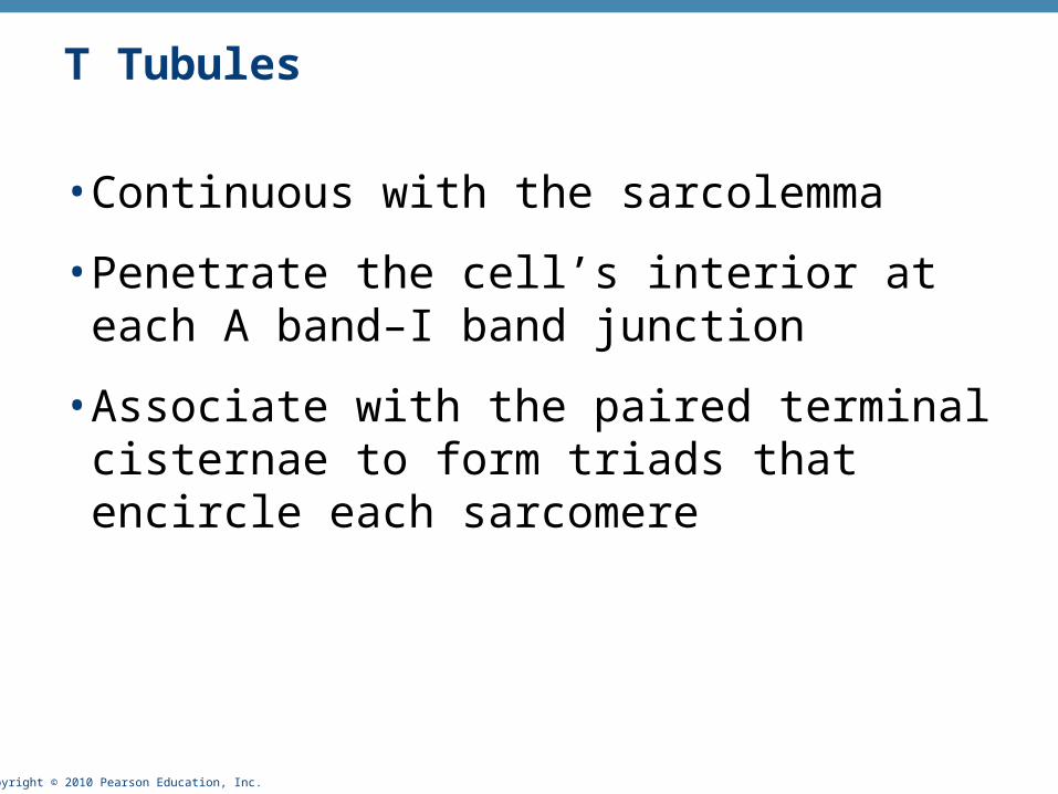
T Tubules
• Continuous with the sarcolemma
• Penetrate the cell’s interior at each A band–I band junction
• Associate with the paired terminal cisternae to form triads that encircle each sarcomere
Copyright © 2010 Pearson Education, Inc. Figure 9.5
Myofibril
Myofibrils
Triad:
Tubules ofthe SR
Sarcolemma
Sarcolemma
Mitochondria
I band I bandA band
H zone Z discZ disc
Part of a skeletalmuscle fiber (cell)
• T tubule• Terminal
cisternaeof the SR (2)
M line

Copyright © 2010 Pearson Education, Inc.
Triad Relationships
• T tubules conduct impulses deep into muscle fiber
• Integral proteins protrude into the intermembrane space from T tubule and SR cisternae membranes
• T tubule proteins: voltage sensors
• SR foot proteins: gated channels that regulate Ca2+ release from the SR cisternae

Copyright © 2010 Pearson Education, Inc.
Contraction
• The generation of force
• Does not necessarily cause shortening of the fiber
• Shortening occurs when tension generated by cross bridges on the thin filaments exceeds forces opposing shortening
Copyright © 2010 Pearson Education, Inc.
Sliding Filament Model of Contraction
• In the relaxed state, thin and thick filaments overlap only slightly
• During contraction, myosin heads bind to actin, detach, and bind again, to propel the thin filaments toward the M line
• As H zones shorten and disappear, sarcomeres shorten, muscle cells shorten, and the whole muscle shortens

Copyright © 2010 Pearson Education, Inc. Figure 9.6
I
Fully relaxed sarcomere of a muscle fiber
Fully contracted sarcomere of a muscle fiber
IA
Z ZH
I IA
Z Z
1
2
Copyright © 2010 Pearson Education, Inc.
Requirements for Skeletal Muscle Contraction
1. Activation: neural stimulation at aneuromuscular junction
2. Excitation-contraction coupling:
• Generation and propagation of an action potential along the sarcolemma
• Final trigger: a brief rise in intracellular Ca2+ levels

Copyright © 2010 Pearson Education, Inc.
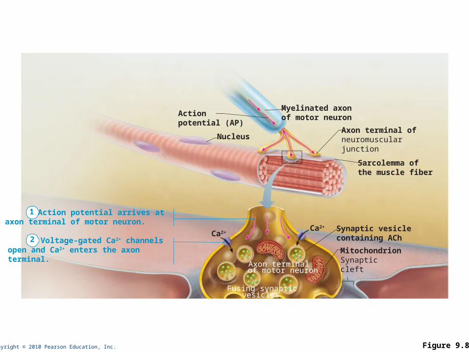
Events at the Neuromuscular Junction
• Skeletal muscles are stimulated by somatic motor neurons
• Axons of motor neurons travel from the central nervous system via nerves to skeletal muscles
• Each axon forms several branches as it enters a muscle
• Each axon ending forms a neuromuscular junction with a single muscle fiber
Copyright © 2010 Pearson Education, Inc.
Nucleus
Actionpotential (AP)
Myelinated axonof motor neuron
Axon terminal ofneuromuscular junction
Sarcolemma ofthe muscle fiber
Ca2+Ca2+
Axon terminalof motor neuron
Synaptic vesiclecontaining ACh
MitochondrionSynapticcleft
Fusing synaptic vesicles
1 Action potential arrives ataxon terminal of motor neuron.
2 Voltage-gated Ca2+ channels open and Ca2+ enters the axon terminal.
Figure 9.8

Copyright © 2010 Pearson Education, Inc.
Neuromuscular Junction
• Situated midway along the length of a muscle fiber
• Axon terminal and muscle fiber are separated by a gel-filled space called the synaptic cleft
• Synaptic vesicles of axon terminal contain the neurotransmitter acetylcholine (ACh)
• Junctional folds of the sarcolemma contain ACh receptors

Copyright © 2010 Pearson Education, Inc.
Events at the Neuromuscular Junction
• Nerve impulse arrives at axon terminal
• ACh is released and binds with receptors on the sarcolemma
• Electrical events lead to the generation of an action potential
PLAYPLAY A&P Flix™: Events at the Neuromuscular Junction

Copyright © 2010 Pearson Education, Inc. Figure 9.8
Nucleus
Actionpotential (AP)
Myelinated axonof motor neuron
Axon terminal ofneuromuscular junction
Sarcolemma ofthe muscle fiber
Ca2+Ca2+
Axon terminalof motor neuron
Synaptic vesiclecontaining AChMitochondrionSynapticcleft
Junctionalfolds ofsarcolemma
Fusing synaptic vesicles
ACh
Sarcoplasm ofmuscle fiber
Postsynaptic membraneion channel opens;ions pass.
Na+ K+
Ach–
Na+
K+
Degraded ACh
Acetyl-cholinesterase
Postsynaptic membraneion channel closed;ions cannot pass.
1 Action potential arrives ataxon terminal of motor neuron.
2 Voltage-gated Ca2+ channels open and Ca2+ enters the axon terminal.
3 Ca2+ entry causes some synaptic vesicles to release their contents (acetylcholine)by exocytosis.
4 Acetylcholine, aneurotransmitter, diffuses across the synaptic cleft and binds to receptors in the sarcolemma.
5 ACh binding opens ionchannels that allow simultaneous passage of Na+ into the musclefiber and K+ out of the muscle fiber.
6 ACh effects are terminated by its enzymatic breakdown in the synaptic cleft by acetylcholinesterase.

Copyright © 2010 Pearson Education, Inc.
Destruction of Acetylcholine
• ACh effects are quickly terminated by the enzyme acetylcholinesterase
• Prevents continued muscle fiber contraction in the absence of additional stimulation

Copyright © 2010 Pearson Education, Inc.
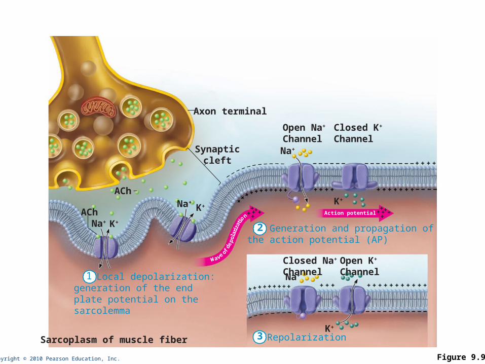
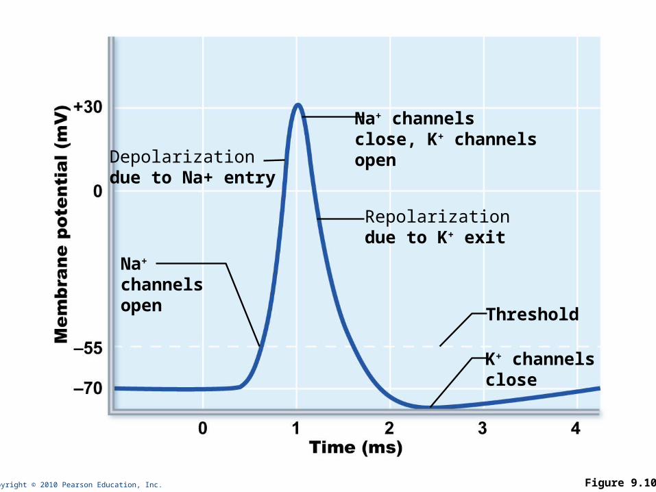
Events in Generation of an Action Potential
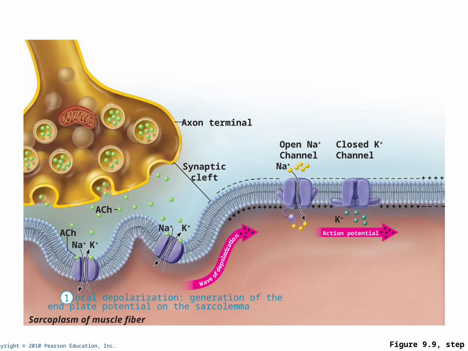
1. Local depolarization (end plate potential):
• ACh binding opens chemically (ligand) gated ion channels
• Simultaneous diffusion of Na+ (inward) and K+ (outward)
• More Na+ diffuses, so the interior of the sarcolemma becomes less negative
• Local depolarization – end plate potential
Copyright © 2010 Pearson Education, Inc.
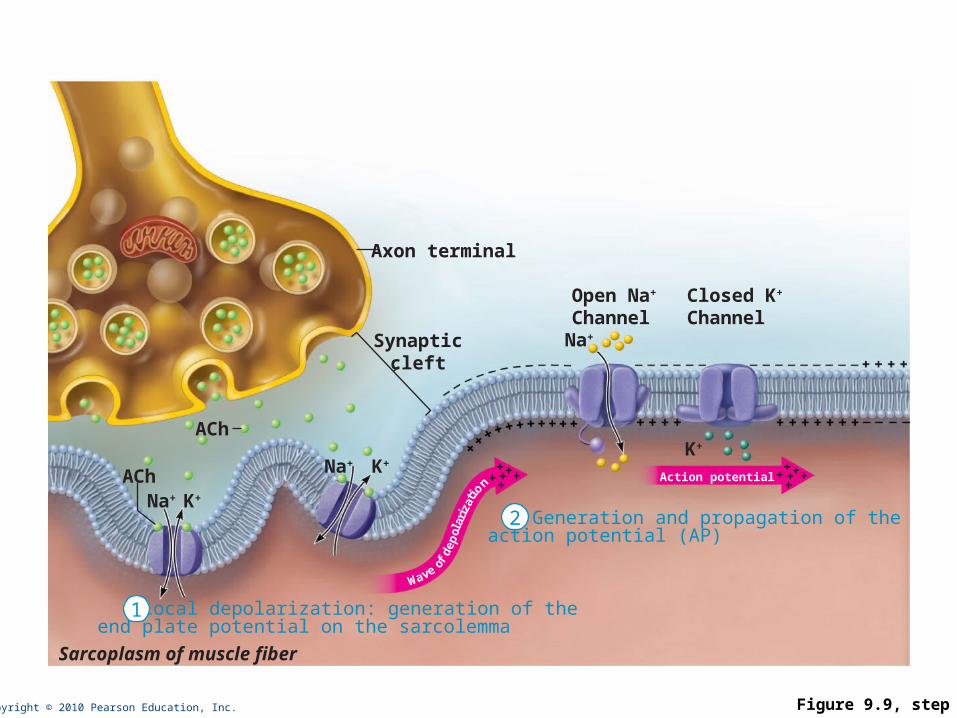
Events in Generation of an Action Potential
2. Generation and propagation of an action potential:
• End plate potential spreads to adjacent membrane areas
• Voltage-gated Na+ channels open
• Na+ influx decreases the membrane voltage toward a critical threshold
• If threshold is reached, an action potential is generated

Copyright © 2010 Pearson Education, Inc.
Events in Generation of an Action Potential
• Local depolarization wave continues to spread, changing the permeability of the sarcolemma
• Voltage-regulated Na+ channels open in the adjacent patch, causing it to depolarize to threshold

Copyright © 2010 Pearson Education, Inc.
Events in Generation of an Action Potential
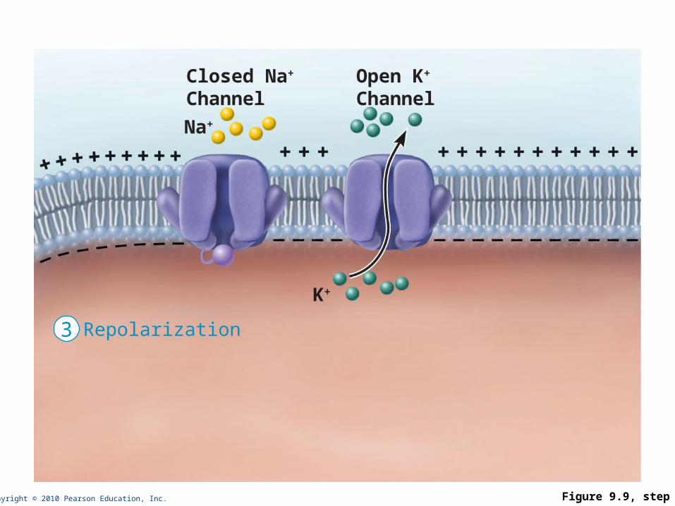
3. Repolarization:
• Na+ channels close and voltage-gated K+ channels open
• K+ efflux rapidly restores the resting polarity
• Fiber cannot be stimulated and is in a refractory period until repolarization is complete
• Ionic conditions of the resting state are restored by the Na+-K+ pump

Copyright © 2010 Pearson Education, Inc. Figure 9.9
Na+
Na+
Open Na+
Channel
Closed Na+
Channel
Closed K+
Channel
Open K+
Channel
Action potential++++++
+++++
+
Axon terminal
Synapticcleft
ACh
ACh
Sarcoplasm of muscle fiber
K+
2 Generation and propagation ofthe action potential (AP)
3 Repolarization
1 Local depolarization: generation of the end plate potential on the sarcolemma
K+
K+Na+
K+Na+
Wave ofde
po
lari
zatio
n
Copyright © 2010 Pearson Education, Inc. Figure 9.9, step 1
Na+
Na+
Open Na+
ChannelClosed K+
Channel
K+
Na+ K+Action potential
++++++
+++++
+
Axon terminal
Synapticcleft
ACh
ACh
Sarcoplasm of muscle fiber
K+
1 Local depolarization: generation of the end plate potential on the sarcolemma
1
Wave ofde
po
lari
zatio
n
Copyright © 2010 Pearson Education, Inc. Figure 9.9, step 2
Na+
Na+
Open Na+
ChannelClosed K+
Channel
K+
Na+ K+Action potential
++++++
+++++
+
Axon terminal
Synapticcleft
ACh
ACh
Sarcoplasm of muscle fiber
K+
Generation and propagation of the action potential (AP)
1 Local depolarization: generation of the end plate potential on the sarcolemma
2
1
Wave ofde
po
lari
zatio
n
Copyright © 2010 Pearson Education, Inc. Figure 9.9, step 3
Na+
Closed Na+
ChannelOpen K+
Channel
K+
Repolarization3
Copyright © 2010 Pearson Education, Inc. Figure 9.9
Na+
Na+
Open Na+
ChannelClosed K+
Channel
Action potential++++++
+++++
+
Axon terminal
Synapticcleft
ACh
ACh
Sarcoplasm of muscle fiber
K+
2 Generation and propagation ofthe action potential (AP)
3 Repolarization
1 Local depolarization: generation of the end plate potential on the sarcolemma
K+
K+Na+
K+Na+
Wave ofde
po
lari
zatio
n
Closed Na+
ChannelOpen K+
Channel
Copyright © 2010 Pearson Education, Inc. Figure 9.10
Na+ channelsclose, K+ channelsopen
K+ channelsclose
Repolarizationdue to K+ exit
Threshold
Na+
channelsopen
Depolarizationdue to Na+ entry

Copyright © 2010 Pearson Education, Inc.
Excitation-Contraction (E-C) Coupling
• Sequence of events by which transmission of an AP along the sarcolemma leads to sliding of the myofilaments
• Latent period:
• Time when E-C coupling events occur
• Time between AP initiation and the beginning of contraction
Copyright © 2010 Pearson Education, Inc.
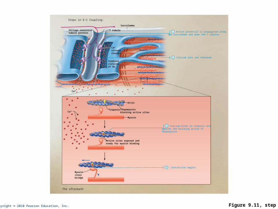
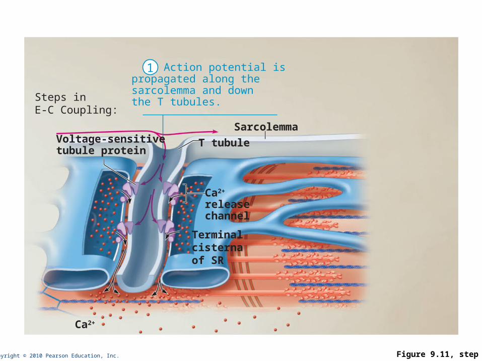
Events of Excitation-Contraction (E-C) Coupling
• AP is propagated along sarcomere to T tubules
• Voltage-sensitive proteins stimulate Ca2+ release from SR
• Ca2+ is necessary for contraction

Copyright © 2010 Pearson Education, Inc. Figure 9.11, step 1
Axon terminalof motor neuron
Muscle fiberTriad
One sarcomere
Synaptic cleft
Setting the stage
Sarcolemma
Action potentialis generated
Terminal cisterna of SR ACh
Ca2+
Copyright © 2010 Pearson Education, Inc. Figure 9.11, step 2
Action potential is propagated alongthe sarcolemma and down the T tubules.
Steps in E-C Coupling:
Troponin Tropomyosinblocking active sites
Myosin
Actin
Active sites exposed and ready for myosin binding
Ca2+
Terminal cisterna of SR
Voltage-sensitivetubule protein
T tubule
Ca2+
releasechannel
Myosincross bridge
Ca2+
Sarcolemma
Calcium ions are released.
Calcium binds to troponin andremoves the blocking action oftropomyosin.
Contraction begins
The aftermath
1
2
3
4
Copyright © 2010 Pearson Education, Inc. Figure 9.11, step 3
Steps inE-C Coupling:
Terminal cisterna of SR
Voltage-sensitivetubule protein
T tubule
Ca2+
releasechannel
Ca2+
Sarcolemma
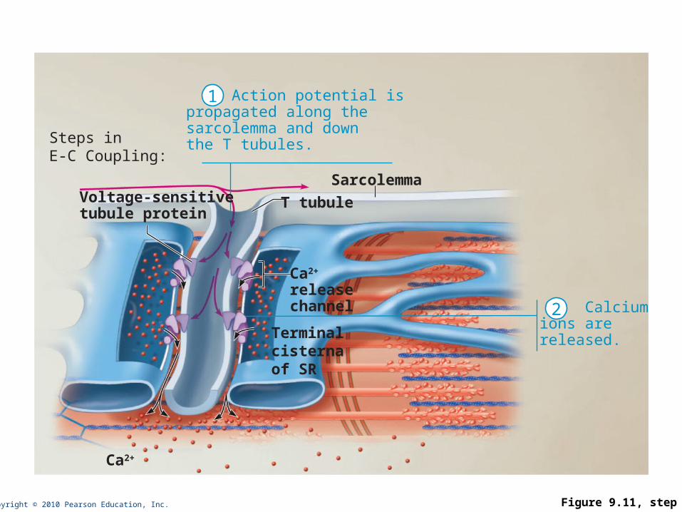
Action potential ispropagated along thesarcolemma and downthe T tubules.
1
Copyright © 2010 Pearson Education, Inc. Figure 9.11, step 4
Steps inE-C Coupling:
Terminal cisterna of SR
Voltage-sensitivetubule protein
T tubule
Ca2+
releasechannel
Ca2+
Sarcolemma
Action potential ispropagated along thesarcolemma and downthe T tubules.
Calciumions arereleased.
1
2
Copyright © 2010 Pearson Education, Inc. Figure 9.11, step 5
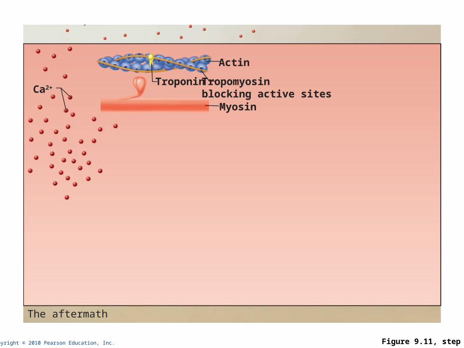
Troponin Tropomyosinblocking active sitesMyosin
Actin
Ca2+
The aftermath

Copyright © 2010 Pearson Education, Inc. Figure 9.11, step 6
Troponin Tropomyosinblocking active sitesMyosin
Actin
Active sites exposed and ready for myosin binding
Ca2+
Calcium binds totroponin and removesthe blocking action oftropomyosin.
The aftermath
3

Copyright © 2010 Pearson Education, Inc. Figure 9.11, step 7
Troponin Tropomyosinblocking active sitesMyosin
Actin
Active sites exposed and ready for myosin binding
Ca2+
Myosincross bridge
Calcium binds totroponin and removesthe blocking action oftropomyosin.
Contraction begins
The aftermath
3
4

Copyright © 2010 Pearson Education, Inc. Figure 9.11, step 8
Action potential is propagated alongthe sarcolemma and down the T tubules.
Steps in E-C Coupling:
Troponin Tropomyosinblocking active sites
Myosin
Actin
Active sites exposed and ready for myosin binding
Ca2+
Terminal cisterna of SR
Voltage-sensitivetubule protein
T tubule
Ca2+
releasechannel
Myosincross bridge
Ca2+
Sarcolemma
Calcium ions are released.
Calcium binds to troponin andremoves the blocking action oftropomyosin.
Contraction begins
The aftermath
1
2
3
4

Copyright © 2010 Pearson Education, Inc.
Role of Calcium (Ca2+) in Contraction
• At low intracellular Ca2+ concentration:
• Tropomyosin blocks the active sites on actin
• Myosin heads cannot attach to actin
• Muscle fiber relaxes

Copyright © 2010 Pearson Education, Inc.
Role of Calcium (Ca2+) in Contraction
• At higher intracellular Ca2+ concentrations:
• Ca2+ binds to troponin
• Troponin changes shape and moves tropomyosin away from active sites
• Events of the cross bridge cycle occur
• When nervous stimulation ceases, Ca2+ is pumped back into the SR and contraction ends
Copyright © 2010 Pearson Education, Inc.
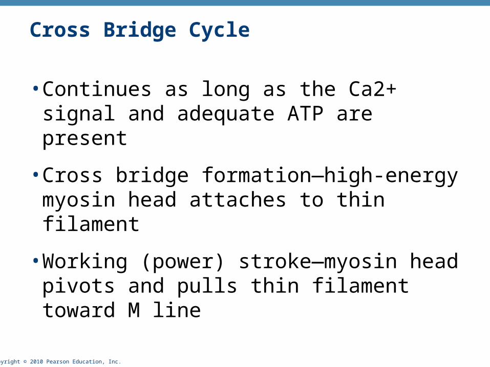
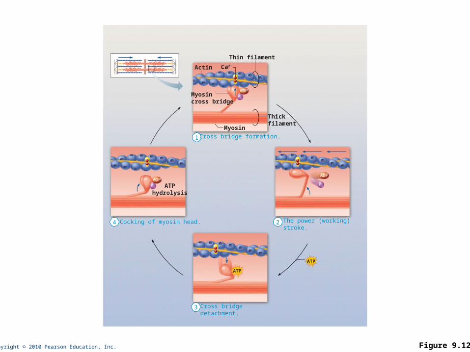
Cross Bridge Cycle
• Continues as long as the Ca2+ signal and adequate ATP are present
• Cross bridge formation—high-energy myosin head attaches to thin filament
•Working (power) stroke—myosin head pivots and pulls thin filament toward M line
Copyright © 2010 Pearson Education, Inc.
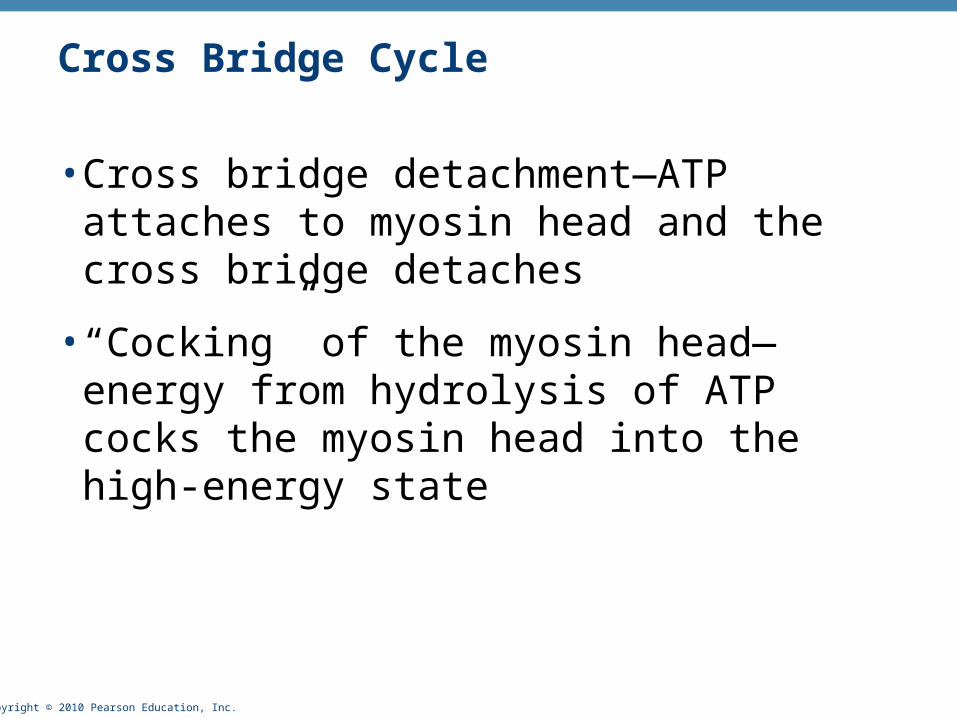
Cross Bridge Cycle
• Cross bridge detachment—ATP attaches to myosin head and the cross bridge detaches
• “Cocking” of the myosin head—energy from hydrolysis of ATP cocks the myosin head into the high-energy state

Copyright © 2010 Pearson Education, Inc. Figure 9.12
1
Actin
Cross bridge formation.
Cocking of myosin head. The power (working) stroke.
Cross bridge detachment.
Ca2+
Myosincross bridge
Thick filament
Thin filament
ADP
Myosin
Pi
ATPhydrolysis
ATP
ATP
24
3
ADP
Pi
ADPPi

Copyright © 2010 Pearson Education, Inc. Figure 9.12, step 1
Actin
Cross bridge formation.
Ca2+
Myosincross bridge
Thick filament
Thin filament
ADP
Myosin
Pi
1

Copyright © 2010 Pearson Education, Inc. Figure 9.12, step 3
The power (working) stroke.
ADP
Pi
2

Copyright © 2010 Pearson Education, Inc. Figure 9.12, step 4
Cross bridge detachment.
ATP
3

Copyright © 2010 Pearson Education, Inc. Figure 9.12, step 5
Cocking of myosin head.
ATPhydrolysis
ADPPi
4
Copyright © 2010 Pearson Education, Inc. Figure 9.12
1
Actin
Cross bridge formation.
Cocking of myosin head. The power (working) stroke.
Cross bridge detachment.
Ca2+
Myosincross bridge
Thick filament
Thin filament
ADP
Myosin
Pi
ATPhydrolysis
ATP
ATP
24
3
ADP
Pi
ADPPi


