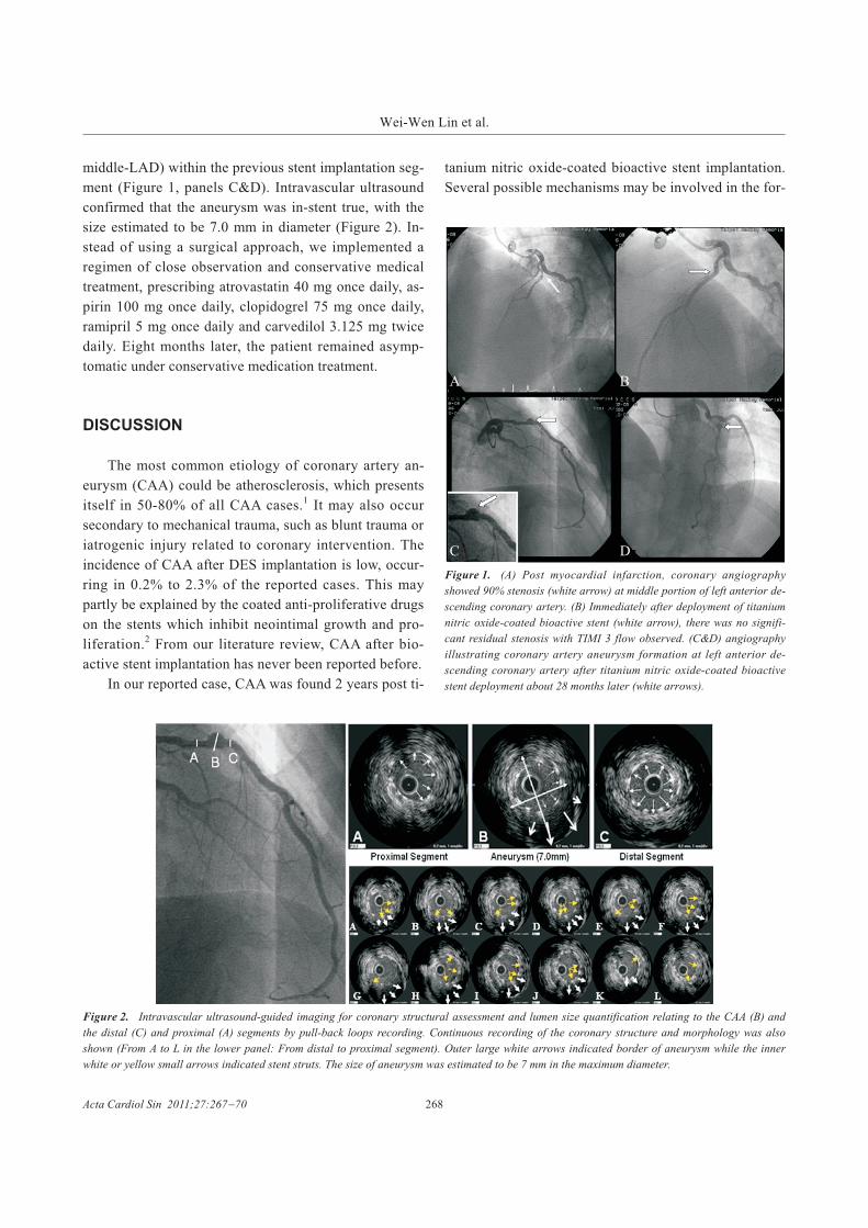
Coronary Aneurysm Formation after Titanium
Nitric Oxide-Coated Stent Implantation
Wei-Wen Lin,1 Jui-Peng Tsai,1,2 Chih-Hsuan Yen,1,2 Jen-Yuan Kuo1 and Chung-Lieh Hung1,2
Coronary artery aneurysm (CAA) formation is a rare complication after percutaneous coronary intervention (PCI)
with stent implantation. Why CAA occurs in these unusual instances is not well understood. Though cases with
drug-eluting stent (DES) induced CAA have been reported before, none was reported to be associated with a
titanium nitric oxide-coated bioactive stent. Herein, we describe the first case where CAA developed after titanium
nitric oxide-coated bioactive stent implantation. Several mechanisms may account for stent-related CAA formation,
including mechanical trauma, vessel remodeling, acute myocardial infarction, long or multiple DES stent
implantation, DES malapposition and hypersensitivity reaction to polymers.
A 53-year-old man presented initially with recent MI and ongoing chest pains. Coronary angiography revealed a
90% occlusion of the middle left anterior descending coronary artery (LAD). PCI was performed on the patient, and
a titanium nitric oxide-coated bioactive stent was implanted. Due to a positive thallium scan 28 months later, a
follow-up coronary angiography scan revealed a true CAA, which was then further confirmed by intravascular
ultrasound. Thereafter, the patient remained asymptomatic at subsequent follow-ups, and continued medical
treatment without further intervention. Herein, we discuss the possible etiologies, mechanisms and further treatments
of CAA formation after titanium nitric oxide-coated bioactive stent implantation.
Key Words: Coronary artery aneurysm � Intravascular ultrasound � Titanium nitric oxide-coated stent
CASE REPORT
In June 2007, a 53-year-old man visited our cardio-
vascular outpatient clinic with intermittent chest tight-
ness which had persisted for 10 days. The patient was an
ex-smoker, and taking medication for hypertension. An
initial thallium scan revealed reversible myocardial
ischemia at the left ventricle (LV) anterior wall. Labora-
tory data showed mildly elevated cardiac enzymes (tro-
ponin I: 1.42 ng/mL), and recent myocardial infarct was
diagnosed. Coronary angiography revealed a high de-
gree of stenosis with irregular surface at the middle por-
tion of the left anterior descending coronary artery
(LAD), with nearly 90 percent stenosis (Figure 1, panel
A). A Sprinter balloon (2.25*20 mm semi-compliant bal-
loon, Medtronic, U.S.A) was initially utilized to pre-
dilate the lesion (pressure: 10 atm), with subsequent
successful Titan 2 stent implantation (3.0*28 mm, tita-
nium nitric oxide-coated bioactive stent, Hexacath,
France). A final coronary angiography showed no re-
sidual stenosis (Figure 1, panel B). After discharge, the
patient remained asymptomatic, and was prescribed dual
anti-platelet agents at regular follow-ups with our outpa-
tient clinic. However, the patient suffered angina symp-
toms almost two years later, and a follow-up thallium
scan showed a small region myocardial ischemia at the
LV anterior wall. Further diagnostic coronary angio-
graphy incidentally revealed an aneurysm formation (at
267 Acta Cardiol Sin 2011;27:267�70
Coronary Aneurysm Formation after Bioactive Stent ImplantationCase Report Acta Cardiol Sin 2011;27:267�70
Received: December 24, 2010 Accepted: March 3, 20111Division of Cardiology, Department of Internal Medicine, Mackay
Memorial Hospital; 2Department of Medicine, Mackay Medical
College, and Mackay Medicine, Nursing and Management College,
Taipei, Taiwan.
Address correspondence and reprint requests to: Dr. Chung-Lieh
Hung, Division of Cardiology, Department of Internal Medicine,
Mackay Memorial Hospital, No. 92, Sec. 2, Zhongshan N. Rd.,
Zhongshan Dist., Taipei City 10449, Taiwan. Tel: 886-2-2543-3535
ext. 2456; E-mail: [email protected]
middle-LAD) within the previous stent implantation seg-
ment (Figure 1, panels C&D). Intravascular ultrasound
confirmed that the aneurysm was in-stent true, with the
size estimated to be 7.0 mm in diameter (Figure 2). In-
stead of using a surgical approach, we implemented a
regimen of close observation and conservative medical
treatment, prescribing atrovastatin 40 mg once daily, as-
pirin 100 mg once daily, clopidogrel 75 mg once daily,
ramipril 5 mg once daily and carvedilol 3.125 mg twice
daily. Eight months later, the patient remained asymp-
tomatic under conservative medication treatment.
DISCUSSION
The most common etiology of coronary artery an-
eurysm (CAA) could be atherosclerosis, which presents
itself in 50-80% of all CAA cases.1 It may also occur
secondary to mechanical trauma, such as blunt trauma or
iatrogenic injury related to coronary intervention. The
incidence of CAA after DES implantation is low, occur-
ring in 0.2% to 2.3% of the reported cases. This may
partly be explained by the coated anti-proliferative drugs
on the stents which inhibit neointimal growth and pro-
liferation.2 From our literature review, CAA after bio-
active stent implantation has never been reported before.
In our reported case, CAA was found 2 years post ti-
tanium nitric oxide-coated bioactive stent implantation.
Several possible mechanisms may be involved in the for-
Acta Cardiol Sin 2011;27:267�70 268
Wei-Wen Lin et al.
Figure 1. (A) Post myocardial infarction, coronary angiography
showed 90% stenosis (white arrow) at middle portion of left anterior de-
scending coronary artery. (B) Immediately after deployment of titanium
nitric oxide-coated bioactive stent (white arrow), there was no signifi-
cant residual stenosis with TIMI 3 flow observed. (C&D) angiography
illustrating coronary artery aneurysm formation at left anterior de-
scending coronary artery after titanium nitric oxide-coated bioactive
stent deployment about 28 months later (white arrows).
Figure 2. Intravascular ultrasound-guided imaging for coronary structural assessment and lumen size quantification relating to the CAA (B) and
the distal (C) and proximal (A) segments by pull-back loops recording. Continuous recording of the coronary structure and morphology was also
shown (From A to L in the lower panel: From distal to proximal segment). Outer large white arrows indicated border of aneurysm while the inner
white or yellow small arrows indicated stent struts. The size of aneurysm was estimated to be 7 mm in the maximum diameter.
A B
C D
mation of a CAA in this case. Previous study suggests
that DES-related CAA may be induced by mechanical
trauma from a coronary intervention, including over-
sized balloon angioplasty or high-pressure inflation lead-
ing to coronary artery dissection. There is no doubt that
a hypersensitive reaction to polymer of the stent could
be one of the mechanisms in this case. Aneurysmal dila-
tion has even been reported at the stented segment in a
case with severe hypersensitivity.3 In addition, weaken-
ing of the medial layer of the vascular wall, which may
be in part be due to chronic overstimulation of endothe-
lium-derived relaxation factor such as endogenous nitric
oxide, has also been postulated to be the cause of an-
eurysm formation.4 Nitric oxide released from the tita-
nium nitric oxide-coated bioactive stent may theoreti-
cally induce coronary artery ectasia and contribute to
CAA formation as well.5
Coronary angiography as a diagnostic tool of CAA
helps portray the silhouette of the lumen, but may fail to
distinguish true from false aneurysms. In this regard,
intravascular ultrasound (IVUS) is a useful tool which
directly images the vessel inside, allowing measurement,
distribution and the determination of exact vessel com-
position. Therefore, IVUS can now provide more accu-
rate measurement of tissue properties than traditional
gray-scale images by different methods of tissue charac-
terization,6 and it was performed in patients with possi-
ble CAA with coronary angiography in order to differen-
tiate the exact type aneurysm (true or false), and the size
of the aneurysms.7 Unlike true aneurysms, pseudoan-
eurysms may lack the normal 3 layers (intima, media,
and adventitia) because of the loss of vessel wall inte-
grity and damage to the adventitia or perivascular tissue.
In this case, intravascular ultrasound was used to depict
the ectasia of the middle portion of LAD which mea-
sured up to 7 mm, with a true aneurysm identified to be
the main pathology.
The appropriate treatment for patients with CAA re-
mains controversial. So far, there is no statistically sig-
nificant association between aneurysm size and long-
term survival rate; instead, the type of the aneurysm
found seems to be the major determinant. A true aneu-
rysm is associated with low morbidity and mortality,
while a pseudo aneurysm has the potential to progres-
sively enlarge, or eventually rupture.8 Possible treatment
options include percutaneous treatment, surgical inter-
vention, and conservative medical management with
continued dual antiplatelet agent therapy. Percutaneous
treatment is a newer option that involves the placement
of a covered stent (ex: a polytetrafluoroethylene-covered
stent) to obstruct blood flow into the aneurysmal sac.
The synthetic membrane of the stent-graft may effec-
tively seal the inlet of the aneurysm � a safer and less in-
vasive alternative in the treatment of coronary an-
eurysms.9 Surgical treatment may be a choice in patients
who have giant saccular-form aneurysms, where the po-
tential incidence of future adverse events is elevated and
concerning,11 such as rupture, thrombosis and fistula for-
mation. The surgical procedures for dealing with CAA
include total resection or plication of the aneurysm, liga-
tion, aneurismal thrombectomy, aneurysmorrhectomy
and isolation of blood flow to the aneurysm.10,11 Earlier
studies indicate that prolonged dual antiplatelet agent
therapy can be effective in DES-related CAA to reduce
stent thrombosis.12 Regarding whether and how often
CAA developed after implantation of a bioactive stent,
however, the published data is limited. In our reported
patient, a 7.0 mm true aneurysm was identified in the
middle portion of the left anterior descending coronary
artery. The patient was kept on medical treatment and
remained asymptomatic during subsequent follow-up.
REFERENCES
1. Sudhir K, Ports TA, Amidon TM, et al. Increased prevalence of
coronary ectasia in heterozygous familial hypercholesterolemia.
Circulation 1995;95:1375-80.
2. Aoki J, Kirtane A, Leon MB, Dangas G. Coronary artery
aneurysms after drug-eluting stent implantation. JACC Car-
diovasc Interv 2008;1:14-21.
3. Virmani R, Guagliumi G, Farb A, et al. Localized hypersensitivity
and late coronary thrombosis secondary to a sirolimus-eluting
stent: should we be cautious? Circulation 2004;109:701-5.
4. Kelly MP, Carver JR. Coronary artery aneurysms. J Invasive
Cardiol 2002;14:461-2.
5. Sorrell VL, Davis MJ, Bove AA. Current knowledge and sig-
nificance of coronary artery ectasia: a chronologic review of the
literature, recommendations for treatment, possible etiologies,
and future considerations. Clin Cardiol 1998;21:157-60.
6. Lin CP, Honye J, Chang CJ, et al. Clinical application of intra-
vascular ultrasound in coronary artery disease: an update. Acta
Cardiol Sin 2011;27:1-13.
7. Ge J, Liu F, Kearney P, et al. Intravascular ultrasound approach to
269 Acta Cardiol Sin 2011;27:267�70
Coronary Aneurysm Formation after Bioactive Stent Implantation
the diagnosis of coronary artery aneurysms. Am Heart J 1995;
130:765-71.
8. Maehara A, Mintz GS, Ahmed JM, et al. An intravascular ultra-
sound classification of angiographic coronary artery aneurysms.
Am J Cardiol 2001;88:365-70.
9. Sharad Baja, Rupen Parikh, Aiman Hamdan, Mahesh Bikkina.
Covered-stent treatment of coronary aneurysm after drug-eluting
stent placement. Tex Heart Inst J 2010;37:449-54.
10. Mawatari T, Koshino T, Morishita K, et al. Successful surgical
treatment of giant coronary artery aneurysm with fistula. Ann
Thorac Surg 2000;70:1394-7.
11. Harandi S, Johnston SB, Wood RE, Roberts WC. Operative ther-
apy of coronary aneurysm. Am J Cardiol 1999;83:1290-3.
12. Alfonso F, Pérez-Vizcayno MJ, Ruiz M, et al. Coronary an-
eurysms after drug-eluting stent implantation: clinical, angio-
graphic, and ntravascular ultrasound findings. J Am Coll Cardiol
2009;53:2053-60.
Acta Cardiol Sin 2011;27:267�70 270
Wei-Wen Lin et al.