53
Coronary Arteries The Basics and Beyond
| Date post: | 07-May-2015 |
| Category: |
Documents |
| Upload: | jmlafroscia |
| View: | 7,418 times |
| Download: | 4 times |
Coronary ArteriesThe Basics and Beyond
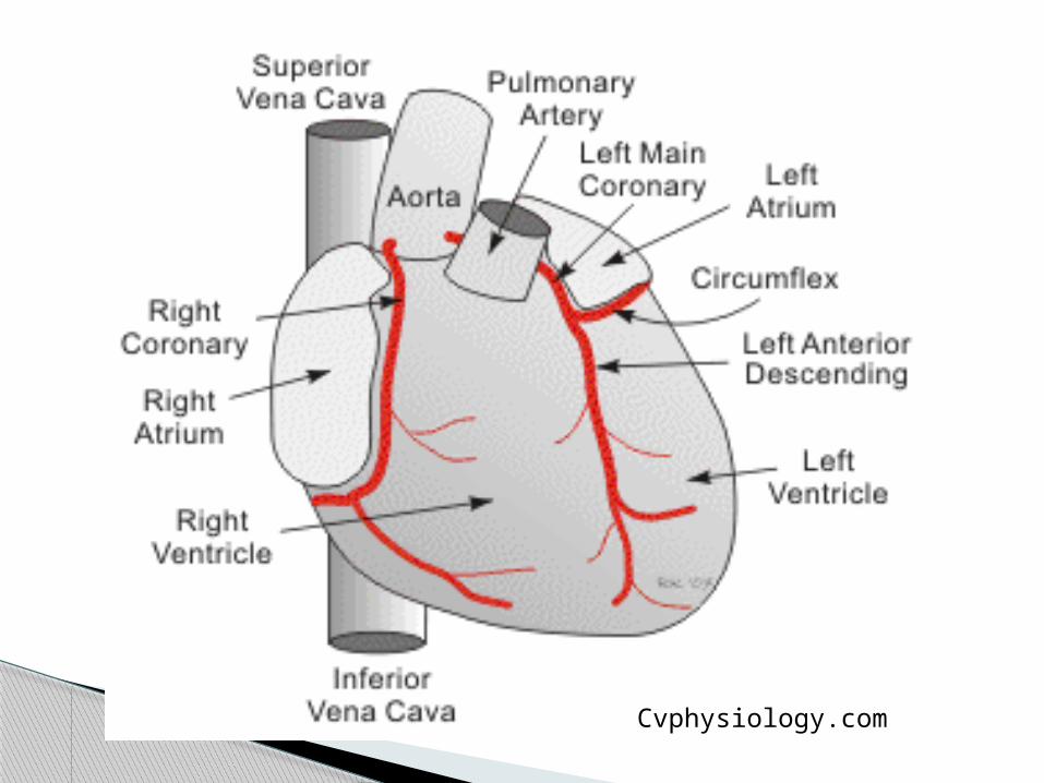
Function of coronary arteries: delivery of oxygenated blood to the heart muscle (myocardium)
The VERY Basics
How-to-draw-cartoons-online.com
Left coronary artery: arises from the left coronary sinus/cusp of the Aortic valve◦ Left main artery branches into:
LAD- Left Anterior Descending CX- Circumflex
Right coronary artery: arises from the right coronary sinus/cusp of the Aortic valve
Main Branches
Coronary arteries lie on top of the myocardium (epicardial) and follow the Atrioventricular (AV) groove and the Interventricular (IV) groove
CX courses along AV groove
LAD and distal RCA follow IV groove
Basics, cont
Cvphysiology.com
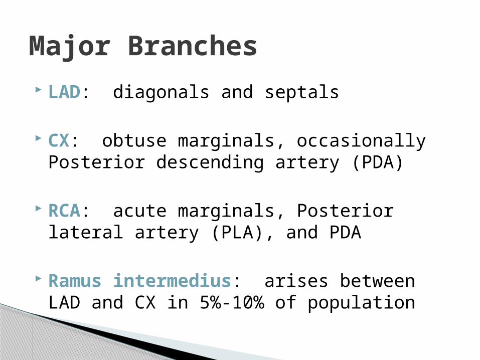
LAD: diagonals and septals
CX: obtuse marginals, occasionally Posterior descending artery (PDA)
RCA: acute marginals, Posterior lateral artery (PLA), and PDA
Ramus intermedius: arises between LAD and CX in 5%-10% of population
Major Branches
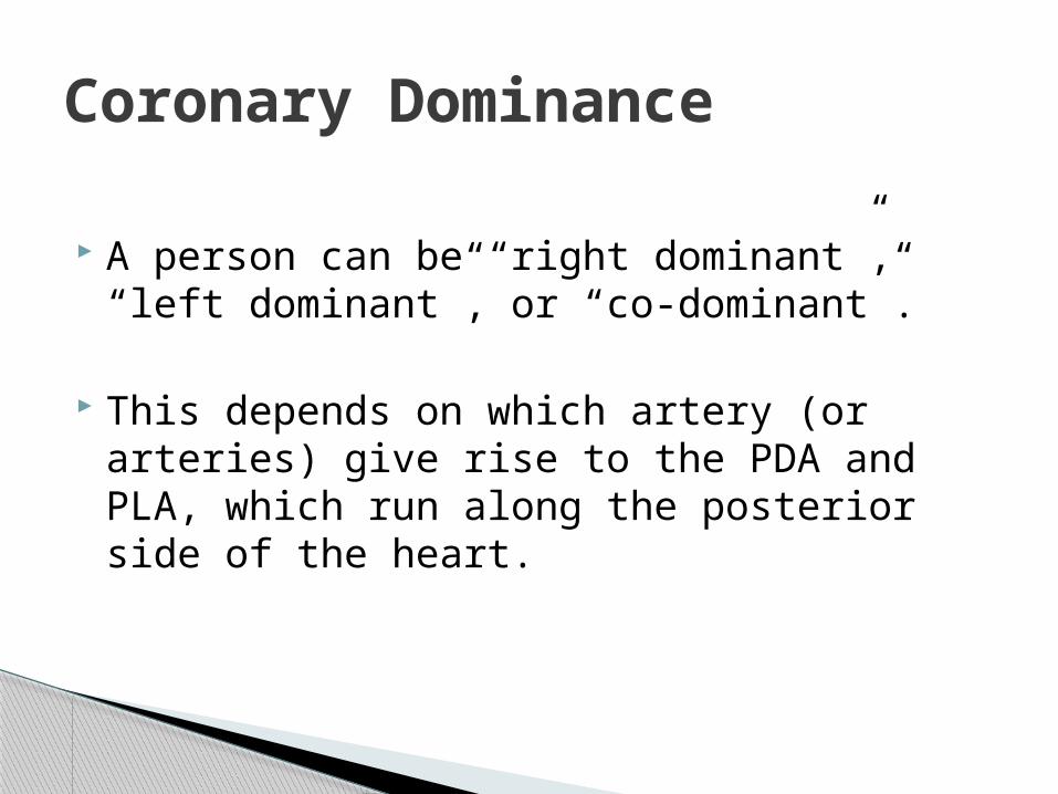
A person can be “right dominant”, “left dominant”, or “co-dominant”.
This depends on which artery (or arteries) give rise to the PDA and PLA, which run along the posterior side of the heart.
Coronary Dominance

Right Dominant
The PDA branch arises from the RCA (60%-70% of population)

Left Dominant
The PDA arises from the LCA (10%-15% of population)
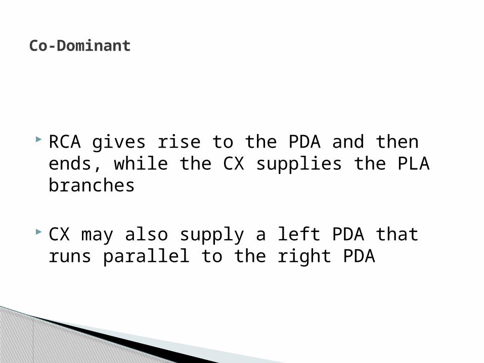
RCA gives rise to the PDA and then ends, while the CX supplies the PLA branches
CX may also supply a left PDA that runs parallel to the right PDA
Co-Dominant

As with all structures in the human body, WEIRD stuff can happen! A few examples:
CX originates with RCA from right sinus LM from right sinus RCA from left sinus Separate originations (ostia) for all three All three arteries from one ostia
Variations and Anomalies
Coronary aneurysms
Fistulas- abnormal communication with venous system
Anomalous origin of LCA from Pulmonary Artery (defect that can have a mortality rate of 90% in first year of life, due to MI or MR leading to CHF)
Anomalies
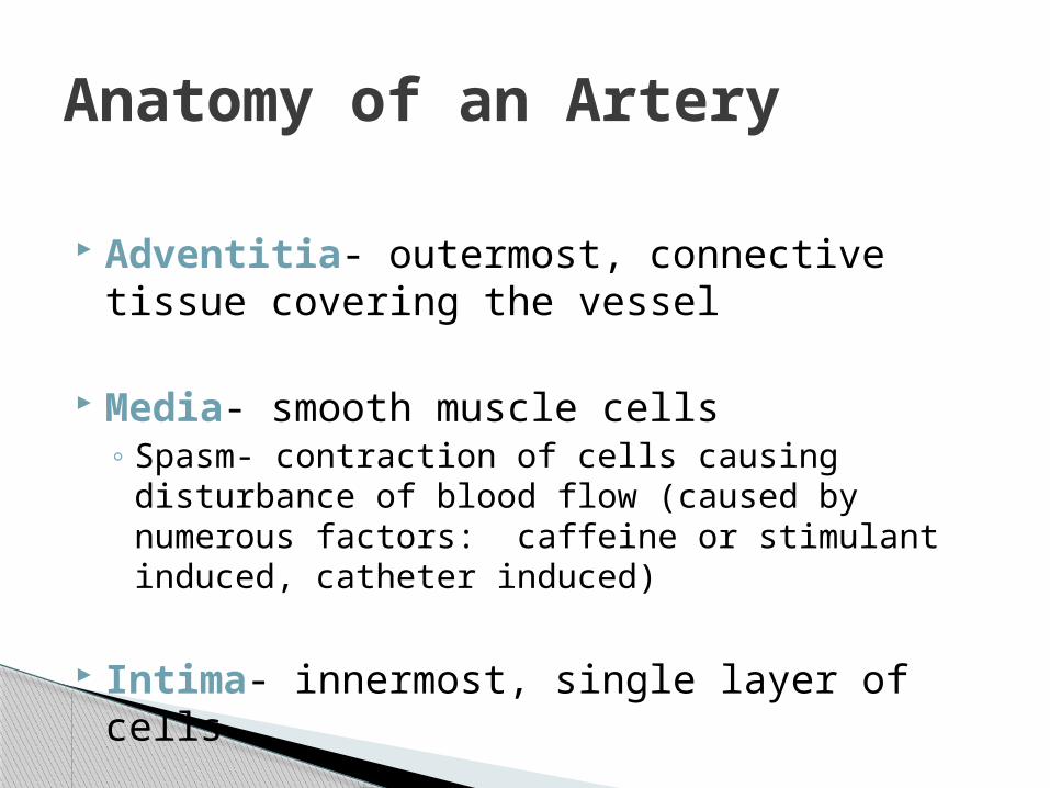
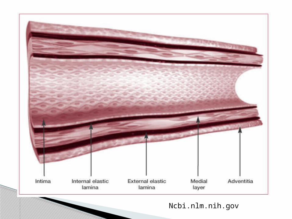
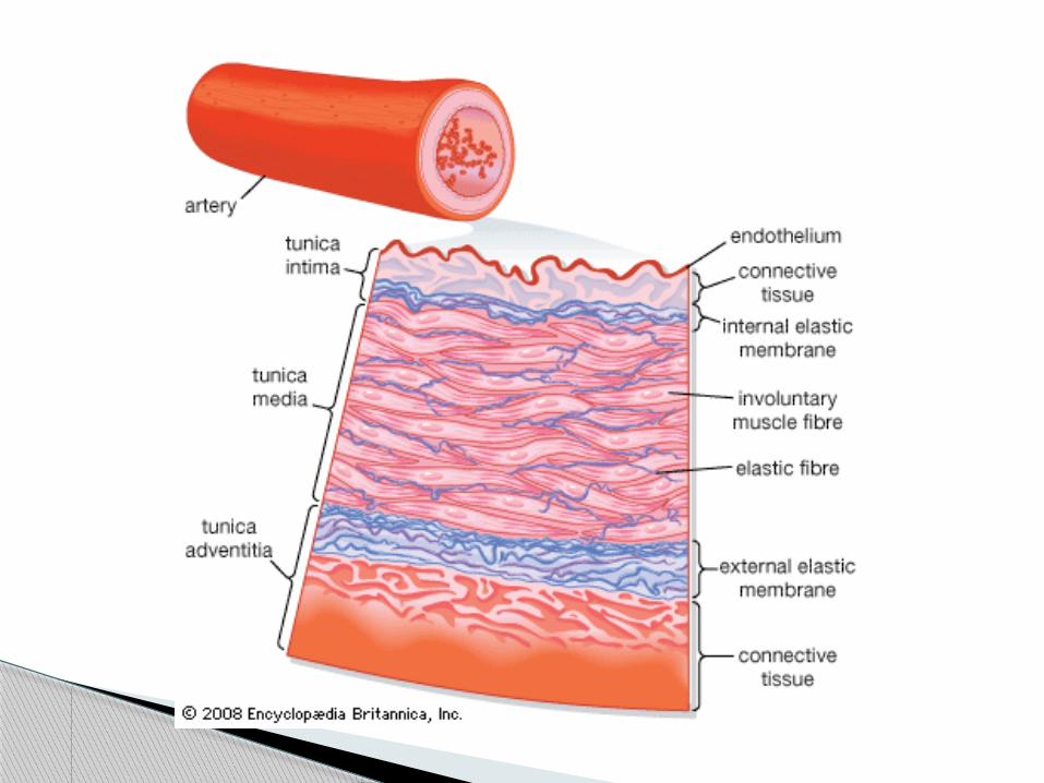
Adventitia- outermost, connective tissue covering the vessel
Media- smooth muscle cells◦ Spasm- contraction of cells causing disturbance of
blood flow (caused by numerous factors: caffeine or stimulant induced, catheter induced)
Intima- innermost, single layer of cells
Anatomy of an Artery
Ncbi.nlm.nih.gov
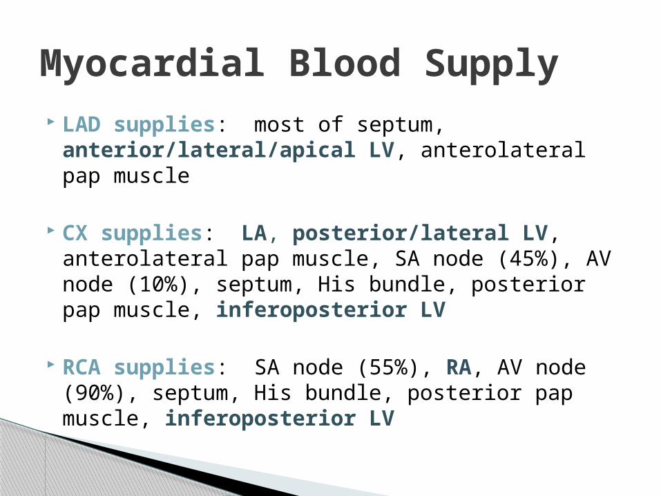
LAD supplies: most of septum, anterior/lateral/apical LV, anterolateral pap muscle
CX supplies: LA, posterior/lateral LV, anterolateral pap muscle, SA node (45%), AV node (10%), septum, His bundle, posterior pap muscle, inferoposterior LV
RCA supplies: SA node (55%), RA, AV node (90%), septum, His bundle, posterior pap muscle, inferoposterior LV
Myocardial Blood Supply
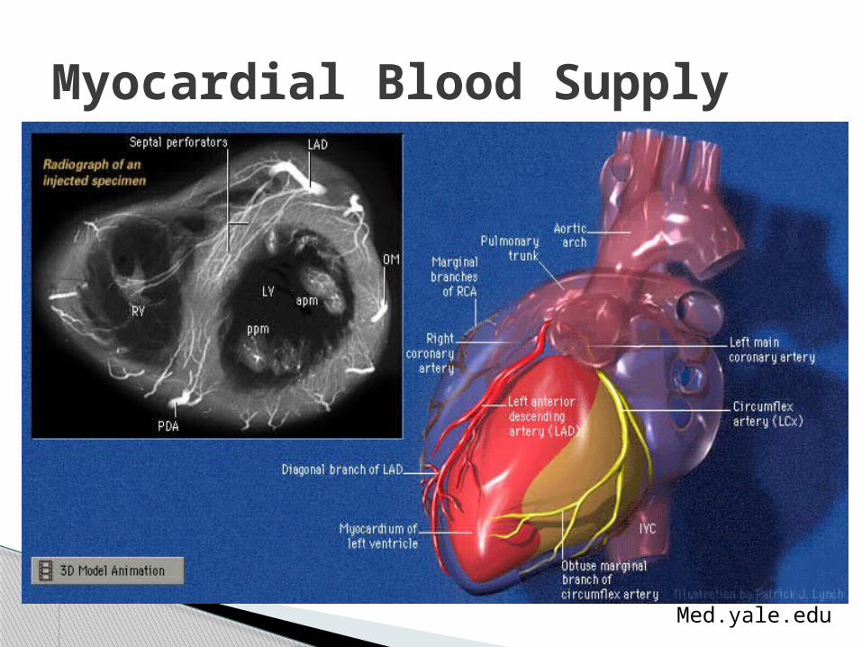
Myocardial Blood Supply
Med.yale.edu
Coronary Arteries perfuse in diastole (this is part of the theory behind the IABP)
Coronary sinus collects used blood from the mycardium to send to lungs for re-oxygenation
Coronary bridging = compression of a coronary artery by the myocardium during systole◦ Usually benign, but can occasionally result in MI or
even death (most common with LAD)
Hemodynamics
Several medications are injected to elicit vasodilation in the arteries during catheterization, such as diltiazem, verapamil, adenosine, nitroglycerine, and nipride
IIbIIIa Inhibitors (abciximab, eptifibitide) are also directly injected in coronary arteries with apparent thrombus
Pharmacology
CAD and Atherosclerosis: What does this mean, exactly?
Build-up of fatty substances, cholesterol, cellular waste products, and calcium within the intima of an artery
With or without symptoms
Coronary Artery Disease
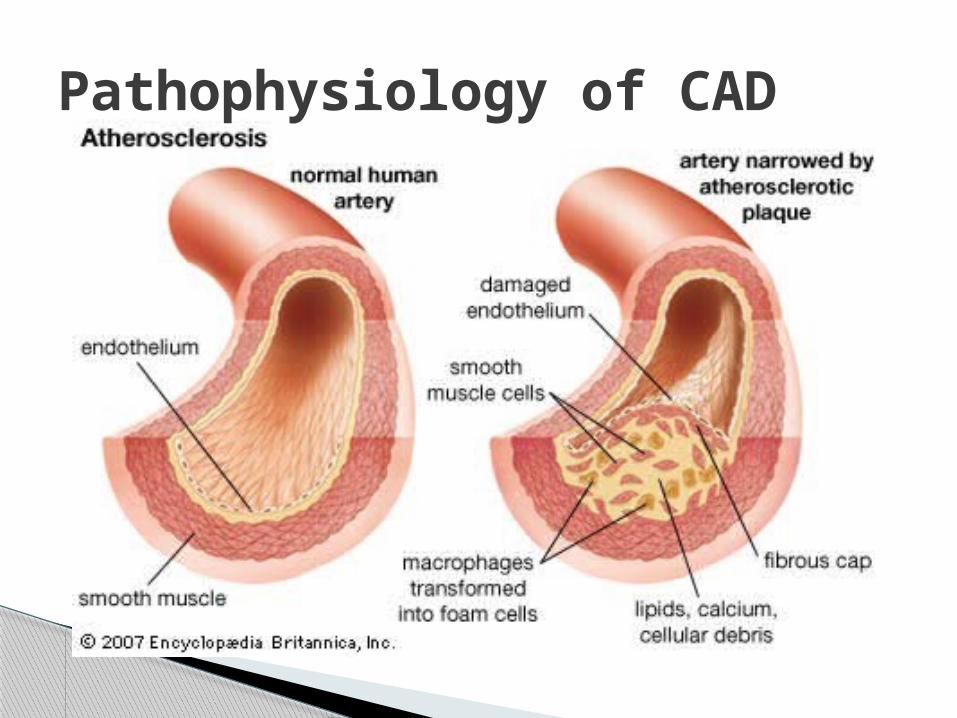
Pathophysiology of CAD

Rupture of fibrous cap
Platelets rush in to fix the vessel
Clot forms
Blood flow obstructed
Damage/death of myocardium
Pathophysiology of Acute MI

Many tests and screening tools are available to help detect CAD
Can be invasive or non-invasive
Can be performed in MD’s office or hospital, depending on type of test
Diagnostic Testing Methods

Treadmill- assesses coronary blood flow by ECG, blood pressure, and signs/symptoms during exercise
Stress Tests

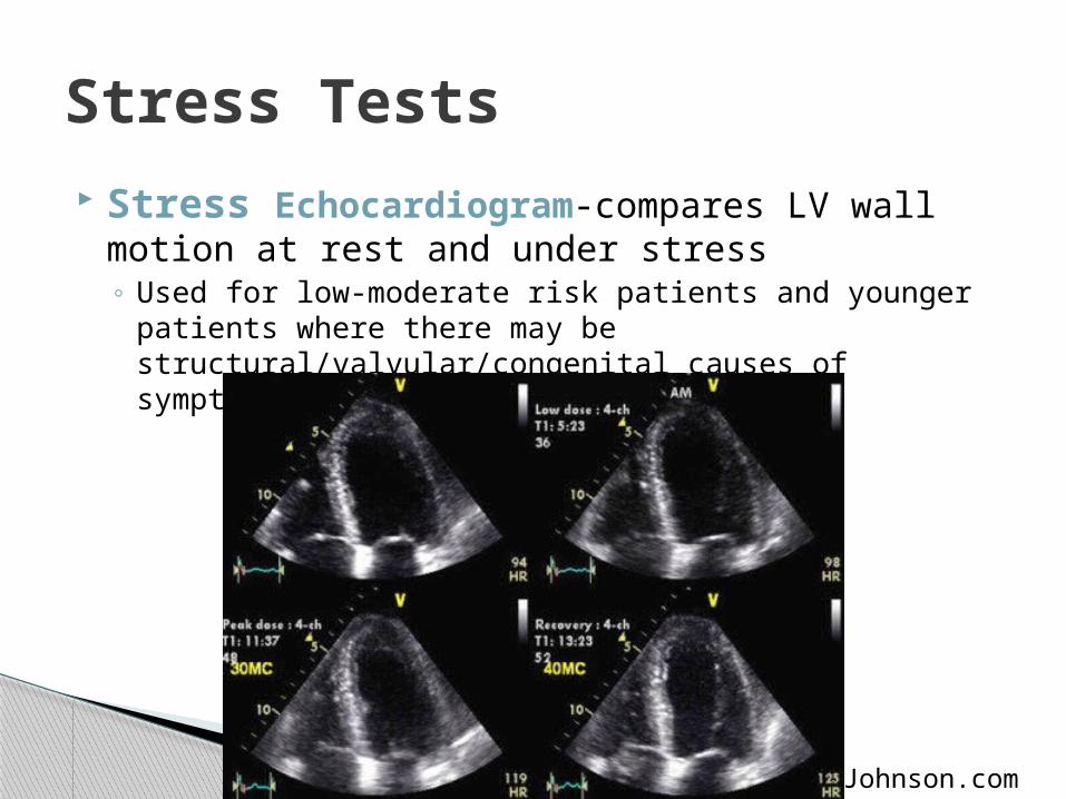
Stress Echocardiogram-compares LV wall motion at rest and under stress◦ Used for low-moderate risk patients and younger patients
where there may be structural/valvular/congenital causes of symptoms
Stress Tests
Johnson.com
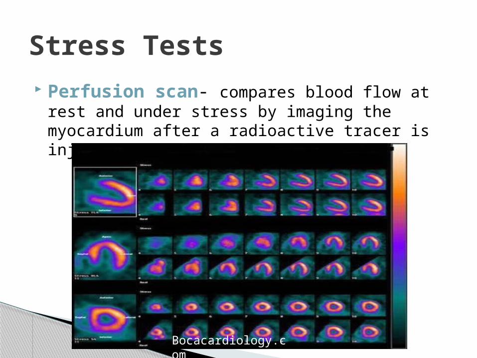
Perfusion scan- compares blood flow at rest and under stress by imaging the myocardium after a radioactive tracer is injected
Stress Tests
Bocacardiology.com

Echocardiogram- assesses structural, valvular, and congenital causes of heart disease
Diagnostic Testing
Brighamandwomens.org
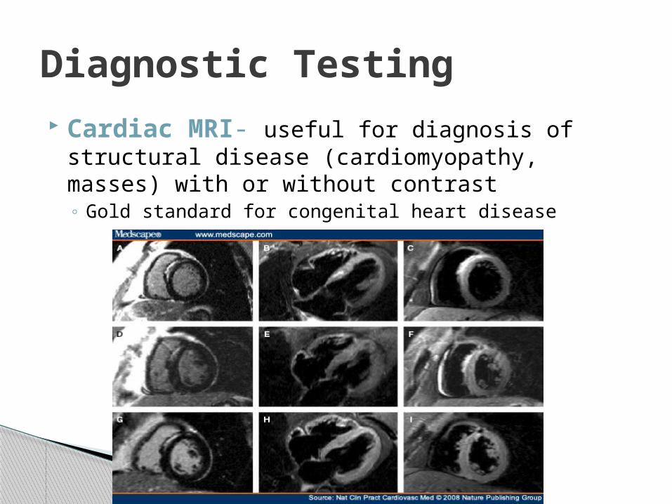
Cardiac MRI- useful for diagnosis of structural disease (cardiomyopathy, masses) with or without contrast◦ Gold standard for congenital heart disease
Diagnostic Testing

Cardiac Computed Tomography (CT)- evaluates coronary arteries as well as LV function, anatomy, and calcification (calcium score)
Diagnostic Testing
Imagingeconomics.com

Cardiac CT Images
64ctscan.comCsmc.edu

Coronary Angiography- used for positive and indeterminate stress tests, assessment of bypass grafts◦ Also for patients with known history of CAD◦ Gold standard for coronary evaluation
Diagnostic Testing
Circulation.or.kr

IVUS- small ultrasound catheter is inserted in the coronary artery to image the vessel and assess plaque◦ Can differentiate between fibrous and calcified plaque◦ Virtual histology
Diagnostic TestingIntravascular Ultrasound
Ptca.org

Technically: the ratio of blood flow in a stenotic artery to normal flow
Essentially: a stress test on a specific artery◦ Flow is measured by a special guidewire, using flow
measurements beyond the lesion and comparing them with flow before the lesion
◦ IV infusion of Adenosine is used to increase HR ◦ Ratio is calculated from a 2-3 minute period◦ Normal value = 1.0◦ Abnormal value = <0.75
Diagnostic TestingFractional Flow Reserve (FFR)
Treatment of CAD
Medical treatment- managing the patient’s medications to help alleviate symptoms
Percutaneous Coronary Intervention (PCI)- used in various situations, from single lesions to complex, high-risk multi vessel disease

Nhlbi.nih.gov

Treatment of CAD Coronary Artery Bypass Grafting- most
often used for severe multi vessel disease and diabetic patients
Beaumonthospitals.com
We’ve come a long way from the first accidental coronary angiogram in 1958…
Diagnosis and treatments continue to evolve
Coronary Arteries Summary
Peripheral Arterial Disease (PAD)
◦ Same arterial anatomy and disease process, but most patients (with exception of CVA) tend to wait much longer to seek treatment
May mimic arthritis, neuropathy Symptoms attributed to “old age”
What about Peripheral Vascular Disease?

Claudication- leg pain with walking that resolves at rest
Decreased temperature of extremity
Non-healing wounds
Symptoms of LE PAD
Sudden numbness or weakness, especially on one side
Sudden confusion, trouble speaking
Sudden trouble seeing in one or both eyes
Sudden dizziness, loss of balance and coordination
Sudden severe headache
Symptoms of CVA
Ultrasound/Doppler studies
MRI/MRA
CT/CTA
Angiography
Diagnostic Testing for PAD

Medications
Percutaneous Transluminal Angioplasty (PTA) with or without stent placement
Atherectomy
Bypass surgery
Treatment of PAD

Interventions done for high-risk, asymptomatic or symptomatic patients (depends on clinical study enrollment)
All patients must be enrolled in a research study to receive a stent
◦ Typical criteria: asymptomatic with >80% stenosis by ultrasound, or symptomatic with >50% stenosis and at least one high-risk factor, such as age>80 years, CHF, severe COPD, previous CEA with restenosis, previous radiation therapy or neck surgery, lesion location
Carotids

Cleveland Clinic Heart and Vascular Institute

Code Stroke
An Interdisciplinary effort to get treatment started for stroke victims ASAP
◦ Emcompasses: ED assessment, activation of Code Stroke protocol, Neurology consults, CT scans, Interventional Cardiology consults, thrombolytic treatment if indicated, invasive intervention if indicated
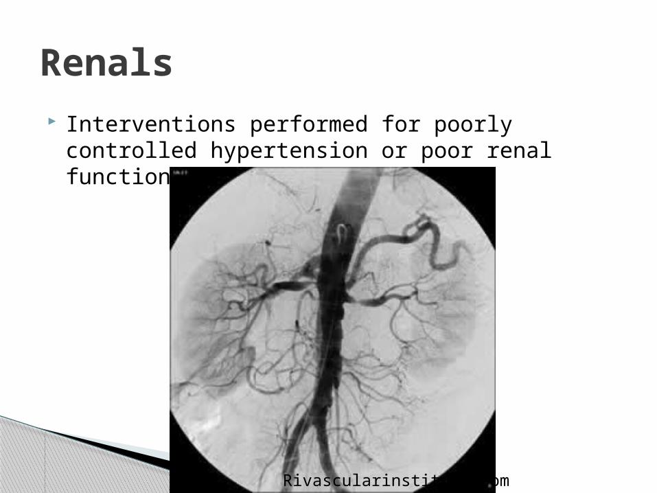
Renals Interventions performed for poorly controlled
hypertension or poor renal function
Rivascularinstitute.com
Interventions performed for claudication, critical limb ischemia (non healing wounds), and limb salvage
Lower Extremity

Radiologyrsnajnls.org

PAD
Basically, if we can get a catheter to an artery, we can take a picture and potentially intervene!
◦ Mesenterics◦ Subclavians◦ ETC…

Fast growing segment of cardiac cath lab procedures
Percutaneous treatments are constantly being developed
PAD Summary

Cvphysiology.com Heartsite.com NEJM, Vol. 360 No.3 Ncbi.nlm.nih.gov Encyclopedia Brittanica Med.yale.edu Johnson.com Bocacardiology.com Brighamandwomens.org Nature Publishing Group Emedicine.medscape.com
Sources

64ctscan.com Csmc.edu Circulation.or.kr Ptca.org Beaumonthospitals.com Nhlbi.nih.gov Cleveland Clinic Heart and Vascular Institute Rivascularinstitute.com Radiologyrsnajnls.org Imagingeconomics.com How-to-draw-cartoons-online.com
Sources


