Correction of Shunt from Right Conal Coronary Artery to Pulmonary Trunk with Relief of Symptoms By GERALD B. LEE, M.D., FREDARICK L. GOBEL, M.D., C. WALTON LILLEHEI, M.D., WALTER S. NEFF, M.D., AND ROBERT S. ELIOT, M.D. SUMMARY A case of a small right coronary conal branch to pulmonary trunk shunt with surgical correction is reported. The patient, a 45-year-old housewife, had chest pain, labile hypertension, and intermittent left bundle-branch block with normal serum cholesterol and triglyceride levels. Selective coronary arteriography was necessary to demonstrate the abnormal communication between the conal branch of the right coronary artery and the pulmonary trunk. Symptoms disappeared after surgical correction of the shunt. Transient left bundle-branch block appeared during exercise after surgery, but the patient was improved subjectively and resumed all household chores without difficulty. Shunting of oxygenated blood away from the myocardium thereby decreasing coronary blood flow to a specific area may have been responsible for significant symptomatology. Additional Indexing Words: Coronary arteriovenous fistula Coronary arteriography CORONARY arteriovenous fistulas are rel- atively uncommon and therefore account for few cases of myocardial ischemia in adults.' Anomalous communications between the conus artery and the pulmonary trunk are even more unusual. When such a shunt is small, it may be overlooked clinically or at necropsy. That such an apparently insig- nificant shunt might bring about major man- ifestations of myocardial ischemia is of significant interest. Case Report A 45-year-old housewife was hospitalized in From the Departments of Medicine and Surgery, University of Minnesota Hospitals, Minneapolis, Min- nesota. Supported by Cardiovascular Research Program Project Grant HE 06314-04; U. S. Public Health Service Research Grant HE 00830; The Max Baer Heart Fund-Fraternal Order of Eagles; Minnesota Heart Association; Sussex County Heart Association, Newton, New Jersey; and Maria and Joseph Gales Ramsay, III, Cardiovascular Research Fund. 244 Conus artery Angina pectoris November 1965 with the initial episode of severe retrosternal chest pain. An electrocardiogram showed left bundle-branch block. Serial enzyme determinations did not suggest acute myocardial necrosis. The blood pressure was 160/85 mm Hg. After discharge the electrocardiogram changed to normal intraventricular conduction but was sug- gestive of left ventricular hypertrophy. During a two-step exercise test left bundle-branch block re- appeared transiently and was accompanied by chest pain and dyspnea. Because of increasing symptoms she was referred to the University of Minnesota Hospitals. The patient related that she was unable to perform her household duties because of chest pain. She gave a 9-year history of labile hypertension. She stated that her menses were normal and regu- lar. There was no known history of premature cardiovascular disease in the family although her mother also has hypertension. Physical examina- tion revealed a slender white female. The blood pressure was 150/90, and the pulse was 82 and regular. The retinal vessels appeared normal. The neck veins were not distended, and the heart was normal in size. A grade II/VI early systolic mur- mur was audible at the left sternal border. Laboratory data included a normal hemoglobin, white blood cell count and differential. A urine Circulation, Volume XXXVII, February 1968 by guest on October 4, 2017 http://circ.ahajournals.org/ Downloaded from
Transcript
Correction of Shunt from Right Conal CoronaryArtery to Pulmonary Trunk with
Relief of SymptomsBy GERALD B. LEE, M.D., FREDARICK L. GOBEL, M.D., C. WALTON LILLEHEI, M.D.,
WALTER S. NEFF, M.D., AND ROBERT S. ELIOT, M.D.
SUMMARYA case of a small right coronary conal branch to pulmonary trunk shunt with surgical
correction is reported. The patient, a 45-year-old housewife, had chest pain, labilehypertension, and intermittent left bundle-branch block with normal serum cholesteroland triglyceride levels. Selective coronary arteriography was necessary to demonstratethe abnormal communication between the conal branch of the right coronary arteryand the pulmonary trunk. Symptoms disappeared after surgical correction of the shunt.Transient left bundle-branch block appeared during exercise after surgery, but thepatient was improved subjectively and resumed all household chores without difficulty.Shunting of oxygenated blood away from the myocardium thereby decreasing coronaryblood flow to a specific area may have been responsible for significant symptomatology.
CORONARY arteriovenous fistulas are rel-atively uncommon and therefore account
for few cases of myocardial ischemia inadults.' Anomalous communications betweenthe conus artery and the pulmonary trunkare even more unusual. When such a shuntis small, it may be overlooked clinically orat necropsy. That such an apparently insig-nificant shunt might bring about major man-ifestations of myocardial ischemia is ofsignificant interest.
Case ReportA 45-year-old housewife was hospitalized in
From the Departments of Medicine and Surgery,University of Minnesota Hospitals, Minneapolis, Min-nesota.
Supported by Cardiovascular Research ProgramProject Grant HE 06314-04; U. S. Public HealthService Research Grant HE 00830; The Max BaerHeart Fund-Fraternal Order of Eagles; MinnesotaHeart Association; Sussex County Heart Association,Newton, New Jersey; and Maria and Joseph GalesRamsay, III, Cardiovascular Research Fund.
244
Conus artery Angina pectoris
November 1965 with the initial episode of severeretrosternal chest pain. An electrocardiogramshowed left bundle-branch block. Serial enzymedeterminations did not suggest acute myocardialnecrosis. The blood pressure was 160/85 mm Hg.
After discharge the electrocardiogram changedto normal intraventricular conduction but was sug-gestive of left ventricular hypertrophy. During atwo-step exercise test left bundle-branch block re-appeared transiently and was accompanied bychest pain and dyspnea. Because of increasingsymptoms she was referred to the University ofMinnesota Hospitals. The patient related thatshe was unable to perform her household dutiesbecause of chest pain.
She gave a 9-year history of labile hypertension.She stated that her menses were normal and regu-lar. There was no known history of prematurecardiovascular disease in the family although hermother also has hypertension. Physical examina-tion revealed a slender white female. The bloodpressure was 150/90, and the pulse was 82 andregular. The retinal vessels appeared normal. Theneck veins were not distended, and the heart wasnormal in size. A grade II/VI early systolic mur-mur was audible at the left sternal border.
Laboratory data included a normal hemoglobin,white blood cell count and differential. A urine
Figure 1The preoperative vectorcardiogram (VCG) shows a slight increase in QRS loop size. (Systemused was that of Schmitt-SVEC III.) An electrocardiogram (ECG) recorded on the same dayshows nonspecific ST-T changes.
culture grew Escherichia coli in significant num-bers and then became sterile after treatment withtetracycline. Urinalysis, blood urea nitrogen, se-rum creatinine, LDH, blood glucose, and electro-lytes were normal. Serum cholesterol was 124mg%. The serum triglycerides were 108 mg%. Thechest x-ray and cardiac fluoroscopy were normal.The electrocardiogram showed nonspecific ST-Tchanges (fig. 1) . Vectorcardiogram revealed a QRSloop that was slightly increased in size but other-wise was normal.Circulation, Volume XXXVII, February 1968
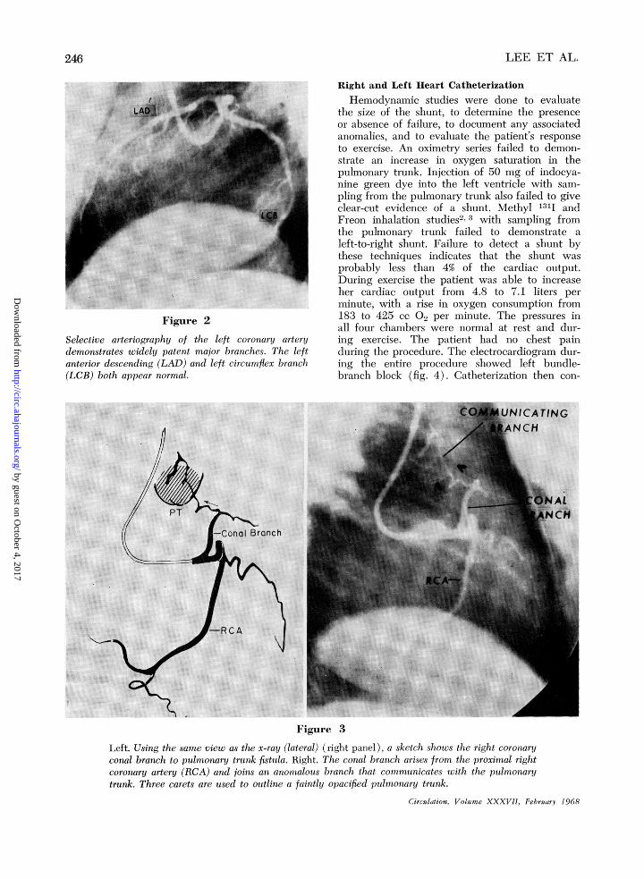
Coronary ArteriographyTwo days after admission, a bilateral selective
coronary arteriogram was performed. The leftcoronary artery and major branches were normal(fig. 2). The right coronary artery (RCA) waswidely patent and preponderant. A moderatelylarge conal branch originated from the proximalportion of the RCA, deviated cephalad and tothe left, and communicated with an accessorycoronary artery from the pulmonary trunk (fig.3).
Selective arteriography of the left coronary arterydemonstrates widely patent major branches. The leftanterior descending (LAD) and left circumflex branch(LCB) both appear normal.
Right and Left Heart CatheterizationHemodynamic studies were done to evaluate
the size of the shunt, to determine the presenceor absence of failure, to document any associatedanomalies, and to evaluate the patient's responseto exercise. An oximetry series failed to demon-strate an increase in oxygen saturation in thepulmonary trunk. Injection of 50 mg of indocya-nine green dye into the left ventricle with sam-pling from the pulmonary trunk also failed to giveclear-cut evidence of a shunt. Methyl 1311 andFreon inhalation studies2 3 with sampling fromthe pulmonary trunk failed to demonstrate aleft-to-right shunt. Failure to detect a shunt bythese techniques indicates that the shunt wasprobably less than 4% of the cardiac output.During exercise the patient was able to increaseher cardiac output from 4.8 to 7.1 liters perminute, with a rise in oxygen consumption from183 to 425 cc 02 per minute. The pressures inall four chambers were normal at rest and dur-ing exercise. The patient had no chest painduring the procedure. The electrocardiogram dur-ing the entire procedure showed left bundle-branch block (fig. 4). Catheterization then con-
Figure 3
Left. Using the same view as the x-ray (lateral) (right panel), a sketch shows the right coronaryconal branch to pulmonary trunk fistula. Right. The conal branch arises from the proximal rightcoronary artery (RCA) and joins an anonwlous branch that communicates with the pulmonarytrunk. Three carets are used to outline a faintly opacified pulmonary trunk.
Figure 4Electrocardiogram during the time of heart catheter-ization showed persistent left bundle-branch block(record was taken at a paper speed of 25 mm per
second).
firmed the facts that the left-to-right shunt dem-onstrated angiographically was small, that therewas no failure, that there were no associatedanomalies, and that the exercise response was
normal.SurgeryOne week following catheterization a left
thoractomy was performed. The communicatingvessel between the conal branch of the RCA andthe pulmonary trunk measured 2 to 3 mm indiameter. After transection of this vessel, pul-satile bright red blood came from the conalbranch, whereas venous blood appeared fromthe portion communicating with the pulmonarytrunk. There were no complications in the post-operative period.
During the 8 months following operation shehad no recurrence of chest pain and resumedall her household chores without difficulties. Theblood pressure remained labile but averaged165/100 mm Hg. The murmur at the leftsternal border could no longer be heard. Herresting electrocardiogram showed nonspecific ST-Tchanges. A left bundle-branch block appearedtransiently during a two-step exercise test, butdyspnea and chest pain were not present.
Discussion
A shunt from the right conal branch ofthe RCA to the pulmonary trunk is anotherunusual cause of nonatheromatous myocardialischemia. The true incidence of this shuntis unknown but it is probably uncommon.
Such a shunt is clinically elusive, as theremay be no characteristic historical or physicalfindings. In the case presented, the symptomswere suggestive of angina pectoris, and themurmur appeared innocent in type. The lab-
Circulation, Volume XXXVII, February 1968
oratory studies may also be noncontributoryas evidenced in this case by normal serumcholesterol, serum triglycerides, and myocar-dial enzymes. Because of its small size, theleft-to-right shunt may be readily missed atroutine heart catheterization. Aortic root ar-teriography probably would not lead to ad-equate visualization of such a small vesselbecause of inadequate filling of the coronaryarteries. Lack of awareness of the conditioncombined with its small size may lead to itsbeing completely overlooked at necropsy.Normally about 4 to 6% of the cardiac
output is diverted to the coronary arterialsystem.4 We do not know the actual bloodflow through this anastomosis or how muchthe remaining vascular bed was compromisedin this patient. The fact that the shunt wassmall, however, was documented by cardiaccatheterization, by coronary arteriography,and by direct visualization of the small vesselat the time of surgery. That such a small shuntcould produce such dramatic symptoms isprovocative. One may only speculate thatoxygenated blood was intermittently divertedaway from a portion of myocardium and fromthe left bundle branch. Studies by James5' 6
have shown that the right coronary artery-when preponderant-does supply the proximalfew millimeters of the left bundle by terminalbranches of the A-V node artery.
In the case reported, postoperative exercisecaused a transient reappearance of leftbundle-branch block. Furthermore, subjectiverelief of anginal pain following operationcannot be considered proof of the importanceof the shunt. The possibility of "small vesseldisease" always exists among hypertensivepatients. When small vessel disease does exist,a small shunt might have a more significantand deleterious hemodynamic effect than inthe normal coronary vascular bed. It is pos-sible that pathological changes in the con-duction system had become fixed or thatdisease in the coronary vessels was presentbut unrecognized.
In approximately half of the specimensexamined by James5 6 and Schlesinger andassociates,7 the conus artery was a direct
branch of the aorta; in these cases the coniusartery ostium was located usually near theostium of the RCA. In the remainder of thespecimens the conus artery was the first ven-tricular branch of the RCA (conal branch ofRCA). Regardless of the origin, the conusartery normally supplied the conus arteriosusand, in addition, anastomosed with a branchof the left coronary artery to form Vieussens'ring.8 We were unable to visualize this ringby selective coronary arteriography. It is pos-sible that, in early development of the heart,anastomosis occurred between the conalbranch of the RCA and an accessory coronaryartery arising from the pulmonary trunk.A review of the literature failed to reveal
a case of an extensively studied and surgicallycorrected conal artery to pulmonary trunkshunt. In most reported cases, the anomalouscoronary artery is found to enter one of thefour cardiac chambers, the pulmonarytrunk,1' 7or the bronchial circulation.9 Thevessel is usually enlarged and elongated, withfocal saccular aneurysms. An increase in oxy-gen saturation in the chamber receiving theanomalous vessel has been reported.' In 1958,Edwards and associates'0 described at nec-ropsy the communication of a branch of theright coronary artery with the right atrium.In 1960, Amplatz and associates"- demonstrat-ed by aortography a left coronary artery tomain pulmonary artery fistula. In 1961, Neufeldand associates' summarized six cases of "coro-nary artery fistula" diagnosed at the Univer-sity of Minnesota Hospitals; two of the sixhad anomalous right coronary arteries thatterminated in the right atrium and rightventricle. These authors described six pre-viously studied cases in the literature withanomalous termination of the RCA into thepulmonary trunk. In four of the six casesreported by Neufeld, there was an increasein oxygen saturation in the cardiac chamberin which the anomalous vessel terminated.In no case reported is there a description ofa communication between an aberrant arterybetween the pulmonary trunk and the conalbranch of the RCA demonstrated by arteriog-raphy.
ConclusionIt is difficult to be certain that the patient's
symptoms were related to the small shuntdemonstrated by arteriography and at sur-gery. Relief of chest pain postoperatively iscommon even in patients who have had shamoperations for coronary artery disease. Theleft bundle-branch block could well be dueto undiagnosed small vessel disease or sec-ondary to systemic hypertension. It is possiblethat a specific portion of cardiac tissue wasintermittently "robbed" of oxygenated bloodthat was diverted to the pulmonary circula-tion.The risk of surgical correction of the shunt
described is that of thoracotomy alone. Whenthe shunt is larger and leads to cardiac failurethe hazard is, of course, greater. The reliefof myocardial ischemia by a corrective op-eration warrants such a risk.
References1. NEUFELD, H. N., LESTER, R. G., ADAMS, P., JR.,
ANDERSON, R. C., LILLEHEI, C. W., AND ED-WARDS, J. E.: Congenital communication of acoronary artery with a cardiac chamber orthe pulmonary trunk. Circulation 24: 171,1961.
2. AMPLATZ, K.: Methyl iodide test in left to rightshunts. Amer J Roentgen 85: 1059, 1961.
4. ROWE, G.: Nitrous oxide method for determiningcoronary blood flow in man. Amer Heart J58: 268, 1958.
5. JAMES, T. N.: Anatomy of the Coronary Arteries.Paul B. Hoeber, Inc., New York, 1961.
6. JAMES, T. N.: Anatomy of the coronary arteriesand veins. In The Heart: Arteries and Veins,edited by J. Willis Hurst and R. Bruce Logue.New York, McGraw-Hill Book Co., 1966,p. 640.
7. SCHLESINGER, M. J., ZOLL, P. M., AND WESSLER,S.: The conus artery: A third coronary artery.Amer Heart J 38: 823, 1949.
8. VIEUSSENS, R.: Nouvelles Decouvertes sur leCoeur. Paris, 1706.
10. EDWARDS, J. E., GLADDING, T. C., AND WEIR,A. B.: Congenital communications between theright coronary artery and the right atrium.J Thorac Surg 35: 662, 1958.
11. AMPLATZ, K., AGUIRRE, J., AND LILLEHEI, C. W.:Coronary arteriovenous fistula into main pul-monary artery. JAMA 172: 1384, 1960.
is published by the American Heart Association, 7272 Greenville Avenue, Dallas, TX 75231Circulation doi: 10.1161/01.CIR.37.2.244
1968;37:244-248Circulation.
http://circ.ahajournals.org/content/37/2/244located on the World Wide Web at:
The online version of this article, along with updated information and services, is
http://circ.ahajournals.org//subscriptions/
is online at: Circulation Information about subscribing to Subscriptions:
http://www.lww.com/reprints Information about reprints can be found online at: Reprints:
document. and Rights Question and Answer
Permissionsthe Web page under Services. Further information about this process is available in thewhich permission is being requested is located, click Request Permissions in the middle column ofClearance Center, not the Editorial Office. Once the online version of the published article for
can be obtained via RightsLink, a service of the CopyrightCirculationoriginally published in Requests for permissions to reproduce figures, tables, or portions of articlesPermissions: