UNIVERSITA’ DEGLI STUDI DELLA TUSCIA DI VITERBO Dipartimento di Scienze Ecologiche e Biologiche CORSO DI DOTTORATO DI RICERCA IN GENETICA E BIOLOGIA CELLULARE XXVI Ciclo Genetic and cytological analysis of two male sterile mutant strains of Drosophila melanogaster BIO/18 Tesi di dottorato di: Fabiana Fabbretti Coordinatore del corso Tutore Prof. Giorgio Prantera Prof. Giorgio Prantera
Transcript
UNIVERSITA’ DEGLI STUDI DELLA TUSCIA DI VITERBO
Dipartimento di Scienze Ecologiche e Biologiche
CORSO DI DOTTORATO DI RICERCA IN GENETICA E BIOLOGIA CELLULARE
XXVI Ciclo
Genetic and cytological analysis of two male sterile
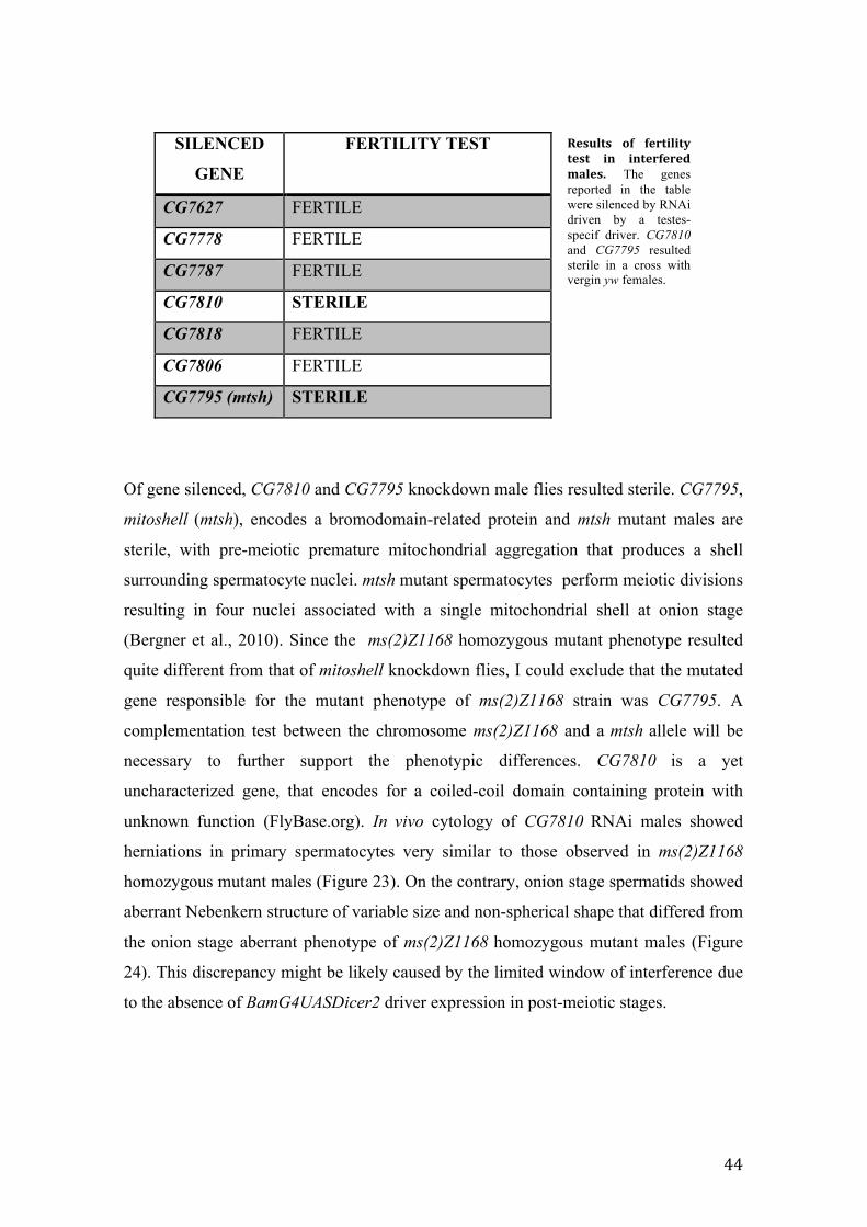
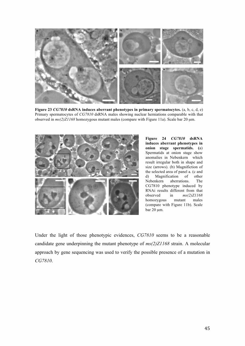
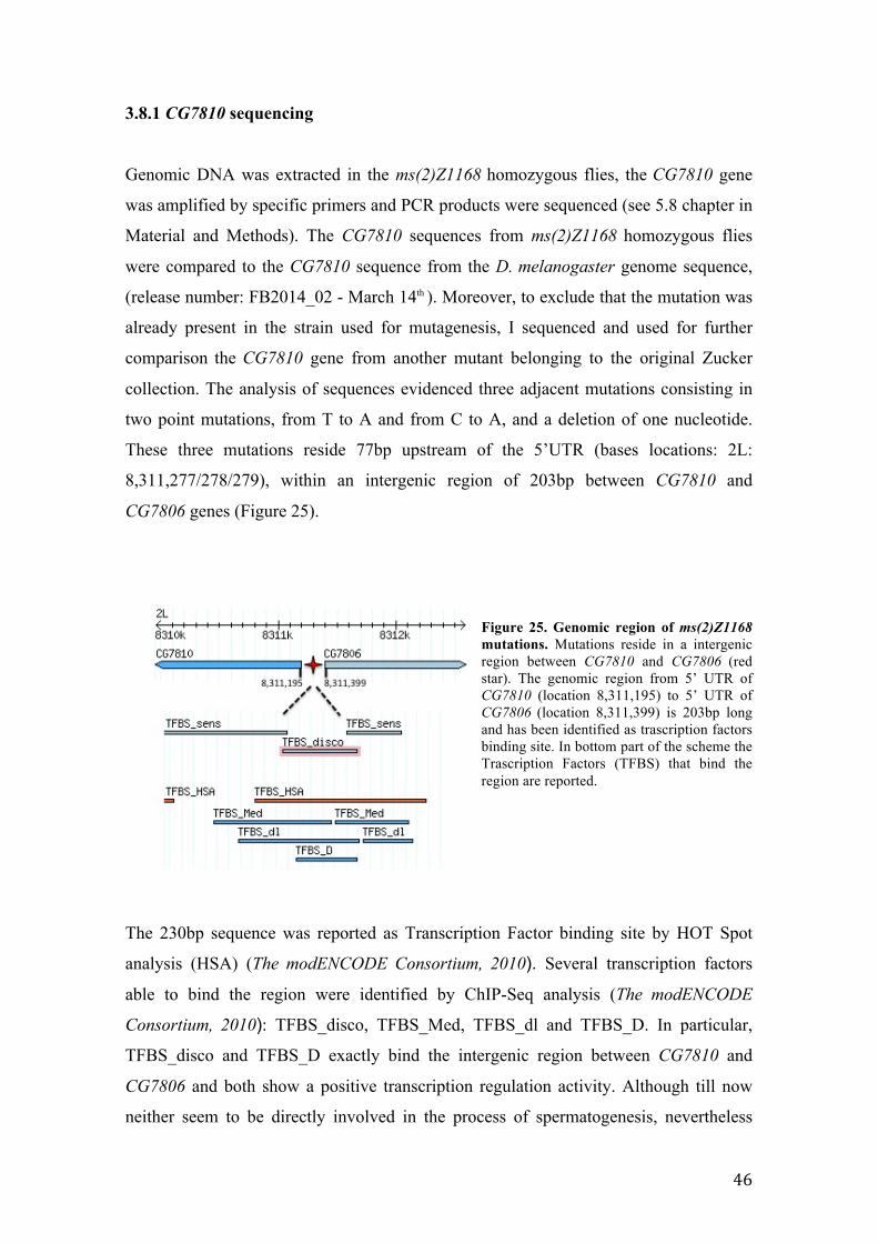
5.7 In vivo cytology of RNA-interfered males for gene identification in ms(2)Z1168
mutant…………………………………………………………………………………..52
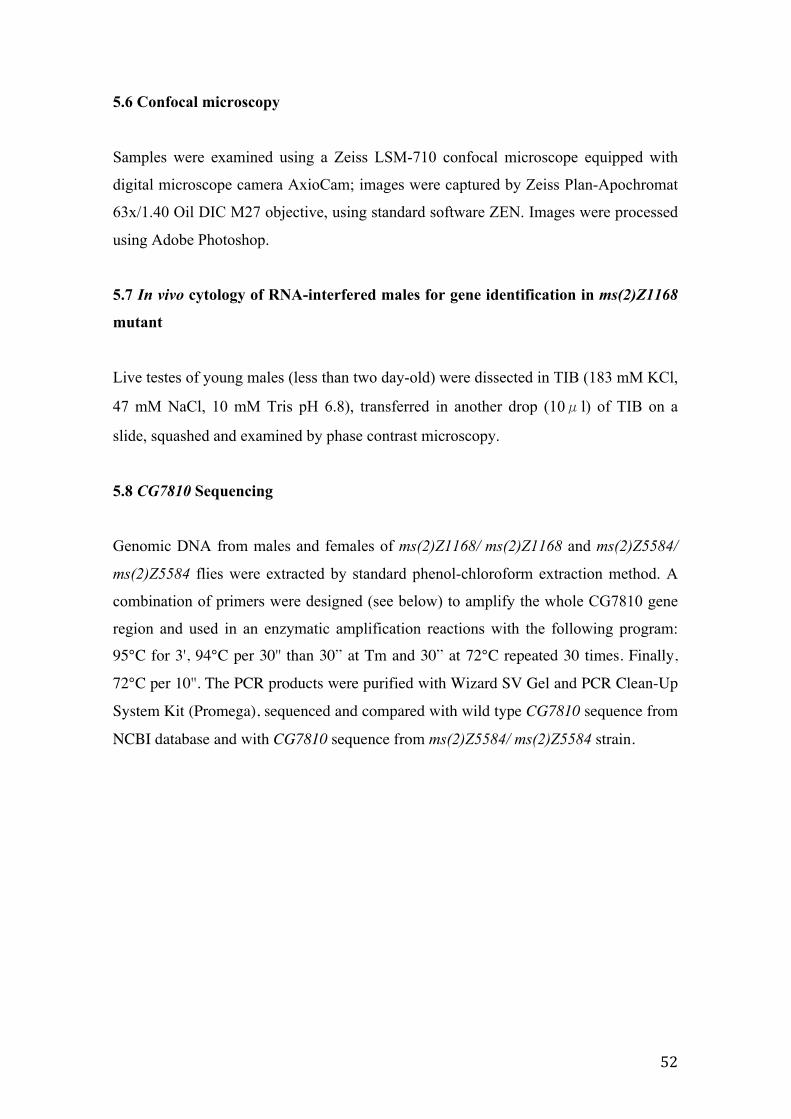
5.8 CG7810 Sequencing………………………………………………………………..52
6. REFERENCES………………………………………………………………………54
II
1
ABSTRACT
Spermatogenesis, the production of male functional gametes from germinal stem cells,
represents one of the most dramatic examples of cell differentiation. The availability of
Drosophila melanogaster mutants defective for specific spermatogenesis stages, the
short Drosophila life cycle, the availability of genetic resources, like a fully sequenced
genome, and the gene and pathway conservation with humans, make this insect
particularly suitable to study the genetic control of the spermatogenesis process. The
aim of my PhD research project was the analysis of two mutant strains (ms(2)Z5584 and
ms(2)Z1168) belonging to a unique collection of 13 ethyl-methansulfonate (EMS)-
induced male sterile recessive mutants identified in a large screening for male-sterile
mutations on chromosome 2 and 3 (Wakimoto et al., 2004). From a preliminary
cytological screen of mutations, a general and common aberrant phenotype affecting the
entry into and progression of the meiotic cell cycle was identified: mutants skipped one
or both meiotic divisions but carried out the differentiation of spermatids although with
anomalies.
The ms(2)Z5584 mutant strain was previously genetically and cytologically
characterized and a point mutation in rae1 gene was identified as responsible of the
aberrant phenotypes (Volpi et al., 2013). Starting from that evidences, I confirmed the
identification of rae1 as the gene underlying the ms(2)Z5584 mutant phenotype, by
RNAi silencing of the wild type rae1. Then, I uncovered the localization pattern of
RAE1 during meiotic cell cycle by GAL4/UAS system allowing the expression of
UASGFP-rae1 transgene under both testis-specific and constitutive drivers. Finally, I
performed the phenotype rescue of the ms(2)Z5584 mutant by using of UASGFP-rae1
transgene.
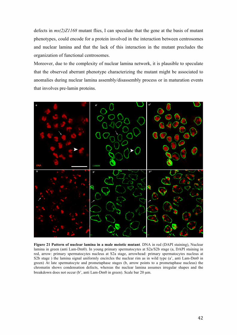
The ms(2)Z1168 mutant strain was cytologically characterized by
immunohistochemistry technique using antibodies against several structures involved in
male meiosis and confocal microscopy. The ms(2)Z1168 mutant exhibited anomalies in
nuclear lamina structure together with chromatin condensation defects. The ms(2)Z1168
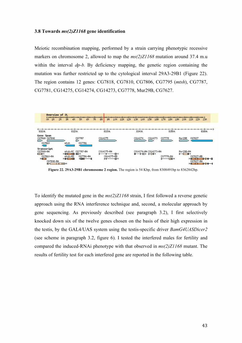
genetic analysis narrowed the gene locus to a genomic region containing 12 genes. The
gene identification was further refined by reverse genetic approach using RNA
interference and by sequencing. Mutations affecting a regulatory region of CG7810
gene were identified in the mutant genome. Finally, since the ms(2)Z1168 mutant
2
exhibited nuclear lamina defects, an accurate nuclear lamina characterization during
wild type meiosis and spermatogenesis was performed.
The study of mutant strains disrupted for some aspects of spermatogenesis
process allows a broader understanding of the genetic and molecular factors involved in
the regulation and in the execution of male meiosis and spermiogenesis.
3
1. INTRODUCTION
Spermatogenesis is one of the most complex differentiation processes leading to
sperm formation starting from an undifferentiated spermatogonial cell. The
process is characterized by a series of mitotic divisions followed by meiotic
divisions to form haploid spermatids and a series of post-meiotic events
involving severe changes in cellular morphology that culminate in the
achievement of the canonical shape of the mature sperm. The availability of
Drosophila melanogaster mutants, that are defective for specific
spermatogenesis stages, makes that insect particularly suitable for the study of
spermatogenesis process. Moreover, Drosophila presents a short generation life
cycle, the availability of genetic resources and a fully sequenced genome.
Finally, about 61% of Drosophila melanogaster genes are conserved in humans
(IHGSC, 2001), as it is the spermatogenesis process in terms of cells
morphology and regulatory pathways.
1.1 Spermatogenesis of Drosophila melanogaster
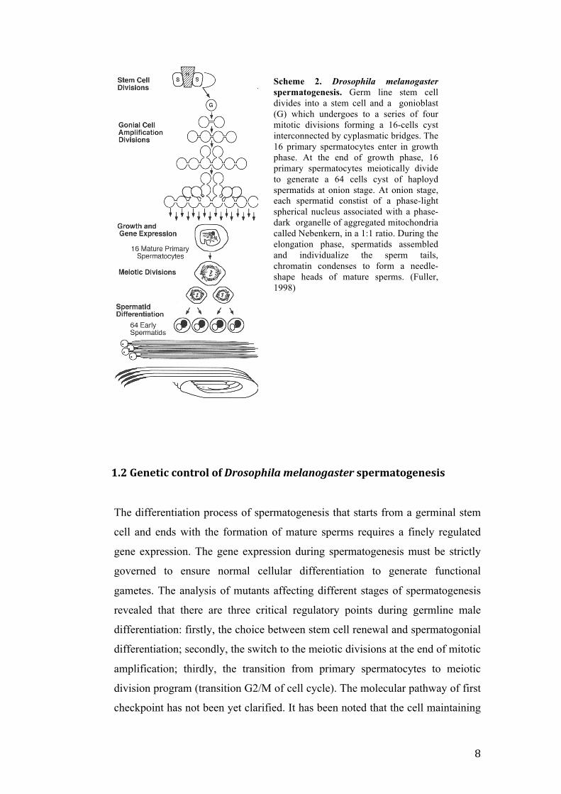
Drosophila spermatogenesis occurs in testis, a blind-ended tube, in which the
stages are well defined in a spatio-temporal manner from the top, containing the
stem cells, to the seminal vesicle at the base where mature sperms are released.
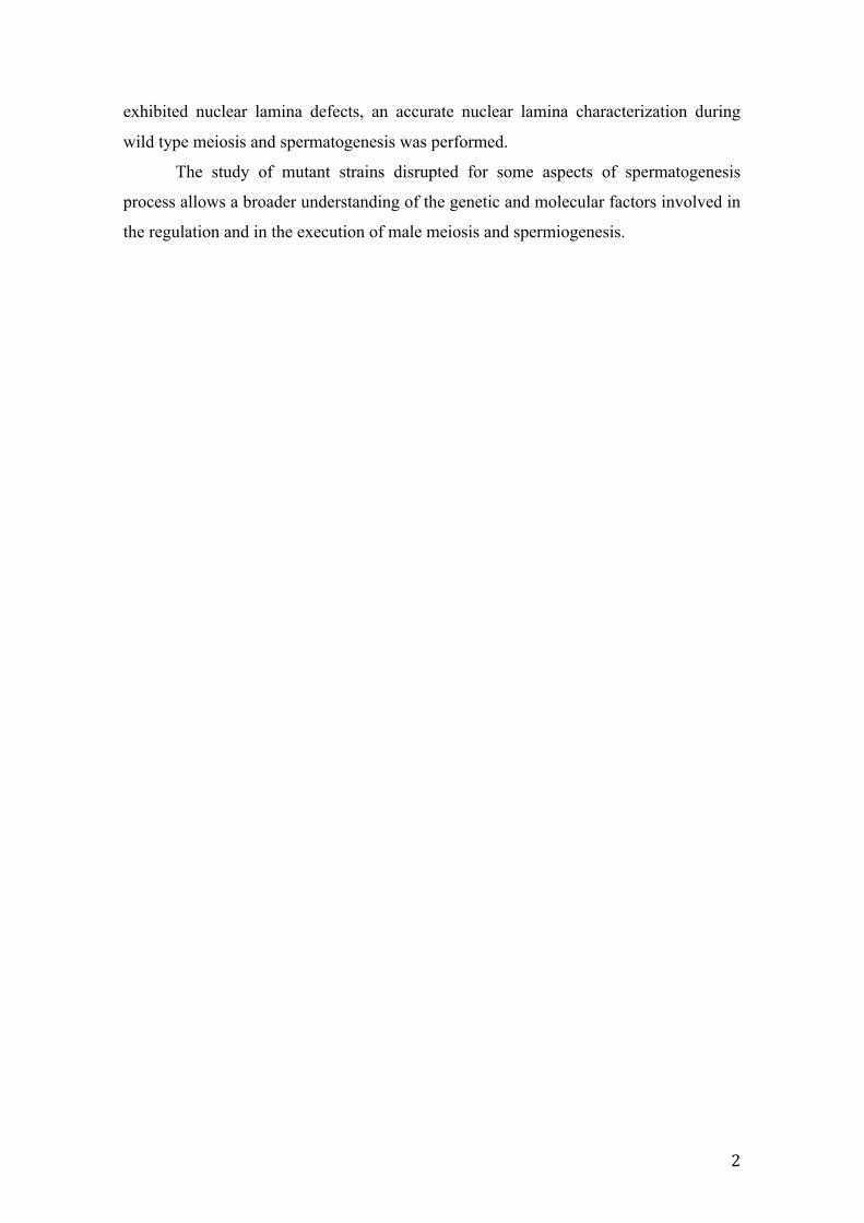
Spermatogenesis starts at the apical tip of testis in the Germinal Proliferation
Center (GPC) when a germ line stem cell divides asimmetrically producing a
stem cell and a gonioblast. The GPC is made up of a group of somatic cells
termed the hub (H), flanked by germ line stem cells (S) each surrounded by a
pair of somatic stem cells called cyst progenitor cells (CP). The germ line stem
cell divides into a stem cell and a gonioblast (G) which undergoes the
differentiation program. At the same time, the cyst progenitor cell divides into
cyst cells (C), that enclose each gonioblast. This group of three cells is termed
cyst and represent the basic unit of spermatogenesis (Fuller, 1998) (Scheme 1).
The gonioblast enters the differentiation program and undergoes four mitotic
divisions, forming a 16-cells cyst. Due to an incomplete cytokinesis, cyst cells
4
are interconnected by cytoplasmic bridges, called ring canals,. The 16 cells
represent the primary spermatocytes which enters a growth phase characterized
by severe morphological changes of nuclear shape.
The growth phase can be considered as a meiotic prophase in which the cells
increase their volume up to 25 times in relation with an extensive gene
expression (Fuller, 1993). At that point a testis-specific gene expression occurs
both for genes involved in spermatocyte differentiation and meiosis and for
genes involved in late stages of spermiogenesis (Fuller, 1993) assuming the
absence of post-meiotic transcription. In according with this view, all the
proteins involved in spermiogenesis need to be transcribed during primary
spermatocytes growth phase and stored until needed (review in White-Cooper,
2010). However, recent evidences suggested that in Drosophila, gene
transcription is turned off in late primary spermatocytes and is reactivated during
spermatid elongation phase when the transition between histone to protammine
takes place (Barreau et al., 2008). Two groups of genes, cup and comet, are
transcribed post-meiotically and do not encode sperm component proteins as in
mammals, but transcripts involved in spermiogenesis as soti, that is required for
spermatid individualization (Barreau et al., 2008). When primary spermatocytes
start to growth, the nucleus assumes an eccentric position and the chromatin
appears highly condensed, whereas mitochondria are positioned at the opposite
pole respect to the nucleus. The polar spermatocytes show a dense network of
microtubules. As the polar spermatocytes grow, chromatin subdivides into three
different chromatin masses (clumps, S2 stage according to Cenci, 1994), with
the progress of growth at S3 stage, the nucleus assumes again a central position
and mitochondria result diffused in the cytoplasm. The two bigger chromatin
masses are the somatically-paired autosomes 2 and 3, the third chromatin mass
Scheme 1. Shematic rappresentation of Germinal Proliferation Center. The hub cells (H) are flanked by germ line stem cells (S) sourronded by cyst progenitor stem cells (CP). A S cell divides to form a gonioblast (G) which is sourronded by two cyst cells. (Fuller, 1998)
5
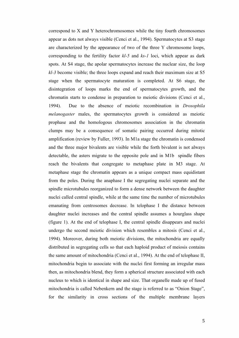
correspond to X and Y heterochromosomes while the tiny fourth chromosomes
appear as dots not always visible (Cenci et al., 1994). Spermatocytes at S3 stage
are characterized by the appearance of two of the three Y chromosome loops,
corresponding to the fertility factor kl-5 and ks-1 loci, which appear as dark
spots. At S4 stage, the apolar spermatocytes increase the nuclear size, the loop
kl-3 become visible; the three loops expand and reach their maximum size at S5
stage when the spermatocyte maturation is completed. At S6 stage, the
disintegration of loops marks the end of spermatocytes growth, and the
chromatin starts to condense in preparation to meiotic divisions (Cenci et al.,
1994). Due to the absence of meiotic recombination in Drosophila
melanogaster males, the spermatocytes growth is considered as meiotic
prophase and the homologous chromosomes association in the chromatin
clumps may be a consequence of somatic pairing occurred during mitotic
amplification (review by Fuller, 1993). In M1a stage the chromatin is condensed
and the three major bivalents are visible while the forth bivalent is not always
detectable, the asters migrate to the opposite pole and in M1b spindle fibers
reach the bivalents that congregate to metaphase plate in M3 stage. At
metaphase stage the chromatin appears as a unique compact mass equidistant
from the poles. During the anaphase I the segregating nuclei separate and the
spindle microtubules reorganized to form a dense network between the daughter
nuclei called central spindle, while at the same time the number of microtubules
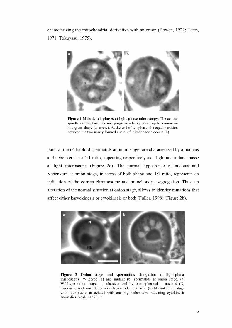
emanating from centrosomes decrease. In telophase I the distance between
daughter nuclei increases and the central spindle assumes a hourglass shape
(figure 1). At the end of telophase I, the central spindle disappears and nuclei
undergo the second meiotic division which resembles a mitosis (Cenci et al.,
1994). Moreover, during both meiotic divisions, the mitochondria are equally
distributed in segregating cells so that each haploid product of meiosis contains
the same amount of mitochondria (Cenci et al., 1994). At the end of telophase II,
mitochondria begin to associate with the nuclei first forming an irregular mass
then, as mitochondria blend, they form a spherical structure associated with each
nucleus to which is identical in shape and size. That organelle made up of fused
mitochondria is called Nebenkern and the stage is referred to as “Onion Stage”,
for the similarity in cross sections of the multiple membrane layers
6
characterizing the mitochondrial derivative with an onion (Bowen, 1922; Tates,
1971; Tokuyasu, 1975).
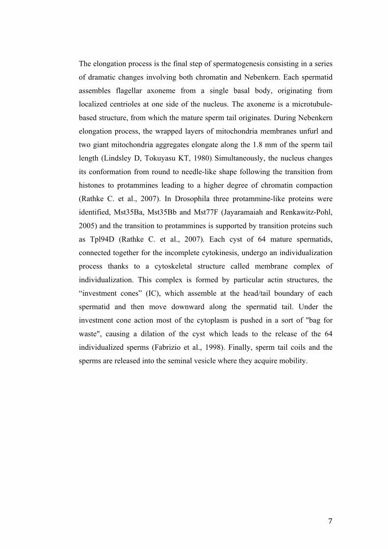
Each of the 64 haploid spermatids at onion stage are characterized by a nucleus
and nebenkern in a 1:1 ratio, appearing respectively as a light and a dark masse
at light microscopy (Figure 2a). The normal appearance of nucleus and
Nebenkern at onion stage, in terms of both shape and 1:1 ratio, represents an
indication of the correct chromosome and mitochondria segregation. Thus, an
alteration of the normal situation at onion stage, allows to identify mutations that
affect either karyokinesis or cytokinesis or both (Fuller, 1998) (Figure 2b).
Figure 2 Onion stage and spermatids elongation at light-phase microscopy. Wildtype (a) and mutant (b) spermatids at onion stage. (a) Wildtype onion stage is characterized by one spherical nucleus (N) associated with one Nebenkern (Nb) of identical size. (b) Mutant onion stage with four nuclei associated with one big Nebenkern indicating cytokinesis anomalies. Scale bar 20um
Figure 1 Meiotic telophases at light-phase microscopy. The central spindle in telophase become progressively squeezed up to assume an hourglass shape (a, arrow). At the end of telophase, the equal partition between the two newly formed nuclei of mitochondria occurs (b).
7
The elongation process is the final step of spermatogenesis consisting in a series
of dramatic changes involving both chromatin and Nebenkern. Each spermatid
assembles flagellar axoneme from a single basal body, originating from
localized centrioles at one side of the nucleus. The axoneme is a microtubule-
based structure, from which the mature sperm tail originates. During Nebenkern
elongation process, the wrapped layers of mitochondria membranes unfurl and
two giant mitochondria aggregates elongate along the 1.8 mm of the sperm tail
length (Lindsley D, Tokuyasu KT, 1980). Simultaneously, the nucleus changes
its conformation from round to needle-like shape following the transition from
histones to protammines leading to a higher degree of chromatin compaction
(Rathke C. et al., 2007). In Drosophila three protammine-like proteins were
identified, Mst35Ba, Mst35Bb and Mst77F (Jayaramaiah and Renkawitz-Pohl,
2005) and the transition to protammines is supported by transition proteins such
as Tpl94D (Rathke C. et al., 2007). Each cyst of 64 mature spermatids,
connected together for the incomplete cytokinesis, undergo an individualization
process thanks to a cytoskeletal structure called membrane complex of
individualization. This complex is formed by particular actin structures, the
“investment cones” (IC), which assemble at the head/tail boundary of each
spermatid and then move downward along the spermatid tail. Under the
investment cone action most of the cytoplasm is pushed in a sort of "bag for
waste", causing a dilation of the cyst which leads to the release of the 64
individualized sperms (Fabrizio et al., 1998). Finally, sperm tail coils and the
sperms are released into the seminal vesicle where they acquire mobility.
8
1.2 Genetic control of Drosophila melanogaster spermatogenesis
The differentiation process of spermatogenesis that starts from a germinal stem
cell and ends with the formation of mature sperms requires a finely regulated
gene expression. The gene expression during spermatogenesis must be strictly
governed to ensure normal cellular differentiation to generate functional
gametes. The analysis of mutants affecting different stages of spermatogenesis
revealed that there are three critical regulatory points during germline male
differentiation: firstly, the choice between stem cell renewal and spermatogonial
differentiation; secondly, the switch to the meiotic divisions at the end of mitotic
amplification; thirdly, the transition from primary spermatocytes to meiotic
division program (transition G2/M of cell cycle). The molecular pathway of first
checkpoint has not been yet clarified. It has been noted that the cell maintaining
Scheme 2. Drosophila melanogaster spermatogenesis. Germ line stem cell divides into a stem cell and a gonioblast (G) which undergoes to a series of four mitotic divisions forming a 16-cells cyst interconnected by cyplasmatic bridges. The 16 primary spermatocytes enter in growth phase. At the end of growth phase, 16 primary spermatocytes meiotically divide to generate a 64 cells cyst of haployd spermatids at onion stage. At onion stage, each spermatid constist of a phase-light spherical nucleus associated with a phase-dark organelle of aggregated mitochondria called Nebenkern, in a 1:1 ratio. During the elongation phase, spermatids assembled and individualize the sperm tails, chromatin condenses to form a needle-shape heads of mature sperms. (Fuller, 1998)
9
contact with the hub tends to maintain stem cells identity while, the cell displace
away from the hub undergo to differentiation program. The apical hub cells
express protein from armadillo locus as Fasciclin III, D-Ecadherin and β-catenin
(Peifer et al., 1993) and hedgehog gene encoding a signaling molecules (Lee et
al., 1992), suggesting a role of the hub as signaling center (Fuller, 1998). One of
the pathways implicated in stem cells differentiation involves the JAK-STAT
signaling activation as a consequence of Upd ligand secretion (Kiger et al.,
2001; Tulina and Matunis, 2001). The transition between sparmatogonial mitotic
divisions to the primary spermatocytes growth and consequently the trigger of
meiotic program is regulated by two genes, bag of marbles (bam) and benign
gonial cell neoplasm (bgcn). Mutant testes for both genes are full of cysts of
germ cells and completely lacking of primary spermatocytes or downstream
stages. The correct function of bam and bgcn are mandatory to ensure the
transition to the onset of meiotic differentiation program (review by Fuller,
1998). The third checkpoint, the transition between the growth period of primary
spermatocytes that can be considerate as an extended G2 phase, to the entry in
meiosis I is regulated by two different classes of genes, “meiotic arrest class”
and “twine class”. Spermatocytes of “meiotic arrest” mutant do not enter into
meiotic divisions and do not execute post meiotic spermatids differentiation.
The meiotic arrest class can be subdivided in two subclasses on the basis of
different phenotypes, always early (aly) class and cannonbal (can) class (White-
Cooper., 1998).
can class includes the following genes: cannonbal (can) (Hiller et al., 2001),
meiosis I arrest (mia) (Hiller et al., 2004), spermatocyte arrest (sa) (Hiller et al.,
2004) and no hitter (nht) (Hiller et al., 2004) which respectively encode for a
testis specific TBP-associated factors (tTAFs) TAF-5, TAF-6, TAF-8 and TAF-
4 playing a role in the interaction between RNA polymerase II and gene
promoter regions. The basal transcription factor complex, TFIID, which is
constitute by TATA-binding protein and (TBP) and several TAFs, is an
ubiquitous transcriptional factor always acting during transcriptional process.
The discovery of testis specific TAFs led to a model in which they can act as
basal transcription factors for promoters of genes required in spermiogenesis
(White-Cooper, 2010). Moreover, the colocalization of TAFs with PRC1, a
10
component of Polycomb repression complex (Chen et al., 2005) led to
hypothesize a “repressor of a repressor” pathway according to which tTAFs
sequester the PRC1 repressor away from testis-specific promoters allowing gene
expression (White-Cooper, 2010).
aly class includes the following genes: always early (aly) (White-Cooper et al.,
2000) whose molecular functions is unknown and lin-9 is its homolog in
C.elegans, cookie monster (comr) whose molecular function is unknown (Jiang
and White-cooper, 2003), tombola (tomb) encoding for a DNA binding protein
and lin-54 is its homolog in C.elegans (Jiang and White-cooper, 2007),
matotopetli (topi) (Perezgazga et al., 2004) and achintya and vismay (achi-vis)
(Ayyar et al., 2013; Wang and Mann 2003) encoding for DNA binding proteins.
aly gene is conserved from plants to animal, except in fungi (White-Cooper et
al., 2000) and it is a paralog of mip130 gene. Mip130 protein, together with Rbf,
E2F2, Dp is a subunit of dREAM/MMB repress gene expression complex
(Lewis et al., 2004). An analogous dRAM/MMB complex and specific for testis,
named testis meiotic arrest complex (tMAC) has been identified in Drosophila
(Beall et al., 2007). Some of the tMAC components are in common or are
paralogs of dRAM/MMB complex subunits, others as Comr and Topi are unique
of tMAC ensuring a testis specific gene activation (White-Cooper, 2010). Even
if dREAM complex is linked with transcriptional inactivation, the role of aly
genes is presumably that of transcriptional activators rather than repressor of a
repressor due to their localization, which determine their function, with
euchromatin in primary spermatocytes (White-Cooper, 2010, Jiang and White-
cooper, 2003, Wang and Mann 2003, Jiang et al., 2007).
Spermatocytes of twine-class mutants skip one or both meiotic divisions and
some peculiar events of meiosis as spindle assembly, chromosomes segregation
and cytokinesis, but spermatids differentiation, although with defects, proceed.
The twine-class includes Dmcdc2, twine (twe), pelota (pelo), boule (bol) genes
and are required for entry into meiotic cell divisions. Studies showed that the
meiotic factors involved in entry into meiosis are the same involved in mitotic
cycle. The cdc2 kinases is responsible for the initiation of mitosis, the activity
of cdc2 is regulated by its association with cyclin and with the phosphorilation
11
of threonine 167 in S. pombe (reviewed by Nurse, 1990). The cdc2 kinases
activity is repressed by phosphorilation of tyrosine 15 in S. pombe (Gould et al.,
1990) and threonine 14 in higher eukaryotes (Krek and Nigg, 1991; Norbury et
al., 1991) by Wee1. The removal of the inhibitory phosphates is mandatory for
the activation of cyclin/cdc2 activation and consequently for the beginning of M
phase (reviewed by Maines and Wasserman 1998); the inactivation of
cyclin/cdc2 complex for the M phase exit occur by cyclin degradation (Glotzer
et al., 1991). Phosphatases belong to cdc25 family remove the inhibitory
phosphates of cdc2 (Dunphy and Kumagai, 1991; Gautier et al., 1991; Strausfeld
et al., 1991). In Drosophila have been identified two cdc25 phosphatases, String
is active in mitosis and cdc25 homolog Twine which are responsible for the
onset of the meiotic divisions (Edgar and O’Farrel, 1989, 1990; Jimenez et al.,
1990; Alphey et al., 1992; Courtout et al., 1992). Dmcdc2 encodes for cdc2
kinase which form a complex with Cyclin B underline the transition G2/M, the
activity of that complex is mediated by a phosphorilation/dephosphorilation of
the cdc2 kinase (reviewed by Nurse, 1990). twine and Dmcdc2 are required for
the transition G2/M during Drosophila spermatogenesis (White-Cooper et al.,
1993; Eberhart and Wasserman, 1995). Mutation in twine leads to sterility but
does not affect somatic development and viability, pre-meiotic stages of
spermatogenesis are phenotipically normal while chromosomes condensation is
incomplete, cyclin A is not degraded, centrosomes do not separate and spindle
does not form. Dmcdc2 mutations leads to developmental defects and larval
lethality suggesting a role also in mitosis (Stern et a., 1993). Mutants for twine
and Dmcdc2 accumulate cysts of 16 undivided nuclei however, many aspects of
post meiotic stages, as elongation of spermatids, still occur leading to the
formation of unbalanced and no motile sperms (White-Cooper et al., 1993).
twine gene is transcribed early during the extended G2 phase therefore, twine
mRNA accumulation, is not sufficient to promove the G2/M transition contrary
with that observed with the transcription of string/cdc25 that trigger the G2/M
transition (Edgar and O’Farrel, 1989, 1990). POLO kinase activates TWINE
consequently triggering CyclinB/cdc2 complex. Once activated the
CyclinB/cdc2 complex activates by phosphorilation TWINE (its same activator)
and repressed its same inhibitor Wee1 by a positive feedback loop. pelota and
boule show a similar phenotypes observed in twine and Dmcdc2, they fail some
12
meiotic aspects as chromosomes congression, nuclear lamina breaks down and
spindle formation but exhibit post-meiotic differentiation (Eberhart and
Wasserman, 1995; Eberhart et al., 1996). boule encodes for a RNA-binding
protein, it is expressed only in testis and mutation in the gene affect only meiosis
(Eberhart et al., 1996). pelota is widely express and acts both in mitosis and in
meiosis (Eberhart and Wasserman, 1995). Finally, Dmcdc2, twine and roughex,
have a role in regulating the second meiotic division. roughex in particular
negatively regulates MII, an excess of rux prevents the second meiotic division,
an low level of rux leads to an extra MII division (Gonczy et al., 1994). A model
for meiotic cell cycle and spermatid differentiation control cordinating by both
meiotic-arrest and twine gene clesses has been proposed (Fuller, 1998). aly
gene could play a role as global regulator of spermatogenesis inasmuch aly
control the transcription or the activity of can, mia and sa and twine, cyclinB and
boule or their products (White-Cooper., 1998). In can, mia and sa mutants a
primary spermatocytes arrested in G2/M phase were observed suggesting that all
the genes involved in spermatids differentiation has to be transcribed. Moreover,
in twine mutant the twine mRNA is present but protein can be not trasleted or
not stabilized (White-Cooper., 1998). The hypotesis is that a gene/genes regulate
by meiotic-arrest class genes can act to regulate and stabilize TWINE protein
(Fuller., 1998).
13
1.3 RAE1: structure and functions
The WD domain containing proteins belongs to a family characterized by a
common sequence repeat enriched of tryptophan (W) and aspartic acid (D)
usually at the end of a 40 residues sequence. The WD domains show a beta
propeller fold. WD proteins are found in all eukariotes and show a very wide
variety of functions: they are involved in signal transduction, RNA processing,
chromatin assembly, vesicular trafficking, cell cycle progression and many
others. A common feature of WD proteins seems to be the ability of interacting
with different proteins to form complexes (for a review see Smith, 2008). RAE1
is a conserved component of WD-40 protein family (Neer et al., 1994) showing
several different functions. RAE1 was first identified in Schizosaccharomyces
pombe (spRae1p) in a screening for temperature-sensitive mutation defective for
RNA exportation from nucleus to cytoplasm. rae1 (ribonucleic acid export 1)
mutant accumulates poly (A)+ RNA in the nucleus together with defects
associated with organization of actin and tubulin pattern and a block at the G2/M
transition in mitosis (Brown et al., 1995). Moreover, when rae1 is inactivated or
depleted the cells arrest in G2 phase without the formation of mitotic spindle
(Whalen et al., 1997). In Saccharomyces cerevisiae, the S.pombe rae1 homolog,
gle2, is associated with nuclear pore complexes. gle2 mutants show an
accumulation of poly (A)+ RNA and a severe perturbation of the structure of
nuclear pore complexes and of nuclear envelope but, contrary to what observed
in S.Pombe, gle2 is required but not essential for cells proliferation (Murphy et
al., 1996). The role of RAE1 in the process of mRNA trafficking between
nucleus and cytoplasm has been demonstrated also in human. The human
protein RAE1 is involved in nuclear cytoplasmic mRNA export (Bharathi et al.,
1997) by its direct binding through the GLEBS-like motif to NUP98 at nuclear
pore complex (Pritchard et al., 1999). In mammalian cells, GLEB motif
modulates also the binding of hRAE1 (and mRAE1) to the mitotic checkpoint
protein mBUB1 indicating an interaction between nuclear cytoplasmic
trafficking and mitotic machinery and a role for RAE1 as mitotic checkpoint
regulator (Wang et al., 2001). Knock-out mice for rae1 and bub3 show mitotic
checkpoint defects and chromosome missegregation, but the lack of rae1 has no
effects on mRNAs export (Babu et al., 2003). Moreover, Rae1 and Nup98 are
14
associated with APC and regulate the transition from metaphase to anaphase in
mammalian mitotic cells (Jeganathan et al., 2005). In Xenopus egg extracts,
RAE1 binds to microtubules and is involved in the spindle assembly regulating
the activity of the spindle-assembly factor Ran. In HeLa cells the interaction
between the Nuclear Mitotic Apparatus protein (NuMA) and RAE1 was
demonstrated showing that a perturbation of protein levels lead to the formation
of spindle defects and consequent chromosome alignment anomalies. The
equilibrium of the two proteins is a critical condition for bipolar spindle
formation (Wong et al., 2006). In Drosophila melanogaster the role of rae1 was
firstly investigated in SL2 culture cells. The dmRae1 protein localizes at nuclear
envelope and shows a higher identity with the human form than with the yeast
form . The depletion of rae1 by a dsRNA interference does not affect the mRNA
export but an accumulation of cells in G1 phase and defects in S phase entry
were observed. Those evidences indicates that in drosophila culture cells
dmRae1 is not involved in nucleocytoplasmic trafficking but is involved in the
progression of the cell cycle through the regulation of G1/S transition (Sitterlin
2004). From those observations the pleiotropic effects of the rae1 gene emerge
that leads to a multiple protein roles among organisms. Further insights onto the
pleiotropic effects of rae1 arise from Drosophila in vivo studies, from which
both an involvement in the regulation of neurogenesis and a role in meiotic cell
cycle emerge (Tian et al., 2011; Volpi et al., 2013). The ubiquitin ligase
Highwire is a member of conserved PHR proteins that regulates the
development of nervous system. Drosophila hiw mutants show an abnormal
overgrowth of synapses at larval neuromuscolar juction (NMJ) in terms of
number and size of boutons and extension of branches (Hong et al., 2000).
Using the tandem affinity purification assay Drosophila Rae1 was identified as
interactor of Hiw (Tian et al., 2011 Like hiw mutant, rae1 mutant flies show
terminal synapsis overgrowth and small boutons. Further evidences of the
interaction between rae1 and hiw arise from the fact that the heterozygosity
conditions of rae1 can enhance the phenotype observed in a hiw ipomorfic
mutant indicating that the two genes collaborate to control the terminal synapsis
overgrowth (Tian et al., 2011). Finally, the authors show that RAE1 regulates in
a positive manner the level of E3 ubiquitin ligase Hiw to restrain the growth of
terminal synapsis (Tian et al., 2011). An interaction of RAE1 with RPM-1,
15
orthologue of Highwire, that regulate the axon termination and synapse
formation, was shown in C. elegans putting in evidence a conserved role
between species of RAE1 as regulator of neuronal development (Grill et al.,
2012). The role of rae1 in meiosis was shown in a study aimed to characterize a
male sterile recessive mutation of Drosophila melanogaster (Volpi et al., 2013).
rae1 mutant males are sterile but completely viable, the meiocytes do not
complete meiosis I and do not progress towards meiosis II, but the unreduced
spermatids progress to the final stages of spermatogenesis although producing
defective sperms. Cytological analysis showed defects in chromatin
condensation from pre-meiotic stage up to metaphase I division where
chromosomes were poorly organized and chromatin fragments are delocalized
respect to metaphase array ending with chromosome lagging in ana/telo phase.
The meiotic spindle is poor of microtubules and the central spindle is
mislocalized. Defects associated with actin structures and centrioles distribution
were also observed (Volpi et al., 2013). Those evidences emphasized the
multifaceted role of RAE1 as protein involved in both the mitotic and the
meiotic cell cycle.
16
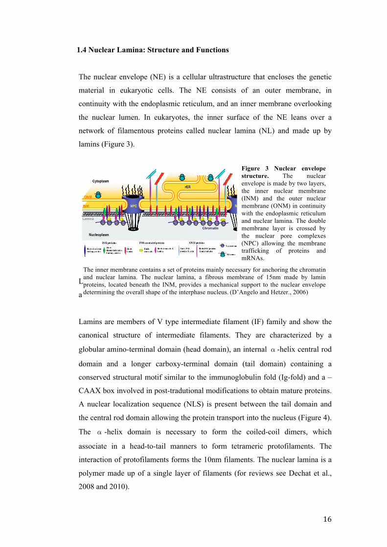
1.4 Nuclear Lamina: Structure and Functions
The nuclear envelope (NE) is a cellular ultrastructure that encloses the genetic
material in eukaryotic cells. The NE consists of an outer membrane, in
continuity with the endoplasmic reticulum, and an inner membrane overlooking
the nuclear lumen. In eukaryotes, the inner surface of the NE leans over a
network of filamentous proteins called nuclear lamina (NL) and made up by
lamins (Figure 3).
L
a
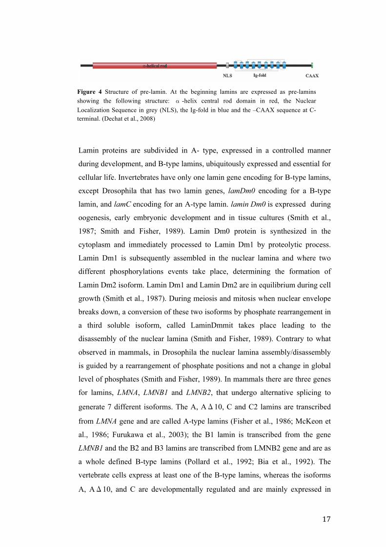
Lamins are members of V type intermediate filament (IF) family and show the
canonical structure of intermediate filaments. They are characterized by a
globular amino-terminal domain (head domain), an internal α-helix central rod
domain and a longer carboxy-terminal domain (tail domain) containing a
conserved structural motif similar to the immunoglobulin fold (Ig-fold) and a –
CAAX box involved in post-tradutional modifications to obtain mature proteins.
A nuclear localization sequence (NLS) is present between the tail domain and
the central rod domain allowing the protein transport into the nucleus (Figure 4).
The α -helix domain is necessary to form the coiled-coil dimers, which
associate in a head-to-tail manners to form tetrameric protofilaments. The
interaction of protofilaments forms the 10nm filaments. The nuclear lamina is a
polymer made up of a single layer of filaments (for reviews see Dechat et al.,
2008 and 2010).
Figure 3 Nuclear envelope structure. The nuclear envelope is made by two layers, the inner nuclear membrane (INM) and the outer nuclear membrane (ONM) in continuity with the endoplasmic reticulum and nuclear lamina. The double membrane layer is crossed by the nuclear pore complexes (NPC) allowing the membrane trafficking of proteins and mRNAs.
The inner membrane contains a set of proteins mainly necessary for anchoring the chromatin and nuclear lamina. The nuclear lamina, a fibrous membrane of 15nm made by lamin proteins, located beneath the INM, provides a mechanical support to the nuclear envelope determining the overall shape of the interphase nucleus. (D’Angelo and Hetzer., 2006)
17
Figure 4 Structure of pre-lamin. At the beginning lamins are expressed as pre-lamins showing the following structure: α -helix central rod domain in red, the Nuclear Localization Sequence in grey (NLS), the Ig-fold in blue and the –CAAX sequence at C-terminal. (Dechat et al., 2008)
Lamin proteins are subdivided in A- type, expressed in a controlled manner
during development, and B-type lamins, ubiquitously expressed and essential for
cellular life. Invertebrates have only one lamin gene encoding for B-type lamins,
except Drosophila that has two lamin genes, lamDm0 encoding for a B-type
lamin, and lamC encoding for an A-type lamin. lamin Dm0 is expressed during
oogenesis, early embryonic development and in tissue cultures (Smith et al.,
1987; Smith and Fisher, 1989). Lamin Dm0 protein is synthesized in the
cytoplasm and immediately processed to Lamin Dm1 by proteolytic process.
Lamin Dm1 is subsequently assembled in the nuclear lamina and where two
different phosphorylations events take place, determining the formation of
Lamin Dm2 isoform. Lamin Dm1 and Lamin Dm2 are in equilibrium during cell
growth (Smith et al., 1987). During meiosis and mitosis when nuclear envelope
breaks down, a conversion of these two isoforms by phosphate rearrangement in
a third soluble isoform, called LaminDmmit takes place leading to the
disassembly of the nuclear lamina (Smith and Fisher, 1989). Contrary to what
observed in mammals, in Drosophila the nuclear lamina assembly/disassembly
is guided by a rearrangement of phosphate positions and not a change in global
level of phosphates (Smith and Fisher, 1989). In mammals there are three genes
for lamins, LMNA, LMNB1 and LMNB2, that undergo alternative splicing to
generate 7 different isoforms. The A, AΔ10, C and C2 lamins are transcribed
from LMNA gene and are called A-type lamins (Fisher et al., 1986; McKeon et
al., 1986; Furukawa et al., 2003); the B1 lamin is transcribed from the gene
LMNB1 and the B2 and B3 lamins are transcribed from LMNB2 gene and are as
a whole defined B-type lamins (Pollard et al., 1992; Bia et al., 1992). The
vertebrate cells express at least one of the B-type lamins, whereas the isoforms
A, AΔ10, and C are developmentally regulated and are mainly expressed in
18
differentiated cells (Rober et al., 1989; Machielis et al., 1996); the C2 and B3
isoforms are expressed only in the germ line (Fukurawa and Hotta, 1993;
Fukurawa et al., 1994; Alsheimer et al., 1999). Due to the main function of
nuclear lamina (NL) to provide support to the nuclear envelope, during the cell
cycle the nuclear lamina undergoes structural changes. The most significant
alteration of nuclear lamina takes place during the transition prophase to
metaphase of mitotic cells, when the nuclear envelope breaks down. At this
stage nuclear lamina disassembles by phosphorylation of residues flanking the
rod domain leading to a depolymerization of lamins polymers. The nuclear
lamina reassembly is guided by dephosphorilation of the same residues (for a
review see Moir et al., 2000). The NL contributes to maintain the mechanical
properties of the cell forming a bridge between the nucleus and the cytoplasm
and is involved in determining the nuclear shape (for a review Moir et al., 2000;
Dechat et al., 2010;). Due to the nuclear lamina position very close to chromatin,
a role of lamins in regulations of gene transcription through chromatin
positioning has been suggested. Microscopy studies demonstrated the
association between heterochromatin and nuclear lamina (Paddy et al., 1990;
Fawcett, 1996) and the interaction of lamins with histones and specific DNA
sequences was reported (for a review Dechat et al., 2010). A decrease in the
expression of lamDm0 in drosophila blocks the nuclear membrane assembly
(Lenz-bohme et al., 1997) and the down regulation of lamin gene leads to
chromatin condensation and chromosome segregation defects in C.elegans (Liu
et al., 2000). Lamin involvement in DNA replication and in transcription was
also suggested: in culture cells, lamin B1 localized at replication foci (Moir et
al., 1994) and lamin depletion in Xenopus egg extracts results in a block of
DNA replication without effects on nuclear envelope behavior although nuclei
are smaller (Newport et al., 1990). Moreover, the association of lamin proteins
with DNA replication factors as PCNA was shown (Shumaker et al., 2008).
Lamins also have a role in the control of gene expression by controlling the
nuclear chromatin organization. Evidences showed that inactive genes are often
allocated close lamina region, it was thus suggested that nuclear lamina could
act to assemble a transcriptionally silent domain interacting directly with
chromatin (for a review see Dechat el al., 2010). In Drosophila melanogaster
specific chromosomal regions of inactive chromatin are associated with nuclear
19
envelope (Mathog and Sedat, 1989). Moreover, changes in lamin expression
lead to histone modification alterations and consequently changes in chromatin
structure, thus pointing out a role of lamins in epigenetic regulation. Finally,
lamins are involved in cell cycle regulation , acting in pathways involved in cell
cycle progression (for a review see Dechat et al., 2010). By all these evidence is
clear, therefore, that the nuclear lamina has not only a role in determining the
architecture of the nucleus but it is also actively involved in gene regulation and
hence in the control of nuclear and cellular process.
20
2. AIM OF THE PROJECT
The proper execution of spermatogenesis process, from stem cell divisions to mature
sperms formation, is a mandatory condition to ensure male fertility. Drosophila
melanogaster is a widely used model organism to genetically and cytologically dissect
developmental processes. Drosophila male sterile mutations affecting any stages of
spermatogenesis represent an excellent study material to explore the genetic mechanism
regulating the whole process. Due to the conservation of developmental mechanisms
between species, evidences obtained in flies should provide insight into genetic and
molecular pathways underpinning male fertility in other organisms, including humans.
The general aim of my PhD research project was the analysis of ms(2)Z5584 and
ms(2)Z1168 mutant strains belonging to a unique collection of 13 ethyl-methansulfonate
(EMS)-induced male sterile recessive mutants on chromosome 2 and 3 identified in a
large screening of male-sterile mutations (Wakimoto et al., 2004). Preliminary
cytological screen of mutations highlighted a general and common aberrant phenotype
affecting the entry into the meiotic cell cycle. In particular mutants skip one or both the
meiotic divisions but carry out the spermatid differentiation, although with anomalies,.
That phenotype reminds that observed in twine class mutants.
The ms(2)Z5584 mutant strain was previously characterized both genetically, by
identifying a point mutation in rae1 gene, and cytologically by a description of aberrant
phenotypes throughout the spermatogenesis process (Volpi et al., 2013). Starting from
those evidences, I first confirmed the identification of rae1 as the gene underling the
ms(2)Z5584 mutant phenotype. The rae1 gene was knocked-down by RNA interference
and the ensuing phenotipic effects on spermatogenesis were analyzed and compared to
ms(2)Z5584 mutant defects. Secondly, the RAE1 localization pattern during meiotic cell
cycle was investigated by GAL4/UAS system allowing the expression of UASGFP-rae1
transgene under either a testes-specific or a constitutive driver. Finally, a ms(2)Z5584
mutant phenotype rescue was performed using an UASGFP-rae1 transgene.
The ms(2)Z1168 mutant strain was previously partially characterized. Preliminary
recombination and deficiency mapping allowed to identify the genetic region containing
21
the mutation. I carried out a complete characterization of the mutant phenotype during
spermatogenesis. The analysis was performed by immunohistochemestry technique
using antibodies against several structures involved in male meiosis and confocal
microscopy. The ms(2)Z1168 mutant exhibited anomalies in nuclear lamina structure
together with chromatin condensation defects. The genetic region containing the
ms(2)Z1168 mutation was further restricted by recombination and deficiency mapping.
The gene identification was made by genetic approach using RNA interference and by
sequencing.
22
3. RESULTS AND DISCUSSIONS
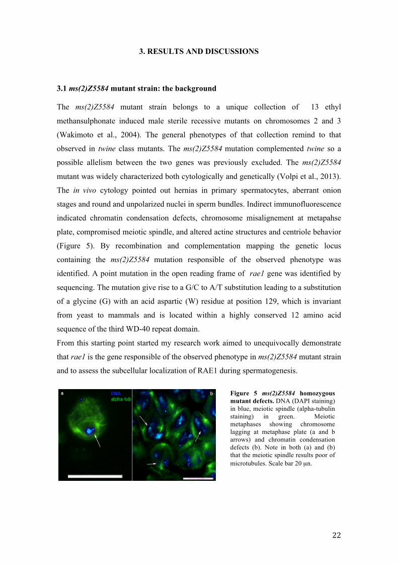
3.1 ms(2)Z5584 mutant strain: the background The ms(2)Z5584 mutant strain belongs to a unique collection of 13 ethyl
methansulphonate induced male sterile recessive mutants on chromosomes 2 and 3
(Wakimoto et al., 2004). The general phenotypes of that collection remind to that
observed in twine class mutants. The ms(2)Z5584 mutation complemented twine so a
possible allelism between the two genes was previously excluded. The ms(2)Z5584
mutant was widely characterized both cytologically and genetically (Volpi et al., 2013).
The in vivo cytology pointed out hernias in primary spermatocytes, aberrant onion
stages and round and unpolarized nuclei in sperm bundles. Indirect immunofluorescence
indicated chromatin condensation defects, chromosome misalignement at metapahse
plate, compromised meiotic spindle, and altered actine structures and centriole behavior
(Figure 5). By recombination and complementation mapping the genetic locus
containing the ms(2)Z5584 mutation responsible of the observed phenotype was
identified. A point mutation in the open reading frame of rae1 gene was identified by
sequencing. The mutation give rise to a G/C to A/T substitution leading to a substitution
of a glycine (G) with an acid aspartic (W) residue at position 129, which is invariant
from yeast to mammals and is located within a highly conserved 12 amino acid
sequence of the third WD-40 repeat domain.
From this starting point started my research work aimed to unequivocally demonstrate
that rae1 is the gene responsible of the observed phenotype in ms(2)Z5584 mutant strain
and to assess the subcellular localization of RAE1 during spermatogenesis.
Figure 5 ms(2)Z5584 homozygous mutant defects. DNA (DAPI staining) in blue, meiotic spindle (alpha-tubulin staining) in green. Meiotic metaphases showing chromosome lagging at metaphase plate (a and b arrows) and chromatin condensation defects (b). Note in both (a) and (b) that the meiotic spindle results poor of microtubules. Scale bar 20 µn.
23
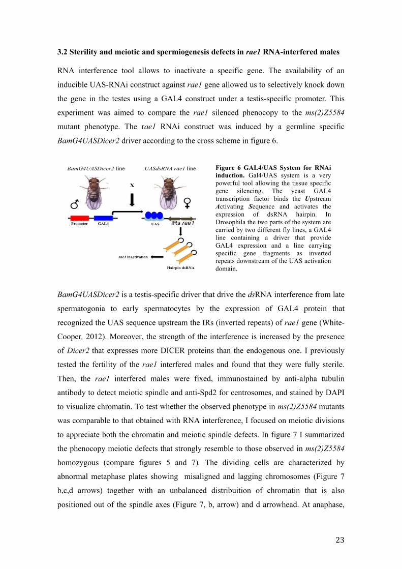
3.2 Sterility and meiotic and spermiogenesis defects in rae1 RNA-interfered males RNA interference tool allows to inactivate a specific gene. The availability of an
inducible UAS-RNAi construct against rae1 gene allowed us to selectively knock down
the gene in the testes using a GAL4 construct under a testis-specific promoter. This
experiment was aimed to compare the rae1 silenced phenocopy to the ms(2)Z5584
mutant phenotype. The rae1 RNAi construct was induced by a germline specific
BamG4UASDicer2 driver according to the cross scheme in figure 6.
BamG4UASDicer2 is a testis-specific driver that drive the dsRNA interference from late
spermatogonia to early spermatocytes by the expression of GAL4 protein that
recognized the UAS sequence upstream the IRs (inverted repeats) of rae1 gene (White-
Cooper, 2012). Moreover, the strength of the interference is increased by the presence
of Dicer2 that expresses more DICER proteins than the endogenous one. I previously
tested the fertility of the rae1 interfered males and found that they were fully sterile.
Then, the rae1 interfered males were fixed, immunostained by anti-alpha tubulin
antibody to detect meiotic spindle and anti-Spd2 for centrosomes, and stained by DAPI
to visualize chromatin. To test whether the observed phenotype in ms(2)Z5584 mutants
was comparable to that obtained with RNA interference, I focused on meiotic divisions
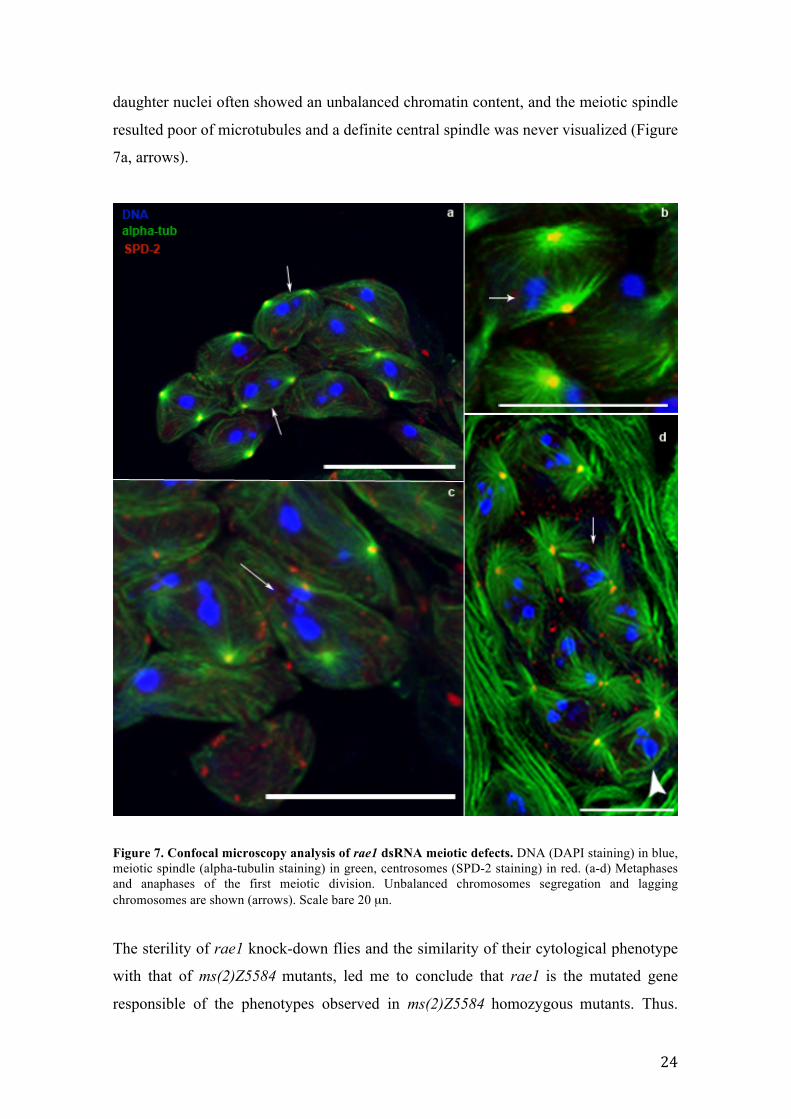
to appreciate both the chromatin and meiotic spindle defects. In figure 7 I summarized
the phenocopy meiotic defects that strongly resemble to those observed in ms(2)Z5584
homozygous (compare figures 5 and 7). The dividing cells are characterized by
abnormal metaphase plates showing misaligned and lagging chromosomes (Figure 7
b,c,d arrows) together with an unbalanced distribuition of chromatin that is also
positioned out of the spindle axes (Figure 7, b, arrow) and d arrowhead. At anaphase,
Figure 6 GAL4/UAS System for RNAi induction. Gal4/UAS system is a very powerful tool allowing the tissue specific gene silencing. The yeast GAL4 transcription factor binds the Upstream Activating Sequence and activates the expression of dsRNA hairpin. In Drosophila the two parts of the system are carried by two different fly lines, a GAL4 line containing a driver that provide GAL4 expression and a line carrying specific gene fragments as inverted repeats downstream of the UAS activation domain.
24
daughter nuclei often showed an unbalanced chromatin content, and the meiotic spindle
resulted poor of microtubules and a definite central spindle was never visualized (Figure
7a, arrows).
Figure 7. Confocal microscopy analysis of rae1 dsRNA meiotic defects. DNA (DAPI staining) in blue, meiotic spindle (alpha-tubulin staining) in green, centrosomes (SPD-2 staining) in red. (a-d) Metaphases and anaphases of the first meiotic division. Unbalanced chromosomes segregation and lagging chromosomes are shown (arrows). Scale bare 20 µn.
The sterility of rae1 knock-down flies and the similarity of their cytological phenotype
with that of ms(2)Z5584 mutants, led me to conclude that rae1 is the mutated gene
responsible of the phenotypes observed in ms(2)Z5584 homozygous mutants. Thus.
25
rae1 plays a fundamental role in the execution of a proper meiosis and spermatogenesis.
Due to meiotic chromosomes segregation defects characterizing the ms(2)Z5584
mutant, it is reasonable to speculate a role of rae1 in chromosome segregation as
previously observed in mitosis. Rae1 haplo-insufficient mice show significant
chromosome missegregation defects which increase in combination with a Bub3 deficit.
This suggests a cooperation of the two proteins for proper chromosome segregation in a
common pathway involving also the mitotic checkpoint control protein BUB1 (Babu et
al., 2003; Basu et al., 1999). The localization of RAE1, BUB1 and BUB3 at mitotic
unattached kinetochores (Wang et al., 2001; Babu et al., 2003) could generate a wait
signal in metaphase before skipping to anaphase (Babu et al., 2003). During Drosophila
spermatogenesis BUB1 results associated with kinetochores at prometaphase I cells,
decrease during metaphase I and disappear during anaphase I moreover, bub1 mutant
show chromosomes missegregation defects (Basu et al., 1999). An equivalent of the
spindle checkpoint in Drosophila meiosis seems to exist even though less efficient than
the mitotic one. It has been conjectured an involvement of the Bub1 pathway in the
delay, but not full arrest, of the meiotic cell cycle also in the presence of unattached
chromosomes (Basu et al., 1999). A reduced severity of the meiotic spindle checkpoint
is also supported by my findings in the ms(2)Z5584 mutants which progress to the final
stages of spermatogenesis notwithstanding the severe meiotic defects. In the light of this
view and considering the colocalization of RAE1 and BUB1 in mitosis, I can likewise
speculate a colocalization of the two proteins at kinetochores of meiotic chromosomes
and their synergistic involvement in meiosis spindle checkpoint. Moreover, in
eukaryotic cells the proper chromosome segregation is guaranteed by a bipolar spindle
formation. In HeLa cells the interaction between RAE1 and NuMa protein are
responsible for a correct formation of mitotic spindle (Wong et al., 2006). The presence
of meiotic spindle anomalies ms(2)Z5584 mutant and in rae1 interfered males could
likewise suggest the interaction between RAE1 and proteins involved in meiotic spindle
formation (Volpi et a., 2013).
26
3.3 Confocal analysis of GFP-RAE1 localization during spermatogenesis By confocal microscopy analysis I followed RAE1 localization pattern during wildtype
spermatogenesis in Drosophila melanogaster. As above reported, the GAL4/UAS
system is composed by two different fly lines, a GAL4 line containing a Bam or a
Tubulin driver that provide, respectively, testis-specific or constitutive GAL4
expression, and a reporter line carrying a coding sequence of targeted gene with a GFP
reporter gene fused to the rae1 sequence, downstream of the UAS activation domain
(see scheme in figure 6 paragraph 3.2). In these experiments, I followed the RAE1
distribution through the whole spermatogenic process, from mitotic stages to mature
sperms, taking advantage of GFP protein autofluorescence .
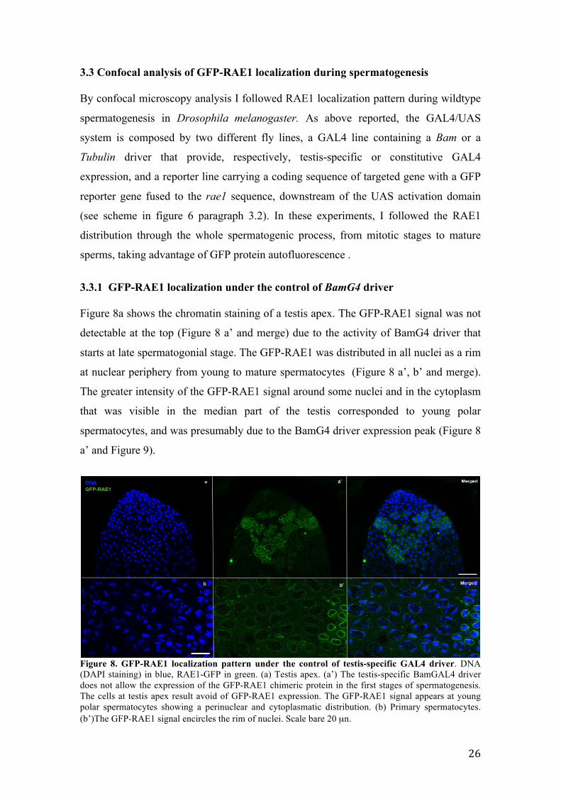
3.3.1 GFP-RAE1 localization under the control of BamG4 driver Figure 8a shows the chromatin staining of a testis apex. The GFP-RAE1 signal was not
detectable at the top (Figure 8 a’ and merge) due to the activity of BamG4 driver that
starts at late spermatogonial stage. The GFP-RAE1 was distributed in all nuclei as a rim
at nuclear periphery from young to mature spermatocytes (Figure 8 a’, b’ and merge).
The greater intensity of the GFP-RAE1 signal around some nuclei and in the cytoplasm
that was visible in the median part of the testis corresponded to young polar
spermatocytes, and was presumably due to the BamG4 driver expression peak (Figure 8
a’ and Figure 9).
Figure 8. GFP-RAE1 localization pattern under the control of testis-specific GAL4 driver. DNA (DAPI staining) in blue, RAE1-GFP in green. (a) Testis apex. (a’) The testis-specific BamGAL4 driver does not allow the expression of the GFP-RAE1 chimeric protein in the first stages of spermatogenesis. The cells at testis apex result avoid of GFP-RAE1 expression. The GFP-RAE1 signal appears at young polar spermatocytes showing a perinuclear and cytoplasmatic distribution. (b) Primary spermatocytes. (b’)The GFP-RAE1 signal encircles the rim of nuclei. Scale bare 20 µn.
27
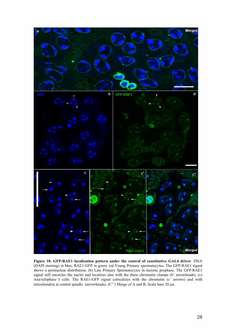
3.3.2 GFP-RAE1 localization under the control of TubulinG4 driver To overcome the temporal limits of of BamG4driver expression, I used a second
ubiquitous driver, TubulinG4. In the early stages of spermatogenesis and in primary
spermatocytes, the distribution of GFP-RAE1 was in accordance with that observed
with the Bam driver. The chimeric protein localized at nuclear rim of the apex testis
cells (Figure 10 a). The disintegration of Y loops defines the end of growth phase of
primary spermatocytes and the beginning of meiotic phase (Cenci et al., 1994) At this
stage, S6, chromatin begin to condense in preparation of meiotic division, the three
chromatin clumps are still close to nuclear envelope but they start to congregate toward
the center of the cells (Figure 10 b, DAPI staining). Notably, at this stage, the GFP-
RAE1 signal not only still encircled the nuclei but resulted also associated with the
chromatin clumps (Figure 10 b’, GFP-RAE1 staining, arrowheads). During
ana/telophase stages of first meiotic division, the two daughter nuclei appeared as
compact chromatin masses (Figure 10 c, DAPI staining, arrows) separated by a central
spindle where mitochondria localized. Note a DAPI staining halo of mitochondrial
DNA colocalizing with the central spindle (Figure 10 c, DAPI staining, arrowheads).
Significantly, the GFP-RAE1 signal localized with both newly formed nuclei and
mitochondria at the central spindle (Figure 10 c’, arrows for nuclei, arrowheads for
central spindle).
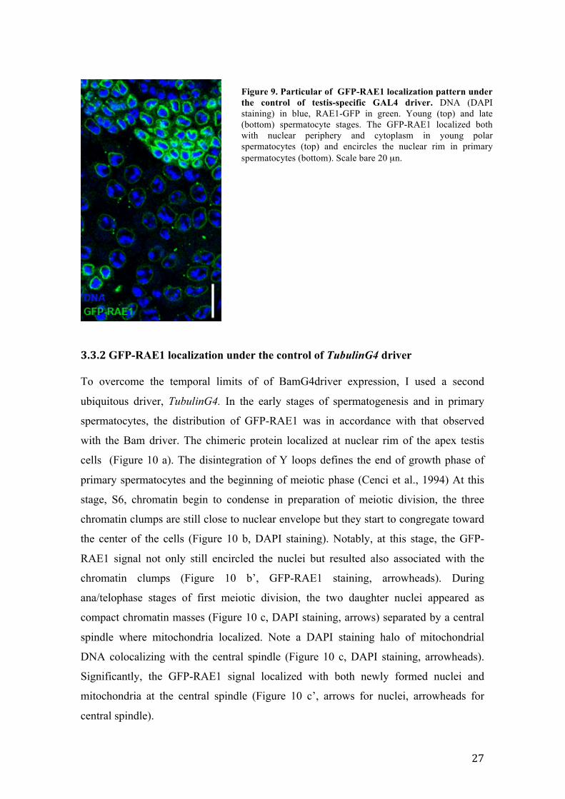
Figure 9. Particular of GFP-RAE1 localization pattern under the control of testis-specific GAL4 driver. DNA (DAPI staining) in blue, RAE1-GFP in green. Young (top) and late (bottom) spermatocyte stages. The GFP-RAE1 localized both with nuclear periphery and cytoplasm in young polar spermatocytes (top) and encircles the nuclear rim in primary spermatocytes (bottom). Scale bare 20 µn.
28
Figure 10. GFP-RAE1 localization pattern under the control of constitutive GAL4 driver. DNA (DAPI staining) in blue, RAE1-GFP in green. (a) Young Primary spermatocytes. The GFP-RAE1 signal shows a perinuclear distribution. (b) Late Primary Spermatocytes in meiotic prophase. The GFP-RAE1 signal still encircles the nuclei and localizes also with the three chromatin clumps (b’ arrowheads). (c) Ana/telophase I cells. The RAE1-GFP signal colocalizes with the chromatin (c’ arrows) and with mitochondria at central spindle (arrowheads). (C’’) Merge of A and B. Scale bare 20 µn.
29
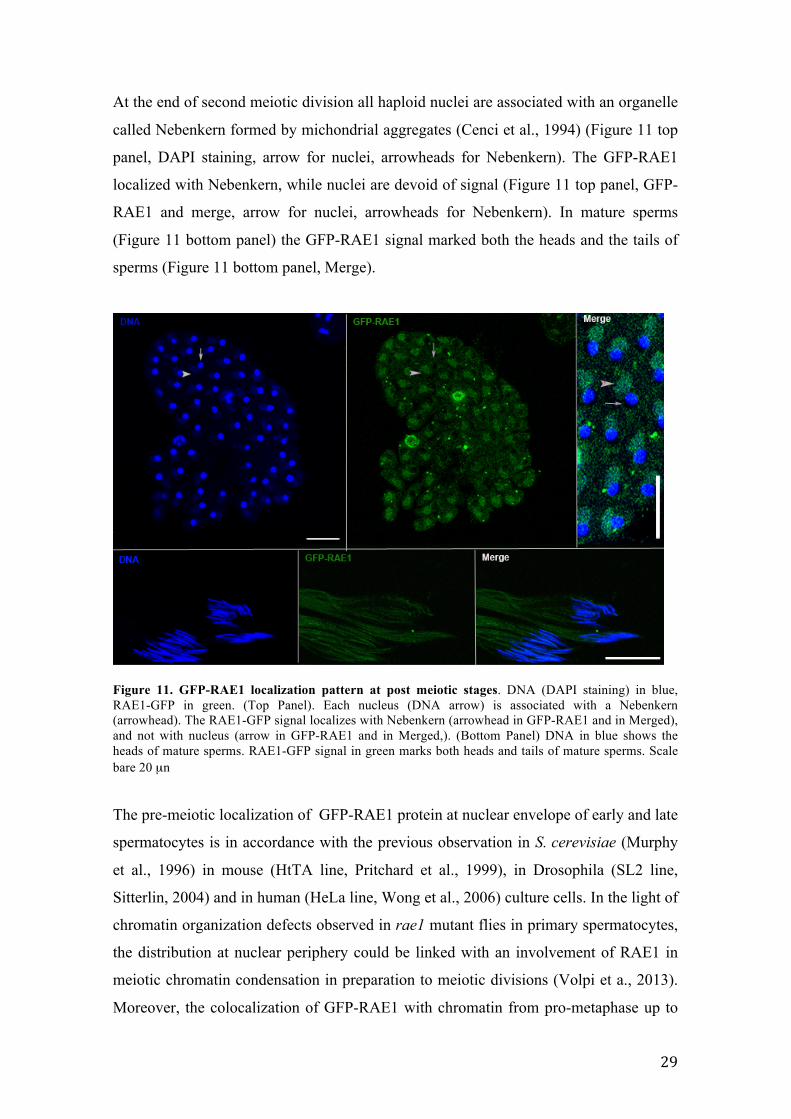
At the end of second meiotic division all haploid nuclei are associated with an organelle
called Nebenkern formed by michondrial aggregates (Cenci et al., 1994) (Figure 11 top
panel, DAPI staining, arrow for nuclei, arrowheads for Nebenkern). The GFP-RAE1
localized with Nebenkern, while nuclei are devoid of signal (Figure 11 top panel, GFP-
RAE1 and merge, arrow for nuclei, arrowheads for Nebenkern). In mature sperms
(Figure 11 bottom panel) the GFP-RAE1 signal marked both the heads and the tails of
sperms (Figure 11 bottom panel, Merge).
Figure 11. GFP-RAE1 localization pattern at post meiotic stages. DNA (DAPI staining) in blue, RAE1-GFP in green. (Top Panel). Each nucleus (DNA arrow) is associated with a Nebenkern (arrowhead). The RAE1-GFP signal localizes with Nebenkern (arrowhead in GFP-RAE1 and in Merged), and not with nucleus (arrow in GFP-RAE1 and in Merged,). (Bottom Panel) DNA in blue shows the heads of mature sperms. RAE1-GFP signal in green marks both heads and tails of mature sperms. Scale bare 20 µn The pre-meiotic localization of GFP-RAE1 protein at nuclear envelope of early and late
spermatocytes is in accordance with the previous observation in S. cerevisiae (Murphy
et al., 1996) in mouse (HtTA line, Pritchard et al., 1999), in Drosophila (SL2 line,
Sitterlin, 2004) and in human (HeLa line, Wong et al., 2006) culture cells. In the light of
chromatin organization defects observed in rae1 mutant flies in primary spermatocytes,
the distribution at nuclear periphery could be linked with an involvement of RAE1 in
meiotic chromatin condensation in preparation to meiotic divisions (Volpi et a., 2013).
Moreover, the colocalization of GFP-RAE1 with chromatin from pro-metaphase up to
30
meiotic divisions and the presence of chromosmes fragmentation in both ms(2)Z5584
mutant and rae1 interfered males further support the connection between RAE1 and
chromatin organization (Volpi et al., 2013). During meiotic divisions the GFP-RAE1
localized with both chromatin and central spindle mitochondria while once the meiotic
phase is terminated at the onion stage, the protein moved to the Nebenkern leaving the
nucleus completely devoid of signal. The presence of meiotic central spindle anomalies
in rae1 mutants and the association of GFP-RAE1 with central spindle mitochondria
might be likely connected to a conserved role of RAE1 as interactor of proteins
involved in spindle assembly in meiosis as in mitosis (Volpi et al., 2013). In this context
it is noteworthy that the knockdown of Tpr nucleoporin results in the enhancement of
chromosomes lagging due to an impaired recruitment of spindle checkpoint proteins to
the dynein complex at kinetochore (Nakano et al., 2010). Notably, the fact that at the
end of spermatogenesis the GFP-RAE1 protein was still present in heads and tails of
mature sperms, indicates its involvement in spermatid differentiation. rae1 mutant flies
showed aberrant sperm head penotype that appeared round and unpolarized suggesting a
RAE1 role in post meiotic chromatin condensation and organization as seen in pre-
meiotic stages (Volpi et al., 2013).
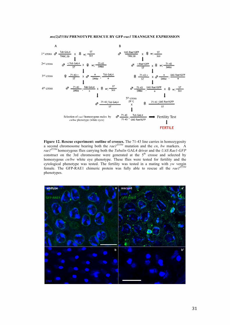
3.4 The GFP-rae1 transgene rescues the mutant phenotype of ms(2)Z5584 To verify if the chimeric GFP-RAE1 protein behavior recapitulated that of wild type
protein, I checked if the transgenic construct UAS-GFP-rae1 was able to rescue the
sterile and cytological phenotype of rae1Z5584 mutant males. To this aim, I conceived a
complex experiment of five successive crosses to generate rae1Z5584 homozygous flies
carrying the Tubulin GAL4 driver and the UAS-GFP-rae1 construct in
transheterozygosity on the 3rd chromosome (Figure 12). I found that the GFP-RAE1
chimeric protein was able to fully rescue the rae1Z5584 sterile phenotype. Rescued fertile
males were cytologically tested to check if the cytological defects of ms(2)Z5584 were
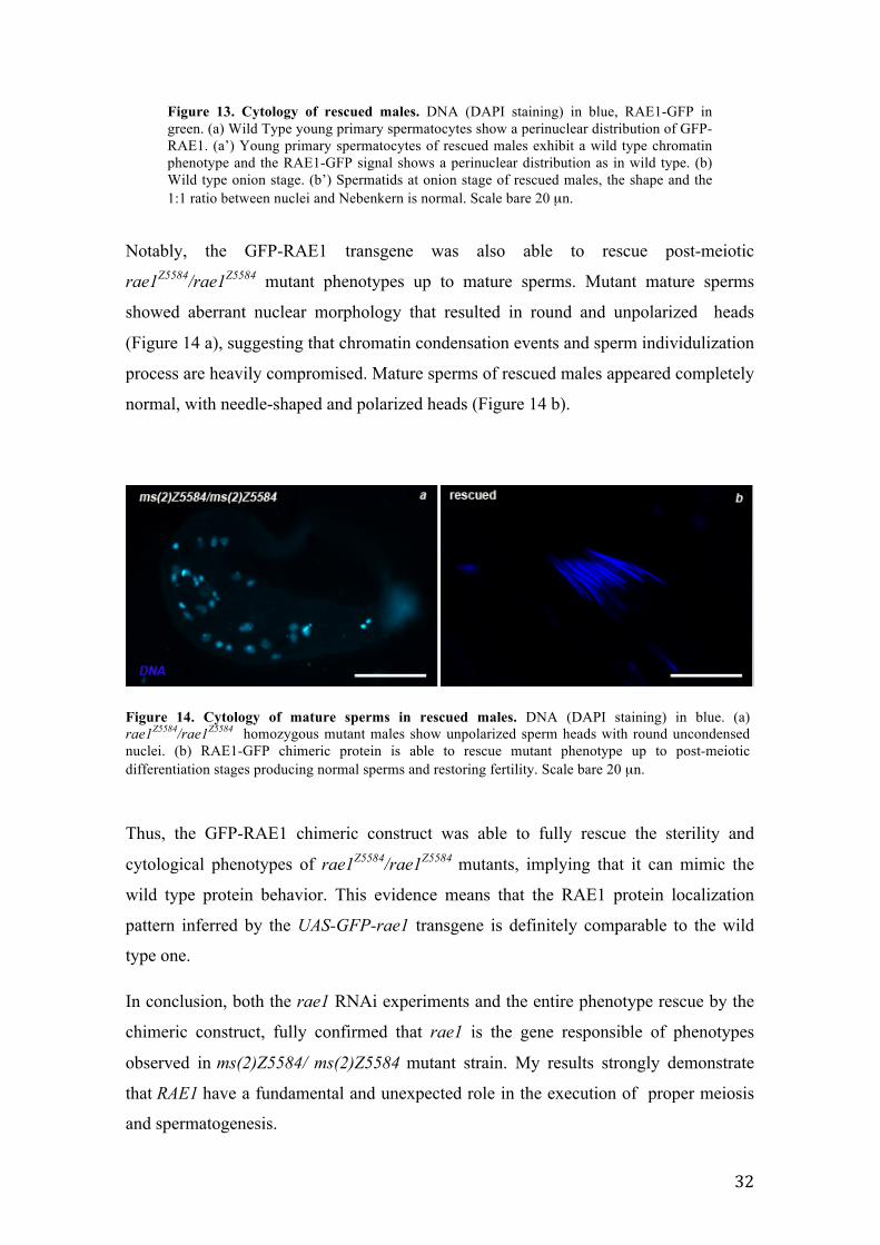
also rescued. Primary spermatocytes of rescued males showed a normal organization of
chromatin with the GFP-RAE1 signal localized at nuclear periphery as in wild type
(Figure 13 a, a’). Rescued spermatids at onion stage showed a normal 1:1 ratio of nuclei
and Nebenkern that appeared of identical size (Figure 13 b, b’). Moreover, at that stage
the distribution of GFP-RAE1 chimeric protein is identical to wild type (not shown).
These results confirm definitely that the cytological defects characterizing ms(2)Z5584
mutants were also completely restored by the GFP-rae1 construct expression.
31
Figure 12. Rescue experiment: outline of crosses. The 71-43 line carries in homozygosity a second chromosome bearing both the rae1Z5584 mutation and the cn, bw markers. A rae1Z5584 homozygous flies carrying both the Tubulin GAL4 driver and the UAS Rae1-GFP construct on the 3rd chromosome were generated at the 5th crosse and selected by homozygous cn/bw white eye phenotype. These flies were tested for fertility and the cytological phenotype was tested. The fertility was tested in a mating with yw vergin female. The GFP-RAE1 chimeric protein was fully able to rescue all the rae1Z5584
phenotypes.
32
Figure 13. Cytology of rescued males. DNA (DAPI staining) in blue, RAE1-GFP in green. (a) Wild Type young primary spermatocytes show a perinuclear distribution of GFP-RAE1. (a’) Young primary spermatocytes of rescued males exhibit a wild type chromatin phenotype and the RAE1-GFP signal shows a perinuclear distribution as in wild type. (b) Wild type onion stage. (b’) Spermatids at onion stage of rescued males, the shape and the 1:1 ratio between nuclei and Nebenkern is normal. Scale bare 20 µn.
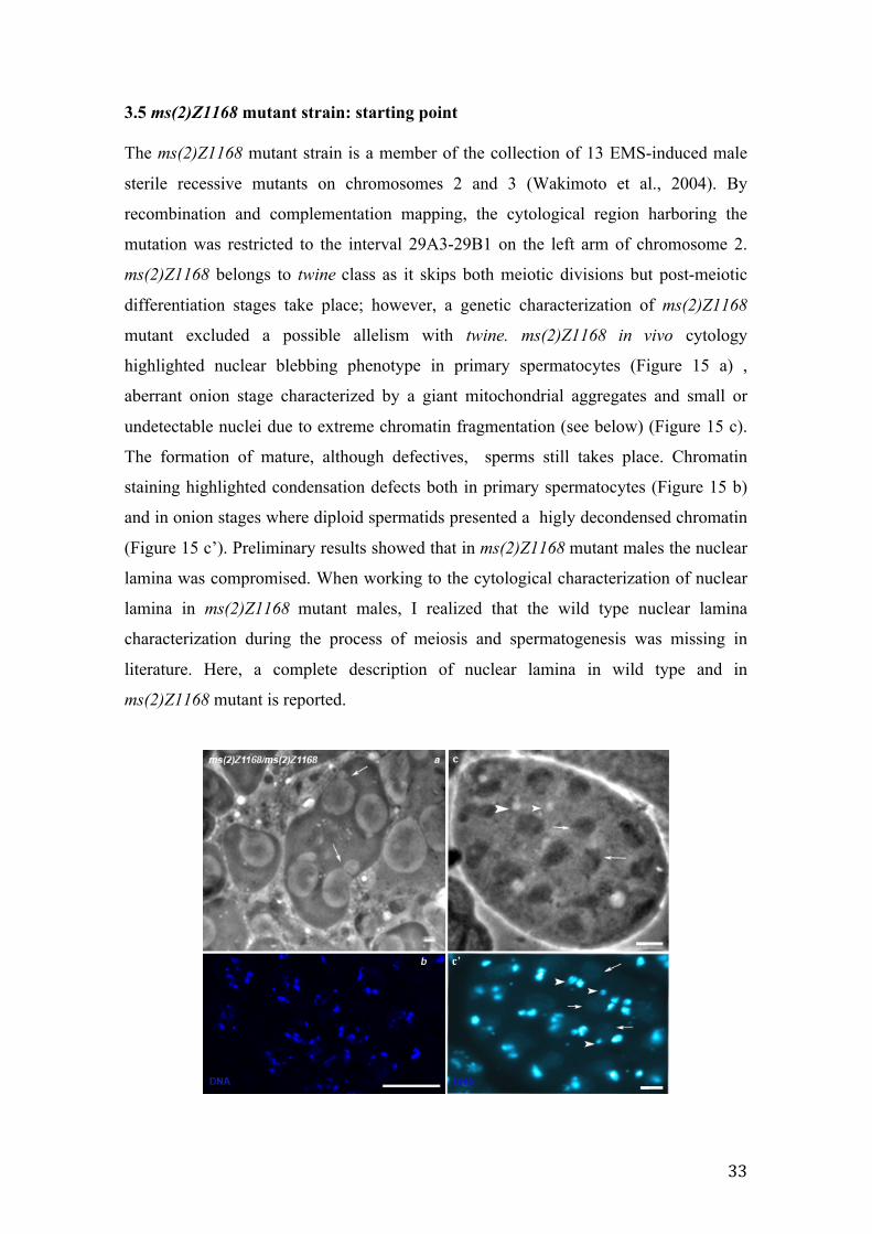
Notably, the GFP-RAE1 transgene was also able to rescue post-meiotic
rae1Z5584/rae1Z5584 mutant phenotypes up to mature sperms. Mutant mature sperms
showed aberrant nuclear morphology that resulted in round and unpolarized heads
(Figure 14 a), suggesting that chromatin condensation events and sperm individulization
process are heavily compromised. Mature sperms of rescued males appeared completely
normal, with needle-shaped and polarized heads (Figure 14 b).
Figure 14. Cytology of mature sperms in rescued males. DNA (DAPI staining) in blue. (a) rae1Z5584/rae1Z5584 homozygous mutant males show unpolarized sperm heads with round uncondensed nuclei. (b) RAE1-GFP chimeric protein is able to rescue mutant phenotype up to post-meiotic differentiation stages producing normal sperms and restoring fertility. Scale bare 20 µn.
Thus, the GFP-RAE1 chimeric construct was able to fully rescue the sterility and
cytological phenotypes of rae1Z5584/rae1Z5584 mutants, implying that it can mimic the
wild type protein behavior. This evidence means that the RAE1 protein localization
pattern inferred by the UAS-GFP-rae1 transgene is definitely comparable to the wild
type one.
In conclusion, both the rae1 RNAi experiments and the entire phenotype rescue by the
chimeric construct, fully confirmed that rae1 is the gene responsible of phenotypes
observed in ms(2)Z5584/ ms(2)Z5584 mutant strain. My results strongly demonstrate
that RAE1 have a fundamental and unexpected role in the execution of proper meiosis
and spermatogenesis.
33
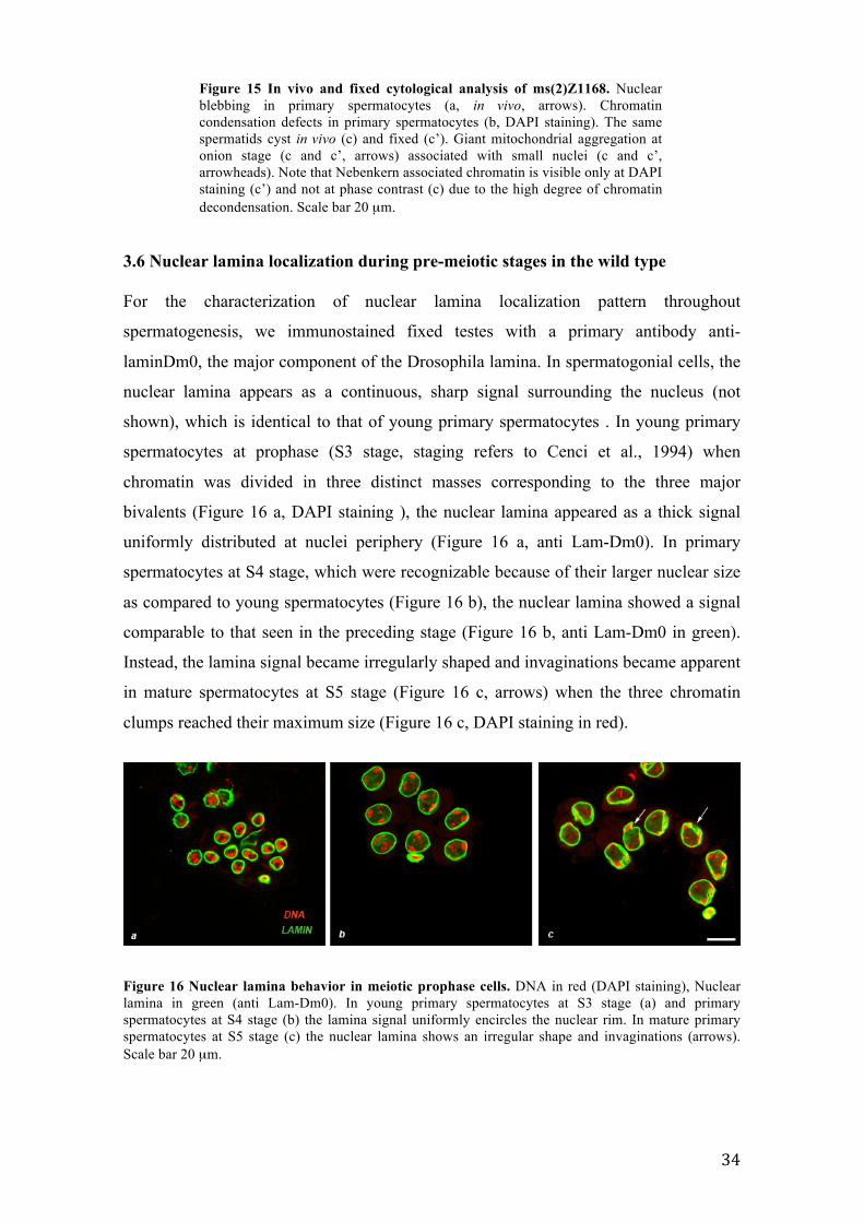
3.5 ms(2)Z1168 mutant strain: starting point The ms(2)Z1168 mutant strain is a member of the collection of 13 EMS-induced male
sterile recessive mutants on chromosomes 2 and 3 (Wakimoto et al., 2004). By
recombination and complementation mapping, the cytological region harboring the
mutation was restricted to the interval 29A3-29B1 on the left arm of chromosome 2.
ms(2)Z1168 belongs to twine class as it skips both meiotic divisions but post-meiotic
differentiation stages take place; however, a genetic characterization of ms(2)Z1168
mutant excluded a possible allelism with twine. ms(2)Z1168 in vivo cytology
highlighted nuclear blebbing phenotype in primary spermatocytes (Figure 15 a) ,
aberrant onion stage characterized by a giant mitochondrial aggregates and small or
undetectable nuclei due to extreme chromatin fragmentation (see below) (Figure 15 c).
The formation of mature, although defectives, sperms still takes place. Chromatin
staining highlighted condensation defects both in primary spermatocytes (Figure 15 b)
and in onion stages where diploid spermatids presented a higly decondensed chromatin
(Figure 15 c’). Preliminary results showed that in ms(2)Z1168 mutant males the nuclear
lamina was compromised. When working to the cytological characterization of nuclear
lamina in ms(2)Z1168 mutant males, I realized that the wild type nuclear lamina
characterization during the process of meiosis and spermatogenesis was missing in
literature. Here, a complete description of nuclear lamina in wild type and in
ms(2)Z1168 mutant is reported.
34
3.6 Nuclear lamina localization during pre-meiotic stages in the wild type
For the characterization of nuclear lamina localization pattern throughout
spermatogenesis, we immunostained fixed testes with a primary antibody anti-
laminDm0, the major component of the Drosophila lamina. In spermatogonial cells, the
nuclear lamina appears as a continuous, sharp signal surrounding the nucleus (not
shown), which is identical to that of young primary spermatocytes . In young primary
spermatocytes at prophase (S3 stage, staging refers to Cenci et al., 1994) when
chromatin was divided in three distinct masses corresponding to the three major
bivalents (Figure 16 a, DAPI staining ), the nuclear lamina appeared as a thick signal
uniformly distributed at nuclei periphery (Figure 16 a, anti Lam-Dm0). In primary
spermatocytes at S4 stage, which were recognizable because of their larger nuclear size
as compared to young spermatocytes (Figure 16 b), the nuclear lamina showed a signal
comparable to that seen in the preceding stage (Figure 16 b, anti Lam-Dm0 in green).
Instead, the lamina signal became irregularly shaped and invaginations became apparent
in mature spermatocytes at S5 stage (Figure 16 c, arrows) when the three chromatin
clumps reached their maximum size (Figure 16 c, DAPI staining in red).
Figure 16 Nuclear lamina behavior in meiotic prophase cells. DNA in red (DAPI staining), Nuclear lamina in green (anti Lam-Dm0). In young primary spermatocytes at S3 stage (a) and primary spermatocytes at S4 stage (b) the lamina signal uniformly encircles the nuclear rim. In mature primary spermatocytes at S5 stage (c) the nuclear lamina shows an irregular shape and invaginations (arrows). Scale bar 20 µm.
Figure 15 In vivo and fixed cytological analysis of ms(2)Z1168. Nuclear blebbing in primary spermatocytes (a, in vivo, arrows). Chromatin condensation defects in primary spermatocytes (b, DAPI staining). The same spermatids cyst in vivo (c) and fixed (c’). Giant mitochondrial aggregation at onion stage (c and c’, arrows) associated with small nuclei (c and c’, arrowheads). Note that Nebenkern associated chromatin is visible only at DAPI staining (c’) and not at phase contrast (c) due to the high degree of chromatin decondensation. Scale bar 20 µm.
cc)
35
These observations are consistent with previous evidences in drosophila embryos and in
mammalian culture cells. In drosophila, early embryo mitotic interphase nuclei show a
continous nuclear lamina structure at their periphery (Harel et al., 1989), changes in the
nuclear envelope structure appear during the between late prophase to metaphase
transition (Fuchs et al., 1983; Hiraoka et al., 1990a; Paddy et al., 1990), when
invaginations of nuclear lamina are visible in a regions close to centrosomes thus
suggesting an interaction between centrosomes and lamins (Paddy et al., 1996). In
mammalian cells during G2/M transition the progressive chromosome condensation is
coupled with invaginations in nuclear lamina leading to a distortion of the entire
structure by microtubules mechanical tension (Beauduin et al., 2002).
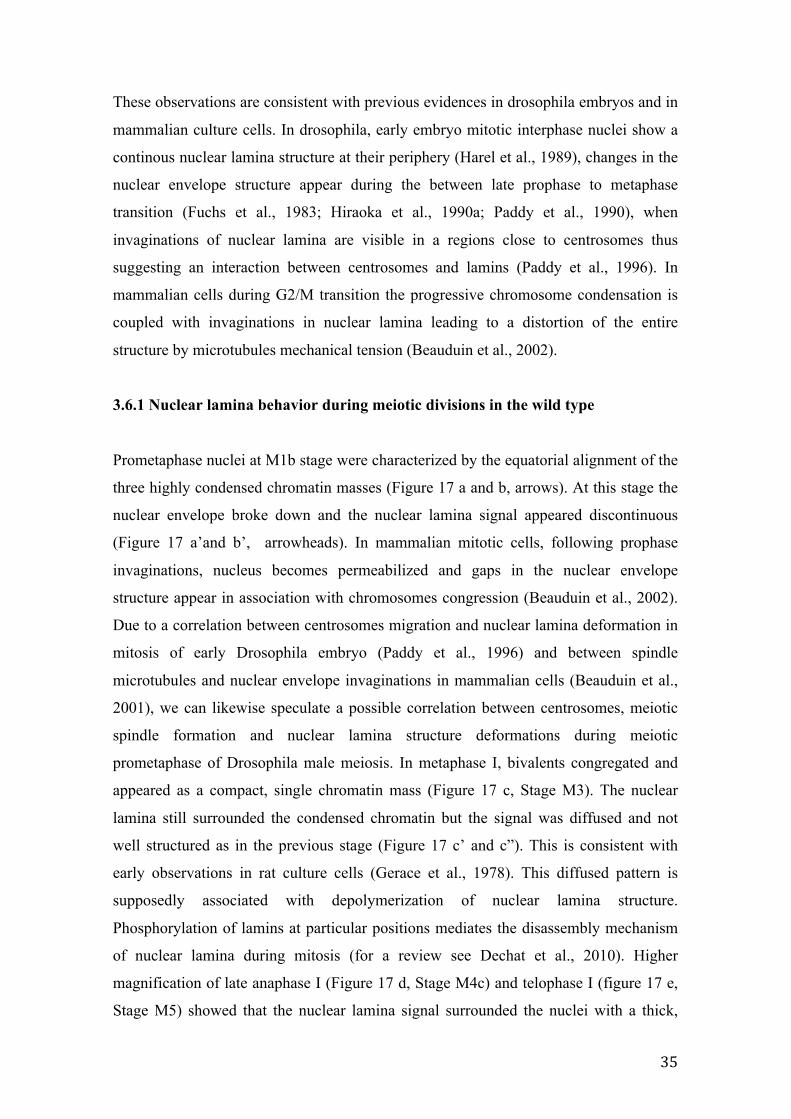
3.6.1 Nuclear lamina behavior during meiotic divisions in the wild type
Prometaphase nuclei at M1b stage were characterized by the equatorial alignment of the
three highly condensed chromatin masses (Figure 17 a and b, arrows). At this stage the
nuclear envelope broke down and the nuclear lamina signal appeared discontinuous
(Figure 17 a’and b’, arrowheads). In mammalian mitotic cells, following prophase
invaginations, nucleus becomes permeabilized and gaps in the nuclear envelope
structure appear in association with chromosomes congression (Beauduin et al., 2002).
Due to a correlation between centrosomes migration and nuclear lamina deformation in
mitosis of early Drosophila embryo (Paddy et al., 1996) and between spindle
microtubules and nuclear envelope invaginations in mammalian cells (Beauduin et al.,
2001), we can likewise speculate a possible correlation between centrosomes, meiotic
spindle formation and nuclear lamina structure deformations during meiotic
prometaphase of Drosophila male meiosis. In metaphase I, bivalents congregated and
appeared as a compact, single chromatin mass (Figure 17 c, Stage M3). The nuclear
lamina still surrounded the condensed chromatin but the signal was diffused and not
well structured as in the previous stage (Figure 17 c’ and c”). This is consistent with
early observations in rat culture cells (Gerace et al., 1978). This diffused pattern is
supposedly associated with depolymerization of nuclear lamina structure.
Phosphorylation of lamins at particular positions mediates the disassembly mechanism
of nuclear lamina during mitosis (for a review see Dechat et al., 2010). Higher
magnification of late anaphase I (Figure 17 d, Stage M4c) and telophase I (figure 17 e,
Stage M5) showed that the nuclear lamina signal surrounded the nuclei with a thick,
36
irregularly shaped design (Figure 17 d’, d’’ and 17 e’, e”, respectively) that thus differed
from the thin, circular signal of premeiotic stages. Moreover, at these stages, the nuclear
lamina showed also a punctuate signal associated to the mitochondria of the central
meiotic spindle (Figure 17 d”, e” arrows). The sequence of these events appears
different from that observed in Drosophila embryo mitotic cells in which the lamina
signal results well localized as a rim at nuclear periphery up to the metaphase and the
lamin delocalization process is completed only when chromosomes moves to anaphase
(Paddy et al., 1996). Second meiotic division advanced very quickly and the nuclear
lamin pattern showed the same pattern observed during the first meiotic division. The
nuclear lamina appeared as a diffuse signal at metaphase II (figure 17 f, merge), and it
reassembled at the periphery of daughter nuclei in anaphase II (Figure 17 g) and
telophase II nuclei (Figure 17 h). However, the signal encircling the nuclei appeared
thicker than in the same stages of the first division. A well-structured assembled lamina
during meiotic division seems to be a mandatory condition to ensure a correct
chromosomes segregation during male meiosis. Interference of lmn-1 gene implies
defects in chromatin condensation and chromosomes segregation in Caenorhabditis
elegans (Liu et al., 2000). In mammalian cells Lamin B is associated with chromosomes
during congression to metaphase (Georgatos et al., 1997). In Drosophila somatic cells
evidences indicate that the nuclear lamin is directly connect with chromatin. Lamins
proteins bind the histone core in vitro (Taniura et al. 1995) and specific DNA sequences
called matrix attachment regions (Luderus et al., 1992). More, Lamin Dm1 and Lamin
Dm2 bind in vivo nucleic acids, the H3- H4 histone tetramer, the mitotic chromosomes
and the heterochromatin protein HP1. Furthermore electron microscopy studies show
that some areas of chromatin are in close contact with the nuclear lamina (Paddy et al.,
1990). In D. melanogaster specific chromosomal regions are associated with nuclear
envelope and some evidence suggests that these regions are inactive chromatin (Mathog
and Sedat, 1989). Recently, it has been proposed a model of nuclear architecture in
which lamins position the chromosomes in the nucleus (Reddy et al. , 2008).
37
Figure 17 Nuclear lamina pattern during prometaphase and meiotic divisions. DNA in red (DAPI staining), Nuclear lamina in green (anti Lam-Dm0). In prometaphase cells at M1b stage, the three chromatin clumps corresponding to the three major bivalents (a and b, arrows) move to the equatorial plate, the nuclear envelope breaks down and the lamina signal becomes discontinous (a’ and b’, arrowheads). In metaphase I, nuclei (stage M3) appear as a single condensed chromatin mass (c), the nuclear lamina signal appears diffused in the nucleoplasm (c’). Anaphase (d) and telophase (e) nuclei, the reformed nuclear lamina exhibits a thick, irregularly shaped signal. Note the punctuated nuclear lamina signals which are associated to the central spindle identified by the typical hourglass configuration of mitochondria. In Metaphases II nuclei at M2 and M3 stages, nuclear lamina appears as diffuse signal around chromatin (f). In anaphase II (g) and telophase II (h), nuclear lamina shows a well-defined structure at daughter nuclei periphery. Scale bar 20 µm.
38
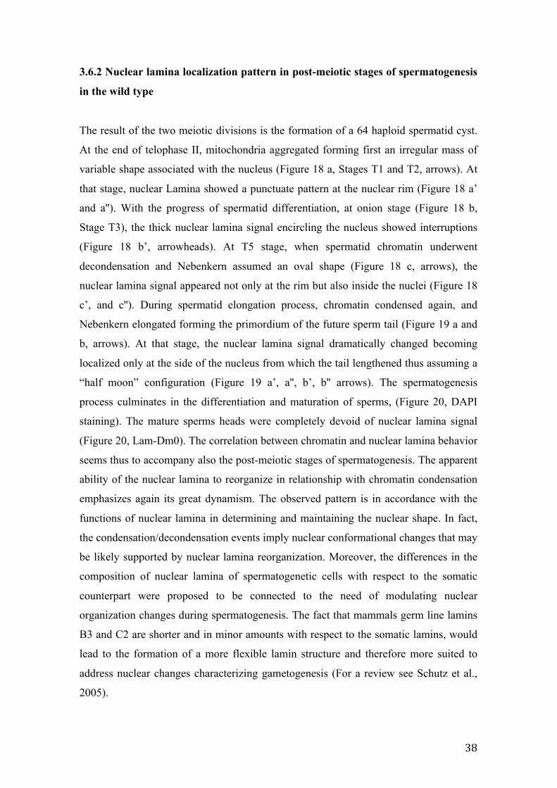
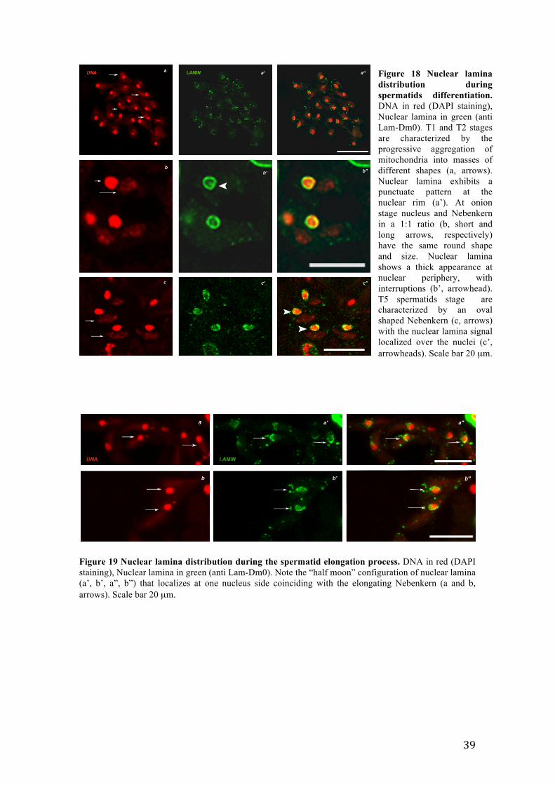
3.6.2 Nuclear lamina localization pattern in post-meiotic stages of spermatogenesis
in the wild type
The result of the two meiotic divisions is the formation of a 64 haploid spermatid cyst.
At the end of telophase II, mitochondria aggregated forming first an irregular mass of
variable shape associated with the nucleus (Figure 18 a, Stages T1 and T2, arrows). At
that stage, nuclear Lamina showed a punctuate pattern at the nuclear rim (Figure 18 a’
and a''). With the progress of spermatid differentiation, at onion stage (Figure 18 b,
Stage T3), the thick nuclear lamina signal encircling the nucleus showed interruptions
(Figure 18 b’, arrowheads). At T5 stage, when spermatid chromatin underwent
decondensation and Nebenkern assumed an oval shape (Figure 18 c, arrows), the
nuclear lamina signal appeared not only at the rim but also inside the nuclei (Figure 18
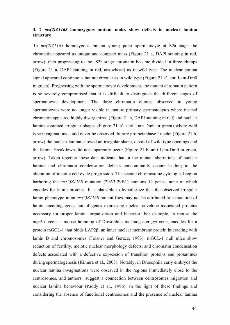
c’, and c''). During spermatid elongation process, chromatin condensed again, and
Nebenkern elongated forming the primordium of the future sperm tail (Figure 19 a and
b, arrows). At that stage, the nuclear lamina signal dramatically changed becoming
localized only at the side of the nucleus from which the tail lengthened thus assuming a
process culminates in the differentiation and maturation of sperms, (Figure 20, DAPI
staining). The mature sperms heads were completely devoid of nuclear lamina signal
(Figure 20, Lam-Dm0). The correlation between chromatin and nuclear lamina behavior
seems thus to accompany also the post-meiotic stages of spermatogenesis. The apparent
ability of the nuclear lamina to reorganize in relationship with chromatin condensation
emphasizes again its great dynamism. The observed pattern is in accordance with the
functions of nuclear lamina in determining and maintaining the nuclear shape. In fact,
the condensation/decondensation events imply nuclear conformational changes that may
be likely supported by nuclear lamina reorganization. Moreover, the differences in the
composition of nuclear lamina of spermatogenetic cells with respect to the somatic
counterpart were proposed to be connected to the need of modulating nuclear
organization changes during spermatogenesis. The fact that mammals germ line lamins
B3 and C2 are shorter and in minor amounts with respect to the somatic lamins, would
lead to the formation of a more flexible lamin structure and therefore more suited to
address nuclear changes characterizing gametogenesis (For a review see Schutz et al.,
2005).
39
Figure 19 Nuclear lamina distribution during the spermatid elongation process. DNA in red (DAPI staining), Nuclear lamina in green (anti Lam-Dm0). Note the “half moon” configuration of nuclear lamina (a’, b’, a”, b”) that localizes at one nucleus side coinciding with the elongating Nebenkern (a and b, arrows). Scale bar 20 µm.
Figure 18 Nuclear lamina distribution during spermatids differentiation. DNA in red (DAPI staining), Nuclear lamina in green (anti Lam-Dm0). T1 and T2 stages are characterized by the progressive aggregation of mitochondria into masses of different shapes (a, arrows). Nuclear lamina exhibits a punctuate pattern at the nuclear rim (a’). At onion stage nucleus and Nebenkern in a 1:1 ratio (b, short and long arrows, respectively) have the same round shape and size. Nuclear lamina shows a thick appearance at nuclear periphery, with interruptions (b’, arrowhead). T5 spermatids stage are characterized by an oval shaped Nebenkern (c, arrows) with the nuclear lamina signal localized over the nuclei (c’, arrowheads). Scale bar 20 µm.
40
With the progress of spermatids elongation, we observed an enrichment of nuclear
lamina at one side of nucleus where the spermatids tail extends. During mammalian
spermiogenesis lamins show different distribution pattern: lamin C2 is expressed in
meiotic stages (Alsheimer and Benevante, 1996) and lamin B3 in later stages of
spermatogenesis (Shutz et al., 2005), whereas lamin B1 is the only isoform always
detectable throughout the whole process (Vester et al., 1993). Notably, during mouse
spermiogenesis lamin B3 polarized at the posterior pole of elongated spermatid nuclei
(Shutz et al., 2005), a localization fully comparable to the one we observed in flies,
suggesting a polarized pattern for lamin proteins at the final stage of spermatogenesis
which is conserved between species. In mammalian post meiotic stages, several nuclear
envelope associated proteins polarized and resulted undetectable in differentiated
mature sperm (Alsheimer et al., 1998). This behavior was observed for proteins that
directly or indirectly interact with chromatin as lamin B1 (Vester et al., 1993), LAPs2
(Alsheimer et al., 1998), germ cell-less (GCL) (Kimura et al., 2003), and lamin B
receptor (LBR) (Mylonis et al., 2004). It is possible that the redistribution of nuclear
envelope proteins to the posterior pole of spermatids, the only site where chromatin is
attached, would contribute to the achievement of chromatin distribution in the sperm
(Alsheimer et al., 1998). The overview emerging from that characterization is a
continuous structural change in the structure of the nuclear lamina from meiotic
prophase until the end of spermatogenesis. These structural changes seem to parallel the
process of condensation/decondensation of chromatin throughout the process. Our
observation are in line with the pioneering immunofluorescence data describing changes
of nuclear lamina distribution throughout the mitotic cell cycle (Gerace and Blobel,
1980; Gerace et al., 1978) thus reinforcing the general idea of the prominent role of
nuclear lamina in modulating the dynamics of the nuclear membrane architecture.
Figure 20 Nuclear lamina in mature sperms. DNA in red (DAPI staining), Nuclear lamina in green (anti Lam-Dm0). The nuclear lamin signal is completely absent from both the needle-shaped sperm heads and the tails. Scale bar 20 µm.
41
3. 7 ms(2)Z1168 homozygous mutant males show defects in nuclear lamina structure In ms(2)Z1168 homozygous mutant young polar spermatocyte at S2a stage the
chromatin appeared as unique and compact mass (Figure 21 a, DAPI staining in red,
arrow), then progressing to the S2b stage chromatin became divided in three clumps
(Figure 21 a, DAPI staining in red, arrowhead) as in wild type. The nuclear lamina
signal appeared continuous but not circular as in wild type (Figure 21 a’, anti Lam-Dm0
in green). Progressing with the spermatocyte development, the mutant chromatin pattern
is so severely compromised that it is difficult to distinguish the different stages of
spermatocyte development. The three chromatin clumps observed in young
spermatocytes were no longer visible in mature primary spermatocytes where instead
chromatin appeared highly disorganized (Figure 21 b, DAPI staining in red) and nuclear
lamina assumed irregular shapes (Figure 21 b’, anti Lam-Dm0 in green) where wild
type invaginations could never be observed. In rare prometaphase I nuclei (Figure 21 b,
arrow) the nuclear lamina showed an irregular shape, devoid of wild type openings and
the lamina breakdown did not apparently occur (Figure 21 b, anti Lam-Dm0 in green,
arrow). Taken together these data indicate that in the mutant aberrations of nuclear
lamina and chromatin condensation defects concomitantly occurr leading to the
alteration of meiotic cell cycle progression. The second chromosome cytological region
harboring the ms(2)Z1168 mutation (29A3-29B1) contains 12 genes, none of which
encodes for lamin proteins. It is plausible to hypothesize that the observed irregular
lamin phenotype in an ms(2)Z1168 mutant flies may not be attributed to a mutation of
lamin encoding genes but of genes expressing nuclear envelope associated proteins
necessary for proper lamina organization and behavior. For example, in mouse the
mgcl-1 gene, a mouse homolog of Drosophila melanogaster gcl gene, encodes for a
protein mGCL-1 that binds LAP2β, an inner nuclear membrane protein interacting with
lamin B and chromosomes (Foisner and Gerace; 1993). mGCL-1 null mice show
reduction of fertility, meiotic nuclear morphology defects, and chromatin condensation
defects associated with a defective expression of transition proteins and protamines
during spermatogenesis (Kimura et al., 2003). Notably, in Drosophila early embryos the
nuclear lamina invaginations were observed in the regions immediately close to the
centrosomes, and authors suggest a connection between centrosomes migration and
nuclear lamina behaviour (Paddy et al., 1996). In the light of these findings and
considering the absence of functional centrosomes and the presence of nuclear lamina
42
defects in ms(2)Z1168 mutant flies, I can speculate that the gene at the basis of mutant
phenotypes, could encode for a protein involved in the interaction between centrosomes
and nuclear lamina and that the lack of this interaction in the mutant precludes the
organization of functional centrosomes.
Moreover, due to the complexity of nuclear lamina network, it is plausible to speculate
that the observed aberrant phenotype characterizing the mutant might be associated to
anomalies during nuclear lamina assembly/disassembly process or in maturation events
that involves pre-lamin proteins.
Figure 21 Pattern of nuclear lamina in a male meiotic mutant. DNA in red (DAPI staining), Nuclear lamina in green (anti Lam-Dm0). In young primary spermatocytes at S2a/S2b stage (a, DAPI staining in red, arrow: primary spermatocytes nucleus at S2a stage, arrowhead: primary spermatocytes nucleus at S2b stage ) the lamina signal uniformly encircles the nuclear rim as in wild type (a’, anti Lam-Dm0 in green) At late spermatocyte and prometaphase stages (b, arrow points to a prometaphase nucleus) the chromatin shows condensation defects, whereas the nuclear lamina assumes irregular shapes and the breakdown does not occur (b’, anti Lam-Dm0 in green). Scale bar 20 µm.
43
3.8 Towards ms(2)Z1168 gene identification
Meiotic recombination mapping, performed by a strain carrying phenotypic recessive
markers on chromosome 2, allowed to map the ms(2)Z1168 mutation around 37.4 m.u
within the interval dp-b. By deficiency mapping, the genetic region containing the
mutation was further restricted up to the cytological interval 29A3-29B1 (Figure 22).
The region contains 12 genes: CG7818, CG7810, CG7806, CG7795 (mtsh), CG7787,
resulting in four nuclei associated with a single mitochondrial shell at onion stage
(Bergner et al., 2010). Since the ms(2)Z1168 homozygous mutant phenotype resulted
quite different from that of mitoshell knockdown flies, I could exclude that the mutated
gene responsible for the mutant phenotype of ms(2)Z1168 strain was CG7795. A
complementation test between the chromosome ms(2)Z1168 and a mtsh allele will be
necessary to further support the phenotypic differences. CG7810 is a yet
uncharacterized gene, that encodes for a coiled-coil domain containing protein with
unknown function (FlyBase.org). In vivo cytology of CG7810 RNAi males showed
herniations in primary spermatocytes very similar to those observed in ms(2)Z1168
homozygous mutant males (Figure 23). On the contrary, onion stage spermatids showed
aberrant Nebenkern structure of variable size and non-spherical shape that differed from
the onion stage aberrant phenotype of ms(2)Z1168 homozygous mutant males (Figure
24). This discrepancy might be likely caused by the limited window of interference due
to the absence of BamG4UASDicer2 driver expression in post-meiotic stages.
Results of fertility test in interfered males. The genes reported in the table were silenced by RNAi driven by a testes-specif driver. CG7810 and CG7795 resulted sterile in a cross with vergin yw females.
45
Figure 23 CG7810 dsRNA induces aberrant phenotypes in primary spermatocytes. (a, b, c, d, e) Primary spermatocytes of CG7810 dsRNA males showing nuclear herniations comparable with that observed in ms(2)Z1168 homozygous mutant males (compare with Figure 11a). Scale bar 20 µm.
Under the light of those phenotypic evidences, CG7810 seems to be a reasonable
candidate gene underpinning the mutant phenotype of ms(2)Z1168 strain. A molecular
approach by gene sequencing was used to verify the possible presence of a mutation in
CG7810.
Figure 24 CG7810 dsRNA induces aberrant phenotypes in onion stage spermatids. (a) Spermatids at onion stage show anomalies in Nebenkern which result irregular both in shape and size (arrows). (b) Magnifiction of the selected area of panel a. (c and d) Magnification of other Nebenkern aberrations. The CG7810 phenotype induced by RNAi results different from that observed in ms(2)Z1168 homozygous mutant males (compare with Figure 11b). Scale bar 20 µm.
46
3.8.1 CG7810 sequencing
Genomic DNA was extracted in the ms(2)Z1168 homozygous flies, the CG7810 gene
was amplified by specific primers and PCR products were sequenced (see 5.8 chapter in
Material and Methods). The CG7810 sequences from ms(2)Z1168 homozygous flies
were compared to the CG7810 sequence from the D. melanogaster genome sequence,
(release number: FB2014_02 - March 14th ). Moreover, to exclude that the mutation was
already present in the strain used for mutagenesis, I sequenced and used for further
comparison the CG7810 gene from another mutant belonging to the original Zucker
collection. The analysis of sequences evidenced three adjacent mutations consisting in
two point mutations, from T to A and from C to A, and a deletion of one nucleotide.
These three mutations reside 77bp upstream of the 5’UTR (bases locations: 2L:
8,311,277/278/279), within an intergenic region of 203bp between CG7810 and
CG7806 genes (Figure 25).
The 230bp sequence was reported as Transcription Factor binding site by HOT Spot
analysis (HSA) (The modENCODE Consortium, 2010). Several transcription factors
able to bind the region were identified by ChIP-Seq analysis (The modENCODE
Consortium, 2010): TFBS_disco, TFBS_Med, TFBS_dl and TFBS_D. In particular,
TFBS_disco and TFBS_D exactly bind the intergenic region between CG7810 and
CG7806 and both show a positive transcription regulation activity. Although till now
neither seem to be directly involved in the process of spermatogenesis, nevertheless
Figure 25. Genomic region of ms(2)Z1168 mutations. Mutations reside in a intergenic region between CG7810 and CG7806 (red star). The genomic region from 5’ UTR of CG7810 (location 8,311,195) to 5’ UTR of CG7806 (location 8,311,399) is 203bp long and has been identified as trascription factors binding site. In bottom part of the scheme the Trascription Factors (TFBS) that bind the region are reported.
47
both, especially TFBS_D, are expressed in testis. In the light of these evidences, it is
reasonable to speculate that the mutations in the sequence of TF binding site can
influence the binding of one or more transcription factors leading to a failure of gene
transcription. Due to the proximity to CG7810 gene, we can speculate that the 230bp
sequence may constitute the promoter of the gene. It is interesting to note that a
canonical TATA box element is not present in the sequence at 5' of the CG7810 gene.
With exceptions, the TATA-containing or TATA–less promoters could discriminate
between housekeeping and tissue-specific genes (Arkhipova, 1994), thus reinforcing the
idea that the CG7810 gene had a tissue-specific function, possibly in spermatogenesis.
48
4. CONCLUSION AND FUTURE PERSPECTIVES
In the present work, I analyzed two Drosophila melanogaster mutant strains,
ms(2)Z5584 and ms(2)Z1168, belonging to a unique collection of 13 EMS-induced male
sterile recessive mutations on chromosome 2 (Wakimoto et al., 2004).
ms(2)Z5584 point mutation was previously identified as residing in a highly conserved
domain of the rae1 gene. I provided further support that rae1 is the gene responsible of
the observed phenotype in ms(2)Z5584 mutant, first by RNA interference approach,
then by phenotype rescue experiments. Moreover the localization of RAE1 during
meiosis and the whole spermatogenesis process was analyzed. The similarities of the
cytological phenotypes observed in rae1 knock-down flies and ms(2)Z5584 mutants
together with the ability of GFP-rae1 transgene to restore the wild type phenotype in
ms(2)Z5584 homozygous flies, led me to conclude that rae1 is precisely the gene
responsible of the phenotypes observed in ms(2)Z5584 homozygous mutant males. On
the basis of these evidences, future research will be directed toward understanding the
RAE1 molecular pathway during the meiotic cell cycle. For this purpose, a molecular
approach by Chromatin Immunoprecipitation (ChIP) should lead to RAE1 co-factors
identification and, consequently, the molecular pattern in which they are involved.
Moreover, a localization analysis of RAE1 in mitotic cells would contribute to a wider
characterization of the protein during Drosophila melanogaster mitotic cell cycle.
The analysis of cytological defects of ms(2)Z1168 homozygous mutant males showed
anomalies affecting nuclear lamina structure and chromatin condensation process. Due
to the absence in bibliography of a description of nuclear lamina behavior during
meiotic cell cycle, I performed a complete characterization of nuclear lamina dynamics
throughout the wild type spermatogenesis process . Moreover, by genetic and molecular
approaches, I identified in the ms(2)Z1168 strain three mutations affecting a supposed
regulatory region of CG7810 gene. To further support the evidences that the improper
CG7810 expression could be the cause of the mutant phenotype in ms(2)Z1168, a gene
expression analysis by RT-PCR technique is scheduled to verify the presence of
differences in CG7810 expression between ms(2)Z1168 homozygous mutant flies and a
control wild type strain.
49
The study of mutant strains in which some steps of spermatogenesis are disrupted
allows a broader understanding of the genetic and molecular factors involved in the
regulation and in the execution of male meiosis and spermiogenesis.
Due to the high between species conservation in terms of genes and regulatory
pathways underlying spermatogenesis, the study of this process in insects represents a
powerful tool for the genetic analysis of human spermatogenesis and for future
applications to human infertility.
50
5. MATERIAL AND METHODS
5.1 Fly strains
The ms(2)Z2-1168 and ms(2)Z5584 lines were identified by a cytological screen from
Zuker collection for EMS-induced male sterile mutations on chromosome 2 and 3
(Wakimoto et al., 2004). The second chromosome lines of Zuker collection carry a
mutagenized second chromosome marked with cn and bw balanced over CyO, Cy cn2.
Oregon-R line was used for cytological analysis of wild type nuclear lamina behavior.
yw line was used for fertility test. The following drivers have been used:
BamG4UASDicer (provided by M. Fuller, Stanford University ), TubG4/TM6: Sb,Tb.
The UAS-GFP-rae1/TM6B;Sb stock for RAE1 localization was provided by Chunlai
Wu (Neuroscience Center, Louisiana State University). The RNA-interfered lines of
were obtained from the VDRC. For phenotype rescue experiment the following stock
have been used: w;ST/Sco (provided by P. Dimitri, University of Rome “Sapienza”), w;
ms(2)Z5584/Sm6a was generated in the lab. Flies were raised on standard Drosophila
medium at 25°C. The crosses involving BamG4UASDicer line were performed at 29°C.
5.2 Cytological dissection of rae1 RNA-interfered males Testes from rae1 RNA-interfered males were dissected in cold TIB (183mM KCl,
47mM NaCl, 10mM Tris pH 6.8). The slide was frozen in liquid nitrogen and cover slip
was removed with a razor blade. Tissues were fixed in cold methanol (-20°C) for 7’ and
permeabilized in PBT (1X PBS 0.1% Tween20) for 10’. For immunostaining of meiotic
spindle and centrosomes testes were incubated in a wet chamber, for 1 hour at room
temperature, respectively with the 1: 50 mouse anti-α tubulin monoclonal (Sigma-
Aldrich, St. Louis, MO), 1:80 rabbit anti-DSpd2 (kindly provided by S. Bonaccorsi,
University of Roma “Sapienza”, Giansanti et al., 2008). The primary antibodies were
detected by 1 hour incubation at room temperature in a wet and dark chamber with
Alexa488-conjugated anti-mouse IgG (Molecular Probes) diluted 1:100 in PBT. Slides
were stained in DAPI (2 mg/ml) in 2X SSC for 10’ and mounted in vectashield.
51
5.3 GFP-RAE1 localization during spermatogenesis
Testes from UASGFPrae;BamG4UASDicer and UASGFPrae1/TubG4 (negatively
selected for Sb marker) males were dissected in cold TIB (183mM KCl, 47mM NaCl,
10mM Tris pH 6.8). The slide was frozen in liquid nitrogen and cover slip was removed
with a razor blade. Tissues were fixed in cold methanol (-20°C) for 7’ and stained in
DAPI (2 mg/ml) in 2X SSC for 10’ and mounted in vectashield.
5.4 ms(2)Z5584 mutant phenotype rescue Males ms(2)Z5584 homozygous carrying on the chromosome 3 both TubG4 and
UASGFPrae1 were obtained according to the crosses outline in figure 8. These males
were used for fertility test in a cross with yw female and for cytological analysis to
verify if the transgene was able to restore the wild type phenotype. The cytology
samples were performed as described for GFP-RAE1 localization.
5.5 Cytological dissection of nuclear lamina behavior during spermatogenesis Testes from OreR and Z2-1168 very young adult males (up to two day-old), were
dissected in cold TIB (183mM KCl, 47mM NaCl, 10mM Tris pH 6.8). Testes were
transferred in a drop (10 μl) of TIB solution on a slide and, once put on a siliconized
cover slip, the slide was frozen in liquid nitrogen and cover slip was removed with a
razor blade. Tissues were fixed in cold methanol (-20°C) for 7’ and permeabilized in
PBT (1X PBS 0.1% Tween20) for 10’. For immunostaining of nuclear lamina, testes
were incubated in a wet chamber, for 1 hour at room temperature, with the monoclonal
mouse anti-laminDm0 IgG (Developmental Studies Hybridoma Bank –DSHB-
Department of Biological Sciences, University of Iowa) diluted 1:50 in PBT. The
primary antibodies were detected by 1 hour incubation at room temperature in a wet and
dark chamber with Alexa488-conjugated anti-mouse IgG (Molecular Probes) diluted
1:100 in PBT. Slides were stained in DAPI (2 mg/ml) in 2X SSC for 10’ and mounted
in vectashield.
52
5.6 Confocal microscopy
Samples were examined using a Zeiss LSM-710 confocal microscope equipped with
digital microscope camera AxioCam; images were captured by Zeiss Plan-Apochromat
63x/1.40 Oil DIC M27 objective, using standard software ZEN. Images were processed
using Adobe Photoshop.
5.7 In vivo cytology of RNA-interfered males for gene identification in ms(2)Z1168
mutant
Live testes of young males (less than two day-old) were dissected in TIB (183 mM KCl,
47 mM NaCl, 10 mM Tris pH 6.8), transferred in another drop (10μl) of TIB on a
slide, squashed and examined by phase contrast microscopy.
5.8 CG7810 Sequencing
Genomic DNA from males and females of ms(2)Z1168/ ms(2)Z1168 and ms(2)Z5584/
ms(2)Z5584 flies were extracted by standard phenol-chloroform extraction method. A
combination of primers were designed (see below) to amplify the whole CG7810 gene
region and used in an enzymatic amplification reactions with the following program: 95°C for 3', 94°C per 30'' than 30” at Tm and 30” at 72°C repeated 30 times. Finally,
72°C per 10''. The PCR products were purified with Wizard SV Gel and PCR Clean-Up
System Kit (Promega), sequenced and compared with wild type CG7810 sequence from
NCBI database and with CG7810 sequence from ms(2)Z5584/ ms(2)Z5584 strain.
53
CG7810 sequence and primers used for PCR amplification