+ All Categories
Home > Documents > Corticosteroid receptors involved in stress regulation in ...

Corticosteroid receptors involved in stress regulation in ...

Date post: 20-Nov-2021
Category:
Upload: others
View: 1 times
Download: 0 times
Share this document with a friend
15
Corticosteroid receptors involved in stress regulation in common carp, Cyprinus carpio Ellen H Stolte 1,2 , Aure ´lia F de Mazon 1,2,3 , Karen M Leon-Koosterziel 1,2,3 , Maria Je ˛siak 1,2,3 , Nic R Bury 3 , Armin Sturm 3 , Huub F J Savelkoul 2 , B M Lidy Verburg van Kemenade 2 and Gert Flik 1 1 Department of Animal Physiology, Radboud University, Toernooiveld 1, 6525 ED Nijmegen, The Netherlands 2 Cell Biology and Immunology Group, Wageningen University, Marijkeweg 40, 6709 PG Wageningen, The Netherlands 3 Department of Biochemistry, King’s College London, Franklin-Wilkins Building, 150 Stamford Street, London SE1 9NH, UK (Correspondence should be addressed to G Flik; Email: g.fl[email protected]) Abstract In higher vertebrates, mineralo- (aldosterone) and glucocor- ticoids (cortisol/corticosterone) exert their multiple actions via specific transcription factors, glucocorticoid (GR) and mineralocorticoid (MR) receptors. Teleostean fishes lack aldosterone and mineral regulatory processes seem under dominant control by cortisol. Despite the absence of the classical mineralocorticoid aldosterone, teleostean fishes do have an MR with cortisol and possibly 11-deoxycortico- sterone (DOC) (as alternative for aldosterone) as predominant ligands. We studied corticoid receptors in common carp (Cyprinus carpio L). Through homology cloning and bioinformatic analysis, we found duplicated GR genes and a single MR gene. The GR genes likely result from a major genomic duplication event in the teleostean lineage; we propose that the gene for a second MR was lost. Transactivation studies show that the carp GRs and MR have comparable affinity for cortisol; the MR has significantly higher sensitivity to DOC, and this favours a role for DOC as MR ligand in fish physiology. mRNA of the GRs and the MR is expressed in forebrain (in pallial areas homologous to mammalian hippocampus), corticotrophin-releasing hor- mone (CRH) cells in the pre-optic nucleus (NPO) and pituitary pars distalis ACTH cells, three key neural/endocrine components of the stress axis. After exposure to prolonged and strong (not to mild acute) stressors, mRNA levels of both GRs and MR become down-regulated in the brain, but not in the NPO CRH cells or pituitary ACTH cells. Our data predicts a function in stress physiology for all CRs and suggest telencephalon as a first line cortisol target in stress. Journal of Endocrinology (2008) 198, 403–417 Introduction The adrenal cortex of mammals produces cortisol (or corticosterone) as glucocorticoid and aldosterone as miner- alocorticoid. The function of these steroids is ultimately specified by the transcription factors (glucocorticoid and mineralocorticoid (MR) receptors) that mediate their actions in the diverse targets and that define which genes will be activated or repressed. Interrenal cells of teleostean fishes produce cortisol as the major steroid; in addition, deoxy- corticosterone (DOC; a possible side product of progesterone conversion by 21-hydroxylase activity in cortisol pathway) is found in fish plasma (Sturm et al. 2005). Aldosterone is an evolutionary more recent steroid (Bridgham et al. 2006), believed to be absent in teleostean fishes (Balment & Henderson 1987). In all vertebrates, glucocorticosteroids play a key regulatory role in stress responses, growth and general metabolism, reproduction and immunity (Mommsen et al. 1999); and in terrestrial vertebrates, a specific subtask in mineral regulation is given to aldosterone. In fish, cortisol is intimately involved in the regulation of water and mineral balance (Gilmour 2005). At least two notions come to mind at the basis of a fundamentally different corticoid endocrinology in fishes: i) cortisol, the main corticosteroid exerts receptor-defined gluco- or mineralocorticoid actions and thus the regulation of water and mineral balance in fishes is not necessarily controlled by a mineralocorticoid and ii) the poorly studied DOC could, via a MR, act as a mineralocorticoid in fishes. The corticoid receptors are promiscuous for ligands; cortisol is bound both by GR and MRs. The evolution of multiple corticosteroid receptors and their signalling pathways in vertebrates was extensively reviewed (Bridgham et al. 2006, Prunet et al. 2006, Baker et al. 2007, Bury & Sturm 2007). An ancestral corticosteroid receptor (AncCR) is assumed to have been an effective receptor for cortisol; the AncCR may further have transmitted a DOC signal in the ancestors of fishes. Duplication of the AncCR gene led to separate GR and MR species over 450 million years ago; it is assumed that the MR retained an ancestral phenotype and that the GR lost sensitivity for aldosterone (Bridgham et al. 2006) in favour of cortisol. A second major genomic duplication event took 403 Journal of Endocrinology (2008) 198, 403–417 DOI: 10.1677/JOE-08-0100 0022–0795/08/0198–403 q 2008 Society for Endocrinology Printed in Great Britain Online version via http://www.endocrinology-journals.org Downloaded from Bioscientifica.com at 11/19/2021 05:34:07PM via free access
Transcript

403

Corticosteroid receptors involved i

n stress regulation in common carp,Cyprinus carpio

Ellen H Stolte1,2, Aurelia F de Mazon1,2,3, Karen M Leon-Koosterziel1,2,3, Maria Jesiak1,2,3, Nic R Bury3,

Armin Sturm3, Huub F J Savelkoul2, B M Lidy Verburg van Kemenade2 and Gert Flik1

1Department of Animal Physiology, Radboud University, Toernooiveld 1, 6525 ED Nijmegen, The Netherlands2Cell Biology and Immunology Group, Wageningen University, Marijkeweg 40, 6709 PG Wageningen, The Netherlands3Department of Biochemistry, King’s College London, Franklin-Wilkins Building, 150 Stamford Street, London SE1 9NH, UK

(Correspondence should be addressed to G Flik; Email: [email protected])

Abstract

In higher vertebrates, mineralo- (aldosterone) and glucocor-

ticoids (cortisol/corticosterone) exert their multiple actions

via specific transcription factors, glucocorticoid (GR) and

mineralocorticoid (MR) receptors. Teleostean fishes lack

aldosterone and mineral regulatory processes seem under

dominant control by cortisol. Despite the absence of the

classical mineralocorticoid aldosterone, teleostean fishes do

have an MR with cortisol and possibly 11-deoxycortico-

sterone (DOC) (as alternative for aldosterone) as predominant

ligands. We studied corticoid receptors in common carp

(Cyprinus carpio L). Through homology cloning and

bioinformatic analysis, we found duplicated GR genes

and a single MR gene. The GR genes likely result from a

major genomic duplication event in the teleostean lineage;

we propose that the gene for a second MR was lost.

Journal of Endocrinology (2008) 198, 403–4170022–0795/08/0198–403 q 2008 Society for Endocrinology Printed in Great

Transactivation studies show that the carp GRs and MR have

comparable affinity for cortisol; the MR has significantly

higher sensitivity to DOC, and this favours a role for DOC as

MR ligand in fish physiology. mRNA of the GRs and the

MR is expressed in forebrain (in pallial areas homologous to

mammalian hippocampus), corticotrophin-releasing hor-

mone (CRH) cells in the pre-optic nucleus (NPO) and

pituitary pars distalis ACTH cells, three key neural/endocrine

components of the stress axis. After exposure to prolonged

and strong (not to mild acute) stressors, mRNA levels of both

GRs and MR become down-regulated in the brain, but not

in the NPO CRH cells or pituitary ACTH cells. Our data

predicts a function in stress physiology for all CRs and suggest

telencephalon as a first line cortisol target in stress.

Journal of Endocrinology (2008) 198, 403–417

Introduction

The adrenal cortex of mammals produces cortisol (or

corticosterone) as glucocorticoid and aldosterone as miner-

alocorticoid. The function of these steroids is ultimately

specified by the transcription factors (glucocorticoid and

mineralocorticoid (MR) receptors) that mediate their actions

in the diverse targets and that define which genes will be

activated or repressed. Interrenal cells of teleostean fishes

produce cortisol as the major steroid; in addition, deoxy-

corticosterone (DOC; a possible side product of progesterone

conversion by 21-hydroxylase activity in cortisol pathway) is

found in fish plasma (Sturm et al. 2005). Aldosterone is an

evolutionary more recent steroid (Bridgham et al. 2006),

believed to be absent in teleostean fishes (Balment &

Henderson 1987).

In all vertebrates, glucocorticosteroids play a key regulatory

role in stress responses, growth and general metabolism,

reproduction and immunity (Mommsen et al. 1999); and in

terrestrial vertebrates, a specific subtask in mineral regulation

is given to aldosterone. In fish, cortisol is intimately involved

in the regulation of water and mineral balance (Gilmour

2005). At least two notions come to mind at the basis of a

fundamentally different corticoid endocrinology in fishes: i)

cortisol, the main corticosteroid exerts receptor-defined

gluco- or mineralocorticoid actions and thus the regulation

of water and mineral balance in fishes is not necessarily

controlled by a mineralocorticoid and ii) the poorly studied

DOC could, via a MR, act as a mineralocorticoid in fishes.

The corticoid receptors are promiscuous for ligands;

cortisol is bound both by GR and MRs. The evolution of

multiple corticosteroid receptors and their signalling pathways

in vertebrates was extensively reviewed (Bridgham et al. 2006,

Prunet et al. 2006, Baker et al. 2007, Bury & Sturm 2007). An

ancestral corticosteroid receptor (AncCR) is assumed to have

been an effective receptor for cortisol; the AncCR may

further have transmitted a DOC signal in the ancestors of

fishes. Duplication of the AncCR gene led to separate GR

and MR species over 450 million years ago; it is assumed that

the MR retained an ancestral phenotype and that the GR lost

sensitivity for aldosterone (Bridgham et al. 2006) in favour of

cortisol. A second major genomic duplication event took

DOI: 10.1677/JOE-08-0100Britain Online version via http://www.endocrinology-journals.org

Downloaded from Bioscientifica.com at 11/19/2021 05:34:07PMvia free access

E H STOLTE and others . Corticoid receptors in carp404

place in teleostean fishes (not in tetrapods) and gave rise to

further diversification of the corticosteroid receptor family. In

the extant teleostean fishes studied so far, two different GR

genes and one MR gene are found (Greenwood et al. 2003,

Bury & Sturm 2007). The interesting picture arises in fish in

that a single ligand (cortisol) may steer three different

receptors; therefore leading to diversification of receptors

rather than of ligands.

Physiological research on the role of these fish receptors

(combined) in stress and osmoregulation is as yet scarce.

Although extensive research effort has been put in aspects of

stress handling, smoltification and reproduction, and the effects

of increased cortisol levels on GR expression (Maule & Schreck

1991, Basu et al. 2003), only a few studies focussed on both GR

and MR activities either simultaneously (Greenwood et al.

2003) or within the same species (Bury et al. 2003, Sturm et al.

2005). A role for MR activity in fish osmoregulation seems

likely (Gilmour 2005, Sturm et al. 2005, Prunet et al. 2006), yet

other MR functionality, for instance in brain function, or stress

axis regulation requires far more research.

The endocrine stress axis is a pivotal and phylogenetically

ancient regulatory system, key in adaptation of vertebrates to

their dynamic environment (Wendelaar Bonga 1997) as well

as in stress handling. The teleostean hypothalamic–pituitary–

interrenal (HPI) axis is comparable with the mammalian stress

axis (Wendelaar Bonga 1997, Mommsen et al. 1999), a result

of convergent evolution. Stressful sensory information is

conveyed to the hypothalamic pre-optic area (NPO) and

results in release of corticotrophin-releasing hormone

(CRH). NPO CRH cells in fish project directly to the

rostral pars distalis adrenocorticotrophic hormone (ACTH)-

producing cells. ACTH released into the general circulation

will then activate interrenal cells of the head kidney to

produce and secrete cortisol. This cortisol will redistribute

energy flows to deal with the stressor(s) and by doing so

guarantees homeostasis (Wendelaar Bonga 1997).

Inmammals, bothGRandMRare involved in the regulation

of cortisol release and coping with stress (De Kloet et al. 1998).

MR activity governs a tonic hippocampal inhibitory control

over the hypothalamus–pituitary–adrenal (HPA) axis; GR

activity exerts a negative feedback on higher brain centres

under conditions of high cortisol (De Kloet et al. 1998, Reul

et al. 2000). Indeed, high cortisol levels, via GR activity, inhibit

CRH and ACTH release from hypothalamus and pituitary

gland respectively, to counteract the stress imposed (De Kloet

et al. 1998). In fish, where such negative feedback has been

described (Wendelaar Bonga 1997, Mommsen et al. 1999),

neither the receptors involved are known nor are the signals

precisely defined (i.e. is it cortisol, DOC or may be both).

We therefore investigated which corticosteroid receptors

were present in common carp and where these receptors were

located in the stress axis and focussed on CRH and ACTH

cells and higher brain centres (telencephalic pallial neurons).

Next, we determined mRNA expression of the three

receptors (GR1, GR2 and MR) and sensitivity for cortisol

by transactivation analysis to assign the possible differential

Journal of Endocrinology (2008) 198, 403–417

functionality under basal or stress conditions (i.e. when

plasma cortisol is low and high). Finally, we showed that

corticosteroid mRNA expression in the stress axis could be

manipulated by a chronic stress paradigm suggesting the

involvement of all three receptors in stress axis regulation.

Materials and Methods

Animals

Common carp (Cyprinus carpio L.) were kept at 23 8C in

recirculating u.v.-treated tap water at ‘De Haar Vissen’ in

Wageningen. Fish were fed with dry food pellets (Promivi,

Rotterdam, The Netherlands) at a daily maintenance ration of

0.7% of their estimated body weight. The cross ‘R3!R8’ is

offspring of Hungarian (R8) and Polish (R3) strains (Irnazarow

1995). Experimental repeats were performed with fish from

different batches of eggs. All experiments were performed

according to national legislation and were approved by the

institutional Ethical Committee.

Identification of GR1, GR2 and MR genes

We screened the Ensembl zebrafish genome database with

sequences ofmammalianGRandMRgenes, using thebasic local

alignment search tool (BLAST) algorithm. This screen revealed

oneGRgene and oneMRgene.These geneswere incorporated

in separate multiple sequence alignments, using CLUSTALW

(Chenna et al. 2003); for both the GR andMR, genes of several

species were used. Primers were designed in regions of high

amino acid identity.We obtained partial cDNA sequences from a

lZAP cDNA library of carp brain. The corresponding full-

length sequences were obtained by RACE (Invitrogen). PCR

was carried out as described previously (Huising et al. 2004) and

the sequences were determined from both strands.

Probe synthesis

Digoxygenine (DIG)-labelled carp GR1,GR2 andMRprobes

(Table 1)were synthesised fromaPGEMTeasy (Promega) vector

with the respective inserts by the use of a DIG RNA-labelling

kit according to the manufacturer’s instructions (Roche). The

GR1 vector contained a 474 base pairs insert, the GR2 vector a

509 base pairs insert and the MR vector a 827 base pairs insert

(primers shown in Table 1). Proper probe labelling was

confirmed by the northern blotting. Probes were constructed

in the AB domain, the least conserved region, to avoid cross-

reactivity between probes (Fig. 1).

Tissue and section preparation

Nine-month-old carp (150–200 g) was anaesthetised with

0.2 g/l tricaine methane sulfonate (TMS) (Cresent Research

Chemicals, Phoenix,AZ,USA)bufferedwith0.4 g/lNaHCO3

(Merck). Blood was collected by puncture of the caudal vessels

www.endocrinology-journals.org

Downloaded from Bioscientifica.com at 11/19/2021 05:34:07PMvia free access

Table 1 Primers used to generate in situ hybridisation probes

Sequence Amplicon length (bp)

GeneGR1 FW: 5 0-AGC-ATC-CAG-GCC-CCT-GAC-AC-3 0 474

RV: 5 0-AGG-ACC-ACC-CAT-CCC-TGA-CAT-CTG-30

GR2 FW: 5 0-CCT-TCA-GTG-GAC-TCC-CTG-ATT-G-3 0 509RV: 5 0-GAA-GTG-GTG-ACG-CCG-CAG-ATG-TTA-A-3 0

MR FW: 5 0-CAY-YGT-GGG-GTC-ACC-TCC-AC-30 827RV: 5 0-TCC-CTT-GCG-CTC-CAA-TCT-GG-30

Corticoid receptors in carp . E H STOLTE and others 405

using a heparinised (Leo Pharmaceuticals Products, Ltd,Weesp,

The Netherlands) syringe fitted with a 21 gauge needle. Next,

fish were killed by spinal transsection, and organs and tissues for

RNA extraction were carefully removed, snap frozen in dry ice

or liquid N2, and stored at K80 8C. Organs for in situ

hybridisation were removed and fixed overnight in 4% freshly

prepared paraformaldehyde (PFA) in PBS. Next, the tissues

were transferred to 1.5% agarose in 15% sucrose in PBS, snap

frozen in liquid N2 and stored atK80 8C. Serial 7 mm cryostat

sections were made (Frigocut 2800, Reichert-Jung) and

mounted on poly-L-lysine coated microscope slides (BDH

Laboratory Supplies, Poole, UK).

In situ hybridisation

In situ hybridisation was adapted from Engelsma et al. (2001) and

included a proteinase K (5 mg/ml) (Promega) treatment for

10 min at 37 8C to improve probe accessibility and a 10-min

exposure to 0.25% acetic acid anhydride in 0.1 M triethanola-

mine (TEA) to stopRNAse activity. Probes (sense 0.5 ng/ml andantisense 1.5 ng/ml) were dissolved in HybC buffer with 2%

blocking reagent (Roche Applied Science) and denatured for

5 min at 80 8C, added to every glass slide, covered with parafilm

and incubatedovernight at 55 8C.HybCbufferwas replacedwith

4!SSCT (4!SCC with 0.01% Tween 20) buffer and sections

were rinsed (2!15 min) in 4!SSCT buffer at 60 8C. An

RNAse treatment with 10 ug/mlRNAseA (Qiagen) for 30 min

at 37 8C removed unbound RNA fragments. Next, slides were

washed (2!with 2!SSCT, for 5 min) at 60 8C, once with 1!SSCT for 10 min at 60 8C, once with 0.5!SSCT for 10 min at

60 8Candfinallyoncewith0.1!SSCTfor30 minduringwhich

the samples were allowed to cool to room temperature. Washing

and colour reaction were performed as described previously

(Engelsma et al. 2001).

Immunohistochemistry

The tissue on glass slides was first fixed in 4% PFA in PBS for

15 min. The slides were washed oncewith PBST for 5 min and

once with aquadest for 5 min. Subsequently, they were

incubated 10 min with methanol plus 0.3% H2O2, after

which the slides were rinsed twice for 10 min in PBST.

Subsequently, non-specific antigenic sites were blocked with

10% normal goat serum (NGS) in PBS for 30 min. The slides

www.endocrinology-journals.org

were incubatedovernightwithpolyclonal antiserumagainstGH

(1:4000) or ACTH (1:2000) in PBS with 10% NGS. The next

day the slides were rinsed twice for 10 min in PBST and

incubated for 1 h with goat anti-rabbit secondary antibody at a

dilution of 1:200.

Imaging

Pictureswere takenwithZeiss Axiovert tv 135microscopewith

a 5.0 Q-imaging colour camera and Leitz orthoplan cool snap

colour camera (Roper Scientific). The pictures were edited

(cropped and background colour compensation) using Adobe

Photoshop.

Stress experiments

Restraint stress Prolonged restraint (24 h) was given by

netting the fish and suspending the nets with the fish in the

tanks (Huising et al. 2004). After 24 h, the experimental

group was transferred all at once to a tank with 0.2 g/l TMS,

resulting in rapid (!1 min) and deep anaesthesia prior to

blood sampling and killing. A control group was housed in an

identical tank but left undisturbed. Control fish were sampled

following rapid netting and anaesthesia, immediately before

sampling of the experimental group.

Cold water stress Fish were netted and transferred from a

tank with 23 8C water to an identical tank with 10 8C water,

and left there for 15 min, after which they were returned to

their original tank. This transfer was repeated thrice a day for

3 days. On day 4, the fish were transferred once more and

sampled 30 min after return to their original (warm) tank. For

sampling, fish of a tank were all at once transferred to a tank

with 0.2 g/l TMS, resulting in rapid anaesthesia. Sham-

treated fish were housed in identical tanks and transferred as

mentioned above, but to tanks with 23 8C water. Control fish

were housed in identical tanks and left undisturbed. The sham

and control fish were sampled just before sampling of the

experimental fish.

Physiological parameters and plasma hormone determination

Freshly collected, heparinised bloodwas centrifuged for 10 min

at 2000 g at 4 8C, after which plasma was transferred to a new

Journal of Endocrinology (2008) 198, 403–417

Downloaded from Bioscientifica.com at 11/19/2021 05:34:07PMvia free access

Figure 1 (continued )

E H STOLTE and others . Corticoid receptors in carp406

Journal of Endocrinology (2008) 198, 403–417 www.endocrinology-journals.org

Downloaded from Bioscientifica.com at 11/19/2021 05:34:07PMvia free access

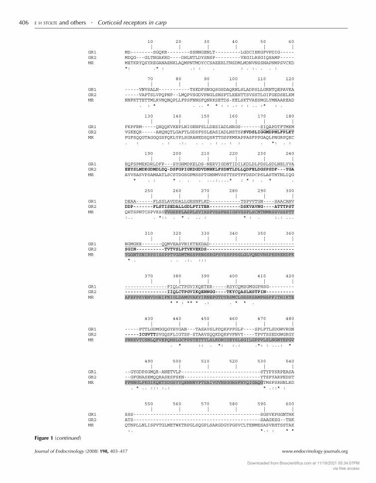
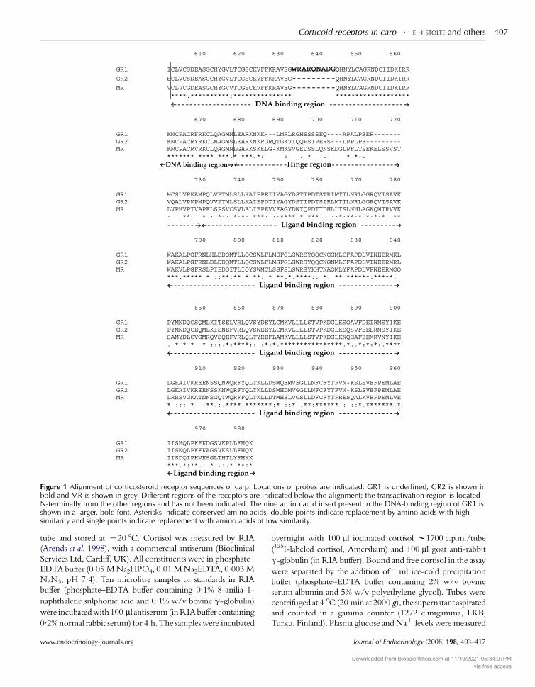
Figure 1 Alignment of corticosteroid receptor sequences of carp. Locations of probes are indicated; GR1 is underlined, GR2 is shown inbold and MR is shown in grey. Different regions of the receptors are indicated below the alignment; the transactivation region is locatedN-terminally from the other regions and has not been indicated. The nine amino acid insert present in the DNA-binding region of GR1 isshown in a larger, bold font. Asterisks indicate conserved amino acids, double points indicate replacement by amino acids with highsimilarity and single points indicate replacement with amino acids of low similarity.

Corticoid receptors in carp . E H STOLTE and others 407

tube and stored at K20 8C. Cortisol was measured by RIA

(Arends et al. 1998), with a commercial antiserum (Bioclinical

Services Ltd, Cardiff, UK). All constituents were in phosphate–

EDTAbuffer (0.05 MNa2HPO4, 0.01 MNa2EDTA, 0.003 MNaN3, pH 7.4). Ten microlitre samples or standards in RIA

buffer (phosphate–EDTA buffer containing 0.1% 8-anilia-1-

naphthalene sulphonic acid and 0.1% w/v bovine g-globulin)

were incubatedwith100 ml antiserum(inRIAbuffer containing

0.2% normal rabbit serum) for 4 h. The samples were incubated

www.endocrinology-journals.org

overnight with 100 ml iodinated cortisol w1700 c.p.m./tube

(125I-labeled cortisol, Amersham) and 100 ml goat anti-rabbit

g-globulin (in RIA buffer). Bound and free cortisol in the assay

were separated by the addition of 1 ml ice-cold precipitation

buffer (phosphate–EDTA buffer containing 2% w/v bovine

serum albumin and 5% w/v polyethylene glycol). Tubes were

centrifuged at 4 8C (20 min at 2000 g), the supernatant aspirated

and counted in a gamma counter (1272 clinigamma, LKB,

Turku, Finland). Plasma glucose andNaC levels were measured

Journal of Endocrinology (2008) 198, 403–417

Downloaded from Bioscientifica.com at 11/19/2021 05:34:07PMvia free access

E H STOLTE and others . Corticoid receptors in carp408

with a Stat Profile pHOx Plus L Analyser (Nova Biochemical,

Waltham, USA).

RNA isolation

RNA was isolated from tissues after extraction in Trizol

reagent (Invitrogen), as suggested by the manufacturer. Total

RNA was precipitated in isopropanol, washed with 75%

ethanol and dissolved in nuclease-free water. RNA of

separated pituitary glands (pars distalis and pars intermedia

separate) was isolated as described by the RNeasy Mini Kit

(Qiagen) strictly according to the manufacturer’s instructions.

RNA concentrations were measured by spectrophotometry

and integrity was ensured by analysis on a 1.5% agarose gel

before proceeding with cDNA synthesis.

DNAse treatment and first strand cDNA synthesis

For each sample, a ‘KRT’ (non-reverse transcriptase) control

was included. One microlitre 10! Dnase-I reaction buffer

and 1 ml Dnase-I (Invitrogen, 18068-015) were added to 1 mgtotal RNA and incubated for 15 min at room temperature in a

total volume of 10 ml. DNase I was inactivated with 1 ml25 mM EDTA at 65 8C for 10 min. To each sample, 300 ng

random hexamers (Invitrogen, 48190-011), 1 ml 10 mM

dNTP mix, 4 ml 5! first strand buffer, 2 ml 0.1 M

dithiothreitol and 40 Units RNAse Out (Invitrogen

10777-019) were added and the mix was incubated for

10 min at room temperature and for an additional 2 min at

37 8C. To each sample (not to the ‘KRT’ controls), 200 U

Superscript-II RNase HK reverese transcriptase (RT;

Invitrogen, 18064-014) was added and the reaction mixtures

were incubated for 50 min at 37 8C. All reaction mixtures

were filled up with demineralised water to a final volume of

100 ml and stored at K20 8C until further use.

Real-time quantitative PCR

PRIMER EXPRESS (Applied Biosystems, Foster City, CA,

USA) and PRIMER3 software were used to design primers

Table 2 Primers used for gene expression studies

Sequence

GeneGR1 FW: 5 0-AGA-CTG-AGA-GGC-GGA-GCT-ACT-G

RV: 5 0-GGC-GGT-GTT-GGC-TCC-AT-3 0

GR2 FW: 5 0-GGA-GAA-CAA-CGG-TGG-GAC-TAA-ARV: 5 0-GGC-TGG-TCC-CGA-TTA-GGA-A-30

MR FW: 5 0-TTC-CCT-GCA-GAA-CTC-AAA-GGA-3 0

RV: 5 0-ACG-GAC-GGT-GAC-AGA-AAC-G-30

PRL FW: 5 0-CAT-CAA-TGG-TGT-CGG-TCT-GA-3 0

RV: 5 0-TGA-AGA-GAG-GAA-GTG-TGG-CA-30

b-actin FW: 5 0-GCT-ATG-TGG-CTC-TTG-ACT-TCG-A-3RV: 5 0-CCG-TCA-GGC-AGC-TCA-TAG-CT-3 0

40S FW: 5 0-CCG-TGG-GTG-ACA-TCG-TTA-CA-30

RV: 5 0-TCA-GGA-CAT-TGA-ACC-TCA-CTG-TCT

Journal of Endocrinology (2008) 198, 403–417

for use in real-time quantitative PCR (RQ-PCR) (Table 2).

For RQ-PCR, 5 ml cDNA and forward and reverse primers

(300 nM each) were added to 12.5 ml Quantitect Sybr Green

PCR Master Mix (Qiagen) and filled up with demineralised

water to a final volume of 25 ml. RQ-PCR (15 min 95 8C,

40 cycles of 15 s at 94 8C, 30 s at 60 8C, and 30 s at 72 8C

followed by 1 min at 60 8C) was carried out on a Rotorgene

2000 real-time cycler (Corbett Research, Sydney, Australia).

Raw data were analysed by use of the comparative

quantitation of the Rotor-gene Analysis Software V5.0.

Basal gene expression in organs and tissues of adult carp

was determined as a ratio of target gene versus reference

gene and was calculated according to the following equation:

RatioZ ðEreferenceÞCt reference=ðEtargetÞ

Ct target, where E is the

amplification efficiency and Ct is the number of PCR

cycles needed for the signal to exceed a predetermined

threshold value. Expression following 24 h of restraint or

cold water transfer was determined relative to the expression

of non-restraint control fish according to the following

equation (Pfaffl 2001), RatioZ ðEtargetÞCt targetðcontrolKsampleÞ=

ðEreferenceÞCt referenceðcontrolKsampleÞ. Dual internal reference

genes (40S ribosomal protein and b-actin) were incorporatedin all RQ-PCR experiments; results were similar following

standardisation to either gene. ‘K RT’ controls were

included in all experiments and no amplification above

background levels was observed. Non-template controls

were included for each gene in each run and no

amplification above background levels was observed.

Specificity of the amplification was ensured by checking

the melting temperature and the profile of each melting

curve. The product of each template was checked at least

once by sequencing.

Transactivation assay

Clones encoding full-length open reading frame of common

carp GR1 and GR2 were excised from pGEM-Teasy vector by

EcoR1 andBamH1 and ligated into pcDNA3expressionvector

cutwith the same enzymes.Orientation and qualityof the insert

was confirmed by sequencing. COS-7 cells (derived from

Amplicon length (bp) Genbank acc. no.

-3 0 113 AJ879149

T-3 0 110 AM183668

117 AJ783704

130 X52881

0 89 M24113

69 AB012087-3 0

www.endocrinology-journals.org

Downloaded from Bioscientifica.com at 11/19/2021 05:34:07PMvia free access

Corticoid receptors in carp . E H STOLTE and others 409

African green monkey kidney) were cultured as described

previously (Sturm et al. 2005). Cells were transiently transfected

using calcium precipitation method (Sambrook & Russell

2001), with the cells growing in log phase at 30–50%

confluence. Cells were co-transfected with the following

plasmids: expression vector with the appropriate hormone

receptor cDNA (1 mg/24-well plate), reporter plasmid

pFC31Luc that contains the mouse mammary tumour virus

promoter upstream of the luciferase gene (10 mg/24-well plate);pSVb (Clontech), a second reporter plasmid under control of

the SV40 promoter and serving as a control for the transfection

efficiency (2 mg/24-well plate); and finally pBluescript (Clon-

tech) (7 mg/24-well plate), an irrelevant plasmid to increase

transfection. Sixteen hours after transfection, medium was

renewed and cortisol added from 1000-fold concentrated stock

solution in ethanol. After 36-h incubation, cells were harvested

using reporter lysis buffer (Promega) following the manufac-

turer’s instructions; luciferase and b-galactosidase activities weredetermined as described previously (Bury et al. 2003). In

addition to solvent controls (receiving only ethanol carrier

instead of hormone), cells were transfected with an empty

expression vector as control for luciferase activity in the absence

of hormone receptor DNA. Experiments were repeated thrice

independently, with triplicate cell cultures per treatment.

Luciferase activity was corrected for ‘well-specific’ transfection

efficiency (determined by b-galactosidase activity) and then

expressed as percentage luciferase activity observed in cells

treated with 10K7 M cortisol.

Half maximum activation concentration of ligand (EC50)

in the transactivation assay were assessed by fitting the data to a

single ligand binding model using the Sigma plotR software.

Only converging data were included in data sets presented.

Ligands were tested in the range of 10 pM to 1 mM.Data were

normalised to maximum (100%) response and corrected for

blanks prior to kinetic analysis.

Figure 2 Similarities between the receptor domains (AD, hinge region; E, ligand binding region) of the commoPercentage of amino acid identity of the different domaiparticular domains is represented by the length of boxes ashown at the right. Grey bar indicates nine amino acid i

www.endocrinology-journals.org

Bioinformatics

Sequences were retrieved from the Swissprot, EMBL and

GenBank databases using SRS and/or BLAST (Altschul et al.

1997). Multiple sequence alignments were carried out using

CLUSTALW (Chenna et al. 2003). Calculation of pairwise

amino acid identities was carried out using the SIM

ALIGNMENT tool (Huang & Miller 1991). Phylogenetic

and molecular evolutionary analyses were conducted using

MEGAversion 3.1 (Kumar et al. 2004). Phylogenetic tree was

constructed based on the neighbour-joining method using

the Poisson correction for evolutionary distance (Nei &

Kumar 2000). Reliability of the tree was assessed by

bootstrapping, using 1000 bootstrap replications.

Statistical analysis

Statistic analysis was performed with SPSS 12.0.1 software

(SPSS Inc., Chicago, IL, USA). Following ANOVA,

differences between treatments were assessed by Mann–

WitneyU test, and P!0.05 was accepted as fiducial limit. For

RQ-PCR data, tests were performed for both internal

reference genes (b-actin and 40S) and statistical significance is

reported only if both reference genes showed a significant

effect, where *indicates P!0.05 and **indicates P!0.01.

Results

CR characterisation

Cloning and characterisation of the MR and GRgenes Full-length sequences of one MR and two different

GR genes were obtained by homology cloning using a

common carp brain cDNA library. The first GR gene (GR1;

acc. no. AJ 879149) comprises 2190 nucleotides and encodes

a protein of 730 amino acids. The second gene (GR2; acc. no.

B, transactivation region; C, DNA-binding region;n carp and other vertebrate corticosteroid receptors.ns is shown in the boxes. Amino acid length ofnd is also mentioned. Total length of each protein isnsert as a result of alternative splicing.

Journal of Endocrinology (2008) 198, 403–417

Downloaded from Bioscientifica.com at 11/19/2021 05:34:07PMvia free access

E H STOLTE and others . Corticoid receptors in carp410

AM183668) contains an open reading frame of 2235

nucleotides and encodes a protein of 745 amino acids

(Fig. 1). The predicted amino acid identity of these two

GRs is 57%; both gene products show moderate sequence

(45–60%) identity when compared with other teleostean fish

and mammalian GR genes (Fig. 2). The MR gene (acc. No.

AJ783704) has an open reading frame of 2913 nucleotides that

codes for a 971 amino acids protein. The predicted MR

Journal of Endocrinology (2008) 198, 403–417

amino acid sequence shows relatively high (65–90%)

sequence identity to the other teleostean fish MR sequences

and moderate (w50%) sequence identity to the African

clawed frog (Xenopus laevis) and mammalian MR sequences.

When the protein domains of the receptors (GRs and MR)

are compared among different species, 85–100% sequence

identity is found for the DNA-binding domain. For the

ligand-binding domain (LDB), 50–60% sequence identity is

www.endocrinology-journals.org

Downloaded from Bioscientifica.com at 11/19/2021 05:34:07PMvia free access

Corticoid receptors in carp . E H STOLTE and others 411

found when GRs are compared with MRs between species,

and 70–90% when LDBs of either GRs or MR of individual

species are compared (Fig. 2). The N-terminal domains of

GRs andMRs constitute the most variable region. Alignment

of carp GR and MR genes yields low sequence conservation

(28% for GR1 compared with GR2 and !15% for MR

compared with either of the GRs; Fig. 1).

Phylogenetic analysis The neighbour-joining phyloge-

netic tree for corticosteroid receptor proteins (Fig. 3) resulted

in a predicted cluster of GRs and MRs on separate branches

together with mammalian orthologues; androgen and

corticosteroid receptors from jawless fish represent an

out-group. Within both the MR and GR branch, teleostean

and tetrapod proteins form separate clades. The teleostean

GR clade has a subdivision as a result of duplication of the

GR gene.

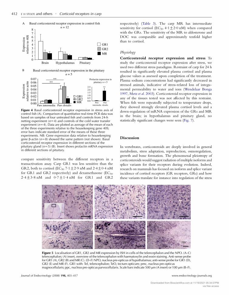
Expression of mRNAs for corticosteroid receptorsExpression of GR1, GR2 and MR genes in the brain

(without hypothalamus and pituitary gland), ventral hypo-

thalamus, pituitary gland of healthy and non-stressed carp was

quantitated by real-time PCR shown in Fig. 4. The highest

GR expression was found in the brain and hypothalamus.

Expression of GR1 and GR2 genes was generally com-

parable; in the brain, however, GR1 expression was higher

than GR2 expression. Receptor expression abundance in

pituitary tissue was about half that was seen in the brain or

hypothalamus. The MR gene also showed an about 50%

lower expression level in pituitary tissue compared with the

brain and hypothalamus (Fig. 4a).

To discriminate gene expression levels in the pars distalis

(pro-opiomelanocortin, POMC cells producing ACTH) and

Figure 3 Phylogenetic tree comparing the amino acid sequences of theMEGA version 3.1 software using the neighbour-joining method. Reliabreplications; values in percentage are indicated at branch nodes. Atlantiand rat androgen receptors (AR) were used as out-group. Common carp(Oncorhynchus mykiss) GR1; P49843, GR2; AY4953720, Burton’s mouAF263739, GR2b; AF263740, Zebrafish (Danio rerio) GR2; EF436284,(O73673), European sea bass (Dicentrarchus labrax) GR1; AY549305, Gminnow (Pimephales promelas) GR; AY533141, Puffer fish (Fugu) (TakifGENSCAN00000029451 (scaffold 4328), GR2; SINFRUG00000143550nigroviridis) GR1; GIDT00024792001 (Chr. 7), GR2; GSTENG0001702ENSGACP00000027400, GR2; ENSGACP00000024074, Japanese KillifiENSORLP00000007570, Mozambique or common tilapia (Oreochromilaevis) GR; P49844, Western clawed frog (X. tropicalis) GR; CR848477partial), Platypus (Ornithorhynchus anatinus) GR; ENSOANP00000009AY238475, Guinea pig (Cavia porcellus) GR; P49115, Mouse (Mus musc(Homo sapiens) GRa; P04150, Human GRb; NP_001018661, Human G(D. rerio) MR; ENSDARP00000053817, Rainbow trout (O.mykiss) MRa; YQ8JJ89, Puffer fish (Fugu) (T. rubripes) MR; NEWSINFRUP00000129848GSTENT00032894001, Stickleback (G. aculeatus)MR;ENSGACP000000ENSORLT00000009439, Chicken (G. gallus) MR;ENSGALP0000001628ENSOANT00000008378, African clawed frog (X. laevis) MR; BC08108MR; M16801, Sea lamprey (Petromyzon marinus) CR; AY028457, AtlanM37890 Human (H. sapiens) AR; P10275. NB: Burton’s mouthbrooderwith common carp and rainbow trout GR2) and GR2 has two splice varGR2b has a nine amino acid insert.

www.endocrinology-journals.org

pars intermedia (POMC cells producing melanocyte-stimu-

lating hormone (MSH)), we dissected pituitary glands and

confirmed tissue separation by assay of prolactin mRNA

expression, a marker for the rostral pars distalis (RPD, insert

Fig. 4b). GR1 and GR2 mRNA expression was significantly

(P!0.01) higher in pituitary pars distalis compared with

pars intermedia. No such difference was observed for MR

mRNA (Fig. 4b).

CR localisation The localisation of mRNA expression was

further studied by in situ hybridisation. In the telencephalon,

mRNA expression of all three receptors was detected,

predominantly in the outer pallial layers. In other brain areas,

GR1 and GR2 show the same distribution pattern, although

relative expression levels were different, whereas MR mRNA

showed a less wide distribution. In transverse sections of the

hypothalamus, mRNA expression of all the three receptors

was observed in themagnocellular part of theNPO (as assessed

by comparison with paramedian sagittal slides (Huising et al.

2004)); expression was less pronounced in the parvocellular

part (Fig. 5). In the pituitary pars intermedia, GR mRNA

expression was low. In the pars distalis, strongest GR

expression was found in the proximal pars distalis, in

GH-producing cells. In the rostral pars distalis, ACTH cells

express both corticosteroid receptors GR1 and GR2 and to a

far higher degree than the prolactin cells (Fig. 6).

Transactivation activity GR2 was more sensitive to the

different hormones tested than GR1. For both receptors,

dexamethasone was the strongest agonist tested, followed by

cortisol, deoxycortisol and corticosterone. Finally, aldoster-

one and DOC were very weak agonists. The physiologically

important stress hormone cortisol was chosen as ligand to

vertebrate corticosteroid receptors. This tree was generated withility of this tree was assessed by bootstrapping using 1000 bootstrapc hagfish and sea lamprey corticosteroid receptors (CR) and human(C. carpio) GR1; AJ879149, GR2; AM183668, Rainbow trout

thbrooder (Haplochromis burtoni) GR1; AF263738, GR2a;GR2b; EF436285 Japanese flounder (Paralichthys olivaceus) GR;R2; AY619996, Brown trout GR (Salmo trutta); AY863149, Fatheadugu rubripes) GR1; GENSCAN00000003615 (scaffold 1264) &(scaffold 59), Green spotted puffer (Tetraodon) (Tetraodon

7001 (Chr. 1) Stickleback (Gasterosteus aculeatus) GR1;sh (Medaka) (Oryzias latipes) GR1; ENSORLP00000001939, GR2;s mossambicus) GR; BAA23662, African clawed frog (Xenopus, Chicken (Gallus gallus) GR; ENSGALP00000011948 (Q8JHA4152, Pig (Sus scrofa) GR; AY779185, Cow (Bos taurus) GR;ulus) GR; P06537, Rat (Rattus norvegicus) GR; NP_036708, HumanRg; NP_001019265, Carp (C. carpio) MR; AJ783704, Zebrafish495584, MRb; AY495585, Burton’s mouthbrooder (H. burtoni) MR;, Green spotted puffer (Tetraodon) (T. nigroviridis) MR;22713, Japanese killifish (Medaka, O. latipes) MR;3 (Q8QHI2, partial), Platypus (O. anatinus) MR;

2, Mouse MR; XP_356093, Rainbow trout MR; AY495584, Humantic hagfish (M. glutinosa) CR; DQ382336 Mouse (M. musculus) AR;nomenclature is different; GR1 has no splice variants (comparableiants (comparable with rainbow trout GR1); Burton’s mouthbrooder

Journal of Endocrinology (2008) 198, 403–417

Downloaded from Bioscientifica.com at 11/19/2021 05:34:07PMvia free access

Figure 4 Basal corticosteroid receptor expression in stress axis ofcontrol fish (A). Comparison of quantitative real-time PCR data wasbased on samples of four untreated fish and controls from 24-hnetting experiment (nZ4) and controls of the cold water transferexperiment (nZ4). Data are plotted as average of the mean of eachof the three experiments relative to the housekeeping gene 40S;error bars indicate standard error of the means of these threeexperiments. NB: Gene expression data relative to housekeepinggene b-actin (nZ8) showed the same pattern (not shown). Basalcorticosteroid receptor expression in different sections of thepituitary gland (nZ5) (B). Insert shows prolactin mRNA expressionin different sections of pituitary.

E H STOLTE and others . Corticoid receptors in carp412

compare sensitivity between the different receptors in a

transactivation assay. Carp GR1 was less sensitive than the

GR2, both to cortisol (EC50 7.1G2.9 nM and 2.4G0.4 nMfor GR1 and GR2 respectively) and dexamethasone (EC50

2.4G3.8 nM and 0.7G1.4 nM for GR1 and GR2

Figure 5 Localisation of GR1, GR2 and MR expression bytelencephalon; (A) insert, overview of the telencephalon wfor GR1 (A), GR2 (B) and MR (C). (D–F) NPO, nucleus pre-oGR2 (E) and MR (F). GR1 with: Tel, telencephalon; TeO, temagnocellularis; ppc, nucleus pre-opticus parvocellularis.

Journal of Endocrinology (2008) 198, 403–417

respectively) (Table 3). The carp MR has intermediate

sensitivity for cortisol (EC50 4.1G2.0 nM) when compared

with the GRs. The sensitivity of the MR to aldosterone and

DOC was comparable and approximately tenfold higher

than to cortisol.

Physiology

Corticosteroid receptor expression and stress To

study the corticosteroid receptor expression after stress, we

used two different stress paradigms. Restraint of carp for 24 h

resulted in significantly elevated plasma cortisol and plasma

glucose values as assessed upon completion of the treatment.

Plasma sodium concentrations had significantly decreased in

stressed animals, indicative of stress-related loss of integu-

mental permeability to water and ions (Wendelaar Bonga

1997, Metz et al. 2003). Corticosteroid receptor expression in

any of the tissues tested was not affected by this restraint.

When fish were repeatedly subjected to temperature drops,

they showed strongly elevated plasma cortisol levels and a

down-regulation of mRNA expression of the GRs and MR

in the brain; in hypothalamus and pituitary gland, no

statistically significant changes were seen (Fig. 7).

Discussion

In vertebrates, corticosteroids are deeply involved in general

metabolism, stress adaptation, reproduction, osmoregulation,

growth and bone formation. The phenomenal pleiotropy of

corticosteroidswould suggest radiation ofmultiple isoforms and

splice variants for their receptors during evolution. Indeed,

research on mammals has focused on isoform and splice variant

incidence of cortisol receptors (GR receptors, GRs) and how

these variants translate for instance into regulation of the stress

ISH in cells of the telencephalon and the NPO. (A–C)ith haematoxylin and eosin staining. Anti-sense probepticus of hypothalamus; anti-sense probe for GR1 (D),ctum opticum; pmc, nucleus pre-opticusScale bars indicate 500 mm (A insert) or 100 mm (B–F).

www.endocrinology-journals.org

Downloaded from Bioscientifica.com at 11/19/2021 05:34:07PMvia free access

Figure 6 Localisation of GR1 and GR2 expression in the cells of pituitary. (A) Sense control for GR1, (B) anti-sense probe for GR1, and (C) detail of rostral pars distalis (RPD) with anti-sense probe for GR1. (D) Sensecontrol for GR2, (E) anti-sense probe for GR2 and (F) detail of RPD with antisense probe for GR2.(G) Overview of pituitary stained with haematoxylin and eosin, (H) detail of proximal pars distalis (PPD) withantibody against GH, and (I) detail of RPD with antibody against ACTH. PI, pars intermedia. Scale barsindicate 500 mm (A, B, D, E and G), 50 mm (H), 100 mm (C, F and I).

Corticoid receptors in carp . E H STOLTE and others 413

axis. The extant teleostean fishes are representatives of the

earliest true vertebrates and exhibit a complex receptor profile.

With two genes encoding functionally different GRs, this

system is even more complex than that observed in mammals,

which warranted investigation into the role of these different

receptors in stress axis regulation.

CR characterisation

Receptor evolution in fishes Different GR genes were

demonstrated in distantly related teleostean species (Bury et al.

2003, Greenwood et al. 2003) and for that very reason not

Table 3 Transactivation capacity of corticosteroid receptorsa

Cortisol Dexamethasone

EC50 (nM) S.E.M. EC50 (nM) S.D.

GR1 7.1 2.9 2.4 3.8GR2 2.4* 0.4 0.7 1.4MR 4.1 2.0

*GR2 is significantly more sensitive to cortisol than GR1 (P!0.05).aEC50 values of GR1, GR2 and MR for different hormones. Cortisol for GRs; averaMR, dexamethasone, aldosterone and DOC; single experiments. DOC, 11-Deoxy

www.endocrinology-journals.org

necessarily result from the tetraploidisation of common carp

(Greenwood et al. 2003, Stolte et al. 2006). In the green-spotted

puffer, two duplicates on different chromosomes are found that

makes a single gene duplication less likely (Stolte et al. 2006).

Moreover, all known teleosteanGR1proteins share a conserved

insert of nine amino acids (WRARQNTDG, orWrarqnadg in

carp) in theDNA-binding domainwhich are not found inother

vertebrates. We rate it highly unlikely that all teleosts

independently duplicated a single gene and inserted every

time again a nine amino acid sequence.Most convincing for this

debate is that our phylogenetic analysis yields two distinct clades

of GR genes in the teleostean lineage that argues against

Aldosterone DOC

EC50 (nM) S.D. EC50 (nM) S.D.

0.46 4.1 0.25 3.6

ge of three separate experiments with standard error of the mean; cortisol forcorticosterone.

Journal of Endocrinology (2008) 198, 403–417

Downloaded from Bioscientifica.com at 11/19/2021 05:34:07PMvia free access

Figure 7 Corticosteroid receptor expression in stress exposed fish. Gene expression of corticosteroidreceptors in stress axis organs after 24-h restraint stress (A) and cold transfer (23 8C to 10 8C) stress (B). Insertsof plasma cortisol, glucose and sodium levels are shown for control and stressed animals for the respectiveexperiments. Gene expression is shown in comparison with unstressed control fish of the respectiveexperiment and relative to b-actin.

E H STOLTE and others . Corticoid receptors in carp414

duplication in the tetrapod lineage. The duplication in all

likelihood results from an early genome duplication 300–450

million years ago, and only after the divergence of the tetrapods

from the fish lineage (Volff 2005). Ifwe proceed froma notion of

a major genomic duplication in fishes, it follows that one MR

has apparently been lost during evolution aswewere not able to

detect a second MR-coding gene in carp or in genomic

databases for zebrafish (Danio rerio), puffers (Fugu species) or rice

fish (Oryzias latipes). Three possibilities arise after gene/genome

duplication: non-functionalisation, the fate of most duplicated

genes (Brunet et al. 2006); neo-functionalisation, the acquire-

ment of a new function; or sub-functionalisation, where each

copy loses part of the ancestral function and both copies are

required to maintain the full function (Force et al. 1999). The

duplicated GR genes of fish escaped a fate as non-functional

pseudogene: expression levels and differential sensitivities for

cortisol are more, so in-line with neo- or sub-functionalisation

(Bury et al. 2003,Greenwood et al. 2003). Interestingly, zebrafish

has only one GR copy that clusters with other fish GR2

Journal of Endocrinology (2008) 198, 403–417

sequences.However, zebrafish has acquired a splicingb-isoformof theGR (Fig. 2) that resembles the dominant negativeGRb ofhumans in structure, expression level and function. This could

reflect an alternative regulatory mechanism to compensate for

the loss of a functional second GR gene (Schaaf et al. 2008).

Receptor functional definition by transactivationcapacity In carp, transactivation capacity of cortisol (capacity

of hormone to initiate or repress CR-mediated transcription

of downstream genes) is about three-fold higher for GR2

(EC50 2.4G0.4 nM) than for GR1 (EC50 7.1G2.9), and thiswould facilitate differential regulation by basal and elevated

cortisol levels. The carp MR sensitivity (EC50 4.1G2.0 nM)

is intermediate to that of the GRs, and this is in stark contrast

to data for Burton’s mouthbrooder and trout, with MRs more

sensitive to cortisol than the GRs (Greenwood et al. 2003,

Sturm et al. 2005). In fish, levels of up to 10 nM DOC were

published (Campbell et al. 1980). The low EC50 of DOC

(0.25 nM) for MR transactivation in carp certainly does not

www.endocrinology-journals.org

Downloaded from Bioscientifica.com at 11/19/2021 05:34:07PMvia free access

Corticoid receptors in carp . E H STOLTE and others 415

exclude a mineralocorticoid function for DOC in fishes. In

carp plasma basal, total levels of cortisol are around 5 ng/ml

(i.e. 13.8 nM); following stress, cortisol levels easily reach

150 ng/ml (O400 nM). As only 20% is available as unbound

cortisol (Flik & Perry 1989), this corresponds to 2.8 and O80 nM respectively, for which in carp apparently specific

receptor subtypes are present: at rest both GR2 and MR may

be occupied and activated, whereas GR1 is preferentially

activated by stress levels of cortisol. In rodent brain, a similar

system was demonstrated: an 80% MR and 10% GR

corticosterone occupancy was established in non-stressed

situations (Reul et al. 1987). Even though the majority of

MRs is occupied at low cortisol levels, it still is a dynamically

regulated receptor. MR activity could be increased by an

agonist to further inhibit HPA axis activity (Buckley et al.

2007). We assume the same applies for common carp GR2

and MR. This means that although both receptors are partly

or even largely occupied with cortisol, continuous modu-

lation of HPI axis activity could be mediated by increasing

cortisol levels after stressful events via GR1.

Receptor functional definition by localisation As we

focus on the involvement of the different receptors in stress axis

regulation, our areas of interest are the HPI axis organs.

Hypothalamus and pituitary gland of unstressed carp showed

comparablemRNA levels for both theGRsandMR, suggesting

functional importance of all the three. Only in the brain

(without hypothalamus and pituitary gland), a consistent two-

fold higher mRNA expression for GR1 over GR2 was seen.

A receptor-defined duality in GR function in the brain offishes

seems of wider occurrence as similar preferential expression was

seen in the brain of rainbow trout and Burtons’ mouthbrooder

(Bury et al. 2003, Greenwood et al. 2003). The higher mRNA

expression level could translate intohigher protein levels butmay

also reflect higher turnover. We have no data on brain cortisol

levels but two receptors with significant difference in receptor

sensitivity would allow differential responses to basal and stress

levels of the steroid. In carp and trout (Sturm et al. 2005), the

brain shows strong MR mRNA expression; unexpectedly,

typical osmoregulatory organs such as gills, kidney and intestine

show far lower MR mRNA expression. This suggests that a

widely accepted but only presumed hypothesis of MR

involvement in osmoregulation may be wrong, and is a

consequence of extrapolation of the situation for aldosterone

andMRfunction inmammals.However, also in themammalian

brain, MRs play a key role in corticosteroid-regulated processes

(De Kloet et al. 1998). The mammalian hippocampus exerts

inhibitory control over the HPA axis activity: a dominant

inhibition by MR activity is attenuated by GR activation (De

Kloet et al. 1998).Our results support such a combined function

for steroidsmediated by GR andMR activities in the regulation

of stress coping and learning, and this is apparently a very early

function developed in our ancestral fishes.

Steroid receptors distribution in telencephalic regions may

contribute to define hippocampal regions, especially involved in

www.endocrinology-journals.org

(learning) behaviour and sentience. Our demonstration of GR

and MR mRNA in carp telencephalon expands and further

substantiates the notion that GR and MR corticosteroid

receptors have the basic functions in the regulation of learning

and memory. In goldfish (Vargas et al. 2006) and cod (Nilsson

et al. 2008) learning and spatial memory may involve the

telencephalic lateral pallium, the homologue of the hippo-

campus of mammals (Meek & Nieuwenhuys 1998).

In the hypothalamus,mRNAof all the three receptor genes is

expressed in parvo- andmagnocellular neurons of the pre-optic

nucleus. We confirmed (data not shown) by immunohisto-

chemistry on adjacent sections that expression of GR- and

MR-mRNA co-localises with CRH (Huising et al. 2004). So

far, the presence of GR protein in these cells was shown for

rainbow trout (Teitsma et al. 1998) and Mozambique tilapia

(Oreochromis mossambicus) (Pepels et al. 2004). However, the

antiserumused in these studies does not distinguish betweenGR

gene species. To the best of our knowledge, no data on MR

distribution in other fish brains are available. In mammals, the

MRwas described in the ventromedian and arcuate nuclei of the

hypothalamus, and theGR in the arcuate nuclei, paraventricular

and supra-optic nucleus of the hypothalamus (Reul & de Kloet

1985); the hypothalamic arcuate nucleus is an important target

for metabolic and hormonal signals controlling food intake and

feeding behaviour (Meister 2007). Clearly, future studies onGR

andMR localisation in the fish analogues of these structures are

warranted as we know that regulation of stress on feeding

(arcuate nucleus in fish) are strongly intertwined, also in fish

(Bernier et al. 2004).

In carp pituitary gland, the two GR genes co-localise in the

same areas. Remarkably, little expression was seen in pars

intermedia and would imply limited feedback by cortisol on

MSH- and somatolactin-producing cells. This reminds of the

situation reported for trout (Teitsma et al. 1998), but not for

tilapia that has significant GR-immunoreactivity in the pars

intermedia (Pepels et al. 2004), although mRNA expression

could not be detected (Kitahashi et al. 2007).The strongest signal

forGRmRNAwas seen in the proximal pars distalisGHcells (in

linewithobservations forGR1on trout (Teitsma et al. 1998) and

tilapia (Pepels et al. 2004, Kitahashi et al. 2007)). Stress/cortisol

induced suppression of growth, for instance seen after handling

or confinement offishes is thus explained by down-regulationof

GH cells (Auperin et al. 1997). In the rostral pars distalis finally,

ACTH cells express significant levels of GR mRNA, as one

could predict as a basis for feedback control in the stress axis.

Interestingly, recent data in tilapia showed no mRNA

expression of either of the duplicatedGRs inACTH-producing

cells (Kitahashi et al. 2007), whereas immuno-histochemical

studieswith an antibodyagainstGRshowedpositivity inACTH

cells in tilapia and rainbow trout (Teitsma et al. 1998, Pepels et al.

2004). Prolactin cells, however, known to become activated

during chronic stress (Auperin et al. 1997), showednodetectable

GRexpression (Teitsma et al. 1998). This only seems at variance

with recent reports on direct effects of cortisol on prolactin cells

(Uchida et al. 2004): a G-protein-coupled membrane receptor

for cortisol should be considered. Indeed, the reported effects of

Journal of Endocrinology (2008) 198, 403–417

Downloaded from Bioscientifica.com at 11/19/2021 05:34:07PMvia free access

E H STOLTE and others . Corticoid receptors in carp416

cortisol on prolactin cells reported are fast, faster than onewould

predict for GR/MR-mediated genomic effects.

MRmRNAwas far more abundant in the pars distalis than in

the pars intermedia. Based on the comparisons with GR

staining, we predict theMR expression to be scarcely expressed

in thea-MSHcells of the pars intermedia and stronglyexpressed

in the GH cells of the pars distalis. Unfortunately, we were

unable to demonstrate MRmRNA in ACTH cells specifically.

Assuming (by analogy to the situation for GRmRNA) absence

of MR mRNA in prolactin cells, our RQ-PCR data would

favour the presence ofMR inACTHcells; this part of our study

requires future attention.

Physiology

Restraint by netting for 24 h resulted in hyperglycaemia,

hyponatraemia, typical signs of strong catecholaminergic

activation and inherent loss of control over integumental

permeability to water and ions (Wendelaar Bonga 1997).

A persistent hypercortisolinaemia was observed, but the

duration of the stress condition proved insufficient to induce

feedback by cortisol. However, a more persistent stress of

repeated cold transfer induced high cortisol levels and

hyperglycaemia and mild hypernatraemia indicative of

adoption to the stressor. Mild hypernatraemia is typical for

enhanced prolactin activity and successful restoration of blood

mineral levels to a new set point (Metz et al. 2003). This longer

term adaptation process did involve measurable down-

regulation of CRs in the brain (without hypothalamus and

pituitary gland). This observation suggests a central initiation

of down-regulation of cortisol release, rather than direct

feedback via NPO or pituitary gland. In tilapia, a similar

regulation system was suggested; cortisol feedback on CRH

release is exerted via the forebrain medial part of dorsal

telencephalon (Pepels et al. 2004). Our observation of

GR and MR mRNA down-regulation only following

the persistent stress is further consistent with experiments on

Coho salmon (Oncorhynchus kisutch) that showed no effect

of acute stress on GR expression, whereas chronically

elevated cortisol levels in several other paradigms did

(Maule & Schreck 1991).

The MR mRNA in carp brain was down-regulated to the

same extent as the mRNA levels of both GRs. This seems in

contrast with the mammalian model where GR stimulates

HPA axis activity, whereas MR has an inhibiting effect (De

Kloet et al. 1998). However, a similar result was found in

hippocampus of rats exposed to increasing the corticosteroid

levels (Hugin-Flores et al. 2004). And in mouse pups with

high corticosteroid levels due to 24-h maternal deprivation,

both GR and MR mRNA expression were significantly

decreased (Schmidt et al. 2003). These discrepancies between

different stressors are thus of wider occurrence and require

further and species specific approaches.

In conclusion, we show that carp express separate GRs for

regulation under basal and stressful conditions. We predict an

ancestral role not only for the duplicated GRs but also for

Journal of Endocrinology (2008) 198, 403–417

MR in stress physiology, based on the receptor localisation

and expression profile after chronic stress and provide a

receptor profile in the brain conform memory and learning

functionalities in fishes as in higher vertebrates. A role for

DOC in stress physiology in fishes has never been considered

but seems a reasonable hypothesis for future research. More

in-depth studies on specific roles of these receptors in learning

and stress physiology would therefore elucidate early strategies

in vertebrate evolution to control the basics of life.

Declaration of Interest

The authors declare that there is no conflict of interest that would prejudice

the impartiality of this scientific work.

Funding

Support of the Smart Mix Programme of the Netherlands Ministry of

Economic Affairs and the Netherlands Ministry of Education, Culture and

Science is gratefully acknowledged.

Acknowledgements

We gratefully acknowledge Ms Sandra Janssen and Mrs Liesbeth Pierson for

their excellent technical assistance during the experiments. F ATom Spanings

is thanked for excellent fish husbandry.

References

Altschul SF, Madden TL, Schaffer AA, Zhang J, Zhang Z, Miller W & Lipman

DJ 1997 Gapped BLAST and PSI-BLAST: a new generation of protein

database search programs. Nucleic Acids Research 25 3389–3402.

Arends RJ, van der Gaag R, Martens GJ, Wendelaar Bonga SE & Flik G 1998

Differential expression of two pro-opiomelanocortin mRNAs during

temperature stress in common carp (Cyprinus carpio L.). Journal of

Endocrinology 159 85–91.

Auperin B, Baroiller JF, Ricordel MJ, Fostier A & Prunet P 1997 Effect of

confinement stress on circulating levels of growth hormone and two

prolactins in freshwater-adapted tilapia (Oreochromis niloticus). General and

Comparative Endocrinology 108 35–44.

Baker ME, Chandsawangbhuwana C & Ollikainen N 2007 Structural analysis

of the evolution of steroid specificity in the mineralocorticoid and

glucocorticoid receptors. BMC Evolutionary Biology 7 24.

Balment RJ & Henderson IW 1987 In Secretion of Endocrine Glands and Their

Relationship to Osmoregulation, Eds I Chester-Jones, PM Ingleton &

JG Phillips. New York: Plenum press.

Basu N, Kennedy CJ & Iwama GK 2003 The effects of stress on the association

between hsp70 and the glucocorticoid receptor in rainbow trout.

Comparative Biochemistry and Physiology. Part A, Molecular and Integrative

Physiology 134 655–663.

BernierNJ,BedardN&PeterRE2004Effects of cortisol on food intake, growth,

and forebrain neuropeptide Yand corticotropin-releasing factor gene

expression in goldfish. General and Comparative Endocrinology 135 230–240.

Bridgham JT, Carroll SM & Thornton JW 2006 Evolution of hormone-

receptor complexity by molecular exploitation. Science 312 97–101.

Brunet FG, Crollius HR, Paris M, Aury JM, Gibert P, Jaillon O, Laudet V &

Robinson-Rechavi M 2006 Gene loss and evolutionary rates following

whole-genome duplication in teleost fishes. Molecular Biology and Evolution

23 1808–1816.

www.endocrinology-journals.org

Downloaded from Bioscientifica.com at 11/19/2021 05:34:07PMvia free access

Corticoid receptors in carp . E H STOLTE and others 417

Buckley TM, Mullen BC & Schatzberg AF 2007 The acute effects of a

mineralocorticoid receptor (MR) agonist on nocturnal hypothalamic–

adrenal–pituitary (HPA) axis activity in healthy controls. Psychoneuro-

endocrinology 32 859–864.

Bury NR & Sturm A 2007 Evolution of the corticosteroid receptor signalling

pathway in fish. General and Comparative Endocrinology 153 47–56.

Bury NR, Sturm A, Le Rouzic P, Lethimonier C, Ducouret B, Guiguen Y,

Robinson-Rechavi M, Laudet V, Rafestin-Oblin ME & Prunet P 2003

Evidence for two distinct functional glucocorticoid receptors in teleost fish.

Journal of Molecular Endocrinology 31 141–156.

Campbell CM, Fostier A, Jalabert B & Truscott B 1980 Identification and

quantification of steroids in the serum of rainbow trout during spermiation

and oocyte maturation. Journal of Endocrinology 85 371–378.

Chenna R, Sugawara H, Koike T, Lopez R, Gibson TJ, Higgins DG &

Thompson JD 2003 Multiple sequence alignment with the Clustal series of

programs. Nucleic Acids Research 31 3497–3500.

Engelsma MY, Stet RJ, Schipper H & Verburg-van Kemenade BM 2001

Regulation of interleukin 1 beta RNA expression in the common carp,

Cyprinus carpio L. Developmental and Comparative Immunology 25 195–203.

Flik G & Perry SF 1989 Cortisol stimulates whole body calcium uptake and

the branchial calcium pump in freshwater rainbow trout. Journal of

Endocrinology 120 75–82.

Force A, Lynch M, Pickett FB, Amores A, Yan YL & Postlethwait J 1999

Preservation of duplicate genes by complementary, degenerative mutations.

Genetics 151 1531–1545.

Gilmour KM 2005 Mineralocorticoid receptors and hormones: fishing for

answers. Endocrinology 146 44–46.

Greenwood AK, Butler PC, White RB, DeMarco U, Pearce D & Fernald RD

2003 Multiple corticosteroid receptors in a teleost fish: distinct sequences,

expression patterns, and transcriptional activities.Endocrinology 144 4226–4236.

Huang X & Miller W 1991 A time-efficient, linear-space local similarity

algorithm. Advances in Applied Mathematics 12 337–357.

Hugin-Flores ME, Steimer T, Aubert ML & Schulz P 2004 Mineralo- and

glucocorticoid receptor mrnas are differently regulated by corticosterone in

the rat hippocampus and anterior pituitary. Neuroendocrinology 79 174–184.

Huising MO, Metz JR, van Schooten C, Taverne-Thiele AJ, Hermsen T,

Verburg-van Kemenade BM & Flik G 2004 Structural characterisation of a

cyprinid (Cyprinus carpio L.) CRH, CRH-BP and CRH-R1, and the role

of these proteins in the acute stress response. Journal of Molecular

Endocrinology 32 627–648.

Irnazarow I 1995 Genetic variability of Polish and Hungarian carp lines.

Aquaculture 129 215–219.

Kitahashi T, Ogawa S, Soga T, Sakuma Y & Parhar I 2007 Sexual maturation

modulates expression of nuclear receptor types in laser-captured single cells

of the cichlid (Oreochromis niloticus) pituitary. Endocrinology 148 5822–5830.

De Kloet ER, Vreugdenhil E, Oitzl MS & Joels M 1998 Brain corticosteroid

receptor balance in health and disease. Endocrine Reviews 19 269–301.

Kumar S, Tamura K & Nei M 2004 MEGA3: integrated software for

molecular evolutionary genetics analysis and sequence alignment. Briefings

in Bioinformatics 5 150–163.

Maule AG & Schreck CB 1991 Stress and cortisol treatment changed affinity

and number of glucocorticoid receptors in leukocytes and gill of coho

salmon. General and Comparative Endocrinology 84 83–93.

Meek J & Nieuwenhuys R 1998 In The Central Nervous System of Vertebrates 2:

Holosteans and Teleosts, Eds R Nieuwenhuys, HJ Ten Donkelaar &

C Nicholson. Heidelberg: Springer-Verlag.

Meister B 2007 Neurotransmitters in key neurons of the hypothalamus that

regulate feedingbehavior andbodyweight.Physiology andBehavior92263–271.

Metz JR, van den Burg EH, Bonga SE & Flik G 2003 Regulation of branchial

Na(C)/K(C)-ATPase in common carp Cyprinus carpio L. acclimated to

different temperatures. Journal of Experimental Biology 206 2273–2280.

Mommsen TP, Vijayan MM & Moon TW 1999 Cortisol in teleosts:

dynamics, mechanisms of action, and metabolic regulation. Reviews in Fish

Biology and Fisheries 9 211–268.

www.endocrinology-journals.org

Nei M & Kumar S 2000 Molecular Evolution and Phylogenetics, New York:

Oxford University Press Inc.

Nilsson J, Kristiansen TS, Fosseidengen JE, Ferno A & van den Bos R 2008

Learning in cod (Gadus morhua): long trace interval retention. Animal -

Cognition 11 215–222.

Pepels PP, Van Helvoort H, Wendelaar Bonga SE & Balm PH 2004

Corticotropin-releasing hormone in the teleost stress response: rapid

appearance of the peptide in plasma of tilapia (Oreochromis mossambicus).

Journal of Endocrinology 180 425–438.

Pfaffl MW 2001 A new mathematical model for relative quantification in real-

time RT-PCR. Nucleic Acids Research 29 e45.

Prunet P, Sturm A & Milla S 2006 Multiple corticosteroid receptors in fish:

from old ideas to new concepts. General and Comparative Endocrinology 147

17–23.

Reul JM & de Kloet ER 1985 Two receptor systems for corticosterone in rat

brain: microdistribution and differential occupation. Endocrinology 117

2505–2511.

Reul JM, van den Bosch FR & de Kloet ER 1987 Relative occupation of

type-I and type-II corticosteroid receptors in rat brain following stress and

dexamethasone treatment: functional implications. Journal of Endocrinology

115 459–467.

Reul JM, Gesing A, Droste S, Stec IS, Weber A, Bachmann C, Bilang-Bleuel

A, Holsboer F & Linthorst AC 2000 The brain mineralocorticoid receptor:

greedy for ligand, mysterious in function. European Journal of Pharmacology

405 235–249.

Sambrook J & Russell DW 2001 Molecular Cloning, New York: Cold Spring

Harbor Laboratory Press.

Schaaf MJ, Champagne D, van Laanen IH, van Wijk DC, Meijer AH, Meijer

OC, Spaink HP & Richardson MK 2008 Discovery of a functional

glucocorticoid receptor b-isoform in zebrafish. Endocrinology 149

1591–1599.

Schmidt MV, Oitzl MS, Muller MB, Ohl F, Wurst W, Holsboer F, Levine S &

De Kloet ER 2003 Regulation of the developing hypothalamic–pituitary–

adrenal axis in corticotropin releasing hormone receptor 1-deficient mice.

Neuroscience 119 589–595.

Stolte EH, van Kemenade BM, Savelkoul HF & Flik G 2006 Evolution of

glucocorticoid receptors with different glucocorticoid sensitivity. Journal of

Endocrinology 190 17–28.

Sturm A, Bury N, Dengreville L, Fagart J, Flouriot G, Rafestin-Oblin ME &

Prunet P 2005 11-deoxycorticosterone is a potent agonist of the rainbow trout

(Oncorhynchus mykiss) mineralocorticoid receptor. Endocrinology 146 47–55.

Teitsma CA, Anglade I, Toutirais G, Munoz-Cueto JA, Saligaut D, Ducouret

B & Kah O 1998 Immunohistochemical localization of glucocorticoid

receptors in the forebrain of the rainbow trout (Oncorhynchus mykiss).

Journal of Comparative Neurology 401 395–410.

Uchida K, Yoshikawa-Ebesu JS, Kajimura S, Yada T, Hirano T & Gordon

Grau E 2004 In vitro effects of cortisol on the release and gene expression of

prolactin and growth hormone in the tilapia, Oreochromis mossambicus.

General and Comparative Endocrinology 135 116–125.

Vargas JP, Bingman VP, Portavella M & Lopez JC 2006 Telencephalon and

geometric space in goldfish. European Journal of Neuroscience 24 2870–2878.

Volff JN 2005 Genome evolution and biodiversity in teleost fish. Heredity 94

280–294.

Wendelaar Bonga SE 1997 The stress response in fish. Physiological Reviews 77

591–625.

Received in final form 25 April 2008Accepted 27 May 2008Made available online as an Accepted Preprint27 May 2008

Journal of Endocrinology (2008) 198, 403–417

Downloaded from Bioscientifica.com at 11/19/2021 05:34:07PMvia free access


Recommended