you hereby accept the Terms. Non-commercial uses of the work are permitted without any further permission from Dove Medical Press Limited, provided the work is properly attributed. For permission for commercial use of this work, please see paragraphs 4.2 and 5 of our Terms (https://www.dovepress.com/terms.php).
Clinical, Cosmetic and Investigational Dermatology 2017:10 11–16
Clinical, Cosmetic and Investigational Dermatology Dovepress
submit your manuscript | www.dovepress.com
Dovepress 11
O R I G I N A L R E S E A R C H
open access to scientific and medical research
Open Access Full Text Article
http://dx.doi.org/10.2147/CCID.S97573
Cosmeceutical product consisting of biomimetic peptides: antiaging effects in vivo and in vitro
Zarema I Gazitaeva1 Anna O Drobintseva2 Yongji Chung3 Victoria O Polyakova2 Igor M Kvetnoy2
1Institute of Beauty Fijie, Moscow, 2Department of Pathomorphology, D.O. Ott Research Institute of Obstetrics, Gynecology and Reproductology, Saint-Petersburg, Russian Federation; 3Caregen Co., Ltd. Research Center, Seoul, South Korea
Background: Biomimetic peptides are synthetic compounds that are identical to amino acid
sequence synthesized by an organism and can interact with growth factor receptors and provide
antiaging clinical effects.
Purpose: The purpose of this study was to investigate the effects of biomimetic peptides on
the repair processes in the dermis using a model of cell cultures and in vivo.
Patients and methods: Five female volunteers were subjected to the injection of biomimetic
peptides 1 month prior to the abdominoplasty procedure. Cell culture, immunocytochemistry,
and confocal microscopy methods were used in this study.
Results: Biomimetic peptides regulate the synthesis of proteins Ki-67, type I procollagen, AP-1,
and SIRT6 in cell cultures of human fibroblasts. They contribute to the activation of regenera-
tion processes and initiation of mechanisms that prevent aging. Intradermal administration of
complex of biomimetic peptides produces a more dense arrangement of collagen fibers in the
dermis and increased size of the fibers after 2 weeks. The complex of biomimetic peptides was
effective in the in vivo experiments, where an increase in the proliferative and synthetic activi-
ties of fibroblasts was observed.
Conclusion: This investigation showed that the studied peptides have biological effects, testify-
ing the stimulation of reparative processes in the skin under their control.
IntroductionSkin aging is a complex process that affects all its layers and structure and changes
the functional properties of the intracellular matrix.
A wide range of biomimetic peptides with different mechanisms of action can be
applied to solve this problem. By imitating the action of naturally occurring growth
factors and cytokines, they are able to bind to the specific receptors, regulate gene
transcription, and provide a stimulating effect on keratinocytes and fibroblasts. How-
ever, the target genes of different peptides are different; hence, the maximum effect is
achieved only by the combined application of several biomimetic peptides.1
Caregen Co., Ltd. (Anyang-si, Gyeonggi-Do, South Korea) has developed bio-
mimetic peptides based on extensive studies of growth factors since 2002. The term
biomimetic peptide as used herein relates to a synthetic agonist of naturally occurring
growth factors and completely mimics the action of the parental molecules. The biomi-
metic peptides are oligopeptides consisting of ten to 15 amino acids and can provide
clinical benefits similar to recombinant growth factors, reduce costs, and have greater
Correspondence: Anna O Drobintseva Department of Pathomorphology, D.O. Ott Research Institute of Obstetrics, Gynecology and Reproductology, Mendeleevskaya Line, 3, 199034 Saint-Petersburg, Russian Federation Tel +7 981 185 3607 Fax +7 812 328 2361 Email [email protected]
Journal name: Clinical, Cosmetic and Investigational DermatologyArticle Designation: ORIGINAL RESEARCHYear: 2017Volume: 10Running head verso: Gazitaeva et alRunning head recto: Biomimetic peptides’ antiaging effect on dermal fibroblastsDOI: http://dx.doi.org/10.2147/CCID.S97573
Clinical, Cosmetic and Investigational Dermatology 2017:10submit your manuscript | www.dovepress.com
Dovepress
Dovepress
14
Gazitaeva et al
the matrix was revealed without any change in the structure of
the fibers after administration of hyaluronic acid ( Figure 1B).
During the histological study, it was revealed that the maxi-
mum area of the fibers relative to the matrix was observed
in the samples after administration of AQ (Figure 1C) that
was determined by the increase in the thickness and density
of collagen fibers.
The study of the extracellular matrix component expres-
sion of the type I collagen precursor – type I procollagen
– was conducted to explore the synthetic activity of dermal
fibroblasts. The optical density of expression was measured,
as it is the most informative for this marker and character-
izes the intensity of immunohistochemical reaction in the
samples. Indirectly, it gives an indication of the amount of
test substance in the samples.
The maximum value of optical density of the type I
procollagen expression was observed in the sample with
double injection of AQ, which was 0.285 cu, it was also
high in the other three samples with a single administration
(0.260 cu, 0.273 cu, and 0.278 cu, respectively), indicating
a dose-dependent stimulation of the procollagen synthesis
and a noticeable effect with an increase in the number of
treatment sessions. The minimum value of optical density
was observed in the control group and in the samples treated
with hyaluronic acid (0.175 cu and 0.194 cu).
The obtained data were confirmed using the confocal
laser scanning microscopy studies. The three-dimensional
reconstruction of collagen fibers was formed by means of
thick slices of the specimens and fluorescent labels conju-
gated with antibodies to type I procollagen.
By comparing the volume ratios, it was found that the
area of newly synthesized type I procollagen with the injec-
tion of AQ is two times larger than that in the comparison
groups (Figure 2).
The formation and maintenance of collagen fibers not
only promotes the synthesis of procollagen by skin fibroblasts
but also slows down the process of collagen degradation. The
transcription factor AP-1 is one of the main factors regulating
the destruction of collagen fibers.
In terms of the area of the transcription factor AP-1
expression, there was a tendency to reduce it in the samples
with the AQ administration, where it ranged between 2% and
6%, while in the samples with the introduction of hyaluronic
acid and in the control group, it was within 7%–14%.
Figure 1 A histological examination of the skin biopsy specimens, stained with H&E (magnification ×100).Notes: (A) Control group. (B) Administration of hyaluronic acid. (C) Administration of AQ preparation. Scale bars 200 μm.Abbreviations: H&E, hematoxylin and eosin; AQ, aquashine.
A B C
Figure 2 Volumetric reconstruction of marker expression of procollagen (CLSM).Notes: (A) Control sample. (B) AQ administration.Abbreviations: CLSM, confocal laser scanning microscopy; AQ, aquashine.
Clinical, Cosmetic and Investigational Dermatology 2017:10 submit your manuscript | www.dovepress.com
Dovepress
Dovepress
15
Biomimetic peptides’ antiaging effect on dermal fibroblasts
We studied the protein SIRT6 to evaluate the activity
of skin protective mechanisms from premature aging. It is
known that this protein is involved in many processes in
the cell, such as DNA repair, telomere elongation, and gly-
colysis. The area of immunopositive nuclei was assessed by
marker for SIRT6 in relation to the total number of nuclei in
the dermis in the field of view and expressed as percentage.
The analysis of the data by the area of immunopositive
nuclei expression to SIRT6 revealed that there was an increase
in the number of immunopositive nuclei compared with that
in the control samples even after a single injection of AQ.
Thus, AQ could improve skin aging by affecting bioactive
molecules such as SIRT6 and AP-1, promoting division of
fibroblasts and expression of collagen de novo. The skin on a
skin is rejuvenation through reducing wrinkles and improving
skin elasticity by generating new skin cells. Pure hyaluronic
acid moisturizes skin and tightens pores through hydration.
Comparative analysis of the injectable preparation effect
on the synthetic activity of fibroblasts of mature and senescent
skin models showed a significant increase in the area of type
I procollagen expression in groups with the administration of
AQ and AQ BR compared to the control group (Table 1). The
introduction of the investigated preparations promotes the
synthesis of collagen by skin fibroblasts de novo. However,
the response of fibroblasts was weaker at passage 15 than in
younger passages.
These findings were confirmed by experiments in vivo,
when the synthesis of collagen in the ultraviolet-protected
skin of old people (80 years) was reduced by ~75% in rela-
tion to collagen synthesis of young people (18–29 years).17
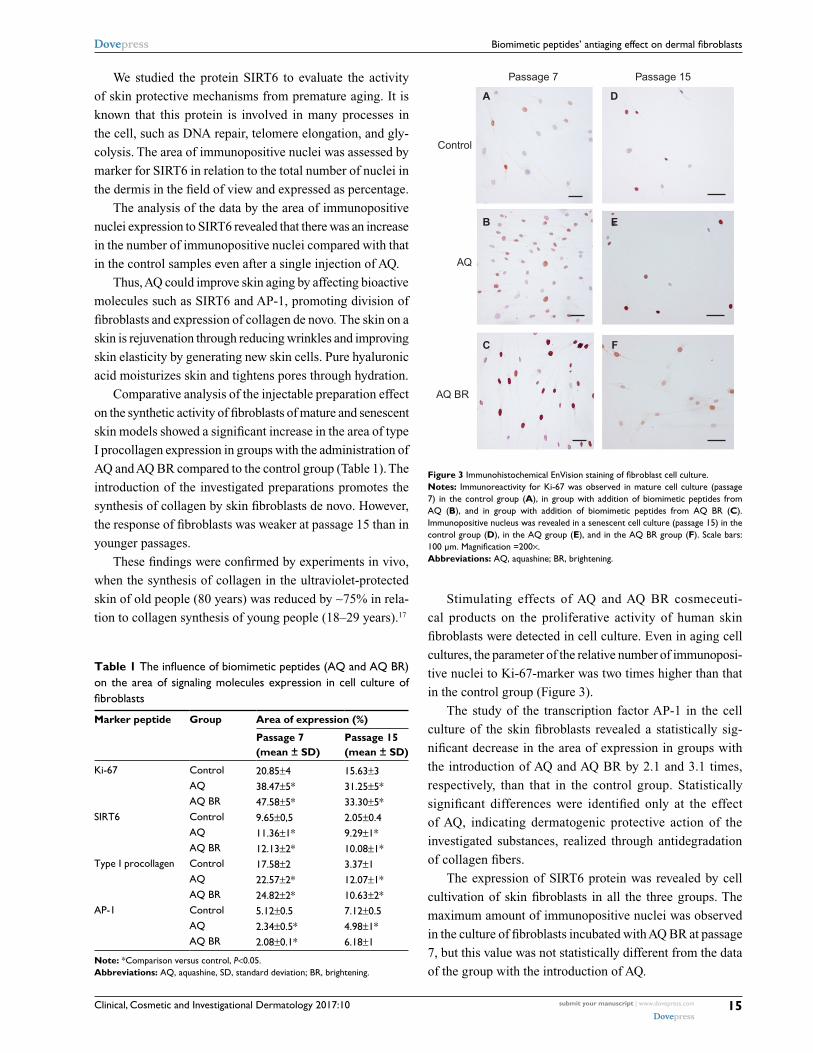
Stimulating effects of AQ and AQ BR cosmeceuti-
cal products on the proliferative activity of human skin
fibroblasts were detected in cell culture. Even in aging cell
cultures, the parameter of the relative number of immunoposi-
tive nuclei to Ki-67-marker was two times higher than that
in the control group (Figure 3).
The study of the transcription factor AP-1 in the cell
culture of the skin fibroblasts revealed a statistically sig-
nificant decrease in the area of expression in groups with
the introduction of AQ and AQ BR by 2.1 and 3.1 times,
respectively, than that in the control group. Statistically
significant differences were identified only at the effect
of AQ, indicating dermatogenic protective action of the
investigated substances, realized through antidegradation
of collagen fibers.
The expression of SIRT6 protein was revealed by cell
cultivation of skin fibroblasts in all the three groups. The
maximum amount of immunopositive nuclei was observed
in the culture of fibroblasts incubated with AQ BR at passage
7, but this value was not statistically different from the data
of the group with the introduction of AQ.
Table 1 The influence of biomimetic peptides (AQ and AQ BR) on the area of signaling molecules expression in cell culture of fibroblasts
Marker peptide Group Area of expression (%)
Passage 7 (mean ± SD)
Passage 15 (mean ± SD)
Ki-67 Control 20.85±4 15.63±3AQ 38.47±5* 31.25±5*AQ BR 47.58±5* 33.30±5*
SIRT6 Control 9.65±0,5 2.05±0.4AQ 11.36±1* 9.29±1*AQ BR 12.13±2* 10.08±1*
Type I procollagen Control 17.58±2 3.37±1AQ 22.57±2* 12.07±1*AQ BR 24.82±2* 10.63±2*
AP-1 Control 5.12±0.5 7.12±0.5AQ 2.34±0.5* 4.98±1*AQ BR 2.08±0.1* 6.18±1
Note: *Comparison versus control, P<0.05.Abbreviations: AQ, aquashine, SD, standard deviation; BR, brightening.
Figure 3 Immunohistochemical EnVision staining of fibroblast cell culture.Notes: Immunoreactivity for Ki-67 was observed in mature cell culture (passage 7) in the control group (A), in group with addition of biomimetic peptides from AQ (B), and in group with addition of biomimetic peptides from AQ BR (C). Immunopositive nucleus was revealed in a senescent cell culture (passage 15) in the control group (D), in the AQ group (E), and in the AQ BR group (F). Scale bars: 100 μm. Magnification =200×.Abbreviations: AQ, aquashine; BR, brightening.
Clinical, Cosmetic and Investigational Dermatology 2017:10submit your manuscript | www.dovepress.com
Dovepress
Dovepress
Clinical, Cosmetic and Investigational Dermatology
Publish your work in this journal
Submit your manuscript here: https://www.dovepress.com/clinical-cosmetic-and-investigational-dermatology-journal
Clinical, Cosmetic and Investigational Dermatology is an interna-tional, peer-reviewed, open access, online journal that focuses on the latest clinical and experimental research in all aspects of skin disease and cosmetic interventions. This journal is included on PubMed. The manuscript management system is completely online
and includes a very quick and fair peer-review system, which is all easy to use. Visit http://www.dovepress.com/testimonials.php to read real quotes from published authors
Dovepress
16
Gazitaeva et al
Biomimetic peptides contributed to an increase in the
area of immunopositive nuclei by 79% after incubation with
AQ BR and by 77% with the introduction of AQ (Table 1).
This indicates the start of the mechanisms of DNA stability
maintenance, protection of telomeres, and other processes
preventing aging.
In the future investigation, it will be interesting to use
real-time polymerase chain reaction and enzyme-linked
immunosorbent assay methods to confirm data obtained in
this study and expand knowledge of action of biomimetic
peptides on skin aging processes.
ConclusionThe immunohistochemical studies revealed the molecular
mechanisms of the clinical effects (lifting effect, remodeling
of the dermis with filling of small wrinkles, dermoprotective
effect on the elastic matrix of the skin, improvement in skin
resistance to aggressive environmental factors and muta-
tions) observed with intradermal administration of AQ both
in vitro (in cell culture of human fibroblasts) and in vivo (in
biopsies of human skin).
DisclosureThe authors report no conflicts of interest in this work.
References 1. Penning A [webpage on the Internet]. Biomimetics: beauty ingredients
that mimic bio functions. GCI Magazine. 2013 Oct 14; 3. Available from: http://www.gcimagazine.com/business/rd/ingredients/Biomimet-ics-Beauty-Ingredients-That-Mimic-Bio-Functions-230110461.html. Accessed August 23, 2015.
2. Caregen Co., LTD. [homepage on the Internet]. Growth Factors and Biomimetic Peptides. Seul: Caregen- Dynamic, Progressive and R&D Based Company; 2015. Available from: http://www.caregen.co.kr/. Accessed September 11, 2015.
3. Oh SJ, Kim K, Lim CJ. Protective properties of ginsenoside Rb1 against UV-B radiation-induced oxidative stress in human dermal keratinocytes. Pharmazie. 2015;70(6):381–387.
4. Grosicki M, Latacz G, Szopa A, Cukier A, Kieć-Kononowicz K. The study of cellular cytotoxicity of argireline - an anti-aging peptide. Acta Biochim Pol. 2014;61(1):29–32.
5. Malinin VV, Durnova AO, Polyakova VO, Kvetnoi IM. Effects of Lys-Glu-Trp peptide on cell-cell interactions and vascular endothelium proliferation under normal conditions and during atherosclerosis. Bull Exp Biol Med. 2014;157(3):324–326.
6. Zamorskii II, Shchudrova TS, Lin’kova NS, Nichik TE, Khavinson VKH. Peptides restore functional state of the kidneys during cisplatin-induced acute renal failure. Bull Exp Biol Med. 2015;159(6):736–739.
7. Bickers DR, Athar M. Oxidative stress in the pathogenesis of skin disease. J Investigative Dermatol. 2006;126(12):2565–2575.
8. Helfrich YR, Sachs DL, Voorhees JJ. Overview of skin aging and photoaging. Dermatol Nurs. 2008;20(3):177–183; quiz 184.
9. Chen YJ, Chang LS. Simvastatin induces NFκB/p65 down-regulation and JNK1/c-Jun/ATF-2 activation, leading to matrix metalloproteinase-9 (MMP-9) but not MMP-2 down-regulation in human leukemia cells. Biochem Pharmacol. 2014;92(4):530–543.
10. Ameyar M, Wisniewska M, Weitzman JB. A role for AP-1 in apoptosis: the case for and against. Biochem. 2003;85(8):747–752.
11. Covas DT, Panepucci RA, Fontes AM, et al. Multipotent mesenchymal stromal cells obtained from diverse human tissues share functional properties and gene expression profile with CD146+ perivascular cells and fibroblasts. Exp Hematol. 2008;36(5):642–654.
12. Grytsenko MA. Fibroblasts in the course of development and aging of an organism. J V.N.Karazin Kharkiv Nat Univ Ser Biol. 2013; 17(1056):10–16.
13. Cristofalo VJ, Allen RG, Pignolo RJ, Martin BG, Beck JC. Relationship between donor age and the replicative lifespan of human cells in culture: a reevaluation. Proc Natl Acad Sci USA. 1998;95(18):10614–10619.
15. Seravallo M, Jagdeo J, Glick SA, Siegel DM, Brody NI. Sirtuins in dermatology: applications for future research and therapeutics. Arch Dermatol Res. 2013;305(4):269–282.
16. Michishita E, McCord RA, Berber E, et al. SIRT6 is a histone H3 lysine 9 deacetylase that modulates telomeric chromatin. Nature. 2008; 452(7186):492–496.
17. Varani J, Dame MK, Rittie L, et al. Decreased collagen production in chronologically aged skin: roles of age-dependent alteration in fibroblast function and defective mechanical stimulation. Am J Pathol. 2006;168(6): 1861–1868.