Gynecomastia is the benign enlargement of the mammary gland in male subjects. The incidence of gynecomastia in the general
population varies from 32 to 65 percent.1 The cause is most frequently idiopathic, although sometimes it may be secondary to hormonal dys-function or medications or it may be syndromic (e.g., Klinefelter syndrome).2–6 In adolescents presenting with breast buds that are not signifi-cantly enlarged, gynecomastia is frequently a tem-porary finding,7 but it can turn into a permanent condition in adulthood. In the presence of other signs—namely, small testis, scarce or no pubic and axillary hair, or micropenis—an endocrine survey is recommended.
In body builders, gynecomastia is generally the result of anabolic steroid use8–10 or ingestion of over-the-counter hormones, frequently sold
in various sport and general nutrition stores. In most cases, gynecomastia becomes a permanent finding, lasting beyond the cessation of steroid use. Patients with gynecomastia typically request surgical treatment for aesthetic purposes, and body builders with gynecomastia who compete on a professional level are no exception. Their sport requires that they have an extremely low percent-age of body fat (9 percent) before a competition, which makes the glandular tissue even more pro-nounced. Moreover, in the senior author’s (M.B.) experience, body builders with gynecomastia suf-fer more frequently than the general population from tenderness, pain, and occasional nipple
AQ1
Disclosure: The authors have no financial interest to declare in relation to the content of this article.
Background: Temporary gynecomastia in the form of breast buds is a com-mon finding in young male subjects. In adults, permanent gynecomastia is an aesthetic impairment that may result in interest in surgical correction. Gyne-comastia in body builders creates an even greater distress for patients seeking surgical treatment because of the demands of professional competition. The authors present their experience with gynecomastia in body builders as the largest study of such a group in the literature.Methods: Between the years 1980 and 2013, 1574 body builders were treated surgically for gynecomastia. Of those, 1073 were followed up for a period of 1 to 5 years. Ages ranged from 18 to 51 years. Subtotal excision in the form of subcutaneous mastectomy with removal of at least 95 percent of the glandular tissue was used in virtually all cases. In cases where body fat was extremely low, liposuction was performed in less than 2 percent of the cases.Results: Aesthetically pleasing results were achieved in 98 percent of the cases based on the authors’ patient satisfaction survey. The overall rate of hemato-mas was 9 percent in the first 15 years of the series and 3 percent in the final 15 years. There were no infections, contour deformities, or recurrences.Conclusions: This study demonstrates the importance of direct excision of the glandular tissue over any other surgical technique when correcting gynecomas-tia deformities in body builders. The novice surgeon is advised to proceed with cases that are less challenging, primarily with patients that require excision of small to medium glandular tissue. (Plast. Reconstr. Surg. 135: 00, 2015.)CLINICAL QUESTION/LEVEL OF EVIDENCE: Therapeutic, IV.
From private practice. Received for publication February 14, 2014; accepted May 28, 2014.
Correction of Gynecomastia in Body Builders and Patients with Good Physique
A Video Discussion by Bahman Guyuron, M.D., accompanies this article. Go to PRS-Journal.com and click on “Video Discus-sions” in the “Videos” tab to watch.
VIDEO DISCUSSION BY BAHMAN GUYURON, M.D., IS AVAILABLE ONLINE FOR THIS ARTICLE.
CODING PERSPECTIVE FOR THIS ARTICLE IS ON PAGE XXX.
cpt
COSMETIClww 18/12/14 18:51 4 Color Fig(s): F1-6 Art: PRSJ-D-14-00812
2
Plastic and Reconstructive Surgery • February 2015
discharge, which lead them to seek assistance from a medical professional. Given the multiple indications for surgical correction of gynecomas-tia in body builders and the growing demand for this procedure in recent years, a large retrospec-tive study of our experience with gynecomastia in body builders is presented.
Surgical treatment of gynecomastia in body-builders is more challenging than in other patients because of various factors. There is an increase in the vascularity of the chest as a result of hyper-trophic pectoralis muscles, the use of various ana-bolic steroids, and an increase in the intake of different omega fatty acids, all of which are known to cause excessive bleeding perioperatively. More-over, these patients are perfectionists with regard to their physique and chest aesthetics; thus, their level of expectations is higher. There is a need to excise the entire gland because of high recurrence if the gland is only partially removed. The pen-etration of the pectoralis muscle has to be avoided by all means because of potential scarring, bleed-ing, and subsequent deformities. The circumareo-lar incision should be in a position that minimizes scar visibility. Lastly, during the competition, the chest is more scrutinized compared with the gen-eral population. Because of these challenges, the senior author (M.B.) developed methods to reli-ably circumvent potential complications and gain an exceedingly high rate of patient satisfaction and virtually no recurrences.
PATIENTS AND METHODSBetween the years 1980 to 2013, well over 5000
patients were treated for various gynecomastia deformities in the senior author’s (M.B.) practice. Of these, 1574 were male body builders who were treated surgically for gynecomastia, 1073 of whom continued to return for follow-up between 1 and 5 years after surgery. A significant number of these 1073 patients (15 percent) were professional body builders with superior physiques. Patient ages ranged from 18 to 51 years. Our patient popu-lation consisted of adult male subjects with no history of hormonal or physical (no signs or symp-toms of pituitary, thyroid, liver, adrenal, renal, or testicular disease) dysfunction.
Each gynecomastia patient with good phy-sique was asked whether he used anabolic steroids or prohormones, and whether the gynecomastia occurred after the consumption of those supple-ments. We asked about medical problems such as hypertension, and recent significant and major weight loss or weight gains. When there was no
history of anabolic steroid intake but a recent his-tory of gynecomastia was evident, the patient was referred to an endocrinologist for further testing. If a testicular mass or tenderness was found, we referred him to a urologist for workup for possible neoplasm.
Routine laboratory tests demonstrated an increase in their hematocrit/hemoglobin levels. Basic metabolic panels showed an increase in the blood urea nitrogen and creatinine higher than normal blood urea nitrogen–to-creatinine ratios.
Patients were given clear preoperative instruc-tions regarding the avoidance of alcoholic bever-ages; strenuous activity 1 week before surgery; and the use of anabolic steroids, prohormones, fish oils, aspirin, nonsteroidal antiinflammatory medi-cations, high doses of vitamin E, or protein snacks that are not regulated by the U.S. Food and Drug Administration containing unreported additives and blood thinners. All cases were performed in a Joint Commission on Accreditation of Healthcare Organizations–approved outpatient ambulatory setting.
TechniqueTumescent solution consisting of lidocaine,
Carbocaine (Sanofi, Paris, France), epineph-rine, and bicarbonate mixed in 250 cc of normal saline solution was used in all cases. The operative technique consisted of a skin incision in the infe-rior periareolar location, 2.0 to 2.5 cm in length (Figs. 1 and 2). Incisions were placed more lat-erally in an attempt to avoid medial scars (e.g., a 4-o’clock to 7-o’clock position). Suction-assisted lipectomy was performed in only 5 percent of the cases using the contralateral breast periareolar incision as reported by Webster,11 Aiache,10 and Huang et al.12 Indications for the use of liposuc-tion were removal of peripheral fatty tissue in
AQ2
F1,F2
Fig. 1. Intraoperative view of the gynecomastia tissue dissected out sharply with the use of a blade and a piercing towel clamp.
lww 18/12/14 18:51 4 Color Fig(s): F1-6 Art: PRSJ-D-14-00812
Volume 135, Number 2 • Gynecomastia in Body Builders
3
patients with mediocre physique. Professional body builders have a much higher ratio of glan-dular to fatty tissue and required no liposuction in this study. In virtually all cases, a subcutaneous mastectomy was performed, leaving behind only 2 to 3 mm of tissue under the skin. In this manner, the nipple-areola complex was flush with the con-tour of the pectoral muscle.
Given the young age and good physique of our patient population, almost all patients had a tight and elastic skin that conformed to an ideal shape after surgery, even when the gynecomastia itself was very large. Sagging skin was extremely rare but was discussed extensively with the patient before the surgical procedure, to ascertain that patients were aware of possible imperfections, and it is also stated in oral and printed consent that is discussed with the patient before surgery. Should ptosis possibly occur, extensive skin undermining was performed, at least a few inches beyond the gland limits, to the surrounding skin. This maneuver helped to lessen and even eliminate this problem.
In patients with low fat content, care was taken to remove all of the glandular tissue. Bodybuilders’ fat content is negligible compared with patients that have a high fat content where the surgeon may have to leave more of the glandular tissue. Sharp undermining was performed initially under direct vision to an area that extended to the area around the nipple-areola complex. The remaining tissue was dissected bluntly when possible, while always staying within the confines of the outer border of the gland. On the deep layer, undermining tis-sues remained strictly above the pectoral fascia. Penetration of the fascia resulted in unnecessary hemorrhage and potential contour deformities.
In addition, excessive bleeding was encountered more frequently with dissection on the far lateral aspect compared with the medial side of the gland. Meticulous hemostasis was achieved throughout with electrocautery. Care was taken to excise the entire gland as one entity, resulting in a single spec-imen. All glandular specimens were sent for patho-logic analysis. This approach, which circumvents the need for piecemeal excisions, resulted in fewer complications and less operative time.
In 3 percent of the cases, a ¼-inch Penrose drain was placed through the surgical incision or the axilla for a total of 24 to 72 hours. Compression dressing was applied over the surgical site for 3 to 5 days. No antibiotics were prescribed postoperatively.
Postoperative instructions consisted of light activity with restricted range of motion of the shoulders for the first week. This is because of the attachment of the pectoral muscle to the proxi-mal humerus. Movement of the pectoralis muscle often results in increased bleeding because of the excessive vascularity in body builders. Motion is permitted in the elbow and wrist early in the post-operative period, as it is unrelated to the pectoralis muscle. Elastic compression dressing was applied over the chest for 5 days. Patients were instructed to keep the dressing intact and dry for that time, with minimal activities. Desk work is allowed after 3 to 5 days. Labor work was restricted for at least 1 to 2 weeks. Modified activity was resumed after 1-week follow-up with light, non–chest-related exercises for 2 to 3 weeks. Any exercise resulting in movement of the pectoralis muscles, includ-ing lifting, extension, or abduction of the shoul-ders, was discouraged. For example, patients were asked to wear button-up shirts to avoid arm lift-ing on the first week after surgery. A regular chest exercise regimen was resumed after 4 to 6 weeks.
Patients were followed up for a minimum of 12 months and up to 5 years. Satisfaction ratings were collected from all patients at a 1-year inter-val. Patients were contacted by phone or e-mail over the past 3 to 4 years. Given the limited data in the literature regarding satisfaction rates in gyne-comastia surgery, each patient was also asked to complete our Joint Commission on Accreditation of Healthcare Organizations patient satisfaction survey (Appendix 1). These surveys were collected over the past 13 years.
RESULTSThe continued use of anabolic steroids was
found in the vast majority of cases. Excised glan-dular tissue weight ranged from to 9 to 110 g. Two
AQ3
AQ4
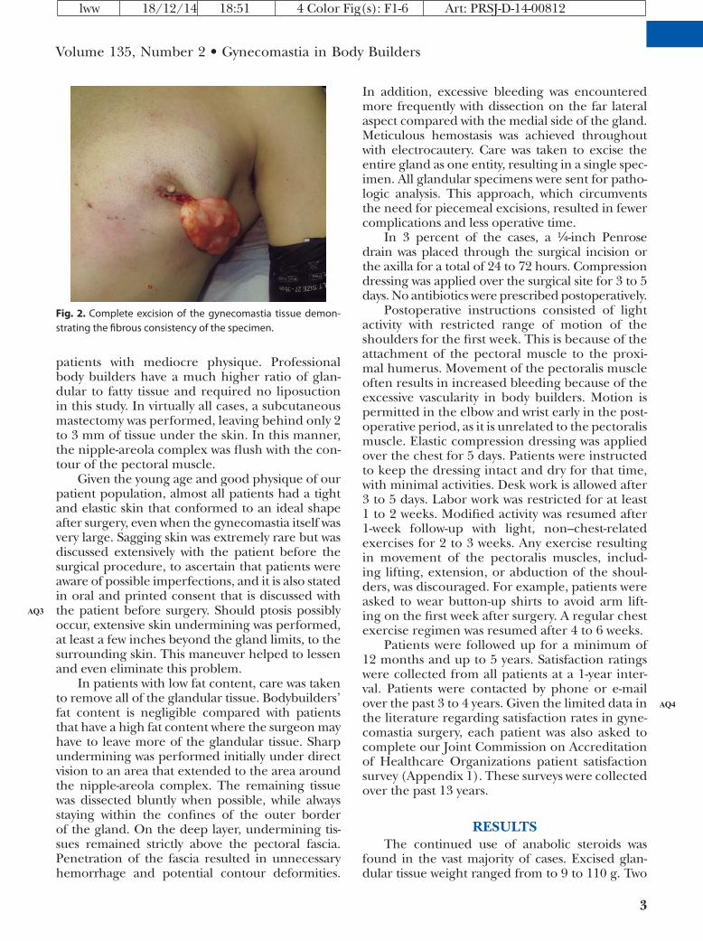
Fig. 2. Complete excision of the gynecomastia tissue demon-strating the fibrous consistency of the specimen.
lww 18/12/14 18:51 4 Color Fig(s): F1-6 Art: PRSJ-D-14-00812lww 18/12/14 18:51 4 Color Fig(s): F1-6 Art: PRSJ-D-14-00812
4
Plastic and Reconstructive Surgery • February 2015
percent of the bodybuilders had a pathology report of gynecomastia with atypical intraductal hyperpla-sia. Patients were than referred to their primary care physician for additional workup and treatment. No reports of disease progression to malignant breast cancer were reported back to our office.
Static postoperative depressions in body build-ers were extremely rare and found in less than 1 percent of patients. Dynamic depressions were found in 7 percent. Permanent resolution of the growth with good symmetry was observed in all patients. Based on a 13-year-long Joint Commis-sion on Accreditation of Healthcare Organizations patient satisfaction survey, aesthetically pleasing results were ranked as high as 98 percent (Fig. 3). All patients presented in these series had a standard 1-inch subareolar incision. With adequate thinning of the areola, the areola shrunk almost instantly in the postoperative phase. There was no need for an areola reduction. The thinner the residual nip-ple-areola complex, the further it shrunk, similar to the principles of skin grafting. No instances of necrosis or sloughing of the areola-nipple complex were seen even after substantial areolar thinning.
In the senior author’s (M.B.) experience, body builders were very cooperative, had realis-tic expectations, and were most concerned with removal of the entire gland. In all cases where these criteria were met, all patients were extremely satisfied with the result (Figs. 3 through 5).
Seroma formation was the most common minor complication in 12 percent of the patients but occurred more frequently in patients who underwent revisions or secondary, corrective pro-cedures (Table 1). The absence of drains did not appear to correlate with the formation of seromas. Fluid collections were resolved after one to three needle (16-gauge) aspirations in the office.
The overall hematoma rate was 6 percent for the entire length of the study; however, an interest-ing trend was noted within that period. Symptom-atic hematomas signified by pain, major swelling, and significant ecchymosis occurred in 9 percent of the cases in the first 15 years, whereas the rate was reduced to 3 percent in the last 15 years of the study. The rate of major hematomas requiring exploratory surgical intervention was 3 percent and 1.5 percent, respectively. Smaller hematomas were treated with a needle aspiration using a 16-gauge needle or by creating a small opening in the sur-gical site and expressing the fluid through the wound. Hematomas were more likely to be found in the noncompliant group of patients. The rate of noncompliance in bodybuilders who admitted to being noncompliant was 6 percent over the past 10 years. It is the senior author’s (M.B.) opinion that the rate is more than 10 percent. A total of 71 per-cent of all hematomas were unilateral.
In the management of postoperative hemato-mas, control of a single bleeding vessel resulted
F3
F3–F5
T1
Fig. 3. (Left) A 30-year-old body builder, 5 feet 10 inches tall and weighing 200 pounds, with severe true gynecomastia resistant to antiestrogen medications after taking prohormones 4 years before presentation. (Right) Eight-month postoperative view. The glandular excision was per-formed through a 1-inch infraareolar incision. A minimal amount of adipose tissue was excised. No liposuction was used.
lww 18/12/14 18:51 4 Color Fig(s): F1-6 Art: PRSJ-D-14-00812
Volume 135, Number 2 • Gynecomastia in Body Builders
5
Fig. 4. (Left) A 25-year-old bodybuilder, 6 feet 0 inches tall and weighing 230 pounds, pre-sented with moderate gynecomastia present since his early teenage years. His gyneco-mastia became more pronounced after he began his bodybuilding career with 9 percent body fat. Antiestrogen therapy was unsuccessful. (Right) The 6-month postoperative view demonstrates excellent chest contour with inconspicuous scars after a 1-inch incision in the infraareolar region. Surgical excision was performed without liposuction. The right breast specimen weighed 5 g and measured 2.9 × 2 × 0.5 cm. The left breast specimen weighed 7.5 g and measured 3.9 × 2.9 × 0.5 cm.
Fig. 5. (Left) A 20-year-old bodybuilder, 5 feet 7 inches tall and weighing 210 pounds, presented with severe gynecomastia. The condition erupted at puberty and worsened progressively after a few cycles of anabolic steroids. The patient’s body fat is 8 percent. (Right) A 6-month postopera-tive view of the patient with an aesthetically pleasing result and no visible scars after a 1-inch incision in the infraareolar region. Surgical excision of the glandular tissue was performed with no liposuction. Pathology report of the right breast specimen measured 7.5 × 6 × 2 cm and the left breast specimen measured 7 × 6 × 3 cm.
lww 18/12/14 18:51 4 Color Fig(s): F1-6 Art: PRSJ-D-14-00812lww 18/12/14 18:51 4 Color Fig(s): F1-6 Art: PRSJ-D-14-00812
6
Plastic and Reconstructive Surgery • February 2015
in a shorter surgical time, whereas treatment of diffuse bleeding increased the operative time and blood loss significantly. However, none of the patients in this series required a transfusion. No major complications were noted in which inpatient hospitalization was necessary, and no surgical-site infections or significant contour deformities were observed in this series.
DISCUSSIONBody builders suffer from gynecomastia because
of the continued use of anabolic steroids.8–10 It has been our observation that approximately 15 per-cent of body builders with permanent gynecomas-tia also had a history of temporary gynecomastia in adolescence. As little as one or several cycles of anabolic steroid use had caused the growth to reap-pear in the form of permanent gynecomastia. The pathologic finding of gynecomastia is hypertrophy of the glandular tissue.15 A typical pathology report reveals benign ductal epithelial hyperplasia and periductal stromal edema.16,17
Based on the senior author’s (M.B.) experi-ence, nearly full excision of the glandular tissue is the most appropriate treatment of gynecomastia in body builders, whereas suction-assisted lipectomy18 should be used only scarcely. Recurrence is fre-quently caused by insufficient primary excision, as the remaining breast tissue will continue to act as a target organ in the face of continued steroid use.14
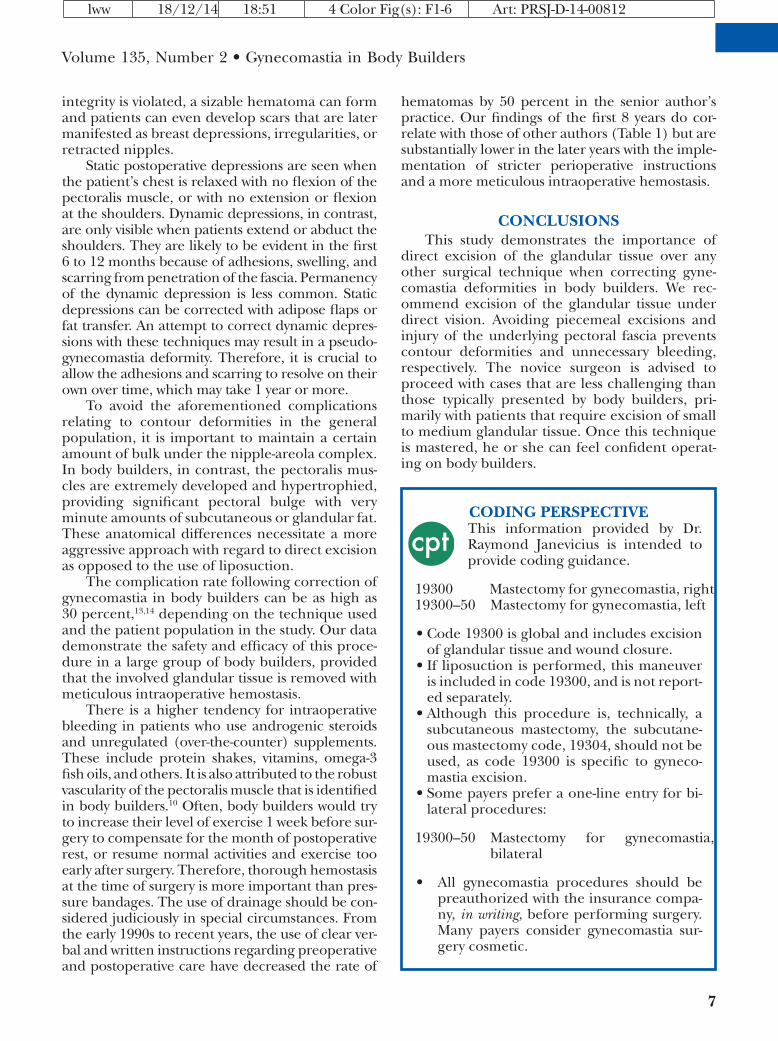
To achieve a nearly complete excision of the glandular tissue, the entire gland as described by the senior author (M.B.), composed of three dif-ferent components, requires surgical attention (Fig. 6). The body of the gland is located imme-diately deep to the nipple-areola complex. The medial expansion of the body is the head of the gland. It extends toward the mid-chest and is usu-ally short and round. The tail of the gland is usu-ally long and narrow in comparison with the head, and is the lateral extension of the gland toward the axilla. This anatomical division of the gland is more noticeable in larger, or more persistent, glands. In the senior author’s (M.B.) opinion,
visualizing the male breast parenchyma in this manner can facilitate a more comprehensive and precise excision of the gland.
Aggressive excision of the glandular tissue is used to the point where the nipple-areola complex thickness superficial to the gland is almost equal to a full-thickness skin graft. Even in cases with a large nipple-areola complex preoperatively, it is the senior author’s (M.B.) observation that it is not necessary to reduce the size of the areola by a cir-cumareolar incision,19 as elasticity of the thin skin will facilitate sufficient contractility. Lastly, if most or all of the tissue to be excised is glandular in nature, suction-assisted lipectomy is seldom used.
The senior author’s experience with ultrasonic liposuction is limited; however, several patients who underwent ultrasonic liposuction in the past required further glandular excision to eliminate the gynecomastia tissue. Therefore, when the gland is the cause of gynecomastia, it should be treated with direct excision of the gland.
In general, unsatisfactory results following the use of liposuction in gynecomastia patients may relate to the inadvertent sharp penetration of the pectoralis muscle fascia. When the fascia’s
F6
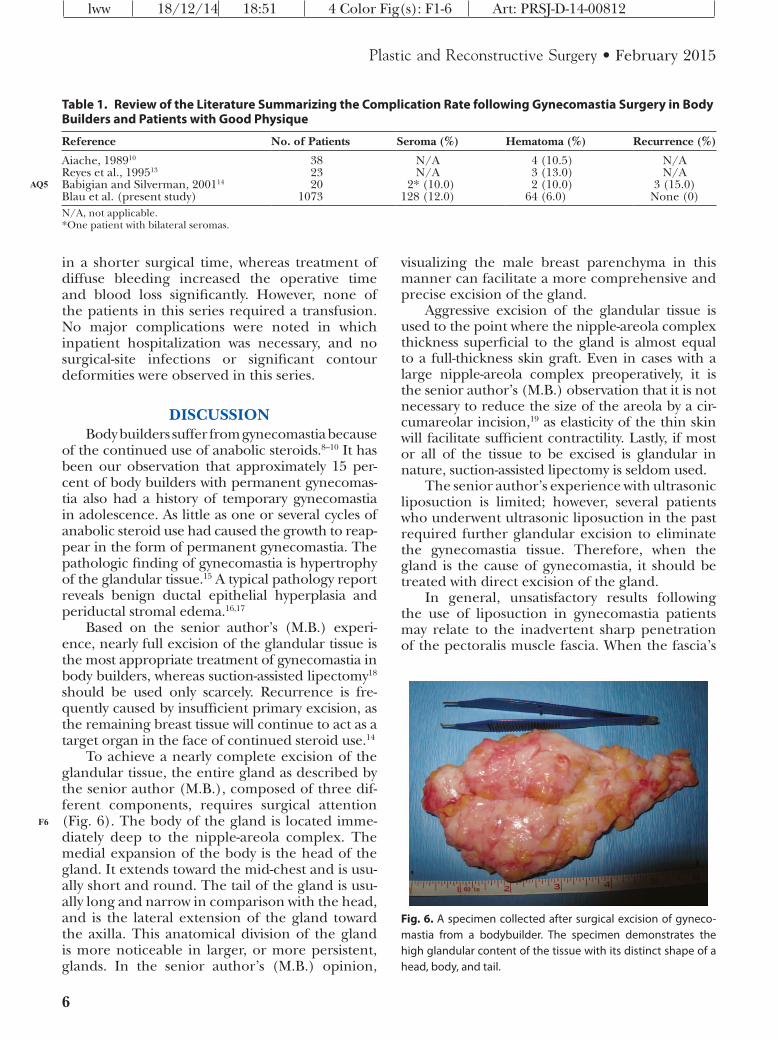
Table 1. Review of the Literature Summarizing the Complication Rate following Gynecomastia Surgery in Body Builders and Patients with Good Physique
Reference No. of Patients Seroma (%) Hematoma (%) Recurrence (%)
Aiache, 198910 38 N/A 4 (10.5) N/AReyes et al., 199513 23 N/A 3 (13.0) N/ABabigian and Silverman, 200114 20 2* (10.0) 2 (10.0) 3 (15.0)Blau et al. (present study) 1073 128 (12.0) 64 (6.0) None (0)N/A, not applicable.*One patient with bilateral seromas.
AQ5
Fig. 6. A specimen collected after surgical excision of gyneco-mastia from a bodybuilder. The specimen demonstrates the high glandular content of the tissue with its distinct shape of a head, body, and tail.
lww 18/12/14 18:51 4 Color Fig(s): F1-6 Art: PRSJ-D-14-00812
Volume 135, Number 2 • Gynecomastia in Body Builders
7
integrity is violated, a sizable hematoma can form and patients can even develop scars that are later manifested as breast depressions, irregularities, or retracted nipples.
Static postoperative depressions are seen when the patient’s chest is relaxed with no flexion of the pectoralis muscle, or with no extension or flexion at the shoulders. Dynamic depressions, in contrast, are only visible when patients extend or abduct the shoulders. They are likely to be evident in the first 6 to 12 months because of adhesions, swelling, and scarring from penetration of the fascia. Permanency of the dynamic depression is less common. Static depressions can be corrected with adipose flaps or fat transfer. An attempt to correct dynamic depres-sions with these techniques may result in a pseudo-gynecomastia deformity. Therefore, it is crucial to allow the adhesions and scarring to resolve on their own over time, which may take 1 year or more.
To avoid the aforementioned complications relating to contour deformities in the general population, it is important to maintain a certain amount of bulk under the nipple-areola complex. In body builders, in contrast, the pectoralis mus-cles are extremely developed and hypertrophied, providing significant pectoral bulge with very minute amounts of subcutaneous or glandular fat. These anatomical differences necessitate a more aggressive approach with regard to direct excision as opposed to the use of liposuction.
The complication rate following correction of gynecomastia in body builders can be as high as 30 percent,13,14 depending on the technique used and the patient population in the study. Our data demonstrate the safety and efficacy of this proce-dure in a large group of body builders, provided that the involved glandular tissue is removed with meticulous intraoperative hemostasis.
There is a higher tendency for intraoperative bleeding in patients who use androgenic steroids and unregulated (over-the-counter) supplements. These include protein shakes, vitamins, omega-3 fish oils, and others. It is also attributed to the robust vascularity of the pectoralis muscle that is identified in body builders.10 Often, body builders would try to increase their level of exercise 1 week before sur-gery to compensate for the month of postoperative rest, or resume normal activities and exercise too early after surgery. Therefore, thorough hemostasis at the time of surgery is more important than pres-sure bandages. The use of drainage should be con-sidered judiciously in special circumstances. From the early 1990s to recent years, the use of clear ver-bal and written instructions regarding preoperative and postoperative care have decreased the rate of
hematomas by 50 percent in the senior author’s practice. Our findings of the first 8 years do cor-relate with those of other authors (Table 1) but are substantially lower in the later years with the imple-mentation of stricter perioperative instructions and a more meticulous intraoperative hemostasis.
CONCLUSIONSThis study demonstrates the importance of
direct excision of the glandular tissue over any other surgical technique when correcting gyne-comastia deformities in body builders. We rec-ommend excision of the glandular tissue under direct vision. Avoiding piecemeal excisions and injury of the underlying pectoral fascia prevents contour deformities and unnecessary bleeding, respectively. The novice surgeon is advised to proceed with cases that are less challenging than those typically presented by body builders, pri-marily with patients that require excision of small to medium glandular tissue. Once this technique is mastered, he or she can feel confident operat-ing on body builders.
CODING PERSPECTIVEThis information provided by Dr. Raymond Janevicius is intended to provide coding guidance.
19300 Mastectomy for gynecomastia, right19300–50 Mastectomy for gynecomastia, left
• Code 19300 is global and includes excision of glandular tissue and wound closure.
• If liposuction is performed, this maneuver is included in code 19300, and is not report-ed separately.
• Although this procedure is, technically, a subcutaneous mastectomy, the subcutane-ous mastectomy code, 19304, should not be used, as code 19300 is specific to gyneco-mastia excision.
• Some payers prefer a one-line entry for bi-lateral procedures:
19300–50 Mastectomy for gynecomastia, bilateral
• All gynecomastia procedures should be preauthorized with the insurance compa-ny, in writing, before performing surgery. Many payers consider gynecomastia sur-gery cosmetic.
cpt
lww 18/12/14 18:51 4 Color Fig(s): F1-6 Art: PRSJ-D-14-00812lww 18/12/14 18:51 4 Color Fig(s): F1-6 Art: PRSJ-D-14-00812
8
Plastic and Reconstructive Surgery • February 2015
Ron Hazani, M.D.250 North Robertson Boulevard, Suite 208
4. LaFranchi SH, Parlow AF, Lippe BM, Coyotupa J, Kaplan SA. Pubertal gynecomastia and transient elevation of serum estradiol level. Am J Dis Child. 1975;129:927–931.
5. Jackson AW, Muldal S, Ockey CH, O’Connor PJ. Carcinoma of male breast in association with the Klinefelter syndrome. BMJ 1965;1:223–225.
8. Spiga L, Gorrini G, Ferraris L, Odaglia G, Frassetto G. Unilateral gynecomastia induced by the use of anabolic
steroids: A clinical case report (in Italian). Minerva Med. 1992;83:575–580.
9. Pope HG Jr, Katz DL. Psychiatric and medical effects of anabolic-androgenic steroid use: A controlled study of 160 athletes. Arch Gen Psychiatry 1994;51:375–382.
10. Aiache AE. Surgical treatment of gynecomastia in the body builder. Plast Reconstr Surg. 1989;83:61–66.
11. Webster JP. Mastectomy for gynecomastia through a semicir-cular intra-areolar incision. Ann Surg. 1946;124:557–575.
12. Huang TT, Hidalgo JE, Lewis SR. A circumareolar approach in surgical management of gynecomastia. Plast Reconstr Surg. 1982;69:35–40.
13. Reyes RJ, Zicchi S, Hamed H, Chaudary MA, Fentiman IS. Surgical correction of gynaecomastia in bodybuilders. Br J Clin Pract. 1995;49:177–179.
14. Babigian A, Silverman RT. Management of gynecomastia due to use of anabolic steroids in bodybuilders. Plast Reconstr Surg. 2001;107:240–242.
15. Lapid O, Jolink F, Meijer SL. Pathological findings in gyne-comastia: Analysis of 5113 breasts. Ann Plast Surg. (in press).
16. Kapila K, Verma K. Cytology of nipple discharge in florid gynecomastia. Acta Cytol. 2003;47:36–40.
17. Kapila K, Verma K. Cytomorphological spectrum in gynaecomastia: A study of 389 cases. Cytopathology 2002;13:300–308.
18. Rosenberg GJ. Gynecomastia: Suction lipectomy as a con-temporary solution. Plast Reconstr Surg. 1987;80:379–386.
19. Peled IJ. Re: Pursestring and skin excision in gynecomastia. Ann Plast Surg. 1999;42:343.
AQ6
AQ7
APPENDIX: An Example of a Patient Satisfaction Form Provided for Each Patient at 1-Year Follow-Up AQ8
How was your procedure?
Dr. Mordcai Blau and staff are always trying to improve patient satisfaction. This inquiry focuses on your satisfaction regarding your procedure. Please take a few minutes to fill out this survey. We greatly appreciate your feedback.
1. In your pre-procedure consultation, do you feel all your questions were answered?
3. After you checked in with the receptionist, how long did you wait to be taken into an exam/procedure room?
Approximately at the same time as my scheduled appointmentApproximately 15 minutes after my scheduled appointmentApproximately 30 minutes after my scheduled appointmentApproximately 45 minutes after my scheduled appointmentAn hour or more after my scheduled appointment
(Continued)
lww 18/12/14 18:51 4 Color Fig(s): F1-6 Art: PRSJ-D-14-00812
Volume 135, Number 2 • Gynecomastia in Body Builders
9
4. While waiting for the procedure to begin were you comfortable? Hot? Cold? Etc.
I was comfortableI did not feel I had enough privacyThe room was hotThe room was coldOther______________________________________________
5. During the procedure were you comfortable or in pain? If you were in pain did the staff do a good job alleviating the pain?
I was comfortable during the procedureI was in pain but the staff did a good job alleviating the painI was in pain and the staff could not alleviate the pain
6. After the procedure were you comfortable or in pain? If you were in pain did the staff do a good job elevating the pain?
I was comfortable after the procedureI was in pain but the staff did a good job alleviating the painI was in pain and the staff could not alleviate the pain
7. Did you feel you were ready to be released from the organization when it was time to go home?
YesNo. (Please provide explanation)_____________________________________
8. Do you feel that your post procedural instructions were clear?
YesNo. (Please provide explanation)_____________________________________
9. Do you feel all your financial responsibilities regarding paying for the procedure were explained to you?
YesNo. (Please provide explanation)_____________________________________
10. Do you feel the staff was respectful and pleasant during your procedure?
YesNo. (Please provide explanation)______________________________________
11. Were you satisfied with the results, so far?
YesNo. (Please provide explanation)____________________________________
Thank you! Please return this survey to the front desk or return in envelope provided.
APPENDIX. (Continued)
lww 18/12/14 18:51 4 Color Fig(s): F1-6 Art: PRSJ-D-14-00812lww 18/12/14 18:51 4 Color Fig(s): F1-6 Art: PRSJ-D-14-00812
AUTHOR QUERIES
AUTHOR PLEASE ANSWER ALL QUERIES
AQ1— Sentence OK as edited (“or medications or it may be syndromic”)? Please advise/revise as needed.
AQ2— Name and location of manufacturer of Carbocaine correct as added? If not, please provide the correct information.
AQ3— Sentence OK as edited (“Sagging skin was extremely rare but…”)? Please advise/revise as needed.
AQ4— 3 to 4 years correct (“over the past 3 to 4 years”)? If not, please revise as needed.AQ5— References 13 through 19 were renumbered according to order of citation in text, including
rights reserved.” Please confirm that you have received permission from IntrepidWare to reprint the Appendix online as well as in print. Please send a signed copy of the letter grant-ing permission to republish this material. Please note that the Appendix cannot be used in this article without permission.