Mechanisms of allergic diseases (Supported by an educational grant from Merck & Co., Inc.) Series editors: Joshua A. Boyce, MD, Fred Finkelman, MD, William T. Shearer, MD, PhD, and Donata Vercelli, MD Costimulation blockade in autoimmunity and transplantation Flavio Vincenti, MD San Francisco, Calif Signaling through the costimulation receptors is a critical pathway in the regulation of T-cell activation. The selective costimulation inhibitor abatacept (cytotoxic T lymphocyte– associated antigen 4—Ig) binds to CD80 and CD86 on antigen- presenting cells, blocking interaction with CD28 on T cells, and is approved for the treatment of moderate to severe rheumatoid arthritis. Belatacept (LEA29Y), currently enrolling phase III trials in renal transplantation, was rationally designed from abatacept to bind with more avidity to CD86, providing the more potent immunosuppressive properties required for immunosuppression in transplantation. This review describes the relevant preclinical studies and summarizes recent clinical findings on these 2 molecules in autoimmune diseases and organ transplantation. Although both inhibit the CD28 costimulatory pathway, they are tailored for specific disease states—abatacept for autoimmune diseases and belatacept for transplantation. (J Allergy Clin Immunol 2008;121:299-306.) Key words: Costimulation, abatacept, belatacept, organ transplan- tation, autoimmune disease, rheumatoid arthritis The costimulation pathway has been one of the most exciting and intensively researched areas in basic immunology in the past quarter century. These efforts culminated in 2005 with the approval by the US Food and Drug Administration (FDA) of abatacept (cytotoxic T lymphocyte–associated antigen 4 [CTLA4]—Ig) for rheumatoid arthritis and the publication of the positive results of the first clinical trial of belatacept, a sec- ond-generation CTLA4-Ig, in renal transplantation (Fig 1). 1-6 Blockade of costimulation offers the advantage of selective inhibition of T-cell responses and has the potential of inducing tolerance to specific antigens in both autoimmunity and trans- plantation. 7-10 In transplantation, costimulation blockade repre- sents a new paradigm in immunosuppression, with biologic agents used as maintenance therapy, replacing current drugs that require frequent therapeutic drug monitoring and are associ- ated with chronic toxicities. 11 T lymphocytes play a central role in the initiation and regulation of the adaptive immune response to antigen, whether foreign or native. Naive T cells require 2 signals for their full activation. 1 The first, Signal 1, is an antigen-specific signal provided by the T-cell receptor interacting with the MHC and antigenic peptide complex on the antigen-presenting cell (APC). The second, or costimula- tory, signal is provided by the interactions between specific recep- tors on the T cell and their ligands on the APC (Fig 2). INFORMATION FOR CATEGORY 1 CME CREDIT Credit can now be obtained, free for a limited time, by reading the review articles in this issue. Please note the following instructions. Method of Physician Participation in Learning Process: The core ma- terial for these activities can be read in this issue of the Journal or online at the JACI Web site: www.jacionline.org. The accompanying tests may only be submitted online at www.jacionline.org. Fax or other copies will not be accepted. Date of Original Release: February 2008. Credit may be obtained for these courses until January 31, 2010. Copyright Statement: Copyright Ó 2008-2010. All rights reserved. Overall Purpose/Goal: To provide excellent reviews on key aspects of allergic disease to those who research, treat, or manage allergic disease. Target Audience: Physicians and researchers within the field of allergic disease. Accreditation/Provider Statements and Credit Designation: The American Academy of Allergy, Asthma & Immunology (AAAAI) is accredited by the Accreditation Council for Continuing Medical Education (ACCME) to provide continuing medical education for physicians. The AAAAI designates these educational activities for a maximum of 1 AMA PRA Category 1 Creditä. Physicians should only claim credit commensu- rate with the extent of their participation in the activity. List of Design Committee Members: Author: Flavio Vincenti, MD Activity Objectives 1. To understand the importance of costimulatory pathways in T-cell activation in autoimmune disease and transplantation. 2. To learn how the pharmacologic targeting of this pathway is being used to develop new therapies. Recognition of Commercial Support: This CME activity has not re- ceived external commercial support. Disclosure of Significant Relationships with Relevant Commercial Companies/Organizations: Flavio Vincenti has declared that he has no significant relationships to disclose. From the University of California, San Francisco, Kidney Transplant Service. F.V. has received grant support from Bristol-Myers Squibb. Disclosure of potential conflict of interest: F. Vincenti has received educational and research grants from Wyeth Pharmaceuticals, Novartis, Roche Laboratories, Genen- tech, Bristol-Myers Squibb, Pfizer, Genzyme, and Ortho Biotech. Received for publication November 29, 2007; revised January 2, 2008; accepted for publication January 2, 2008. Reprint requests: Flavio Vincenti, MD, University of California, San Francisco, Kidney Transplant Service, 505 Parnassus Avenue, Room 884M, San Francisco, CA 94143-0780. E-mail: [email protected]. 0091-6749/$34.00 Ó 2008 American Academy of Allergy, Asthma & Immunology doi:10.1016/j.jaci.2008.01.002 Terms in boldface and italics are defined in the glossary on page 300. 299
Transcript
Mechanisms of allergic diseases(Supported by an educational grant from Merck & Co., Inc.)
Series editors: Joshua A. Boyce, MD, Fred Finkelman, MD, William T. Shearer, MD, PhD, and Donata Vercelli, MD
Costimulation blockade in autoimmunity andtransplantation
Flavio Vincenti, MD San Francisco, Calif
INFORMATION FOR CATEGORY 1 CME CREDIT
Credit can now be obtained, free for a limited time, by reading the review
articles in this issue. Please note the following instructions.
Method of Physician Participation in Learning Process: The core ma-
terial for these activities can be read in this issue of the Journal or online at
the JACI Web site: www.jacionline.org. The accompanying tests may only
be submitted online at www.jacionline.org. Fax or other copies will not be
accepted.
Date of Original Release: February 2008. Credit may be obtained for
these courses until January 31, 2010.
Copyright Statement: Copyright � 2008-2010. All rights reserved.
Overall Purpose/Goal: To provide excellent reviews on key aspects of
allergic disease to those who research, treat, or manage allergic disease.
Target Audience: Physicians and researchers within the field of allergic
disease.
Accreditation/Provider Statements and Credit Designation: The
American Academy of Allergy, Asthma & Immunology (AAAAI) is
accredited by the Accreditation Council for Continuing Medical Education
(ACCME) to provide continuing medical education for physicians. The
AAAAI designates these educational activities for a maximum of 1 AMA
PRA Category 1 Credit�. Physicians should only claim credit commensu-
rate with the extent of their participation in the activity.
List of Design Committee Members: Author: Flavio Vincenti, MD
Activity Objectives
1. To understand the importance of costimulatory pathways in T-cell
activation in autoimmune disease and transplantation.
2. To learn how the pharmacologic targeting of this pathway is being
used to develop new therapies.
Recognition of Commercial Support: This CME activity has not re-
ceived external commercial support.
Disclosure of Significant Relationships with Relevant Commercial
Companies/Organizations: Flavio Vincenti has declared that he has no
significant relationships to disclose.
Signaling through the costimulation receptors is a criticalpathway in the regulation of T-cell activation. The selectivecostimulation inhibitor abatacept (cytotoxic T lymphocyte–associated antigen 4—Ig) binds to CD80 and CD86 on antigen-presenting cells, blocking interaction with CD28 on T cells, andis approved for the treatment of moderate to severe rheumatoidarthritis. Belatacept (LEA29Y), currently enrolling phase IIItrials in renal transplantation, was rationally designed fromabatacept to bind with more avidity to CD86, providing themore potent immunosuppressive properties required forimmunosuppression in transplantation. This review describesthe relevant preclinical studies and summarizes recent clinicalfindings on these 2 molecules in autoimmune diseases and organtransplantation. Although both inhibit the CD28 costimulatorypathway, they are tailored for specific disease states—abatacept
From the University of California, San Francisco, Kidney Transplant Service.
F.V. has received grant support from Bristol-Myers Squibb.
Disclosure of potential conflict of interest: F. Vincenti has received educational and
research grants from Wyeth Pharmaceuticals, Novartis, Roche Laboratories, Genen-
tech, Bristol-Myers Squibb, Pfizer, Genzyme, and Ortho Biotech.
Received for publication November 29, 2007; revised January 2, 2008; accepted for
publication January 2, 2008.
Reprint requests: Flavio Vincenti, MD, University of California, San Francisco,
Kidney Transplant Service, 505 Parnassus Avenue, Room 884M, San Francisco, CA
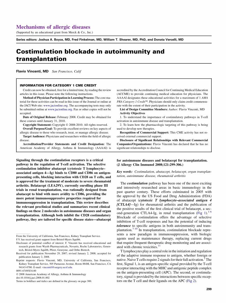
The costimulation pathway has been one of the most excitingand intensively researched areas in basic immunology in thepast quarter century. These efforts culminated in 2005 withthe approval by the US Food and Drug Administration (FDA)of abatacept (cytotoxic T lymphocyte–associated antigen 4[CTLA4]—Ig) for rheumatoid arthritis and the publication ofthe positive results of the first clinical trial of belatacept, a sec-ond-generation CTLA4-Ig, in renal transplantation (Fig 1).1-6
Blockade of costimulation offers the advantage of selectiveinhibition of T-cell responses and has the potential of inducingtolerance to specific antigens in both autoimmunity and trans-plantation.7-10 In transplantation, costimulation blockade repre-sents a new paradigm in immunosuppression, with biologicagents used as maintenance therapy, replacing current drugsthat require frequent therapeutic drug monitoring and are associ-ated with chronic toxicities.11
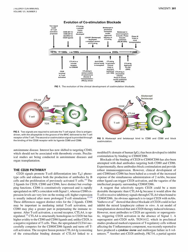
T lymphocytes play a central role in the initiation and regulationof the adaptive immune response to antigen, whether foreign ornative. Naive T cells require 2 signals for their full activation.1 Thefirst, Signal 1, is an antigen-specific signal provided by the T-cellreceptor interacting with the MHC and antigenic peptide complexon the antigen-presenting cell (APC). The second, or costimula-tory, signal is provided by the interactions between specific recep-tors on the T cell and their ligands on the APC (Fig 2).
CTLA4: Cytotoxic T lymphocyte–associated antigen 4
DMARD: Disease-modifying anti-inflammatory drug
FDA: US Food and Drug Administration
IL-2R: IL-2 receptor
MMF: Mycophenolate mofetil
PTLD: Posttransplant lymphoproliferative disease
RA: Rheumatoid arthritis
Following these 2 signals, a number of pathways are activated.They include the calcium-calcineurin pathway, the RAS mito-gen-activated protein kinase pathway, and the subsequent activa-tion of several transcription factors for a number of effectorcompounds, including the cytokine IL-2. IL-2 activates the targetof rapamycin pathway, sometimes referred to as Signal 3. Theseevents induce T-cell proliferation, generation of an effector,CD41 T-cell pool (TH), and the clonal expansion of activatedCD81 or cytotoxic T cells.12 If the T cell does not receive a
costimulation signal because of blockade of this pathway, it be-comes anergic and undergoes apoptosis.
Multiple costimulatory pathways are involved in T-cellregulation; these can either upregulate or downregulate T-cellactivation.13 Perhaps the most critical, and certainly the best char-acterized, costimulatory interactions are between CD40 andCD154 of the TNF:TNF receptor family and between CD28and CD80 and CD86 in the B7 family.13 The CD40-CD154 path-way was initially described as critical for B-cell activation anddifferentiation, but it was subsequently reported to contribute toT-cell activation by upregulating the B7 family ligands CD80and CD86 on APCs. CD154 is expressed on vascular endothelialcells, smooth muscle cells, and macrophages, suggesting anexpanded role for the CD40-CD154 pathway during immunity.2
Prolonged allograft survival was demonstrated in nonhumanprimates treated with anti-CD154 mAbs. The first clinical trial inrecipients of primary renal allografts with hu5C8, a humanizedanti-CD154, was launched with great anticipation that thisregimen might result in operational tolerance. However, therapywith anti-CD154 caused thrombotic events leading to dis-continuation of the trial as well as cessation of the clinicaldevelopment of anti-CD154 in both transplantation and
GLOSSARY
CALCINEURIN: A calcium-dependent phosphatase that is activated in res-
ponse to T-cell activation;dephosphorylates the transcription factornuclear
factor of activated T cells, allowing cytoplasmic escape, entrance into the
cell nucleus, and activation of gene transcription (including the IL-2 gene).
CD28: Expressed on most T cells, provides a second signal for T-cell
activation on binding to CD80/86, resulting in the activation of the
PI-3 kinase–Ras–mitogen-activated protein kinase signaling pathway.
CD40: Present on mature B cells, signal transduction through CD40
causes upregulation of CD80/86, thus facilitating costimulation.
CD154: Also known as CD40 ligand, on activated T cells, required for IgM
class switch. Mutations in CD40 ligand cause X-linked hyper-IgM syn-
drome with absence of germinal centers and IgG, IgA, and IgE.
COSTIMULATION: T-cell activation requires 2 signals: (1) through the
T-cell receptor interacting with antigen-MHC, and (2) a second signal, for
example, through CD28:CD80/86 or CD40:CD40L; T cells that do not
receive a second signal become anergic.
CYTOKINE STORM: The release of multiple inflammatory mediators
resulting in fever, pain, hypotension, and multiorgan failure; can cause
systemic inflammatory response syndrome. Cytokine storm in patients
treated with anti-CD28 was associated with increased TNF-a, IL-2, IL-6, IL-
10, and IFN-g levels and increased numbers of CD41, CD81 T cells, B cells,
and natural killer cells.
CYTOTOXIC T LYMPHOCYTE–ASSOCIATED ANTIGEN 4 (CTLA4): Also
known as CD152, expressed on activated T cells, member of the immu-
noglobulin superfamily, cytoplasmic tail contains an immunoreceptor
clude etanercept (Enbrel), infliximab (Remicade), and adalimumab (Hu-
mira), used in RA, psoriasis/psoriatic arthritis, and Crohn disease.
TOLERANCE: Depletion of autoreactive T and B cells in the thymus and
bone marrow constitutes central tolerance; peripheral antigens can be
expressed in the thymus under the control of AIRE, a transcription factor
involved in autoimmune polyendocrinopathy–candidiasis–ectodermal
dystrophy syndrome. Autoreactive T cells are inactivated through an-
ergy, deleted through apoptosis, or suppressed by regulatory T cells,
all of which is termed peripheral tolerance.
The Editors wish to acknowledge Seema Aceves, MD, PhD, for preparing this glossary.
J ALLERGY CLIN IMMUNOL
VOLUME 121, NUMBER 2
VINCENTI 301
FIG 1. The evolution of the clinical development of costimulation blockade.
autoimmune disease. Interest has now shifted to targeting CD40,which should not be associated with thrombotic events. Preclin-ical studies are being conducted in autoimmune diseases andorgan transplantation.
THE CD28 PATHWAYCD28 signals promote T-cell differentiation into TH1 pheno-
type cells and enhance both the production of antibodies by Bcells and the proliferation of previously activated T cells.14 The2 ligands for CD28, CD80 and CD86, have distinct but overlap-ping functions. CD86 is constitutively expressed and is rapidlyupregulated on APCs coincident with Signal 1, whereas CD80 ex-pression levels are very low on the resting cell; higher expressionis usually induced after more prolonged T-cell stimulation.15,16
These differences suggest distinct roles for the 2 ligands. CD86may be important in mediating initial T-cell activation, andCD80 may play a greater part in perpetuating the immune re-sponse. After T-cell activation, a second receptor, CTLA4, is up-regulated.14 CTLA4 is structurally homologous to CD28 but hashigher avidity to the CD80 and CD86 ligands and, unlike CD28, isa negative regulator of T cells. Thus, the upregulated CLTA4 suc-cessfully competes for the CD80/CD86 ligands and turns off T-cell activation. The receptor fusion protein CTLA4-Ig (consistingof the extracellular binding domain of CTLA4 linked to a
FIG 2. Two signals are required to activate the T-cell signal. One is antigen-
driven, with the allopeptide in the groove of the MHC delivered to the T-cell
receptor of the T cell. The second or costimulation signal is provided through
the binding of the CD28 receptor with its ligands CD80 and CD86.
modified Fc domain of human IgG1) has been developed to inhibitcostimulation by binding to CD80/CD86.
Blockade of the binding of CD28 to CD80/CD86 has also beenattempted with dual antibodies targeting both CD80 and CD86.Experimentally, these antibodies block costimulation and providerobust immunosuppression. However, clinical development ofanti-CD80/anti-CD86 has been halted as a result of the increasedexpense of the simultaneous administration of 2 mAbs, becauseeither ligand can trigger CD28 activation, and the vagaries of theintellectual property surrounding CD80/CD86.
A reagent that selectively targets CD28 could be a moredesirable therapeutic than CTLA4-Ig because it would allow theT cell to receive inhibitory signals through CTLA4 when bound toCD80/CD86. An obvious approach is to target CD28 with mAbs.Vanhove et al17 showed that direct blockade of CD28 could in factinhibit the mixed lymphocyte culture in vitro. A rat model oftransplantation showed that anti-CD28 therapy induced tolerance.However, most antibodies to CD28 are agonistic or superagonis-tic, triggering CD28 activation in the absence of Signal 1. Asuperagonist anti-CD28 mAb, TGN1412, which in preclinicalstudies produced marked expansion of T-regulatory cells withoutaffecting the T inflammatory component, was recently reported tohave produced a cytokine storm and multiorgan failure in 6 vol-unteers.18 Another anti-CD28 antibody, FK734, a partial agonist
FIG 3. Abatacept and belatacept bind to CD80 and CD86 and block
costimulation.
J ALLERGY CLIN IMMUNOL
FEBRUARY 2008
302 VINCENTI
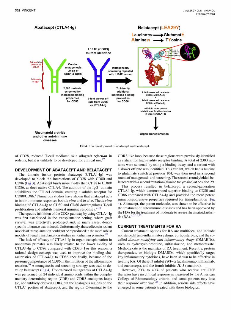
FIG 4. The development of abatacept and belatacept.
of CD28, reduced T-cell–mediated skin allograft rejection inrodents, but it is unlikely to be developed for clinical use.19
DEVELOPMENT OF ABATACEPT AND BELATACEPTThe dimeric fusion protein abatacept (CTLA4-Ig) was
developed to block the interactions of CD28 with CD80 andCD86 (Fig 3). Abatacept binds more avidly than CD28 to CD80/CD86, as does native CTLA4. The addition of the IgG1 domainsolubilizes the CTLA4 domain, creating a soluble receptor forCD80/CD86.7 Numerous studies have shown that abatacept actsto inhibit immune responses both in vitro and in vivo. The in vitrobinding of CTLA4-Ig to CD80 and CD86 downregulates T-cellproliferation and inhibits humoral immune responses.7-10
Therapeutic inhibition of the CD28 pathway by using CTLA4-Igwas first established in the transplantation setting, where graftsurvival was effectively prolonged and, in many cases, donor-specific tolerance was induced. Unfortunately, these effects in rodentmodels of transplantation could not be reproduced in the more robustmodels of renal transplantation studies in nonhuman primates.20
The lack of efficacy of CTLA4-Ig in organ transplantation innonhuman primates was likely related to the lower avidity ofCTLA4-Ig to CD86 compared with CD80. For this reason, arational design concept was used to improve the binding cha-racteristics of CTLA4-Ig to CD86 specifically, because of thepresumed importance of CD86 in the initiation of the alloimmunereaction.20 A mutagenesis and screening strategy was used to de-velop belatacept (Fig 4). Codon-based mutagenesis of CTLA4-Igwas performed on 24 individual amino acids within the comple-mentary determining region (CDR) and CDR3 analogous loops(ie, not antibody-derived CDRs, but the analogous regions on theCTLA4 portion of abatacept), and the region C-terminal to the
CDR3-like loop, because these regions were previously identifiedas critical for high-avidity receptor binding. A total of 2300 mu-tants were screened by using a binding assay, and a variant witha slower off rate was identified. This variant, which had a leucineto glutamate switch at position 104, was then used in a secondround of mutagenesis and screening. The second round yielded be-latacept with a second mutation (alanine to tyrosine) at position 29.
This process resulted in belatacept, a second-generationCTLA4-Ig, which demonstrated superior binding to CD80 andCD86 compared with CTLA4-Ig and provided the more potentimmunosuppressive properties required for transplantation (Fig4). Abatacept, the parent molecule, was shown to be effective inthe treatment of autoimmune diseases and has been approved bythe FDA for the treatment of moderate to severe rheumatoid arthri-tis (RA).4,5,21,22
CURRENT TREATMENTS FOR RACurrent treatment options for RA are multifocal and include
nonsteroidal anti-inflammatory drugs, corticosteroids, and the so-called disease-modifying anti-inflammatory drugs (DMARDs),such as hydroxychloroquine, sulfasalazine, and methotrexate.Methotrexate is the mainstay of RA treatment. Recently, proteintherapeutics, or biologic DMARDs, which specifically targetkey inflammatory cytokines, have been shown to be effective intreating RA. Of these, 3 inhibit TNF-a (adalimumab, infliximab,and etanercept), and the fourth inhibits IL-1 (anakinra).
However, 20% to 40% of patients who receive anti-TNFtherapies have no clinical response as measured by the AmericanCollege of Rheumatology criteria, and some patients may losetheir response over time.23 In addition, serious side effects haveemerged in some patients treated with these biologics.
J ALLERGY CLIN IMMUNOL
VOLUME 121, NUMBER 2
VINCENTI 303
TABLE I. Efficacy of abatacept in patients with RA with an inadequate response to methotrexate and/or TNF-a inhibitors
Clinically meaningful improvement in physical function
(% HAQ improvement �0.3 from baseline)
64 39 47.3 23.3
Mean change from baseline in the physical component
summary score of the SF-36
9.1 5.0 6.59 1.12
Mean change from baseline in the mental component
summary score of the SF-36
6.9 4.7 5.15 2.11
Radiographic scores (mean change from baseline)
Erosions 0.63 1.14
Joint space narrowing 0.58 1.18
Total score 1.21 2.32
Discontinuation due to lack of efficacy (%) 3 9 20 5
ACR, American College of Rheumatology; AIM, Abatacept in Inadequate responders to Methotrexate; ATTAIN, Abatacept Trial in Treatment of Anti-TNF Inadequate responders;
DAS28, Disease Activity Score 28; HAQ, Health Assessment Questionnaire; SF-36, Short Form 36.
COSTIMULATION MODULATION IN AUTOIMMUNE
DISEASESThe efficacy of abatacept in treating human autoimmune
diseases was first demonstrated in patients with psoriasis vulga-ris, in whom a beneficial effect was demonstrated on psoriatic le-sions.21 Phase II and III trials of abatacept plus methotrexate inpatients with RA with an inadequate response to methotrexatedemonstrated statistically significant and clinically meaningfulimprovements in the signs and symptoms of RA comparedwith methotrexate alone24,25 (Table I). Statistically significantand clinically meaningful improvements were also seen inphysical function and both the mental and physical aspectsof Health Related Quality of Life. These benefits were sus-tained and, in some cases, augmented in patients who contin-ued through 5 years of open-label treatment during a long-term extension of the phase II trial.26 Abatacept also inhibitsthe progression of structural damage at 1 year compared withmethotrexate.27
As with the anti-TNF agents, not all patients responded toabatacept, although the proportion of responders was similar tothat seen in studies of anti-TNFs. However, abatacept is the firstagent to be studied and approved for the treatment of patientswith an inadequate response to treatment with TNF-a antago-nists, on the basis of the data from the phase III Abatacept Trialin Treatment of Anti-TNF Inadequate responders study.5 Inthese patients, abatacept demonstrated statistically significantand clinically meaningful improvements in the reduction ofsigns and symptoms and improvements in physical functionand Health Related Quality of Life (Table I). When data from5 clinical trials of abatacept in 4000 patients with RA represent-ing more than 8000 patient-years were integrated, abatacept wasfound to be well tolerated, with a consistent safety profile.28 Theincidence rates of total malignancies and individual malignan-cies in the abatacept clinical program were within the rangesfound in patients with RA, analyzed by using several well
established databases representing large RA cohorts from differ-ent sources.
Abatacept is approved for use in patients with an inadequateresponse to anti-TNF agents and also for patients with aninadequate response to 1 or more DMARDs such as methotrexate.Thus, it is not exclusively approved or used after TNF agents. It istypically used with DMARDs, as are the TNF agents.
The simultaneous use of abatacept and anti-TNF therapy is notcurrently advised because the few patients treated with thiscombination had a higher incidence of serious infections withoutan apparent improvement in the outcome.
Antibodies to the immunoglobulin portion or CTLA4-bindingportions were reported in 1.3% of patients and showed low-levelreactivity.
SOLID-ORGAN TRANSPLANTATION
OverviewRecipients of solid-organ transplants generally require lifelong
immunosuppression to maintain a state of low immunorespon-siveness or nonimmunoresponsiveness to the allograft. Unfortu-nately, advances in the regimens and therapies used to preventrejection have not been matched by similar improvements inlong-term patient or graft survival.29
In the 1990s, several immunosuppression agents, small mol-ecules and biologics, were successfully introduced in the clinic.These new immunosuppression regimens produced a dramaticdecrease in antirejection rates from the 40% range to the 10% to15% range.29 Because rejection is a strong risk factor for graftloss, the decrease in the incidence of acute rejection was antici-pated to lead to a significant improvement in long-term graftsurvival. Yet to date, outcome data from the Scientific Registryof Transplant Recipients have not demonstrated an increase inlong-term graft survival in the recipients of primary kidneytransplants.29
J ALLERGY CLIN IMMUNOL
FEBRUARY 2008
304 VINCENTI
Paradoxically, the long-term survival of both the allografts andthe recipients are affected by nonimmune toxicities caused, inpart, by the immunosuppressive therapies used to prevent graftrejection. Current immunosuppressive regimens are associated withincreased risks of diabetes mellitus, cardiovascular disease, andmalignancies. Perhaps most critically, calcineurin inhibitors (CNIs),currently the cornerstone therapy for chronic maintenance ofimmunosuppression, are almost invariably associated with nephro-toxicity, resulting in a high incidence of renal failure and intensi-fying the already considerable supply/demand imbalance.30,31
Current therapeutics in transplantationThe immune response to transplantation of an allograft is
primarily mediated through T-cell–dependent mechanisms, in-cluding cytokine expression, T-cell proliferation, and clonalexpansion, as well as some forms of antibody-mediated rejectionthat require T-cell interaction for initiation.
The cascade of events that lead to organ rejection presents anumber of targets for inhibiting T cells, T-cell activation, and thesubsequent effects of activated T cells. Current immunosuppres-sive strategies inhibit T-cell effects by depleting or modulatingT cells, inhibiting T-cell signaling pathways, blocking T-cellproliferation, or interrupting the trafficking of T cells intoallografts/injured tissue.
Regimens generally use more potent immunosuppression inthe early posttransplant or induction phase, when immuneresponses to the allograft are at their highest. Induction agentsinclude protein therapeutics (either polyclonal antibodies ormurine, chimeric, or humanized mAbs), which act mainly bydepleting lymphocytes or suppressing proliferation and bluntingthe effects of T-cell activation. Protein therapeutics target specificcell surface glycoproteins or membrane-bound receptors and fallinto 2 broad categories: depleting and nondepleting (ie, modu-lating) agents.
Depleting biologics such as the antithymocyte globulin andOKT3 are used to reduce early rejection by depleting T cells at thetime of antigen presentation and thus preventing activation ofalloreactive T-cell clones. They can be associated with cytokinerelease syndrome as well as the immunologic consequences ofprolonged T-cell depletion, such as infectious complicationsand posttransplant lymphoproliferative disease (PTLD).
Daclizumab and basiliximab are nondepleting mAbs. Theseagents specifically target the a-chain of the IL-2 receptor (IL-2R),inhibiting the signal for proliferation resulting from initial T-cellactivation. Unlike the T-cell–depleting agents, these compoundsare associated with a low incidence of toxicities and side effects.32
However, despite their specificity, the anti–IL-2R mAbs have lim-ited efficacy because of the redundancy of the cytokine receptorsystem and are generally inappropriate for long-term use.33 Thedepleting agents act too broadly, compromising safety; the anti-IL-2R antibodies act too narrowly (targeting only 1 aspect of T-cell activation) effectively to block rejection when used withoutcalcineurin inhibitors.
After the induction phase, transplant patients transition towhat is known as the maintenance phase of immunosuppressivetreatment.34 Effective maintenance immunosuppression is thekey to preventing rejection throughout the life of the graft. Main-tenance immunosuppressive strategies typically include the so-called cornerstone therapies, CNIs, along with antiproliferativeagents (mycophenolate mofetil [MMF]/enteric-coated mycophe-nolate acid or sirolimus) and steroids.
The CNI regimens necessitate frequent therapeutic drug mon-itoring and dosage adjustment to remain within the narrowtherapeutic windows and are associated with multiple toxicities.34
These limitations have motivated a search for novel agents/regimens with greater selectivity and improved toxicity profiles.Recently, as with autoimmune diseases, the focus of research hasturned toward costimulation blockade.
Thus, the promise of costimulation blockade is to provideselective but durable immunosuppression without the nephrotox-icity and metabolic toxicity of current agents that can ultimatelytranslate into improved long-term patient and graft survival.
Costimulation blockade in transplantationPreclinical studies demonstrated CTLA4-Ig–mediated inhibi-
tion of T-cell–dependent antibody responses and prolongation oftransplanted organ survival.35 However, CTLA4-Ig was found to beinadequate to maintain a hyporesponsive state to an allograft insome models.20 It has been demonstrated that CD80 and CD86may differentially control the immune response because of the dis-tinct properties of each molecule.36 The more rapid dissociation ofCTLA4-Ig from CD86 than from CD80 may have resulted in lesseffective inhibition of CD86-dependent responses than of CD80-dependent responses.37 In addition, anti-CD86 antibodies, but notanti-CD80 antibodies, prevented the rejection of allogeneic islettransplants, although the combination of both antibodies was bestable to prolong allograft survival.38 Therefore, it was hypothesizedthat a compound that bound to CD86 with higher avidity thanCTLA4-Ig would provide the inhibition of T-cell costimulationnecessary to prevent allograft rejection. This led to the develop-ment of belatacept (LEA29Y), a modified version of abatacept,which was rationally designed to provide the CD86-binding prop-erties required for immunosuppression in transplantation (Fig 4).20
In nonhuman primate renal transplant studies, belatacept dem-onstrated better efficacy in preventing acute rejection compared withabatacept. When combined with drugs typically used in humantransplant immunosuppressive regimens—basiliximab (an anti IL-2R antibody), steroids, and MMF (an antiproliferative)—renalallograft function was prolonged.20 Belatacept also inhibited the for-mation of antidonor antibodies, thought to contribute to the develop-ment of chronic rejection and a major barrier to retransplantation.
The findings in nonhuman primate renal transplantation wereused to design a phase II multicenter clinical study comparing thesafety and efficacy of belatacept versus cyclosporine.6 The design ofthis trial was novel, because previous biologic agents have been usedfor a short term as induction agents perioperatively, whereas belata-cept was to be administered chronically in maintenance therapy.
De novo renal transplant recipients received either a more orless intensive belatacept dosing regimen or cyclosporine. Allpatients received basiliximab induction, MMF, and corticoste-roids. At 6 months, there was no significant difference in the inci-dence of clinically suspected, biopsy-proven acute rejectionamong the 3 treatment groups, despite the complete avoidanceof CNIs in the belatacept arms.
Importantly, renal function at 12 months, shown to be the mostimportant predictor of the long-term survival of renal allografts,39
was significantly better preserved in patients receiving belataceptthan in patients receiving cyclosporine, possibly because of theavoidance of CNIs. Furthermore, belatacept-treated patients dem-onstrated significantly lower rates of tubular atrophy and intersti-tial fibrosis (also known as chronic allograft nephropathy) onhistologic examination. Chronic allograft nephropathy is almost
J ALLERGY CLIN IMMUNOL
VOLUME 121, NUMBER 2
VINCENTI 305
universally present 10 years posttransplant in patients treated withCNIs and is an important factor in allograft loss.29 Therefore, therenal function benefits seen with belatacept, if borne out in thelong term and in the current phase III studies with higher patientnumbers, are likely to prove extremely significant.
Three patients treated with belatacept PTLD. All were in themore intense treatment arm 1. In 2 patients, the PTLD wasassociated with primary EBV infection. Although this is a seriouscomplication, it is not yet known whether this is an important safetysignal or a random cluster of PTLD cases. The complication willrequires confirmation from the phase III studies. Some reassurancewas recently provided by the long-term follow-up of the phase IIpatients who consented to be in the long-term extension trial.40 Atotal of 102 patients on belatacept were followed with the same reg-imen for a median of 48 months. No additional cases of PTLD werereported. At 1 year, antibodies to belatacept were not detected.
The ability to use an immunospecific maintenance immuno-suppression regimen that improves long-term outcomes mayherald a new era in patient care after transplantation. Phase IIIclinical trials of belatacept are ongoing in renal transplantation,both in primary renal allograft recipients and in recipients usingorgans from extended criteria donors. The latter recipients have aparticularly high unmet need because of the increased suscepti-bility of these organs to the nephrotoxic effects of CNIs. Theresults of these phase III studies and trials in additional organs areeagerly anticipated in 2008 and 2009.
Future trials with costimulation blockadeBeyond RA, costimulation blockade is likely to be tested or used
off-label in other autoimmune diseases including SLE, multiplesclerosis, and potentially allergic conditions such as asthma.41
An important challenge with belatacept therapy in organtransplantation is the determination of the optimum concomitantimmunosuppression to facilitate operational tolerance. It is likelythat in addition to belatacept, other costimulation or adhesionmolecules may need to be targeted for optimal results.21 Experi-mentally costimulation blockade and anti-LFA1 therapy resultedin prolonged graft survival. However, these interesting combina-tions of biologics will likely be tested clinically after FDAapproval of belatacept.
REFERENCES
1. Bretscher PA. A two-step, two-signal model for the primary activation of precursor
helper T cells. Proc Natl Acad Sci U S A 1999;96:185-90.
2. Sayegh MH, Turka LA. The role of T-cell costimulatory activation pathways in
transplant rejection. N Engl J Med 1998;338:1813-21.
3. Linsley PS, Ledbetter JA. The role of the CD28 receptor during T cell responses to
antigen. Annu Rev Immunol 1993;11:191-212.
4. Kremer JM, Westhovens R, Leon M, Di Giorgio E, Alten R, Steinfeld S, et al.
Treatment of rheumatoid arthritis by selective inhibition of T-cell activation with
fusion protein CTLA4-Ig. N Engl J Med 2003;349:1907-15.
5. Genovese M, Becker J-C, Schiff M, Luggen M, Sherrer Y, Kremer J, et al. Abata-
cept for rheumatoid arthritis refractory to tumor necrosis factor alpha inhibition.
N Engl J Med 2005;353:1114-23.
6. Vincenti F, Larsen C, Durrbach A, Wekerle T, Nashan B, Blancho G, et al. Costi-
mulation blockade with belatacept in renal transplantation. N Engl J Med 2005;
353:770-81.
7. Judge TA, Tang A, Spain LM, Deans-Gratiot J, Sayegh MH, Turka LA. The in vivo
mechanism of action of CTLA4-Ig. J Immunol 1996;156:2294-9.
8. Webb LM, Walmsley MJ, Feldmann M. Prevention and amelioration of collagen-
induced arthritis by blockade of the CD28 costimulatory pathway: requirement for
both B7-1 and B7-2. Eur J Immunol 1996;26:2320-8.
9. Lenschow DJ, Zeng Y, Thistlethwaite JR, Montag A, Brady W, Gibson MG, et al.
Long-term survival of xenogeneic pancreatic islet grafts induced by CTLA4 Ig.