Cottonseed Malate Synthase'PURIFICATION AND IMMUNOCHEMICAL CHARACTERIZATION
Received for publication March 18, 1987 and in revised form May 14, 1987
RICHARD N. TRELEASE*, CHERYL A. HERMERATH, RICKIE B. TURLEY, AND CHRISTINE M. KUNCEDepartment ofBotany and Microbiology, Arizona State University, Tempe, Arizona 85287
ABSTRACT
Malate synthase (EC 4.13.2), an enzyme unique to the glyoxylatecycle, was purified to homogeneity from cotyledons of 72-hours, dark-grown cotton (Gossypium hirsutum L.) seedlings. Homogeneity of theenzyme was assessed by silver staining SDS-PAGE gels. Purificationwas accomplished by using a single buffer medium through six stepsinvolving one ammonium sulfate fractionation and chromatography onthree columns (Sephacryl S-300, DEAE Sephacel, Phenyl Sepharose).Large-scale preparation of glyoxysomes, a main step in all other pub-lished procedures, was not involved. The purified enzyme and thatextracted from glyoxysomes appears to be a dodecamer with a nativemolecular weight of 750,000 (sedimentation coefficient of >20 Svedbergunits [SI on sucrose gradients) composed of identical subunits (molecularweight approximately 63,000). The monomer (5S) occurs in the cytosol.Polyclonal antibodies raised in rabbits were judged to be monospecificfor malate synthase by immunotitration, double immunodiffusion, andwestern blotting. Double immunodiffusion experiments revealed onlypartial immunological identity between the 5S (cytosolic) and 20S (glyox-ysomal forms, although complete identity was observed between the 5Sform in immature and germinated seeds, and the 20S form in immatureand germinated seeds. Cross-reactivity of the cotton antimalate synthaseserum was observed with extracts from five other oilseeds. Western blotanalyses showed that malate synthase protein was not present in imma-ture seeds prior to appearance of enzyme activity, but when present,subunit molecular weight was indistinguishable in immature, desiccated,and germinated seeds.
Malate synthase (EC 4.1.3.2), an enzyme specifically involvedin glyoxylate cycle metabolism, catalyzes the aldol condensationofacetyl-CoA with glyoxylate to form malate and CoA. In higherplants, MS2 is localized in glyoxysomes (a type of peroxisome)which occur almost exclusively in storage tissues of oilseedswhere they are directly involved in reserve oil mobilizationfollowing germination (8, 27). Enzyme activity is not restrictedto germinated seeds, but first appears during seed maturation (3,6, 9).Recent research on glyoxysomes and their enzymes has fo-
cused on their ontogeny and biosynthesis, mostly following seedgermination (26). This work has not only been important forlearning more about oilseed metabolism, but has contributedsignificantly toward the overall understanding of intracellularprotein trafficking within eukaryotic cells (1, 16,23). An essential
'Supported by National Science Foundation grant DMB-8414857.2 Abbreviations: MS, malate synthase; PMSF, phenylmethylsulfonyl
fluoride; DPA, days postanthesis; PBS, 10 mm Na phosphate, 0.9% w/vNaCI (pH 7.2); MOPS, morpholinopropane sulfonic acid.
element for studying certain aspects of protein trafficking is theavailability of monospecific antibodies to specific organellar pro-teins. As part of our ongoing efforts to elucidate mechanisms ofglyoxysomal enzyme acquisition in cotton seeds, we have puri-fied and raised antibodies to isocitrate lyase (4) and catalase (13)and now report results for MS.MS has been partially purified from cottonseeds (20), purified
to apparent homogeneity from only three oilseed species (cucum-ber 10, 22; castor bean 2, 5; corn 24), and purified to varyingdegrees in bacteria, yeast, and Euglena (Ref. 20 for references).In all procedures described for oilseeds, the enzyme was purifiedfrom glyoxysome fractions. In the procedure presented here theenzyme was extracted from homogenized cotyledons and puri-fied using the same buffer medium for all steps. The final productwas free of contaminating proteins as judged from silver-stainedgels; this criterion for purity had not been applied previously toother purified MS preparations. New information is also pre-sented on the immunological characteristics and cross-reactivityof the polyclonal antibodies raised in rabbits relative to differentforms ofMS in germinated and maturing seeds.
MATERIALS AND METHODSChemicals. PMSF, DTT, aprotinin, benzamidine-HCl, iodoa-
cetamide, Sephacryl S-300, DEAE Sephacel, Phenyl SepharoseCL-4B, Protein-A Sepharose CL-4B, bovine thyroglobulin, BSA(98-99%), bovine heart L-lactic dehydrogenase, bovine livercatalase (C-I 0), glyoxylic acid (free acid and sodium salt), alkalinephosphatase conjugated goat anti-rabbit IgG, fast red violet LBsalt, naphthol AS-BI phosphate (sodium salt), MOPS, Triton X-100, and PVP-10 were purchased from Sigma Chemical Co.Coenzyme A was from P-L Biochemicals, Inc. Acetic anhydride,sucrose (RNase free), hydrogen peroxide, ethylene glycol, K-phosphate, EDTA, and magnesium chloride were obtained fromJ. T. Baker Chemical Co. Acrylamide, bis-acrylamide, bovineserum gamma globulin, mol wt protein standards, and agaroseimmunodiffusion tablets were from Bio-Rad Laboratories. ServaFine Biochemicals supplied SDS (research grade) and Serva BlueR (Coomassie blue R-250), and ammonium sulfate (specialenzyme grade) came from Schwartz/Mann. Silver nitrate waspurchased from Accurate Chemical and Scientific Corp. Deion-ized water (Barnstead Co.) was used to prepare all aqueoussolutions.
Plant Material. Cotton plants, Gossypium hirsutum L. cvDeltapine 62 were grown under glasshouse conditions as de-scribed (15) for obtaining maturing seeds at varying ages. Com-mercial seeds were soaked in water with aeration for 4 to 6 h,then scrolled in moistened filter paper for germination andgrowth in the dark at 30°C (14). Cucumber seeds (Cucumissativus L. cv Improved Long Green) were provided by W. M.Becker. D. Randall provided castor beans (Ricinus communisL), J. Yamaguchi provided pumpkin seeds (Cucurbita sp. Amak-uri Nankin), and sunflower seeds (Helianthus annuus L. Giant
Greystripe) were purchased locally.Enzyme Purification. Cotton seedlings grown in the dark for
72 h were used as starting material. Cotyledon pairs (250) wereremoved from the scrolled seedlings and added to 125 ml ofhomogenizing medium in a prechilled glass blender. All steps upthrough (NH4)2SO4 fractionation were done at 4°C. The mediumconsisted of 100 mm K-phosphate, 8 mM MgCl2, 2 mm EDTA(pH 7.2), plus 1 mm PMSF (final concentration, added from a100 mm stock solution in isopropanol just before blending).Cotyledons were homogenized at low speed until slurried, thenat high speed for approximately 30 s more. This mixture wascentrifuged at 27,000g for 60 min in a Beckman JA-20 rotor.Surface lipid layers were removed and the supernatant (approx-imately 140 ml) poured through one Whatman No. 1 filter.Sodium glyoxylate was added to a final concentration of 2 mmto prevent possible aggregation of MS (10), then membranefragments and other debris were removed by centrifugation atapproximately 200,000g for I h in a Beckman 50.2 Ti rotor. Thesupernatant was brought to 30% (NH4)2SO4 (16.4 g/l00 mlsample) slowly over a 2-h period with mixing by adding powdered(NH4)2SO4. This was mixed for an additional hour, then centri-fuged at 27,000g for 60 min (JA-20 rotor). The pellets wereresuspended to approximately 5 ml with buffer A (100 mM K-phosphate, 8 mM MgCI2, 2 mM glyoxylate [pH 7.2], fresh 1 mMPMSF) which also was used for all subsequent steps unlessindicated otherwise. These resuspended fractions were held at4°C overnight without any appreciable loss of MS activity. Allsubsequent steps were done at room temperature.Resuspended (NH4)2SO4 fractions were desalted by passage
over Sephadex G-25M (Columns PD-10, Pharmacia Fine Chem-icals). The protein was applied to a 2.5 x 70 cm column ofSephacryl S-300 (superfine) equilibrated with degassed buffer A,and 5.0 ml fractions were collected by gravity flow. MS activitybegan to appear after collecting 25 fractions (void volume). Twopeaks of MS activity were consistently recovered from this col-umn. Typically 4 to 5 fractions exhibiting the highest specificactivity in the second peak, were pooled for chromatography ona 2.5 x 28 cm DEAE Sephacel column equilibrated in buffer A,carefully checked at pH 7.2. Fractions (3.4 ml) were collected bypumping buffer A through the column at 50 ml/h. CottonseedMS does not bind to this matrix under these conditions. Toprepare antigen for antibody production, five peak activity frac-tions were pooled and applied to a 1.5 x 9 cm column of PhenylSepharose equilibrated with buffer A. The column with appliedsample was washed thoroughly overnight with bufferA by pump-ing at 4 ml/h. MS activity was not in any of the wash fractions.The enzyme was eluted in 2.3-ml fractions at the same pumprate with a 100-ml gradient formed by equal volumes of bufferA and ethylene glycol. All fractions with MS activity, usually 5to 6 in approximately 40% ethylene glycol, were pooled and usedfor antigen.
Protein was determined using the Coomassie brilliant bluedye-binding method (Bio-Rad Lab) with bovine serum gammaglobulin as the standard. MS activity was assayed as described indetail by Miernyk et al. (21), except 70 mM MOPS (pH 8.2) and0.04% Triton X-100 were substituted for 70 mm Tris-HCl (pH8.0).Antibody Production. The pooled fractions from Phenyl Se-
pharose chromatography were vacuum dialyzed and concen-trated (-p Micro-ProDiCon, BIO-Molecular Dynamics) to 1 mlin 50 mM Tris-HCI, 8 mM MgCl2, 2 mm glyoxylate, 20% ethyleneglycol (pH 7.5). Concentration of MS in dialysis membranesoften resulted in loss of protein, presumably due to binding tothe membrane. Enzyme (55 ,tg in 0.25 ml) was mixed with 0.75ml PBS and 2 ml complete Freund's adjuvant (Difco, Lab),emulsified in a coupler, and injected subcutaneously in thelymph node area of all four legs of New Zealand White rabbits.
The same procedures and amounts were repeated on d 13. On d36, 66 ,g antigen (in 0.3 ml) was mixed with 1 ml PBS andinjected intravenously into the marginal ear vein. Blood wascollected via heart punctures on d 41 through 43, and antiserawas pooled and stored at -20°C or -80°C.
IgGs were purified from 1 ml antiserum or preimmune serumusing a Protein A-sepharose CL-4B column (1.4 x 1.0 cm)equilibrated with PBS. Column effluent after antisera additionwas reapplied three times before extensive washing with PBS.Bound IgGs were eluted with 4 ml 0.1 M citrate-Na phosphate(pH 3.2), and immediately vacuum dialyzed to 1.0 ml againstPBS. Preparations typically contained 4 to 5 mg/ml IgG.Immunochemical Analyses. Immature seeds and cotyledons of
germinated seeds were homogenized with a motorized Teflonpestle at 4°C typically at a ratio of 1.5 to 2.0 vol medium per gfresh weight. The homogenates were centrifuged at 27,000g, 60min; supernatants with lipid layer removed were used as extractsfor immunotitration, Ouchterlony double immunodiffusion, andWestern blotting experiments. Media used for homogenizationwere either 100 mm K-phosphate, 8 mM MgCl2, 2 mM EDTA(pH 7.2) (±0.05% Triton X-100) or 100 mM Tris-HCl, 8 mMMgCl2, 2 mM EDTA (pH 7.5) (±0.05% Triton X-100). PMSFwas always added just before use to 1 mM. In some experiments,combinations of other protease inhibitors were added (see Figurelegends).For immunotitration experiments, 1 ml portions of extracts
prepared in the K phosphate medium (minus Triton X-100)were incubated in 1.5 ml microfuge tubes for 1 h at roomtemperature, then overnight at 4°C following addition of varyingamounts of anti-MS or preimmune serum. The samples werecentrifuged for 15 min at 13,000 rpm (4°C) in a microfuge(model 235B, Fisher Scientific). Supernatants were assayed forcatalase (14), isocitrate lyase (21), and MS activities which werecompared to activities in samples incubated with PBS or preim-mune serum. These control samples did not exhibit an apprecia-ble loss in activity overnight.
Double-diffusion immunoprecipitation was performed in 1%(w/v) agarose incubated for 48 h in a humid atmosphere. Vis-ualization of precipitation bands was enhanced by washing 1 to2 d in PBS, then staining for 30 to 45 min in 0.125% (w/v)Coomassie blue R, 50% (v/v) methanol 10% (v/v) acetic acid.Slabs were destained first in 50% methanol plus 10% acetic acidfor 1 h, then in 7% acetic acid, 5% methanol until bands wereclearly resolved. Formation of precipitin bands was optimizedfor the various samples tested by placing 9 ul of a 1:8 dilution ofanti-MS serum, or 1:6 dilution (in PBS) of IgGs prepared fromthis serum, in the center wells and 25 to 35 nmol/min MSactivity in outer wells. For samples having low activity per ml,larger diameter outer wells were made in the agarose, and samplewas repeatedly added until sufficient activity had been applied.Extracts of other oilseed cotyledons or endosperm (3 d) also weremade in K phosphate medium containing 0.05% Triton X-100.Cottonseed extracts referred to as 'transition' are from seedsgerminated and grown in the dark for 48 h, then put in theglasshouse under natural light for 24 h.
Electrophoretic (Western) blotting from SDS-PAGE gels wasdone as described in detail by Kunce and Trelease (14), exceptwith anti-MS serum. The extracts used for the blots shown inthe Figures were prepared with K phosphate buffer (- Triton X-100). Extracts prepared in Tris-HCl yielded no distinguishabledifferences after blotting. Extracts boiled in SDS after freezingwith liquid nitrogen, and glyoxysome isolation were accom-plished as in Kunce and Trelease (14). Solubilized glyoxysomalsamples were prepared by slowly diluting glyoxysomes in 51%sucrose to 40% sucrose with Hepes gradient buffer, sedimentingthem at 37,000g for 30 min, then resuspending the pellet inbuffer A plus 1% Triton X-100. Routinely 95% of the gradient
MS was recovered in the glyoxysomal pellets following the su-crose dilution.Gel Electrophoresis. SDS-PAGE was performed as described
(14), except that proteins reduced with DTT were also alkylatedwith iodoacetamide for 30 to 60 min. Proteins on the gels werestained with silver according to the procedure ofWray et al. (29).
Rate-Zonal Centrifugation. Linear sucrose gradients (14 ml,5-25% w/w) constructed on 1 ml 50% sucrose and having 0.8to 1.0 ml applied sample were centrifuged for 19 h at 24,000rpm (4.32 x 10" w2t) at 5°C in a Sorvall AH-627 rotor. Sucrosesolutions were made in 100 mm K-phosphate, 8 mM MgCl2, 2mM glyoxylate (pH 7.2). Approximate sedimentation coefficientswere calculated according to the method ofMcEwen (18). Proteinstandards (lactate dehydrogenase-10 ,ug, bovine catalase-10 ,g,and bovine thyroglobulin-2 gg) were run on separate gradients,and in some cases with 94 gg purified MS. All samples wereprepared in the same solution as were the sucrose solutions,except extracts of germinated seeds included 1 mM PMSF and1% (w/v) PVP- 10.
RESULTS AND DISCUSSION
Purification ofMS from other oilseeds for antibody productionhas involved isolation of large quantities of glyoxysomes requir-ing the use of zonal rotors (2, 10, 12), which are not commonlyfound in laboratories, or numerous tube gradients on swing-outrotors (5, 22). In these procedures, the enzyme was extractedfrom pelleted glyoxysomes or membrane pellets of osmoticallyshocked organelles using relatively high salt (0.6 M MgC92, 10,12, 22; 0.2 M KCI, 2, 5) concentrations. The exception was thework by Servettaz et al. (24) who solubilized the maize enzymefrom pelleted glyoxysomes in 10 mm phosphate buffer. Laterstudies on maize glyoxysomes indicated that MS was not mem-brane bound, therefore it was nearly quantitatively releasedwithout salt treatments (17). We had difficulty in consistentlysolubilizing more than 50% of the MS from cottonseed glyoxy-somes isolated on sucrose gradients. Therefore, we chose todevelop a relatively simple procedure for purifying MS fromexcised cotyledons without the need to purify glyoxysomes on alarge scale.A summary of the purification scheme and data for the steps
are given in Table I. Purification to apparent homogeneityinvolved six main steps, including chromatography on threecolumns. Features ofthe procedure that make it relatively simpleare (a) the same buffer medium (with minor modification) isused throughout, (b) only one ammonium sulfate fractionationis required, -(c) all column chromatography is done at room
temperature, (d) the ion-exchange chromatographic step does
not require fractionation with salt-gradient elutions, and (e) theenzyme is nearly homogeneous after passage through the DEAESephacel, such that this preparation can be used for most pur-
poses other than antibody production.The enzyme was purified at least 10 times in our laboratory
with modifications for improvements. We consistently recoveredapproximately 80% of the enzyme (3-fold purification) in thesupernatant following the two centrifugations (Table I). Thishigh recovery is important because MS aggregates relatively easily(2, 7, 12) and usually requires high-salt solubilization fromisolated glyoxysomes (see references above). It appears that mostof the cotton enzyme can be solubilized from homogenates inK-phosphate media buffer with low salt. When other bufferswere used for homogenization, the activity was reduced (20); webelieve this was due to differential extractions of the enzyme andnot enzyme inactivation.Ammonium sulfate fractionation was specifically avoided in
preparing castor bean MS because it caused aggregation (2, 5).Under our conditions, i.e. with MS in K-phosphate, fractionationbetween 0 to 30% saturation effectively concentrated the enzymefrom large-volume supernatants and provided 4-fold more puri-fication (Table I). However, we also experienced a 40% decreasein yield. Attempts to improve the yield by fractionation at higherconcentrations were possible (up to about 45% saturation), butthe specific activity was significantly decreased and resulted inour inability in later steps to produce a homogeneous MS prep-aration. Desalting on G-25 prior to sieving on Sephacryl S-300proved necessary for partially clarifying the sample and givingconsistent resolution on the S-300 column. Rate-zonal centrifu-gation of the desalted sample in the same buffer indicated thatthe enzyme was not aggregated, but was mostly in the 20S form(not shown).
Molecular sieving always yielded two MS peaks (Fig. 1), thefirst one eluting with most of the protein and shown to be highlyaggregated MS (Fig. 2, panel B), whereas the enzyme in thesecond peak had a sedimentation coefficient of approximately20S (data not shown). Attempts at varying chromatographyconditions to eliminate this aggregation, which apparently occursduring the molecular sieving, were not successful. Servettaz et al(24) showed only one MS peak (with a sedimentation coefficientof20S) after (NH4)2SO4 fractionation and filtration on Sepharose6B. Koller and Kindl (10) resolved one MS peak from Sepharose6B columns equilibrated with Tris buffer containing magnesiumand glyoxylate (referred to as an 'oligomer') and obtained onepeak from another Sepharose column lacking magnesium andglyoxylate which yielded an active 70 kD monomer. Thus,conditions and species sot)rce can influence the degree of aggre-
Table I. Summary ofMalate Synthase Purification from a Homogenate ofCotyledons (250 pairs) Excisedfrom Etiolated (3-D) Cotton Seedlings
Procedure Enzyme Units Protein Specific Activity Yield Purification
a Medium- 00 mm K-phosphate, 8 mM MgC92, 2 mm EDTA, 1 mM PMSF (pH 7.2). b Added 2 mMglyoxylate to above medium; used this medium without EDTA in all subsequent steps. c Enzyme (15 Agprotein) appears homogeneous on Coomassie-stained SDS gels. d Enzyme (4 ug protein) appears homoge-neous on silver-stained SDS gels.
Ion-exchange chromatography was also employed by others.The maize enzyme was recovered as an elutant from a batchDEAE cellulose procedure (24), whereas the cucumber enzymewas chromatographed and eluted with salt gradients from thecation exchanger CM-Sephadex (10, 22). The flow-through frac-tions from the DEAE Sephacel column yielded a cotton enzymepreparation (about 3 mg) that was judged homogeneous onCoomassie-blue stained SDS-PAGE gels. The specific activitywas about 5.1 ,umol substrate/min-mg protein with about 100-fold purification from the original homogenate. This specificactivity is comparable to those obtained for the purified castorbean enzyme, i.e. 2.6 (2) and 5.75 (5), yielding approximately0.2 and 2.2 mg, respectively. Staining proteins on gels with silver,however, showed that our preparation was still contaminated,mostly with lower mol wt polypeptides. Although this enzymepreparation was acceptable for use as standards and so forth, itwas not deemed suitable as antigen for production of potentiallymonospecific antibodies.
Phenyl sepharose chromatography was required to obtain ourfinal enzyme preparation. Kruse and Kindl (12) employed thisas a final step in the purification of 5S cytosolic MS fromcucumber; the enzyme was eluted with an increasing gradient ofethylene glycol and decreasing gradient of KCL. We were able toelute the cotton enzyme with only an increasing ethylene glycolgradient. Elution occurred at approximately 40% ethylene glycol.The yield was reduced to about 0.8 mg with a specific activity ofabout 8 gmol substrate/min mg protein. Values for the purifiedenzymes from corn and cucumber ranged from 15 to 25 gmolsubstrate/min mg protein (10, 22, 24). Silver staining SDS-PAGE gels containing 3 or 4 gg protein per lane indicated thefinal preparation was not detectably contaminated with otherproteins (Fig. 3). The purified enzyme preparations described byothers were assessed for purity on Coomassie blue-stained gels.
Following rate-zonal centrifugation, the purified MS exhibiteda sedimentation coefficient of about 20S (Fig. 2, panel C). This
S-300Fract. Purified MS
kDa492.5
466.2qfl _ _ 63
445.0
431.0
3 ug 4 ug
FIG. 3. Silver-stained gel (10%) after SDS-PAGE of pooled fractions(five) from the 20S malate synthase peak (Fig. 1) (15 Ag) and the PhenylSepharose column (purified MS). With 4 ,ug protein applied, only a 63kD subunit was observed in the final enzyme preparation used for raisingantibodies in rabbits.
corresponded to the predominant form found in crude homog-enates used as starting material (Fig. 2A) and to the MS releasedfrom glyoxysomes isolated previously on sucrose gradients (Fig.2D). Notably, a small portion of 5S MS occurred in homogenateswhich did not appear in the purified preparation, and someaggregated MS occurred in solubilized glyoxysome samples thatdid not appear in the homogenate or purified MS. The mediumfor sample preparation and gradients was the same in all cases,allowing one to compare the occurrence of various forms of MS.The effects of varying media components on MS forms has beenwell documented by Kruse and Kindl (12).The 5S form in homogenates likely was lost during (NH4)2SO4
fractionation. In cucumber, the 5S monomeric form does notreadily aggregate (12), hence this may be evidence that the cotton5S form was not lost during the molecular sieving step. Theoccurrence of some aggregated MS in the glyoxysome fractionappeared to be a consequence of isolating glyoxysomes in sucrosegradients. Aggregates of MS were shown to be associated withER fractions isolated from castor bean (7) or cucumber (12)when they were isolated in Tricine buffer. Cottonseed homoge-nates prepared and centrifuged in K phosphate buffer did notreveal MS associated with ER (see Ref. 28 for further discussion).
Values for the sedimentation coefficient and mol wt of purifiedMS determined by rate-zonal centrifugation are as variable asthe number of studies reporting them. For cucumber MS (10),estimations of 18.6S and 540 kD were presented. Sedimentationand gel filtration behavior of the maize enzyme (24) yieldedvalues of 20S and 500 kD. Using the same two criteria, Bowdenand Lord (2) reported values of 21.6S and 575 kD for castorbean MS. Variations also appeared for determinations of subunitmol wt on SDS-PAGE gels; values for castor bean MS were 59kD (5) and 64 kD (2), for cucumber MS they were 57 kD (22)and 63 kD (10). No data were given for the corn enzyme (24).Others (2, 24) concluded that MS was a multimeric enzyme ofidentical subunits, whereas Koller and Kindl (10) indicated thatcucumber MS was an octamer of identical subunits. Our contri-bution to these data on oilseed MS are in agreement in terms ofsubunits, i.e. we observe a single subunit with an estimated molwt of 63 kD (Figs. 2, 6, 7; Ref. 28), but believe the cotton enzymeexists in glyoxysome as a dodecamer. Previous work with thepartially purified enzyme (20) indicated a native mol wt of 750± 8 kD from rate-zonal centrifugations and calculations ofMartin and Ames (20). In those experiments, cotton MS mi-grated further than f3-galactosidase (540 kD). Gel filtration onBio-Gel A-1 5 indicated a mol wt of 730 kD (20) wherein MSalso eluted ahead of P-galactosidase. Bowden and Lord (2) statedthat the castor bean MS eluted ahead of ribulose-1,5-bisphos-phate carboxylase (550 kD) on Sepharose 6B. In the presentstudy, we included thyroglobulin (699 kD) as a standard inseparate rate-zonal gradients and in gradients with purified MS.The position of thyroglobulin is depicted in Figure 2A, illustrat-ing that cotton MS migrates further down the gradient underidentical conditions. Our S values from numerous experiments(calculated from Tables in McEwen, 18) averaged near 21S, butbecause of variability in our data and those in the literature (seeabove), we rounded the value to indicate approximately 20S inthe Figures. At the present time we believe that native cottonseedMS is a dodecamer with a mol wt of approximately 750 kD,consisting of 12 identical subunits, each with mol wt of approx-imately 63 kD.Antibody Characterization. The fidelity and activity of the
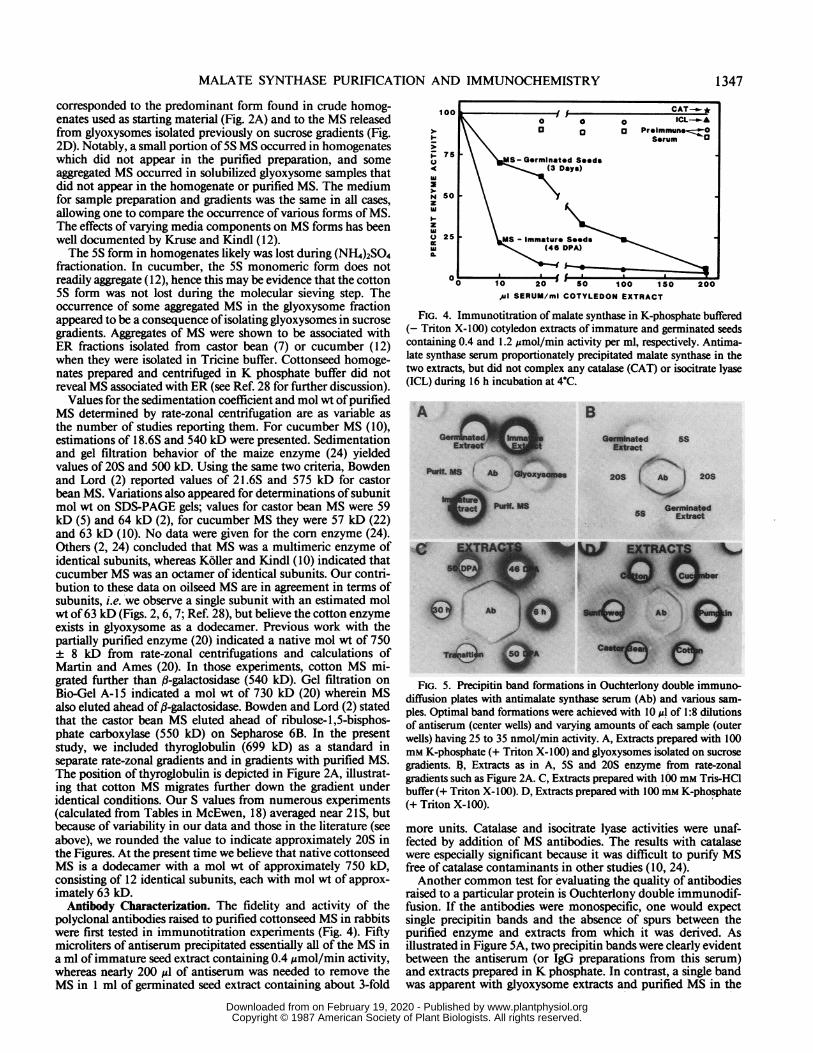
polyclonal antibodies raised to purified cottonseed MS in rabbitswere first tested in immunotitration experiments (Fig. 4). Fiftymicroliters of antiserum precipitated essentially all of the MS ina ml of immature seed extract containing 0.4 ,umol/min activity,whereas nearly 200 usl of antiserum was needed to remove theMS in 1 ml of germinated seed extract containing about 3-fold
100
1u 13 Preimmnune..zz1_\\ ~~~~~~~~SarumC
z75 \ \IMS-Germinated SeedsA 50 \ - (3 Days)
N sozwI~-z
Q 25 Ms - immature Seeds
0 10 20 So 100 1SO 200
,~u SERUM/ml COTYLEDON EXTRACT
FIG. 4. Immunotitration of malate synthase in K-phosphate buffered(- Triton X-100) cotyledon extracts of immature and germinated seedscontaining 0.4 and 1.2 jsmol/min activity per ml, respectively. Antima-late synthase serum proportionately precipitated malate synthase in thetwo extracts, but did not complex any catalase (CAT) or isocitrate lyase(ICL) during 16 h incubation at 4"C.
A_
ft. e( *:_-Ecu_,.i s_
)U ..".
B
86-
Ss
XOSI. 20S
Mi.
Extd
Ab
hi
)
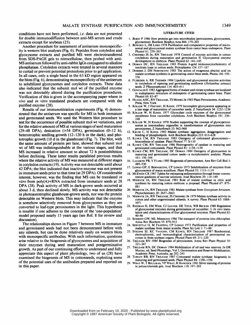
FIG. 5. Precipitin band formations in Ouchterlony double immuno-diffusion plates with antimalate synthase serum (Ab) and various sam-ples. Optimal band formations were achieved with 10 Ml of 1:8 dilutionsof antiserum (center wells) and varying amounts of each sample (outerwells) having 25 to 35 nmol/min activity. A, Extracts prepared with 100mm K-phosphate (+ Triton X-100) and glyoxysomes isolated on sucrosegradients. B. Extracts as in A, 5S and 20S enzyme from rate-zonalgradients such as Figure 2A. C, Extracts prepared with 100 mm Tris-HClbuffer (+ Triton X-100). D, Extracts prepared with 100 mm K-phosphate(+ Triton X-100).
more units. Catalase and isocitrate lyase activities were unaf-fected by addition of MS antibodies. The results with catalasewere especially significant because it was difficult to purify MSfree of catalase contaminants in other studies (10, 24).Another common test for evaluating the quality of antibodies
raised to a particular protein is Ouchterlony double immunodif-fusion. If the antibodies were monospecific, one would expectsingle precipitin bands and the absence of spurs between thepurified enzyme and extracts from which it was derived. Asillustrated in Figure SA, two precipitin bands were clearly evidentbetween the antiserum (or IgG preparations from this serum)and extracts prepared in K phosphate. In contrast, a single bandwas apparent with glyoxysome extracts and purified MS in the
outer wells. An obvious interpretation is that a non-MS contam-inant is being recognized in the extracts that is absent or perhapshas a low titer in glyoxysomes and/or the purified preparation.The absence of spurs, however, indicated complete serologicalidentity. Close inspection of Figure 5A shows that the purifiedand glyoxysomal bands are completely fused with the outer bandsin the extracts. This leads one to-suspect that different forms ofMS, rather than a contaminant, are responsible for the bands inextracts. The results shown in Figure 5B confirmed this suspicion.Germinated-seed extracts again revealed two bands, the outerone being contiguous and fused with 20S MS, and the inner onecontiguous and fused with 5S MS. The data in Figure 5, A andB, therefore, demonstrate that the antibodies can recognize the5S and 20S forms as separate entities in the immunodiffusionplates when the samples are prepared in K-phosphate. Thisconfirmed results presented in Figure 2; 5S and 20S formsoccurred in seed extracts whereas only the 20S form occurred insolubilized glyoxysomes and the purified preparation. The datain Figure 5A indicated serological identity between the 5S formin immature and germinated seeds, and the 20S form in imma-ture and germinated seeds. However, Figure 5B reveals spursbetween the 5S and 20S forms, indicating incomplete identitybetween the two forms. An explanation for this is not obvious;but the immunological nonidentity may be an important clue inunderstanding the transport of the monomer into glyoxysomes.That is, nonidentity may indicate conformational change(s) fol-lowing oligomerization of the 5S monomer to the 20S dodeca-mer.Another interesting phenomenon is illustrated in Figure 5C.
Immunodiffusion of the same antiserum against immature (46and 50 DPA), imbibed (6 h), germinated (30 h), and greening(transition) seed extracts prepared in Tris-HCl buffer yieldedonly one precipitin band showing complete identity among thesamples. We are uncertain ofthe reason for the disparity betweenFigure 5, A and C, but the results are repeatable. We haveunpublished data showing that 5S and highly aggregated MSoccurred in rate-zonal gradients when cotton seeds were homog-enized and centrifuged in Tris-HCl. The Ouchterlony results(Fig. 5C) may be due to only the 5S form diffusing in the agarose,or perhaps to a single large aggregate producing a single band.
Antibodies to cottonseed MS showed widespread cross-reactiv-ity with MS from other oilseeds (Fig. SD). It was difficult toproduce uniformly clear results when applying samples fromdifferent species with varying enzyme units per ml, but precipitinbands were observed with all species shown in Figure SD andwith watermelon (not shown) and soybean (25). Soybean andcottonseed extracts were made in Tris-HCl containing 0.5 MNaCl (25); under these conditions, a single precipitin bandoccurred between cotton anti-MS and the two extracts. In addi-tion, no spurs were observed between the soybean and the cottonextracts. The results in Figure SD were complicated by theappearance of multiple bands and spurs. Complete identity wasobserved between the two MS forms in the cucurbits (cucumberand pumpkin). Partial identity was apparent with both theseforms and cotton (see spurs). The sunflower and castor beanextracts exhibited only one inner band. This suggested that onlya SS form of MS was extracted with the K phosphate. Thisinterpretation was confirmed when rate-zonal centrifugation ofthe castor bean extract (similar to that used for Fig. SD) revealedonly aSS form on the gradient (data not shown). However, whencastor beans were extracted with K phosphate containing 0.2 MKCI, two bands showing partial identities with both bands fromcotton extracts were observed (data not shown). Similar studieswith the sunflower were not done.
Immunological variations described above for MS have notbeen reported by any others. Dommes and Northcote (5) showedone immunodiffusion band between their antiserum andpurified
A B C D E
63kDa.- eM rn-
-0 A*
*I-
o
CI
E _
0 I X
. W U3
, 0
FIG. 6. Western blot stained for malate synthase following transferfrom a SDS-PAGE gel (4% stacking, 8% separating, 17 cm long),demonstrating monospecificity of the antiserum and comparing subunitmol wt of purified malate synthase with enzyme extracted under variedconditions. Each lane had 3 to 5 nmol/min MS activity except C whichhad 100 ,ug protein. Inhibitors included in lane D sample were 1 mMbenzamidine-HCl and PMSF, 5 mm EDTA and iodoacetamide, and 2,ug/ml aprotinin.
63 kD
29 46 Dry 24 48 72 96 120 Grr
DPA Germinated (h)
FIG. 7. Western blot prepared as in Figure 6 comparing subunit molwt and the relative amount of malate synthase in maturing (DPA), dry,germinated and green seeds. Each lane contained 25 Atg protein of extractsprepared with 100 mm K-phosphate, 8 mM MgCl2, 2 mM EDTA (pH7.2), and 1 mm PMSF and benzamidine-HCI.
MS. Koller and Kindl (1 1) stated in their "Methods" section thatOuchterlony plates were used to show that they were dealingwith monovalent serum, but conditions or results were notpresented. Riezman et al (22) showed an immunoelectrophore-togram with two joining arcs which indicated complete indentitybetween a basic and acidic form. Bowden and Lord (2) statedthat single precipitin bands formed between their antiserum andKCl extracts ofglyoxysomes and purified MS; both these sourceswere shown to contain only a 30S form of MS. Thus, it is notthat the interesting phenomena reported here are inconsistentwith previous results, but that the same experiments under these
conditions have not been performed, i.e. data are not presentedfor double immunodiffusion between anti-MS serum and crudeextracts except for soybean (25).Another procedure for assessment of antiserum monospecific-
ity is western blot analyses (Fig. 6). Peptides from cotyledon andglyoxysome extracts and purified MS were electrotransferredfrom SDS-PAGE gels to nitrocellulose, then probed with anti-MS antiserum followed by anti-rabbit IgGs conjugated to alkalinephosphatase. Cotyledon extracts were treated in several mannersto curtail or prevent potential proteolysis prior to gel application.In all cases, only a single band in the 63 kD region appeared onthe blots (Fig. 6), demonstrating monospecificity ofthe antiserumto solubilized glyoxysomes and cotyledon extracts. These dataalso indicated that the subunit mol wt of the purified enzymewas not detectably altered during the purification procedures.Verification of this is given in the accompanying paper where invivo and in vitro translated products are compared with thepurified enzyme (28).
Results of our immunotitration experiments (Fig. 4) demon-strated that the antiserum was specific for MS in both immatureand germinated seeds. We used the Western blot procedure totest for the occurrence of possible subunit mol wt variations, andfor the relative amount ofMS at various stages ofseed maturation(29-48 DPA), desication (>54 DPA), germination (0-12 h),heterotrophic seedling growth (12-120 h in the dark), and pho-totrophic growth (10 d in the glasshouse) (Fig. 7). The blot, withthe same amount of protein per lane, showed that subunit molwt of MS was indistinguishable at the various stages, and thatMS increased in relative amount to about 72 h postimbibitionbefore declining. These latter results paralleled previous resultswhere the relative activity of MS was measured at different stagesin cotyledon extracts ( 19). Activity was not detectable until about42 DPA; the blot indicated that inactive enzyme was not presentin immature seeds prior to that time (at 29 DPA). Ofconsiderableinterest, however, was the finding that MS can be translated invitro from poly(A)+RNA extracted from immature seeds at 28DPA (28). Peak activity of MS in dark-grown seeds occurred atabout 3 d, then declined slowly. MS activity was not detectablein photoautotrophic glasshouse-grown seeds, nor was the proteindetectable on Western blots. This may indicate that the enzymeis somehow selectively removed from glyoxysomes as they areconverted to leaf-type peroxisomes in the light. This hypothesisis tenable if one adheres to the concept of the 'one-population'model proposed nearly 15 years ago (see Ref. 8 for review anddiscussion).The relationships shown in Figure 7 between MS in immature
and germinated seeds had not been demonstrated before withany oilseeds, but can be done relatively easily on western blotswith monospecific antibodies. With such information, questionsarise relative to the biogenesis of glyoxysomes and acquisition oftheir enzymes during seed maturation and postgerminativegrowth. As part of our continuing efforts to understand and fullyappreciate this aspect of plant physiology, two of us (28) haveexamined the biogenesis of MS in cottonseeds, exploiting someof the potential uses of the antibodies prepared and reported onin this paper.
LITERATURE CITED
1. BORST P 1986 How proteins get into microbodies (peroxisomes, glyoxysomes,glycosomes). Biochem Biophys Acta 866: 179-203
2. BOWDEN L, JM LORD 1978 Purification and comparative properties of micro-somal and glyoxysomal malate synthase from castor bean endosperm. PlantPhysiol 61: 259-265
3. CHOINSKI JS JR, RN TRELEASE 1978 Control of enzyme activities in cottoncotyledons during maturation and germination II. Glyoxysomal enzymedevelopment in embryos. Plant Physiol 62: 141-145
4. DOMAN DC, RN TRELEASE 1985 Protein A-gold immunocytochemistry ofisocitrate lyase in cotton seeds. Protoplasma 124: 157-167
5. DOMMES J, DH NORTHCOTE 1985 The action of exogenous abscisic acid onmalate-synthase synthesis in germinating castor-bean seeds. Planta 166: 550-556
6. FUSSEDER A, RR THEIMER 1984 Lipolytic and glyoxysomal enzyme activitiesin cotyledons of ripening and germinating sunflower (Helianthus annuus)seeds. Z Pflanzenphysiol 114: 403-411
7. GONZALES E 1982 Aggregated forms ofmalate and citrate synthase are localizedin endoplasmic reticulum of endosperm of germinating castor bean. PlantPhysiol 69: 83-87
8. HUANG AHC, RN TRELEASE, TS MOORE JR 1983 Plant Peroxisomes. AcademicPress, New York
9. KOLLER W, J FREVERT, H KINDL 1979 Incomplete glyoxysomes appearing ata late stage of maturation of cucumber seeds. Z Naturforsch 34: 1232-1236
10. KOLLER W, H KINDL 1977 Glyoxylate cycle enzymes of the glyoxysomalmembrane from cucumber cotyledons. Arch Biochem Biophys 181: 236-248
11. KOLLER W, H KINDLE 1978 Studies supporting the concept of glyoxyperoxi-somes as intermediary organelles in transformation of glyoxysomes intoperoxisomes. Z Naturforsch 33: 962-968
12. KRUSE C, H KINDL 1983 Malate synthase: aggregation, deaggregation andbinding of phospholipids. Arch Biochem Biophys 223: 613-628
13. KUNCE CM, RN TRELEASE 1985 Ontogeny of cottonseed glyoxysomes-biosynthesis of catalase. Plant Physiol 77: S-lII
14. KUNCE CM, RN TRELEASE 1986 Heterogeneity of catalase in maturing andgerminated cottonseeds. Plant Physiol 81: 1134-1139
15. KUNCE CM, RN TRELEASE, DC DOMAN 1984 Ontogeny of glyoxysomes inmaturing and germinated cotton seeds-a morphometric analysis. Planta161: 156-164
16. LAZAROW PB, Y FUJIKI 1985 Biogenesis of peroxisomes. Ann Rev Cell Biol 1:489-530
17. LONGO GP, E BERNASCONI, CP LONGO 1975 Solubilization of enzymes fromglyoxysomes of maize scutellum. Plant Physiol 55: 1115-1119
18. MCEWEN CR 1967 Tables for estimating sedimentation through linear concen-tration gradients of sucrose solutions. Anal Biochem 20: 114-149
19. MIERNYK JA, RN TRELEASE 1981 Role of malate synthase in citric acidsynthesis by maturing cotton embryos: a proposal. Plant Physiol 67: 875-881
21. MIERNYK JA, RN TRELEASE, JS CHOINSKI JR 1979 Malate synthase activity incotton and other ungerminated oilseeds. A survey. Plant Physiol 63: 1068-1071
22. RIEZMAN H, EM WEIR, CJ LEAVER, DE TITUS, WB BECKER 1980 Regulationof glyoxysomal enzymes during germination of cucumber. 3. In vitro trans-lation and characterizations of four glyoxysomal enzymes. Plant Physiol 65:40-46
23. SCHMIDT GW, ML MISHKIND 1986 The transport of proteins into chloroplast.Annu Rev Biochem 55: 879-912
24. SERVETTAZ 0, M FILIPPINI, CP LONGO 1973 Purification and properties ofmalate synthase from maize scutella. Plant Sci Lett 1: 71-80
25. STEGINK SJ, KC VAUGHN, CM KUNCE, RN TRELEASE 1987 Biochemical,electrophoretic, and immunological characterization of peroxisomal en-zymes in three soybean organs. Physiol Plant 69: 211-220
26. TRELEASE RN 1984 Biogenesis of peroxisomes. Annu Rev Plant Physiol 35:321-347
27. TRELEASE RN, DC DOMAN 1984 Mobilization of oil and wax reserves. In DRMurray, ed, Seed Physiology, Vol 2, Germination and Reserve Mobilization.Academic Press, Australia, pp 202-245