28
COVERA™ Vascular Covered Stents in the Management of Dysfunctional AV Access Bart L. Dolmatch, M.D., FSIR Palo Alto Medical Foundation Mountain View, CA USA BPV/SGF3/0118/0005

COVERA™ Vascular Covered Stents in the Management of
Dysfunctional AV Access
Bart L. Dolmatch, M.D., FSIR
Palo Alto Medical Foundation
Mountain View, CA
USA
BPV/SGF3/0118/0005
• This presentation is being made on behalf of Bard Peripheral Vascular, Inc. Any discussion regarding BARDproducts during the presentation today is limited to information that is consistent with the clearances for those products. Please consult Bard Peripheral Vascular, Inc. product labels and inserts for any indications, contraindications, hazards, warnings, cautions and instructions for use.
• Dr. Dolmatch has been compensated by Bard Peripheral Vascular, Inc. to participate in this presentation.
BPV/SGF3/0118/0005

10 Circuit Patency after Creation AV Fistulae and AV Grafts
Huber et al., Patency of autogenous and polytetrafluoroethylene upper extremity arteriovenous
hemodialysis accesses: A systematic review (J Vasc Surg 2003;38:1005-11.)
BPV/SGF3/0118/0005
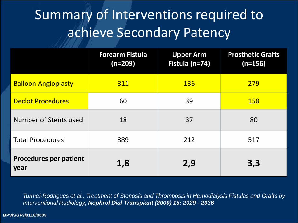
Summary of Interventions required to achieve Secondary Patency
Turmel-Rodrigues et al., Treatment of Stenosis and Thrombosis in Hemodialysis Fistulas and Grafts by
Interventional Radiology, Nephrol Dial Transplant (2000) 15: 2029 - 2036
Forearm Fistula(n=209)
Upper ArmFistula (n=74)
Prosthetic Grafts(n=156)
Balloon Angioplasty 311 136 279
Declot Procedures 60 39 158
Number of Stents used 18 37 80
Total Procedures 389 212 517
Procedures per patientyear 1,8 2,9 3,3
BPV/SGF3/0118/0005

Bard FLAIR® Pivotal Study (Code HXM HM)The results from case studies may not be predictive for all patients. Individual results may vary depending on a variety of patient specific attributes.
Pre Treatment PTA Post PTA 2 months later
Recurrent Stenosis after PTA is Common
BPV/SGF3/0118/0005
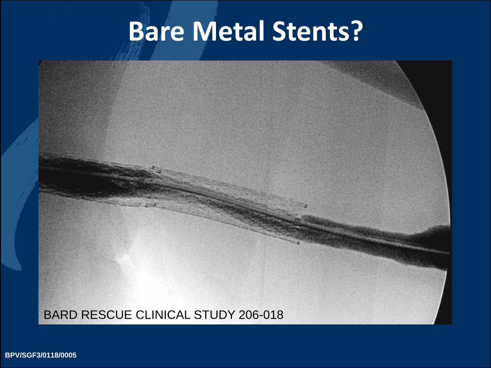
Bare Metal Stents?
BARD RESCUE CLINICAL STUDY 206-018
BPV/SGF3/0118/0005
Four Level 1 Multicenter, Prospective Randomized Controlled Clinical Trials
(RCT) evaluated technologies for patency improvement over standard
of care balloon angioplasty in patients dialyzing with an AV Graft
BPV/SGF3/0118/0005

BSI Peripheral Cutting Balloon (PCB)
Patients• N=340
• Stenotic (≥ 50 %), AVG [N=195] & thrombosed AV Grafts [N=145]
Follow-Up• 14 days, 1, 3, 6 Months
• Clinically driven follow-up
• No mandatory diagnostic imaging during follow up w/o clinical indicator
P = 0.373
Studies vary by design and patient population, and may yield different outcomes.
Vesely et al., J Vasc Interv Radiol 2005 Dec;16(12):1593-603
BPV/SGF3/0118/0005
No Significant Improvement

BARD FLAIR® Stent Graft “PIVOTAL” Trial
Patients• N=190
• Stenotic (≥ 50 %), dysfunctional AVG
• No thrombosed grafts
Follow-Up• 2 and 6 Months
• Mandatory magnified angiogram at 2 and 6 months
P < 0.001
Haskal et al., N Engl J Med 2010; 362: 494-503
BPV/SGF3/0118/0005Studies vary by design and patient population, and may yield different outcomes.
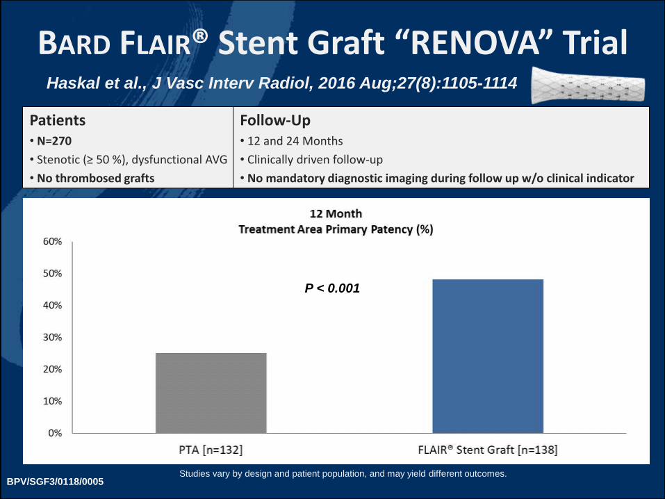
BARD FLAIR® Stent Graft “RENOVA” Trial
P<0.001
Haskal et al., J Vasc Interv Radiol, 2016 Aug;27(8):1105-1114
Patients• N=270
• Stenotic (≥ 50 %), dysfunctional AVG
• No thrombosed grafts
Follow-Up• 12 and 24 Months
• Clinically driven follow-up
• No mandatory diagnostic imaging during follow up w/o clinical indicator
P < 0.001
Studies vary by design and patient population, and may yield different outcomes. BPV/SGF3/0118/0005

Gore ViabahnTM Endoprosthesis “REVISE”
Patients• N=293
• Stenotic (≥ 50 %) AVG [N=164] &thrombosed AV Grafts [N=129]
Follow-Up• 1,3, 6, 12, 18 and 24 Months
• Clinically driven follow-up
• No mandatory diagnostic imaging during follow up w/o clinical indicator
P = 0.006
Vesely et al., J Vasc Surg 2016 Nov;64(5):1400-1410
Studies vary by design and patient population, and may yield different outcomes. BPV/SGF3/0118/0005
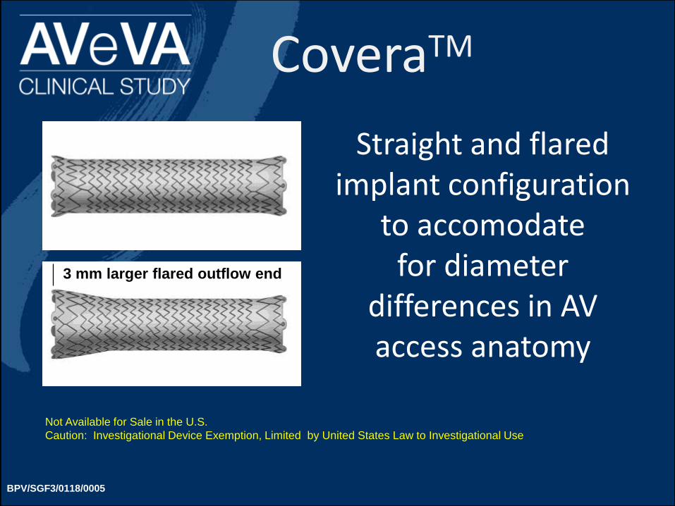
Straight and flared implant configuration
to accomodatefor diameter
differences in AV access anatomy
3 mm larger flared outflow end
Not Available for Sale in the U.S.
Caution: Investigational Device Exemption, Limited by United States Law to Investigational Use
BPV/SGF3/0118/0005
CoveraTM

CoveraTM Vascular Covered Stent
• Dual layer ePTFE covering
• Carbon impregnation on the luminal (blood contacting) surface
• Diameters 6 mm – 10 mm
• Lengths 30 mm – 100 mm
Not Available for Sale in the U.S.
Caution: Investigational Device Exemption, Limited by United States Law to Investigational Use BPV/SGF3/0118/0005

COVERATM Vascular Covered Stent Ongoing Clinical Studies
Prospective, Multi-Center, Randomized, Concurrently-Controlled Clinical Study of
the Bard® Arteriovenous (AV) Stent Graft in the
Treatment of Stenosis in the Venous Outflow of AV Fistula Access Circuits
Prospective, Multi-Center, Clinical Study of the Bard® COVERA™ Arteriovenous
(AV) Stent Graft in the Treatment of Stenosis at the Graft-Vein Anastomosis of
AV Graft Circuits
(AV Fistula) (AV Graft)BPV/SGF3/0118/0005

Study Design Prospective, Multi-Center, Single-Arm
Objective
To assess the safety and effectiveness of the BARD® COVERA™ Vascular Covered Stent for the treatment of stenotic lesions at the graft-vein anastomosis of hemodialysis patients dialyzing with an AV graft.
Number of Patients
110 patients enrolled at 14 US sites
BPV/SGF3/0118/0005

Key Inclusion Criteria
CLINICAL ANGIOGRAPHIC
• AV access graft in an arm
that has been implanted
for 30 days, undergone
≥ 1 dialysis session
• Thrombosed and non
thrombosed AV grafts
• Stenosis ≥ 50% at graft-
vein anastomosis with
clinical or hemodynamic
evidence of AV graft
dysfunction
• Target lesion ≤ 9 cm long
• Reference vessel
diameter 5.0 - 9.0 mm
BPV/SGF3/0118/0005

Primary Safety Endpoint
Freedom from any protocol defined safety event(s) involving the AV access circuit
through 30 days: Performance Goal = 88%
Primary Effectiveness Endpoint
Target Lesion Primary Patency (TLPP) - 6
months: Performance Goal = 40%
Follow Up 30 & 90 days, 6, 12, 18, & 24 months
Status 6 Month Follow Up completed
BPV/SGF3/0118/0005

Bard AVeVA Clinical Study 401-017
• Case Description
• Male, 47 Y
• Pre dilatation with 7 x 40 PTA balloon
• 9 x 60 flared COVERA® Vascular Covered Stent
The opinions and clinical experiences presented herein are for informational purposes only. The results from case studies may not be predictive for all patients. Individual results may vary depending on a variety of patient specific attributes.
BPV/SGF3/0118/0005

Demographics
Age Categories
<65 years 47.3%
≥ 65 and < 75 years 28.2%
≥ 75 years 24.5%
Gender
Male 45.5%
Female 54.5%
BMI Categories
< 30 61.8%
≥ 30 38.2%
BPV/SGF3/0118/0005

Lesion and Circuit Characteristics
BPV/SGF3/0118/0005
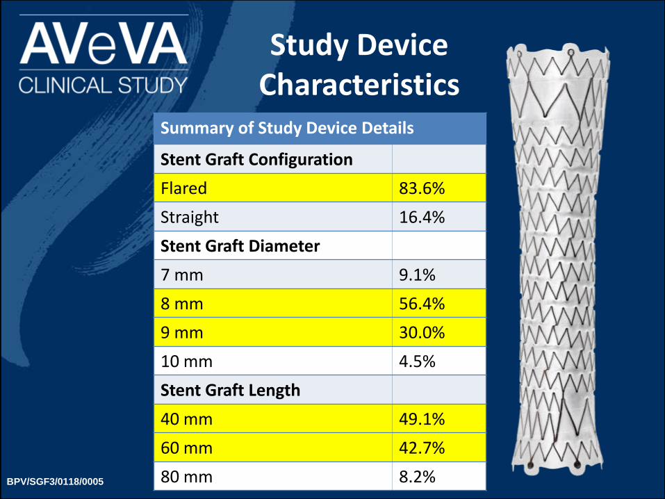
Summary of Study Device Details
Stent Graft Configuration
Flared 83.6%
Straight 16.4%
Stent Graft Diameter
7 mm 9.1%
8 mm 56.4%
9 mm 30.0%
10 mm 4.5%
Stent Graft Length
40 mm 49.1%
60 mm 42.7%
80 mm 8.2%
Study Device Characteristics
BPV/SGF3/0118/0005
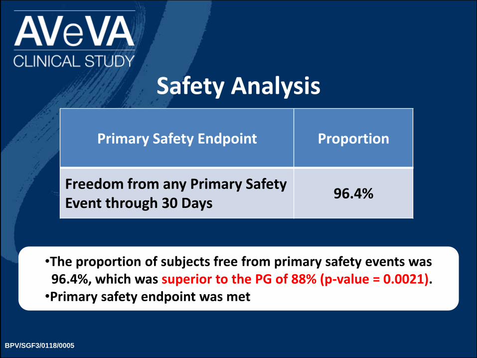
Safety Analysis
Primary Safety Endpoint Proportion
Freedom from any Primary Safety Event through 30 Days
96.4%
•The proportion of subjects free from primary safety events was 96.4%, which was superior to the PG of 88% (p-value = 0.0021).
•Primary safety endpoint was met
BPV/SGF3/0118/0005
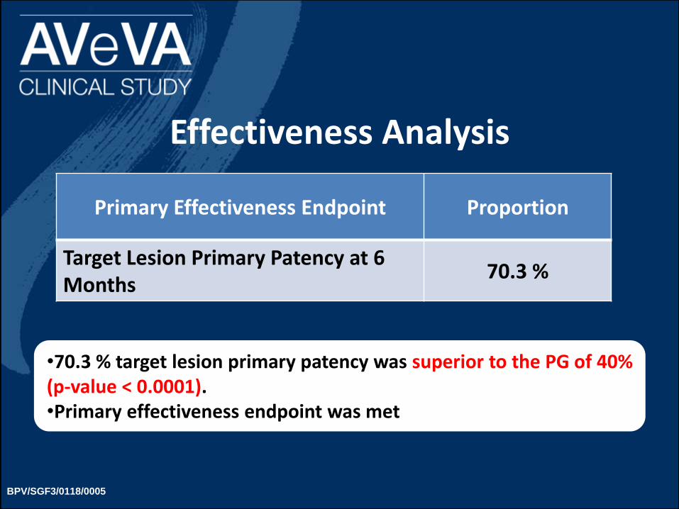
Effectiveness Analysis
Primary Effectiveness Endpoint Proportion
Target Lesion Primary Patency at 6 Months
70.3 %
•70.3 % target lesion primary patency was superior to the PG of 40% (p-value < 0.0001).•Primary effectiveness endpoint was met
BPV/SGF3/0118/0005

Target Lesion Primary Patency to 6 Months (Kaplan-Meyer)
0.1
0.2
0.3
0.4
0.5
0.6
0.7
0.8
0.9
1.0
Surv
ival Pro
bability
0 30 60 90 120 150 180
Time to Event (Days)
+ Censored
Covera™ Vascular Covered StentTreatment:
C.R.Bard Inc. Page 1 of 1
Protocol: BPV-15-001
Interim 6-Month ReportFigure 14.2-1.1
Kaplan Meier Curve of TLPP During the 6-Month Post Procedure Follow-Up Period
All Treated Population
Data Source: ADSL, ADEF
Program name: ftlpp.sas Cutoff Date: Oct 5, 2017 Date: 11DEC2017 20:20
PG = 40%
71.1%
BPV/SGF3/0118/0005

Conclusions
• The AVeVA Clinical Study is the 4th large multicenter clinical study that demonstrated a primary patency benefit when covered stents are used for the treatment of AV Access Graft dysfunction.
• At 6 months, the COVERA™ Vascular Covered Stent was an effective therapy for the treatment of stenosis at the graft – vein anastomosis in patients with thrombosed and non-thrombosed AV Grafts.
• The target lesion primary patency was significantly higher compared to the performance goal of 40%.
BPV/SGF3/0118/0005
Disclaimer
• Trademarks are the property of their respective owners.
• © 2018 BD. BD, the BD Logo, Bard, Flair and Coveraare trademarks of Becton, Dickinson and Company.
BPV/SGF3/0118/0005

Prescriptive InformationCOVERA™ Vascular Covered Stent
Prescriptive Information
Prior to use, please see the complete „Instructions for Use“ for more information on Indications, Contraindicaions,
Warnings, Precautions, Adverse Events and Operator‘s Instructions.
INDICATIONS
The COVERA™ Vascular Covered Stent is indicated for
the treatment of stenoses in the upper extremity venous
outflow of patients dialyzing with an arterio-venous (AV)
access graft or fistula.
CONTRAINDICATIONS
There are no known contraindications for the COVERA™
Vascular Covered Stent.
SELECT WARNINGS DO NOT use in patients whose AV access grafts have been
implanted less than 30 days or in an immature fistula.
DO NOT use the device in patients where full expansion of an
appropriately sized PTA balloon catheter could not be achieve
during pre-dilation with an angioplasty balloon.
Placing a covered stent across a vessel side branch may
impede blood flow and hinder or prevent future procedures.
Covered stent placement beyond the ostium of the cephalic vein
into the axillary/subclavian vein may hinder or prevent future
access.
DO NOT place a flared covered stent with the flared end in a
straight vessel segment since this may lead to flow turbulences.
The device has not been tested for tracking and deployment
around an AV loop graft.
SELECT PRECAUTIONS The safety and effectiveness of the device when placed across
an aneurysm or a pseudo-aneurysm has not been evaluated.
The safety and effectiveness of the device when used in central
veins has not been evaluated.
The safety and effectiveness of the device when placed across a
previously placed bare metal stent has not been evaluated.
The safety and effectiveness of the device when placed across
the antecubital fossa has not been evaluated.
The safety and effectiveness of the device when used in
pediatrics has not been evaluated.
The effects of direct cannulation of the covered stent have not
been evaluated. Notify the patient that the covered stent should
not be directly cannulated for hemodialysis and that applying
pressure to the implant area should be avoided.
The device has not been tested for use in an overlapped
condition with a bare metal stent or covered stent.
BPV/SGF3/0118/0005
COVERA™ Vascular Covered Stents in the Management of
Dysfunctional AV Access
Bart L. Dolmatch, M.D., FSIR
Palo Alto Medical Foundation
Mountain View, CA
USA
BPV/SGF3/0118/0005


