CRANFIELD UNIVERSITY Cranfield Health PhD Thesis AGAPI I. DOULGERAKI MONITORING THE SUCCESSION OF BACTERIAL COMMUNITIES DURING STORAGE OF RAW MEAT Supervisors: Dr. David Aldred Prof. Naresh Magan Prof. George - John Nychas 2010
Transcript
CRANFIELD UNIVERSITY
Cranfield Health
PhD Thesis
AGAPI I. DOULGERAKI
MONITORING THE SUCCESSION OF BACTERIAL
COMMUNITIES DURING STORAGE OF RAW MEAT
Supervisors:
Dr. David Aldred
Prof. Naresh Magan
Prof. George - John Nychas
2010
i
Abstract
Fresh meat is exposed to various factors which cause microbiological
contamination during handling, processing, packaging and storage. Furthermore, the
storage conditions applied may affect the microbial association of the product and
consequently the spoilage process. Therefore, the purpose and importance of this
study was to identify areas that should be addressed to monitor the succession of
bacterial communities during storage of raw meat. The improvement of the
microbiological quality and safety of meat was also studied.
The observed differences in microbial quality of samples showed that
packaging can play a significant role in extending the shelf life of fresh meat, since
the growth of aerobic microorganisms was prevented in meat under modified
atmosphere packaging (MAP). When the minced beef was stored aerobically all the
microbial groups showed viable counts higher than those of the other packaging
conditions adopted (MAP-, MAP+). More specifically, total viable counts levels were
suppressed for 1.9 and 2.15 log cfu g-1
under MAP- and MAP+ at 0°C, respectively.
Additionally, growth of Listeria monocytogenes occurred in minced beef stored
aerobically, although limited growth was observed under MAP with or without
volatile compounds of oregano essential oil. These results revealed that, volatile
compounds of oregano essential oil in conjunction with MAP could be used to control
the microbial loads and colour change to acceptable levels and as a more effective
system for extending the shelf life and increase the safety of meat.
Culture – dependent (PFGE, species specific PCR, SDS-PAGE, sequencing
analysis) and - independent (PCR-DGGE) methods were applied to provide an insight
of the population dynamics of bacteria in relation to storage conditions. Nevertheless,
the main findings of the present study were based on PFGE. This culture dependent
approach has provided important information in relation to the strain distribution of
the microbiota which would have not been acquired if strain typing had not been
ii
performed. In the latter case, a modified PFGE protocol i.e. addition of thiourea after
the proteinase treatment, was successfully developed with all Enterobacteriaceae
isolates that were previously untypeable now producing high quality fingerprints; this
is the first time that thiourea was introduced in a step during the preparation of
agarose inserts.
It has been shown that storage temperature combined with packaging
conditions induced the selectivity of the spoilage microbiota at a species and/or strain
level, while the microbiota recovered from the initial stage of storage was markedly
different from that at the final stage of storage at chill temperatures. More accurately,
within the LAB population obtained, Leuconostoc spp. (Ln. mesenteroides in the case
of beef fillets) and Lactobacillus sakei were identified as significant members of the
microbiota at abuse and chill temperatures, respectively. Moreover, Serratia
liquefaciens represented the dominant isolate of Enterobacteriaceae during storage of
minced beef for most conditions adopted, but 10 and 15 °C under MAP + and 10 °C
under MAP –; in the latter case, Hafnia alvei represented the dominant fingerprint. In
the case of beef fillets, S. liquefaciens, Serratia spp., Klebsiella oxytoca, Enterobacter
ludwigii and E. cloacae were common at 0, 5, 10, 15 and 20°C. Additionally, four
Enterobacteriaceae strains isolated from beef fillets could not be assessed to genus
level, leading to the possibility that new bacterial species were detected. Furthermore,
different pseudomonads strains dominated the Pseudomonas Agar Base (PAB)
community of beef, whereas Ps. fragi was recovered from fresh beef.
The overall outcome of the present study has been clearly demonstrated that
certain species and/or strains are present or dominant only under certain conditions.
These observations seem to be of great importance and fundamental in understanding
the spoilage process in order to widen the knowledge of the spoilage related bacterial
succession during storage of meat.
iii
Acknowledgements
I would like to express my appreciation to Prof. Naresh Magan, Dr. David
Aldred and Prof. George – John Nychas for their advice and time all the way through
this thesis.
I am grateful to my colleagues and best friends Anthoula A. Argyri and
Vasiliki A. Blana for our collaboration, good atmosphere and the meaningful time that
we spent throughout.
A big thank to my closest friends for being a great soul and for their love,
patience and understanding in the last years.
I wish to thank my parents Ioannis and Evaggelia and my lovely brother
Markos for their continual support and for providing me with the opportunity to
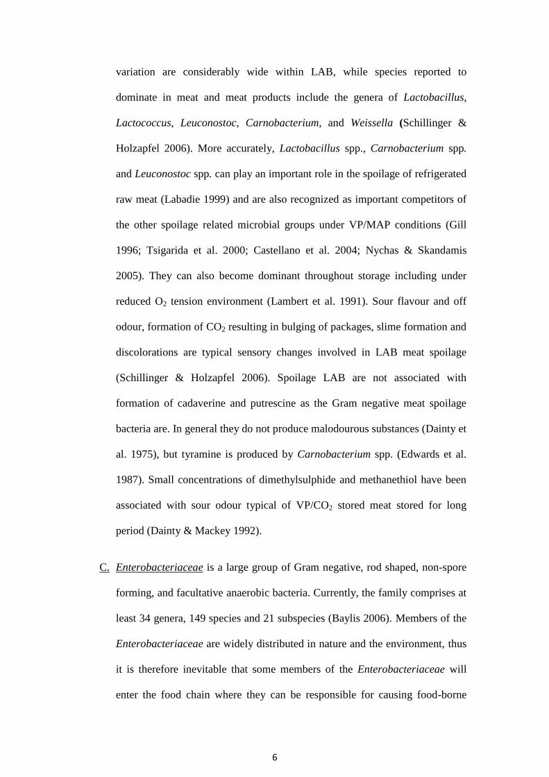
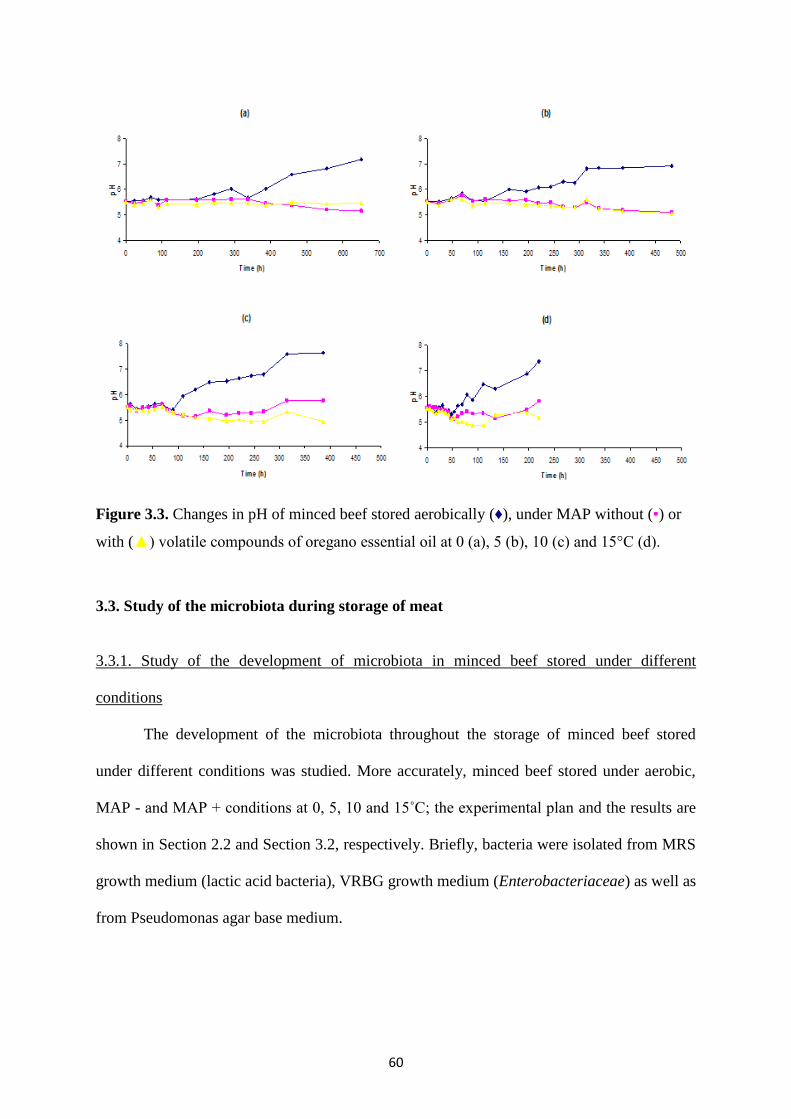
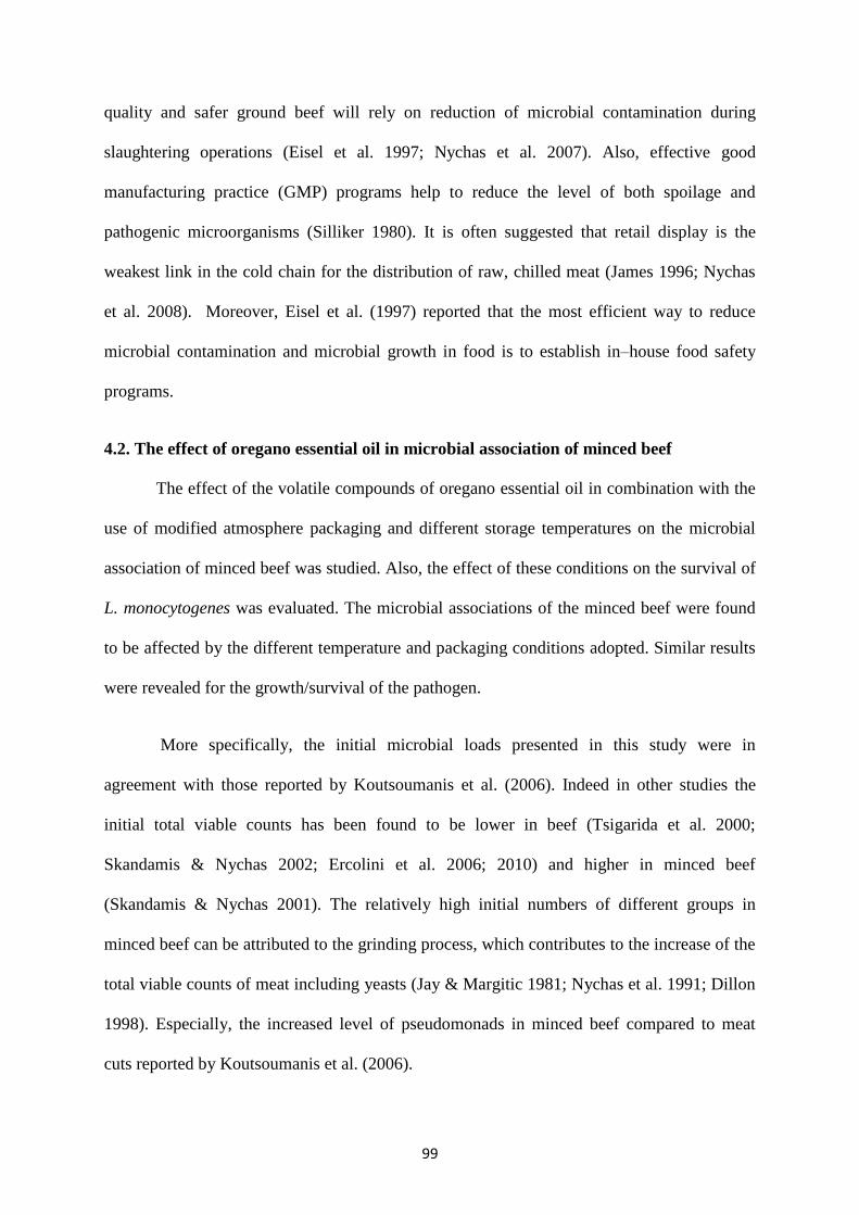
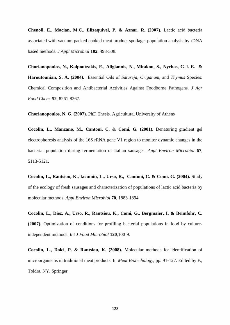
Figure 3.1. Growth of Pseudomonas spp. (▪), Br. thermosphacta (▲),
Enterobacteriaceae (*), lactic acid bacteria (x) and yeasts (♦) on minced beef stored
under MAP without (a) and with (b) volatile compounds of oregano essential oil 5 °C.
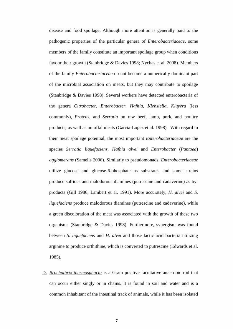
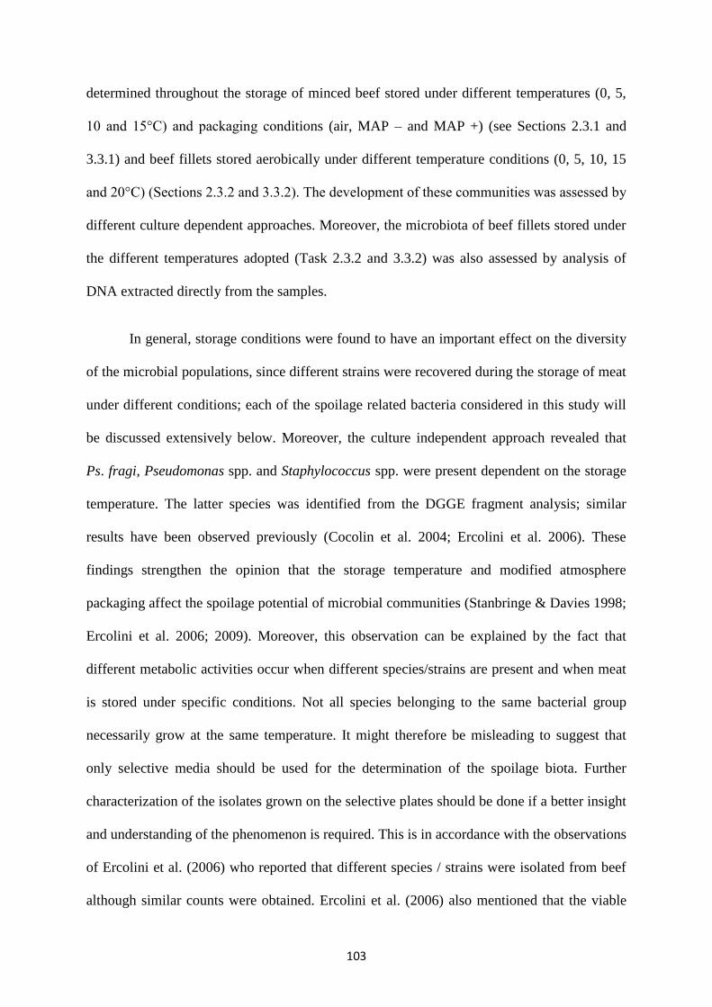
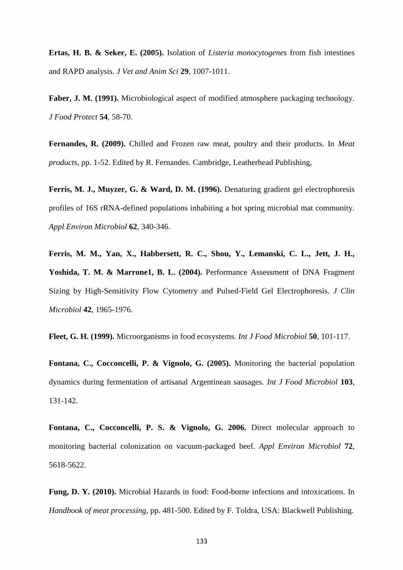
Figure 3.2 Growth of Pseudomonas spp. (▪), Br. thermosphacta (▲),
Enterobacteriaceae (*), lactic acid bacteria (x) and yeasts (♦) on minced beef stored
under MAP without (a) and with (b) volatile compounds of oregano essential oil 10
°C.
(a)
2
3
4
5
6
7
8
9
10
0 50 100 150 200 250 300 350 400 450 500
Time (h)
Lo
g cfu
/g
(b)
2
3
4
5
6
7
8
9
10
0 50 100 150 200 250 300 350 400 450 500
Time (h)L
og
cfu
/g
(a)
2
3
4
5
6
7
8
9
10
0 50 100 150 200 250 300 350 400 450 500
Time (h)
Lo
g cfu
/g
(b)
2
3
4
5
6
7
8
9
10
0 50 100 150 200 250 300 350 400 450 500
Time(h)
Lo
g cfu
/g
57
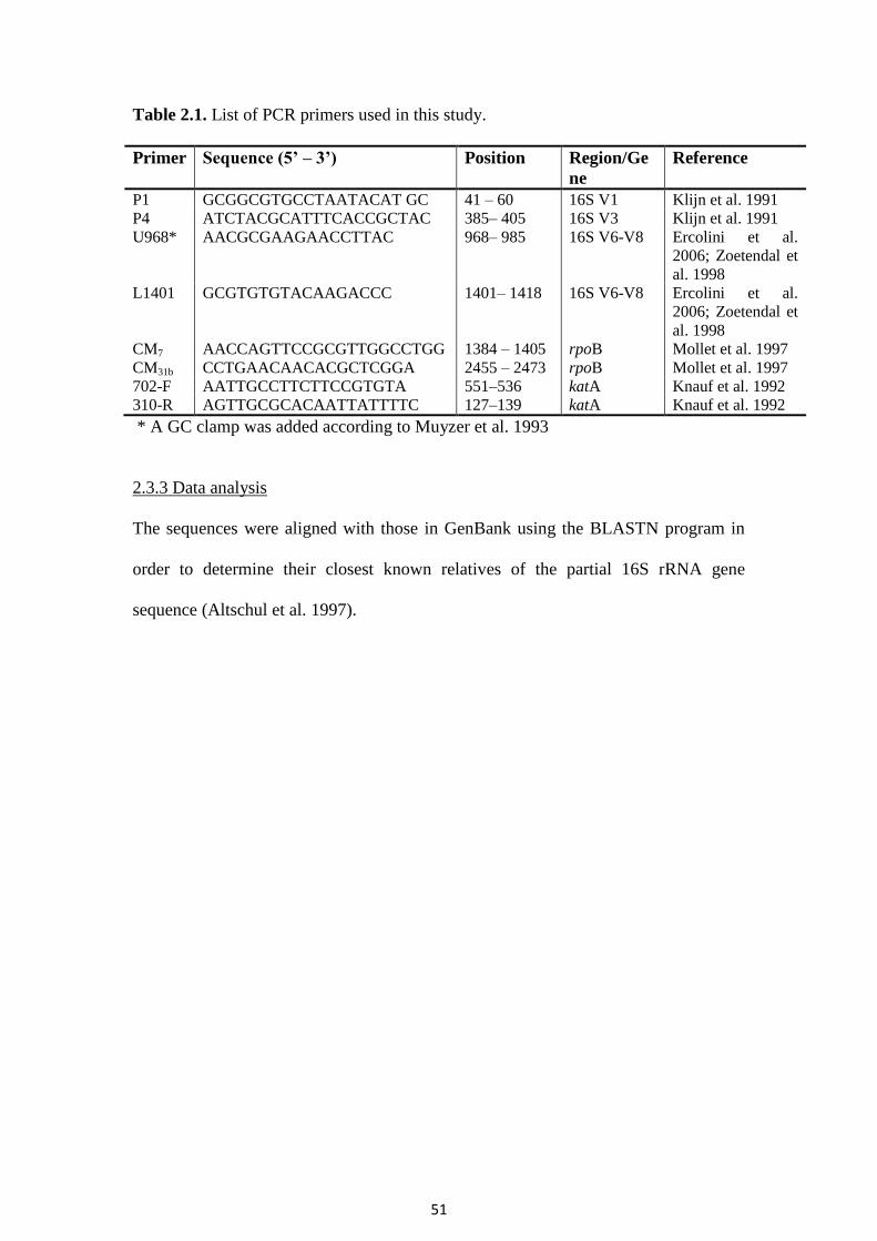
Table 3.7. The effect of packaging and volatile compounds of oregano essential oil on the final population, lag phase and maximum specific growth rate of
spoilage microorganisms of minced beef stored at 0, 5, 10 and 15°C estimated by the Baranyi model.
Yeasts and moulds 4.18 7.95 (8.21) 110.0 5.23 0.1716 7.99 (7.99) 220.0 0 0.0535 7.89 (7.89) 220.0 20.40 0.0603 1 Determined experimentally (values recorded at the end of storage period for each condition) 2 Estimated by the Baranyi model 3 time needed to reach the upper asymptote
4 Fitted curve did not present upper asymptote (semisigmoidal)
58
3.2.2. Growth of Listeria monocytogenes Scott A
L. monocytogenes was inoculated on minced beef in order to evaluate the effect of
volatile compounds of oregano essential oil on its growth. Prior to inoculation, the presence
of L. monocytogenes was tested and this pathogen was not present. This pathogen grew only
in samples stored aerobically (Table 3.5).
The modified atmosphere controlled the growth of L. monocytogenes (Table 3.5),
although the presence of volatile compounds of oregano essential oil did not affect further the
growth/survival of this pathogen. Indeed, in all samples stored under MAP -/ MAP + there
was a reduction of about 1 log cfu g-1
in samples stored aerobically. The storage temperature
also affected the growth of this pathogen (Table 3.5).
Table 3.8. The effect of packaging and volatile compounds of oregano essential oil on the
final population, lag period and maximum specific growth rate of Listeria monocytogenes
Scott A of minced beef stored at 0, 5, 10 and 15°C estimated by the Baranyi model.
1 Determined experimentally
3.2.3. Sensory analysis
The microbial shelf life (TVC 7 log cfu g-1
) and the sensory shelf life (score 2) of
minced beef stored at 0, 5, 10 and 15°C, are shown in Table 3.6. It was evident that in all
cases the volatile compounds of oregano essential oil increased shelf life of minced beef
compared to the samples stored in air or MAP -. The presence of oil affected the odour and
colour (photographs not shown) of minced beef. It needs to be noted that sometimes, oregano
249 and B 251) were equally contributed. Lb. curvatus (B 245), Lb. casei and Lb. sakei (B
248, B 250 and B 253) were also recovered during storage at 0°C.
65
Table 3.7. Identity of lactic acid bacteria isolates obtained from minced beef.
Code1 Closest relative
Accession
Number
B 225 Lactobacillus sakei GU998856
B 226 Lb. sakei GU998877
B 227 Lb. sakei GU998857
B 228 Lb. sakei GU998850
B 229 Lb. sakei GU998851
B 230 Lb. sakei GU998852
B 231 Leuconostoc spp. GU998853
B 232 Leuconostoc spp. GU998854
B 233 Leuconostoc spp. GU998855
B 234 Weissella viridescens GU998858
B 235 Ws. viridescens GU998859
B 236 Lb. sakei GU998860
B 237 Lb. sakei GU998861
B 238 Lb. sakei GU998862
B 239 Lb. sakei GU998863
B 258 Leuconostoc spp GU998864
B240 Leuconostoc spp. GU998865
B 241 Leuconostoc spp. GU998866
B 242 Ln. mesenteroides GU998867
B 243 Ln. mesenteroides GU998868
B 244 Leuconostoc spp. GU998869
B 245 Lb. curvatus GU998870
B 246 Lb. curvatus GU998871
B 247 Lb. casei GU998872
B 248 Lb. sakei GU998873
B 249 Lb. sakei GU998874
B 250 Lb. sakei GU998875
B 251 Lb. sakei GU998876
B 252 Lb. sakei GU998878
B 253 Lb. sakei GU998879
B 254 Lb. sakei GU998880
B 255 Lb. sakei GU998881 1 Code of different PFGE patterns of Figure 3.5
66
Table 3.8. Frequency (%) of isolation and distribution of lactic acid bacteria isolates recovered from minced beef stored under aerobic, MAP -
and MAP + conditions.
67
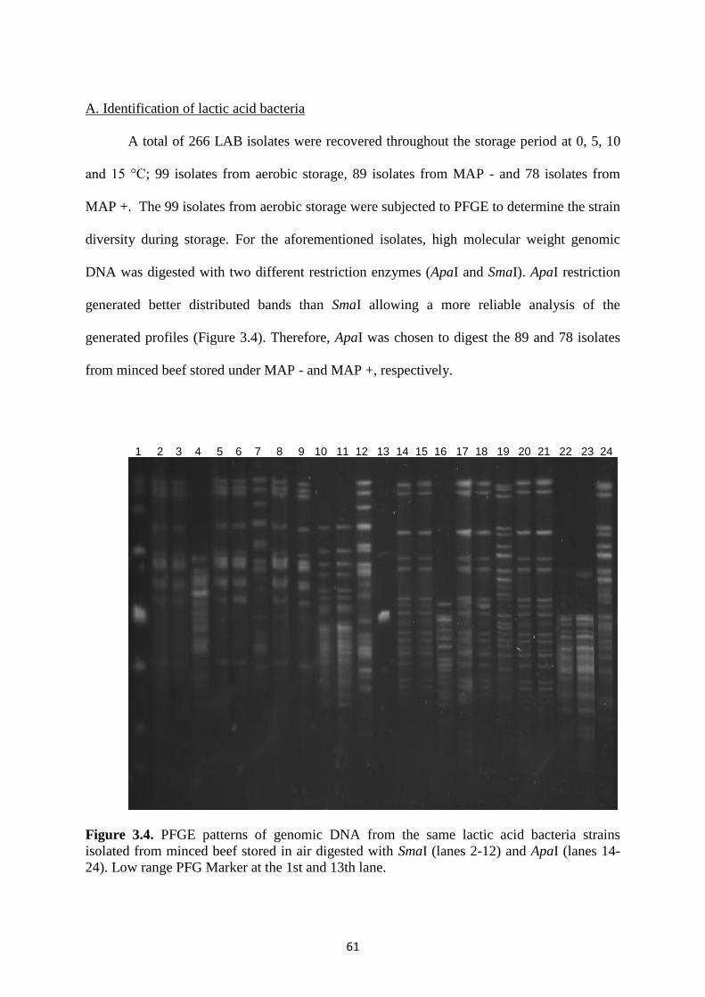
B. Identification of Enterobacteriaceae
A total of 232 Enterobacteriaceae isolates were recovered throughout the storage
period (Table 3.9) and subjected to SDS – PAGE of whole cell proteins and PFGE in order to
determine the species and strain diversity, respectively.
Whole cell protein profiling
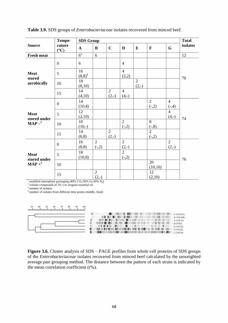
Enterobacteriaceae isolates were clustered into seven groups on the basis of their
SDS – PAGE profile obtained from whole cell proteins. The protein profile of each group is
shown in Figure 3.6 whereas the number of the isolates as well as the storage condition and
time points of isolation is presented in Table 3.9. In fresh meat, presence of two profiles,
namely A and B, was detected. Profile A consisted of 139 isolates and was common for all
packaging and temperature conditions, but 10°C and 15°C under MAP +. Similarly, profiles
C and D, consisting of 4 and 18 isolates, respectively, were recovered from both aerobic and
MAP storage; the former only at 15°C during both aerobic and MAP- storage, while
omnipresence of the latter met the exceptions of 10°C aerobically, 0°C, 5°C and 15°C under
MAP – and 10°C and 15°C under MAP +. Regarding the rest of the profiles, one was
recovered only during aerobic storage and only 3 during MAP storage. The former, namely
profile E, consisted of 2 isolates that were only isolated at 10°C, whereas the latter can be
further subdivided according to the presence of oregano essential oil. Thus, profile B
consisted of 10 isolates, recovered only during MAP+ storage at 0°C and 15°C, while profiles
F and G consisted of 44 and 10 isolates that were isolated regardless of the presence of
oregano essential oil; the former at 0°C under MAP- and 10°C and 15°C under both MAP-
and MAP+, while the latter at 5°C under MAP- and at 0°C under both MAP- and MAP+.
68
Table 3.9. SDS groups of Enterobacteriaceae isolates recovered from minced beef.
Source
Tempe-
rature
(°C)
SDS Group Total
isolates A B C D E F G
Fresh meat 63 6 12
Meat
stored
aerobically
0 6 4
70 5
16
(8,8)4
4
(2,2)
10 18
(8,10)
2
(2,-)
15 14
(4,10)
2
(2,-)
4
(4,-)
Meat
stored under
MAP –1
0 14
(10,4)
2
(-,2)
4
(-,4)
74 5
12
(2,10)
4
(4,-)
10 10
(10,-)
2
(-,2)
8
(-,8)
15 14
(6,8)
2
(2,-)
2
(-,2)
Meat
stored under
MAP +2
0 16
(8,8)
2
(-,2)
2
(2,-)
2
(2,-)
76 5
18
(10,8)
2
(-,2)
10 20
(10,10)
15 2
(2,-)
12
(2,10)
1 modified atmosphere packaging (40% CO2/30% O2/30% N2) 2 volatile compounds of 2% v/w oregano essential oil 3 number of isolates 4 number of isolates from different time points (middle, final)
G (VK101)
D (VK108)
A (VK23)
B (VK6)
E (VK32)
F (VK20)
C (VK19)
10090807060504030
Figure 3.6. Cluster analysis of SDS – PAGE profiles from whole cell proteins of SDS groups
of the Enterobacteriaceae isolates recovered from minced beef calculated by the unweighted
average pair grouping method. The distance between the pattern of each strain is indicated by
the mean correlation coefficient (r%).
69
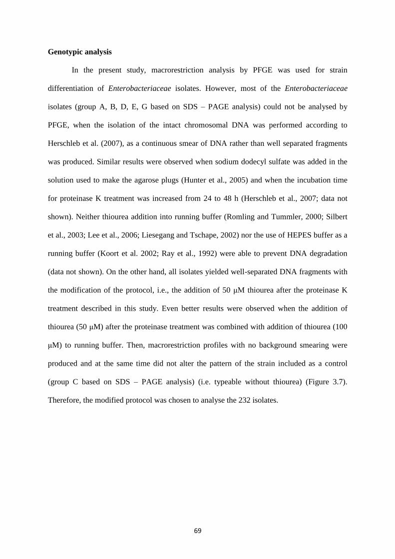
Genotypic analysis
In the present study, macrorestriction analysis by PFGE was used for strain
differentiation of Enterobacteriaceae isolates. However, most of the Enterobacteriaceae
isolates (group A, B, D, E, G based on SDS – PAGE analysis) could not be analysed by
PFGE, when the isolation of the intact chromosomal DNA was performed according to
Herschleb et al. (2007), as a continuous smear of DNA rather than well separated fragments
was produced. Similar results were observed when sodium dodecyl sulfate was added in the
solution used to make the agarose plugs (Hunter et al., 2005) and when the incubation time
for proteinase K treatment was increased from 24 to 48 h (Herschleb et al., 2007; data not
shown). Neither thiourea addition into running buffer (Romling and Tummler, 2000; Silbert
et al., 2003; Lee et al., 2006; Liesegang and Tschape, 2002) nor the use of HEPES buffer as a
running buffer (Koort et al. 2002; Ray et al., 1992) were able to prevent DNA degradation
(data not shown). On the other hand, all isolates yielded well-separated DNA fragments with
the modification of the protocol, i.e., the addition of 50 μΜ thiourea after the proteinase K
treatment described in this study. Even better results were observed when the addition of
thiourea (50 μM) after the proteinase treatment was combined with addition of thiourea (100
μM) to running buffer. Then, macrorestriction profiles with no background smearing were
produced and at the same time did not alter the pattern of the strain included as a control
(group C based on SDS – PAGE analysis) (i.e. typeable without thiourea) (Figure 3.7).
Therefore, the modified protocol was chosen to analyse the 232 isolates.
70
Figure 3.7. PFGE pattern of Enterobacteriaceae isolates recovered from minced beef after
XbaI digestion of their genomic DNA performed a) with the standard protocol (Lanes 1-5), b)
after minimize the culturing time (Lanes 6-10) and c) after thiourea treatment (Lanes 11-15).
(Lanes M: Low range PFG marker, Lanes 1, 6 and 11 positive control).
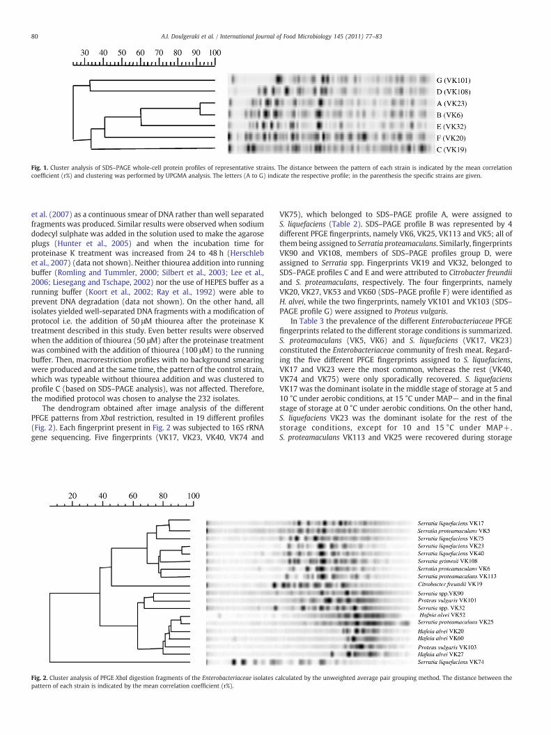
The dendrogram obtained after image analysis of the different PFGE patterns from
XbaI restriction, resulted in 19 different profiles (Figure 3.8). Each fingerprint present in
Figure 3.8 was subjected to 16S rRNA gene sequencing. Five fingerprints (VK17, VK23,
VK40, VK74 and VK75), which belonged to SDS – PAGE profile A, were assigned to
Serratia liquefaciens (Table 3.10). SDS-PAGE profile B was represented by 4 different
PFGE fingerprints, namely VK6, VK25, VK113 and VK5; all of them being assigned to
Serratia proteamaculans. Similarly, fingerprints VK90 and VK108, members of SDS-PAGE
profiles group D, were assigned to Serratia spp.. Fingerprints VK19 and VK32, belonged to
SDS-PAGE profiles C and E, were attributed to C. freundii and S. proteamaculans,
respectively. Four fingerprints, namely VK20, VK27, VK53 and VK60 (SDS-PAGE profile
71
F) were identified as H. alvei, while two fingerprints, namely VK101 and VK103 (SDS-
Tenover, F. C., Arbeit, R. D., Goering, R. V., Mickelsen, P. A., Murray, B. E., Persing,
D. H. & Swaminathan, B. (1995). Interpreting chromosomal DNA restriction patterns
produced by pulsed-field gel electrophoresis: Criteria for bacterial strain typing. J Clin
Microbiol 33, 2233-2239.
154
Teske, A., Wawer, C., Muyzer, G. & Ramsing, N. B. (1996). Distribution of sulfate-
reducing bacteria in a stratified fjord (Mariager Fjord, Denmark) as evaluated by most-
probablenumber counts and denaturing gradient gel electrophoresis of PCR-amplified
ribosomal DNA fragments. Appl Environ Microbiol 62, 1405-1415.
Thomas, L. V., Wimpenny, W. T. & Barker, G. C. (1997). Spatial interactions between
subsurface bacteria colonies in a model system: a territory model describing the inhibition of
Listeria monocytogenes by a nisin-producing lactic acid bacterium. Microbiol 143, 2575-
2582.
Tompkin, R. B. (1973). Refrigeration temperature as an environmental factor influencing the
microbial quality of food. A review. J Food Techn 27, 54-58.
Torriani, S., Felis, G. E. & Dellaglio, F. (2001). Differentiation of Lactobacillus plantarum,
L. pentosus, and L. paraplantarum by recA gene sequence analysis and multiplex PCR assay
with recA gene-derived primers. Appl Environ Microbiol 67, 3450-3454.
Tryfinopoulou, P., Drosinos, E. H. & Nychas, G-J. E. (2001). Performance of
Pseudomonas CFC-selective medium in the fish storage ecosystems. J Microbiol Meth 47,
243-247.
Tryfinopoulou, P., Tsakalidou, E. & Nychas, G-J. E. (2002). Characterization of
Pseudomonas spp. Associated with Spoilage of Gilt-Head Sea Bream Stored under Various
Conditions. Appl Environ Microbiol 68, 65-72.
Tsigarida, E., Skandamis, P. N. & Nychas, G-J. E. (2000). Behaviour of Listeria
monocytogenes and autochthonous flora on meat stored under aerobic, vacuum and modified
atmosphere packaging conditions with or without the presence of oregano essential oil at
5°C. J Appl Microbiol 89, 901-909.
155
Urso, R., Comi, G. & Cocolin, L. (2006). Ecology of lactic acid bacteria in Italian
fermented sausages: Isolation, identification and molecular characterization. Syst Appl
Microbiol 29, 671-680.
Vasilopoulos, C., De Maere, H., De Mey, E., Paelinck, H., De Vuyst, L. & Leroy, F.
(2010). Technology-induced selection towards the spoilage microbiota of artisan-type cooked
ham packed under modified atmosphere. Food Microbiol 27, 77-84.
Vandamme, P., Pot, B., Gillis, M., De Vos, P., Kersters, K. & Swings, J. (1996).
Polyphasic taxonomy, a consensus approach to bacterial systematic. Microbiol reviews 60,
407-438.
Vauterin, L., Hoste, B., Yang, P., Alvarez, A., Kersters, K. & Swings, J. (1993).
Taxonomy of the genus Xanthomonas. In Xanthomonas, pp. 157-192. Edited by J. Swings,
E. L. Civerolo, London:Chapman and Hall, Ltd.
Ventura, M., Meylan, V. & Zink, R. (2003). Identification and tracing of Bifidobacterium
species by use of enterobacterial repetitive intergenic consensus sequences. Appl Environ
Microbiol 69, 4296-4301.
Versalovic, J., Koeuth, T. & Lupski, J. R. (1991). Distribution of repetitive DNA
sequences in eubacteria and application to fingerprinting of bacterial genomes. Nucleic Acids
Res 19, 6823-6931.
Villani, F., Casaburi, A., Pennacchia, C., Filosa, L., Russo, F. & Ercolini, D. (2007). The
microbial ecology of the Soppressata of Vallo di Diano, a traditional dry fermented sausage
from southern Italy, and in vitro and in situ selection of autochthonous starter cultures. Appl
Environ Microbiol 73, 5453-5463.
156
von Holy, A., Holzapfel, W. H. & Dykes, G. A. (1992). Bacterial populations associated
with vienna sausage packaging. Food Microbiol 9, 45-53.
Ward, D. M., Weller, R. & Bateson, M. M. (1990). 16S rRNA sequences reveal numerous
uncultured microorganisms in a natural community. Nature 345, 63-65.
Warriss, P. D. (2000). Meat Hygiene, Spoilage and Preservation. In Meat science: an
introductory text, pp. 182-208. Edited by P. D. Warriss, Wallingford: CABI Publishing.
Welsh, J. & McClelland, M. (1992). PCR-amplified length polymorphisms in tRNA
intergenic spacers for categorizing staphylococci. Mol Microbiol 6, 1673-1680.
Wildmer, F., Seidler, R. J., Gillevet, P. M., Watrud, L. S. & Di Giovanni, G. D. (1998). A
highly selective PCR protocol for detecting 16S rRNA genes of the genus Pseudomonas
(sensu stricto) in environmental samples. Appl Environ Microbiol 64, 2545-2553.
Yamamoto, S., Kasai, H., Arnold, D. L., Jackson, R. W., Viavian, A. & Harayama, S.
(2000). Phylogeny of the genus Pseudomonas: intrageneric structure reconstructed from the
nucleotide sequences of gyrB and rpoD genes. Microbiol 146, 2385-2394.
Yeung, P. S. M., Kitts, C. L., Cano, R., Tong, P. S & Sanders, M. E. (2004). Application
of genotyping and phenotyping analyses to commercial probiotic strain identity and
relatedness. J Appl Microbiol 97, 1095-1104.
Yost, C. K. & Nattress, F. M. (2002). Molecular typing techniques to characterize the
development of a lactic acid bacteria community on vacuum-packaged beef. Int J Food
Microbiol 72, 97-105.
Young, L. L., Reviere, R. D. & Cole, A. B. (1988). Fresh meat red meat: a place to apply
modified atmosphere. Food Techn 42, 65-69.
157
Zoetendal, E. G., Akkermans, A. D. L. & de Vos, W. M. (1998). Temperature gradient gel
electrophoresis analysis of 16S rRNA from human faecal samples reveals stable and host-
specific communities of active bacteria. Appl Environ Microbiol 64, 3854-3859.
Appendix I
The following manuscript has been published in Food Microbiology
Lactic acid bacteria population dynamics during minced beef storageunder aerobic or modified atmosphere packaging conditions
Agapi I. Doulgeraki a,c, Spiros Paramithiotis b, Dafni Maria Kagkli a, George-John E. Nychas a,*
aDepartment of Food Science, Technology and Human Nutrition, Laboratory of Microbiology and Biotechnology of Foods, Agricultural University of Athens,Iera Odos 75, Athens 11855, GreecebDepartment of Food Science, Technology and Human Nutrition, Laboratory of Food Quality Control and Hygiene, Agricultural University of Athens,Iera Odos 75, Athens 11855, GreececApplied Mycology Group, Cranfield Health, Cranfield University, College Road, Cranfield, Bedfordshire, MK43 0AL, UK
a r t i c l e i n f o
Article history:Received 13 November 2009Received in revised form27 June 2010Accepted 6 July 2010Available online 14 July 2010
A total of 266 lactic acid bacteria (LAB) have been isolated from minced beef stored at 0, 5, 10 and 15 �Caerobically and under modified atmosphere packaging consisting of 40% CO2e30% O2e30% N2 in thepresence MAP (þ) and absence MAP (�) of oregano essential oil. Sequencing of their 16S rRNA genealong with presence of the katA gene demonstrated dominance of the LAB microbiota by Leuconostocspp. during aerobic storage at 5, 10 and 15 �C, as well as during MAP (�) and MAP (þ) storage at 10 and15 �C; Lactobacillus sakei prevailed during aerobic storage at 0 �C, as well as at MAP (�) and MAP (þ)storage at 0 and 5 �C. The sporadic presence of other species such as Leuconostoc mesenteroides, Weisellaviridescens, Lactobacillus casei and Lactobacillus curvatus has also been determined. Pulsed-Field GelElectrophoresis of high molecular weight genomic DNA revealed the dynamics of the isolated LABstrains. Prevalence of Leuconostoc spp. was attributed to one strain only. On the other hand, packagingconditions affected Lb. sakei strain spoilage dynamics.
� 2010 Elsevier Ltd. All rights reserved.
1. Introduction
Food spoilage microbiota has been the subject of several studiesconducted so far; the ones focused on meat and meat productswere based on the identification and/or characterization of thedominantmicrobiota at different storage conditions. The concept of‘succession’ of spoilage-related microbial groups i.e. ephemeral/specific spoilage organisms (E/SSO), was only recently, taken intoconsideration (Ercolini et al., 2006; Chenoll et al., 2007; Nychaset al., 2008).
Lactic acid bacteria (LAB) for instance are considered to be theSpecific Spoilage Organisms (SSO) that contribute to the meatspoilage stored under packaging conditions in which the concen-tration of carbon dioxide is increased (Axelsson, 1998; Holzapfel,1998; Nychas and Skandamis, 2005). Lactobacillus, Leuconostocand Carnobacterium are among the most frequently encounteredgenera on vacuum ormodified atmosphere packagedmeat and playan important role in the spoilage of refrigerated rawmeat (ShawandHarding, 1984; Dainty and Mackey, 1992; Hugas et al., 1993;McMullen and Stiles, 1993; Rovira et al., 1997; Holzapfel, 1998;
Labadie, 1999; Parente et al., 2001; Nychas and Skandamis, 2005).Species of Leuconostoc sp. and Lb. sakei have been associated withthe spoilage of vacuumormodified atmosphere packedmeat storedat chill temperatures (Champomier-Verges et al., 2001; Yost andNattress, 2002; Ercolini et al., 2006). The lack of consistency e.g.why these two species were not always found at the end of storageperiod even if the conditionswere similar can be possibly attributednot only to the limitation of the appliedmethodologies used but alsoto the potential effect of the man imposed preservation system onthe development of the microbial association, e.g. EphemeralSpoilage Organism (Stanbridge and Davies, 1998; Nychas et al.,2008; Vasilopoulos et al., 2010). In this case the word ‘ephemeral’does describe the situation where these specific spoilage bacteriacontribute to meat spoilage for a very short period of time till thenext climax population is established. The identification and char-acterization of these ESOs in raw meat under different storageconditions remains still to be elucidated (Jones, 2004; Ercolini et al.,2006, 2009; Fontana et al., 2006; Vasilopoulos et al., 2010).
Oregano essential oil, as a potential ‘hurdle’, was found to affectthe contribution of spoilage microorganisms to the microbial asso-ciation aswell as to the physicochemical changes of themincedmeat(Skandamis and Nychas, 2001; Burt, 2004). Skandamis and Nychas(2002) reported that the oregano essential oil effect on microbialpopulation, including LAB, on active packaging conditions. Axelsson
0740-0020/$ e see front matter � 2010 Elsevier Ltd. All rights reserved.doi:10.1016/j.fm.2010.07.004
Food Microbiology 27 (2010) 1028e1034
(1998) concluded that the addition of oregano essential oil influencedthe metabolic activity of LAB. More specifically, the initial hetero-fermentativemicrobiotawas substituted by a homofermentative oneat the end of storage. However, despite the antimicrobial action ofessential oil on biota, there is less information about the effect of suchcompounds on the microbial diversity of the LAB isolated frommeatat species and strain level. The only information available relates theessential oil effect on growth of meat spoilage bacteria such as Lb.sakei, Lb. curvatus and Carnobacteriumpiscicola (Ouattara et al.,1997).
The use of conventional phenotypic methods does not alwaysallow efficient characterization of the microbiota at species level(Holzapfel, 1998; Stanbridge and Davies, 1998). On the contrary,molecular identification and characterization tools are far moreconsistent, rapid, reliable and reproducible and can discriminateeven between closely related groups of species, which are other-wise indistinguishable on the basis of their phenotype. Theadvances in molecular techniques are expected to widen theknowledge of spoilage-related bacterial succession during storageof foods (Chenoll et al., 2003; Ercolini et al., 2006). Several molec-ular typing techniques have been developed during the past decadefor the identification and classification of bacteria at strain level.Among them, Pulsed-Field Gel Electrophoresis (PFGE) of DNAfragments resulting from the digestion of whole genomic DNAswith rare-cutting restriction endonucleases has proved to be reli-able for bacterial typing. This method has been used to differentiatemembers of several genera including Lactococcus (Tanskanen et al.,1990), Clostridia (Hielm et al., 1998), Streptomyces (Leblond et al.,1990), probiotic lactobacilli (Yeung et al., 2004), and to comparethe genomic restriction patterns of five Bifidobacterium brevestrains (Bourget et al., 1993). It is considered to be a discriminatingand reproducible method to differentiate strains of intestinalbacteria (O’Sullivan, 1999) and for chromosome size estimation inLb. acidophilus (Roussel et al., 1993; Sanders et al., 1996), Lb. plan-tarum (Daniel, 1995), and other LAB (Tanskanen et al., 1990).Furthermore, PFGE in association with PCR-based methods arecommonly used for strain monitoring (Singh et al., 2009).
The aim of the present study was to systematically monitor themicrobial diversity of LAB, isolated from meat stored at differenttemperatures and under different packaging e.g. aerobic or MAPconditions, at strain level, by using modern molecular tools.
2. Materials and methods
2.1. Sample preparation and storage conditions
Minced beef was purchased from the central market of Athensand prepared according to Argyri et al. (submitted for publication).Briefly, minced beef samples were stored at 0, 5, 10 and 15 �C,aerobically or under modified atmospheres packaging (MAP) con-sisting of 40% CO2e30% O2e30% N2 with MAP (þ) or without MAP(�) the application of volatile compounds of oregano essential oil(2% v/w). The samples were placed on Styrofoam trays; all trayswere performed to allow the diffusion of the volatile compounds ofthe essential oil with both sides of the samples. In the case of thetreated samples (MAPþ), the essential oil was distributed ona whatman paper that was placed on the bottom side of the tray.
2.2. Sampling of the meat
Minced beef was sampled at appropriate time intervals,depending on storage temperature; the incubation lasted 650, 482,386 and 220 h at 0, 5, 10 and 15 �C, respectively and all sampleswere analysed in dublicate. A detailed description of the method-ology employed for the enumeration of the total viable counts,Pseudomonas spp., Br. thermosphacta, LAB, Enterobacteriaceae,
yeasts and molds in this work is presented elsewhere (Argyri et al.submitted for publication). Briefly, LAB counts were determined onMRS agar (Biolife, Italiana S.r.l., Milano, Italy) (pH ¼ 5.8) overlaidwith the same medium and incubated at 30 �C for 72 h. LAB wereisolated from the highest dilution from three different time points(initial, middle and final stage of storage) for further analysis; 10%of the colonies (6e10 colonies) derived from plate culture of thehighest sample dilution. They were randomly selected and purifiedby successive subculture on MRS agar at 30 �C. Gram positive,catalase and oxidase negative isolates were stored at�80 �C inMRSbroth (Biolife, Milano, Italy) supplemented with 20% (v/v) glycerol(Merck, Darmstadt, Germany) until further use. Before experi-mental use each strain was grown twice in MRS broth at 30 �C for24 and 16 h respectively. Purity of the culture was always checkedon MRS agar plates before use.
2.3. Pulsed-field Gel Electrophoresis (PFGE)
PFGE was performed according to Kagkli et al. (2007). Briefly,cells were harvested by centrifugation at 10,000�g for 5 min andwashed with 10 mM TriseHCl (pH 7.6) containing 1 M NaCl;resuspended in 100 mL of the same solution, heated at 37 �C for10 min and mixed with an equal volume of 2% (w/v) low melting-point agarose (Bio-Rad, Hercules, CA, USA) in 0.125 M EDTA pH 7.6before letting them to solidify in moulds (Bio-Rad). The cells werelysed in situ in a solution containing 10 mg mL�1 of lysozyme in ECbuffer (6 mM TriseHCl, 1 M NaCl, 100 mM EDTA, 1% (w/v) Sarkosyl,pH 7.6) for 16 h at 37 �C. The lytic treatment was repeated with thesame solution containing 2 U mL�1 mutanolysin. After treatmentwith proteinase K (0.5MEDTA containing 1% sarkosyl, pH 8) for 24 hat 55 �C, the agarose blocks were washed twice for 1 h with 1 mMphenylmethylsulfonyl fluoride (PMSF) in 10 mM TriseHCl contain-ing 1 mM EDTA, (pH 8.0) at 37 �C and then stored at 4 �C in 10 mMTriseHCl containing 100 mM EDTA (pH 8.0) until further use.
The agarose blocks were cut with sterile coverslips and slices(1e2 mm thick) of the blocks were washed three times at roomtemperature in 10 mM TriseHCl containing 0.1 mM EDTA (pH 8.0)for 30 min with gentle agitation. The restriction enzymes ApaI andSmaI (10 U) (New England Biolabs, Ipswich, MA, USA) were initiallyselected to digest the slices of a limited number of strains. Theenzyme that resulted in the production of clearer and sharper PFGEdigestion profile was used for the digestion of all isolates. Diges-tions were performed according to the recommendations of themanufacturer.
Following digestion, slices were loaded into wells of a 1% PFGEgrade agarose gel (Bio-Rad) and the gel was run in 0.5 mMTriseBorate buffer (45 mM TriseHCl, 45 mM Boric acid, 1 mMEDTA) using a CHEF-DRII PFGE apparatus and cooling module (Bio-Rad) at 6 V cm�1 for 16 h, with a pulse time ramped from 1 to 10 s.Gels were then stained with ethidium bromide (0.5 mg ml�1) inwater for 1 h and destained for 2 h before being photographedusing a GelDoc system (Bio-Rad). Conversion, normalization andfurther analysis were performed using the Pearson coefficient andUPGMA cluster analysis with Gel compare software, version 4.0(Applied Maths, Sint-Martens-Latem, Belgium; kindly provided byE. Tsakalidou, Dairy Laboratory, Agricultural University of Athens).
2.4. DNA extraction and species identification
DNA was extracted according to the protocol described by themanufacturer of GenElute Bacterial Genomic DNA Kit (Sigma,Chemical Co., St. Louis, Mo. USA). Representative number of isolatesper distinct PFGE cluster were selected and subjected to speciesidentification by sequencing the V1eV3 variable region of the 16SrRNA gene as described previously (Paramithiotis et al., 2008). PCR
products were purified using the QIAquick� PCR Purification Kit(Qiagen, Hilden, Germany) according to the manufacturer’sinstructions and directly sequenced with an ABI 3730 XL automaticDNA sequencer by Macrogen (http://www.macrogen.com). Theresults were aligned with those in GenBank using the BLASTNprogram in order to determine their closest known relatives ofthe partial 16S rRNA gene sequence (Altschul et al., 1997). TheGenBank/EMBL/DDBJ accession numbers for the 16S rRNA genesequences are GU998850 to GU998881 (Table 2).
2.5. Detection of the heme-dependent catalase (katA) gene
All isolates were screened by PCR for the presence of the katAgene, encoding heme-dependent catalase (Knauf et al., 1992; Hertelet al., 1998). For this purpose the specific primers 702-F (50-AATTGCCTTCTTCCGTGTA-30, position 551e536) and 310-R (50-AGTTGCGCACAATTATTTTC-30, position 127e139) were used.
3. Results
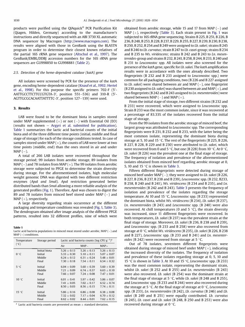
LAB were found to be the dominant biota in samples storedunder MAP supplemented (þ) or not (�) with Essential Oil (EO)(results not shown e Argyri et al. submitted for publication).Table 1 summarizes the lactic acid bacterial counts of the initialbiota and of the three different time points (initial, middle and finalstage of storage) for each of the storage conditions tested. In case ofsamples stored underMAP (þ), the counts of LABwere lower at twotime points (middle, end) than the ones stored in air and underMAP (�).
A total of 266 LAB isolates were recovered throughout thestorage period; 99 isolates from aerobic storage, 89 isolates fromMAP (�) and 78 isolates fromMAP (þ). The 99 isolates from aerobicstorage were subjected to PFGE to determine the strain diversityduring storage. For the aforementioned isolates, high molecularweight genomic DNA was digested with two different restrictionenzymes (ApaI and SmaI). ApaI restriction generated betterdistributed bands than SmaI allowing amore reliable analysis of thegenerated profiles (Fig. 1). Therefore, ApaI was chosen to digest the89 and 78 isolates from minced beef stored under MAP (�) andMAP (þ), respectively.
A large diversity regarding strain occurrence at the differentpackaging and temperature conditions was revealed (Fig. 1, Table 3).The dendrogram obtained after image analysis of the different PFGEpatterns, resulted into 32 different profiles, nine of which were
obtained from aerobic storage, while 15 and 17 from MAP (�) andMAP (þ), respectively (Table 3). Each strain present in Fig. 1 wassubjected to 16S rRNA gene sequencing. Strains B 225, B 251, B 226, B236, B 248, B 253, B 228, B 237, B 229, B 255, B 227, B 239, B 230, B 238,B 250, B 252, B 254 and B 249were assigned to Lb. sakei; strains B 245and B 246 to Lb. curvatus; strain B 247 to Lb. casei-group; strains B 234and B 235 to Ws. viridescens; strains B 242 and B 243 to Ln. mesen-teroides-group and strains B 232, B 241, B 258, B 244, B 233, B 240 andB 231 to Leuconostoc spp. All isolates were also screened for thepresenceof the katAgene, specific for Lb. sakei. The katAamplificationresults were in accordance with the ones already described. Twofingerprints (B 232 and B 233 assigned to Leuconostoc spp.) werecommon for all packaging conditions, two (B 226 and B 227 assignedto Lb. sakei) were shared between air and MAP (�), one fingerprint(B 230 assigned to Lb. sakei)was sharedbetween air andMAP (þ), andtwofingerprints (B 242 andB 243 assigned to Ln.mesenteroides)wereshared between MAP (�) and MAP (þ).
From the initial stage of storage, two different strains (B 232 andB 233) were recovered, which were assigned to Leuconostoc spp.Strain B 233was themost common isolate, since it was recovered ata percentage of 83.33% of the isolates recovered from the initialstage of storage.
From the 99 isolates from the aerobic storage of minced beef, thelargest group was attributed to Leuconostoc spp. The correspondingfingerprints were B 231, B 232 and B 233, with the latter being themost common isolate, representing the dominant biota duringstorage at 5, 10 and 15 �C. The rest of the fingerprints (B 225, B 226,B 227, B 228, B 229 and B 230) were attributed to Lb. sakei, whichwere recovered from 0 and 5 �C, but one (B 228) from 10 �C. At 0 �C,Lb. sakei (B 226) was the prevalent one at the final stage of storage.The frequency of isolation and prevalence of the aforementionedisolates obtained from minced beef regarding aerobic storage at 0,5, 10 and 15 �C is shown in Table 3.
Fifteen different fingerprints were detected during storage ofminced beef under MAP (�); they were assigned to Lb. sakei (B 226,B 227, B 236, B 237, B 238 and B 239), Leuconostoc spp. (B 232, B233,B 258, B 240 and B 241), Ws. viridescens (B 234 and B 235), and Ln.mesenteroides (B 242 and B 243). Table 3 presents the frequency ofisolation and prevalence of the isolates regarding the storagetemperature. At 10 and 15 �C, Leuconostoc spp. (B 233) representedthe dominant biota, whilstWs. viridescens (B 234), Lb. sakei (B 237),Ln. mesenteroides (B 243) and Leuconostoc spp. (B 240) were alsorecovered. At chill temperatures (0 and 5 �C), the strain diversitywas increased, since 11 different fingerprints were recovered. Inboth temperatures, Lb. sakei (B 237) was the prevalent strain at thefinal stage of storage. Moreover, Lb. sakei (B 236, B 238 and B 239),and Leuconostoc spp. (B 233 and B 258) were also recovered fromstorage at 0 �C, whilstWs. viridescens (B 235), Lb. sakei (B 226, B 238and B 227), Leuconostoc spp. (B 233 and B 241) and Ln. mesenter-oides (B 242) were recovered from storage at 5 �C.
Out of 78 isolates, seventeen different fingerprints wereobtained during storage of minced beef under MAP (þ), indicatingthe increased diversity of the isolates. The frequency of isolationand prevalence of these isolates regarding storage at 0, 5, 10 and15 �C is shown in Table 3. At 10 and 15 �C, Leuconostoc spp. (B 233)was the most common isolate, representing the dominant strain,whilst Lb. sakei (B 252 and B 255) and Ln. mesenteroides (B 243)were also recovered. Lb. sakei (B 254) was the dominant strain atthe final stage of storage at 5 �C, while Lb. sakei (B 248 and B 255),and Leuconostoc spp. (B 233 and B 244) were also recovered duringthe storage at 5 �C. At the final stage of storage at 0 �C, Leuconostocspp. (B 233), Ln. mesenteroides (B 242), Lb. curvatus (B 246) and Lb.sakei (B 249 and B 251) were equally contributed. Lb. curvatus(B 245), Lb. casei and Lb. sakei (B 248, B 250 and B 253) were alsorecovered during storage at 0 �C.
Table 1Lactic acid bacteria populations in minced meat stored under aerobic, MAP (�) andMAP (þ) conditions.
Temperature(�C)
Storage period Lactic acid bacteria counts (log CFU g�1)a
a Lactic acid bacteria counts are presented as mean � standard deviation.
A.I. Doulgeraki et al. / Food Microbiology 27 (2010) 1028e10341030
4. Discussion
Spoilage and spoilage progress of meat and meat products havebeen the subject of several studies conducted so far (Borch et al.,1996; Stanbridge and Davies, 1998; Labadie, 1999; Skandamis andNychas, 2002; Nychas and Skandamis, 2005; Nychas et al., 2008).Nevertheless, meat spoilage has only been associated with thephysicochemical and microbiological analysis of the bacterial loadsignoring the spoilage potential of a specific bacterial species orstrain (Skandamis and Nychas, 2002). Only recently did researchtook into consideration the specific characteristics of the spoilagemicrobiota of the meat products and its contribution to the dete-rioration of the product (Cocolin et al., 2004b; Rantsiou et al., 2005;Ercolini et al., 2006, 2009; Vasilopoulos et al., 2010).
The present study focused on the evaluation of the microbialdiversity of LAB isolated from minced beef stored under different
storage conditions at strain level. Storage conditions had an impor-tant effect on the diversity of themicrobial population, since differentstrains were recovered during the storage of meat under differentconditions. These findings strengthen the opinion that the storagetemperature and the modified atmosphere packaging affect thespoilage potential of LAB (Stanbridge and Davies,1998; Ercolini et al.,2006, 2009). Moreover, this observation can be explained by the factthat different metabolic activities occur when different species/strains are present and when meat is stored under specific condi-tions. Not all species belonging to the same bacterial group, e.g. LAB,necessarily grow at the same temperature. It might therefore bemisleading the fact that selective media only are used for the deter-mination of the spoilage biota. Further characterization of the isolatesgrown on the selective plates should be demanded if a better insightand understanding of the phenomenon is required. This is in accor-dance with the observations of Ercolini et al. (2006) who reported
Fig. 1. Cluster analysis of PFGE ApaI digestion fragments of the lactic acid bacteria isolates calculated by the unweighted average pair grouping method. The distance between thepattern of each strain is indicated by the mean correlation coefficient (r%). Strain identity is indicated by the lower and upper case letters.
that different species/strains were isolated from beef althoughsimilar countswere determined. Ercolini et al. (2006) alsomentionedthat the viable counts alonemay not be enough to highlight the shiftsof the bacterial communities depending on the environmentalchanges and species that are actually involved in meat spoilage.
Among the species listed in Table 2, several meat associatedones were identified. Holzapfel (1998) reported that more rarelyLb. plantarum and Lb. casei are associated with meat systems and inlower frequency and numbers than Lb. curvatus and Lb. sakei; thepresence of Ws. viridescens in raw meat has been also described.Moreover, Lb. curvatus, Lb. sakei and Leuconostoc spp. have beenfound to indicate a mixture community of vacuum packed (vp) beef(Yost and Nattress, 2002). Leuconostocs have been identified aspredominant organisms in beef stored under vp/MAP (Stanbridgeand Davies, 1998; Yost and Nattress, 2002) and their presence inthe initial mesophilic bacterial microbiota is very frequent (Borchet al., 1996). Lb. sakei has been associated with fresh meat(Champomier-Verges et al., 2001) as well as spoilage of a variety ofmeat products both under vacuum and modified atmospherepackaging (Ercolini et al., 2006, 2009) and it is known to be amongthe most psychrotrophic lactobacilli. It has also been found to bethe dominant spoilage LAB during storage at chill temperatures(Ercolini et al., 2006; Chenoll et al., 2007).
PFGE has also provided important information in relation to thestrain distribution of the LAB population which would have notbeen acquired if strain typing had not been performed. Within theLAB population of the present study, Leuconostoc spp. and Lb. sakeiwere identified as significant members of the microbiota at abuseand chill temperatures, respectively. More accurately, Leuconostocspp. (B 233) that was initially present at high levels, dominatedeventually the microbiota of the minced beef stored at abuse
temperatures at all packaging conditions. Although, it was persis-tent throughout storage at chill temperatures, Lb. sakei strainsdominated the LAB population only at the final stage of storage.However, some degree of microbial variability was detected at thefinal stage of storage of meat at chill temperatures, since differentLb. sakei strains were the most prevalent ones at the differentpackaging conditions. Indeed, Lb. sakei (B 226), (B 237) and (B 245)dominated the LAB population at 0 �C under aerobic conditions, at0 and 5 �C underMAP (�) and at 5 �C underMAP (þ). This finding isof great importance since it shows the intraspecies variability of Lb.sakei and the ability of certain strains to adapt to the differentstorage conditions outgrowing the other.
Dominance of Leuconostoc spp. at relatively higher temperaturescan be partially attributed to the favourable environmental condi-tions and partially to the shorter generation time (Harris, 1998),both of which enabled it to outgrow Lb. sakei strains which wereindeed detected as a secondary microbiota. On the other hand,dominance of Lb. sakei strains at chill temperatures can be attrib-uted partly to its psychrotrophic nature.
From the different LAB detected throughout the storage underMAP (�) and MAP (þ), a wide range of strains were sporadicallypresent, especially at chill temperatures. This finding indicates thatmodified atmosphere packaging resulted in a development ofa totally different spoilage ecosystem. It has been previouslyreported (Jay, 2000), that during storage of meat under MAP, theinitial heterofermentative microbiota was substituted by a homo-fermentative one at the end of storage. Moreover, the MAP and thepresumed activity of oregano essential oil against hetero-fermentative LAB species (Axelsson, 1998) seem to have providedthe latter with an ecological advantage over leuconostocs.
The findings of the present study were based on the culture-dependent approach, most frequently applied when storage studiesare performed. Selective media have been used for isolation andsubsequent characterization of the microbiota; stressed or injuredcells might not have managed to recover and grow, resulting intheir non isolation from the plates and giving therefore theimpression that they were absence from the system under inves-tigation. A bias is therefore inserted which could have an effect onthe description of the microbial community present. Except fromthe factors mentioned above, random selection of colonies isrequired to have a representative sample. This is not alwayspossible because it depends on the person performing the task andit is therefore not objective.
A culture-independent approach could have been an alternativeto the plates used for the characterization of the different micro-biota (Cocolin et al., 2004a,b). Nevertheless, even this approach hasdrawbacks which lie in the fact that species have to be above thedetection limit (104 cfu g�1) and very frequently, the dominantspecies prevents evidence of the less abundant ones. Primer affinityto the target has also an effect on the amplification and therefore onthe species identified. In order to clarify possible discrepanciesbetween culture-dependent and independent methods, and toevaluate whether these differences would give a different overviewof the ecology of the meat stored at the conditions mentioned,a similar study could be performed applying both approaches in thefuture. This investigation lies beyond the scope of the presentstudy, which was actually focused on elucidating the effect of thedifferent storage conditions with or without the presence ofessential oil on the dynamics of LAB strains.
The present study did provide an insight of the populationdynamics of LAB strains in relation to the temperature and thepackaging conditions. It has been clearly demonstrated that certainspecies and/or strains are present or dominant only under certainconditions. This finding is extremely important since studies con-ducted so far had only taken into consideration the microbiological
Table 2Identity of isolates obtained from minced beef.
Numberof isolates
Closest relative Selected strainsequenceda
AccessionNumber
1 Lactobacillus sakei B 225 GU9988566 Lb. sakei B 226 GU9988772 Lb. sakei B 227 GU9988571 Lb. sakei B 228 GU9988501 Lb. sakei B 229 GU9988512 Lb. sakei B 230 GU9988521 Leuconostoc spp. B 231 GU9988539 Leuconostoc spp. B 232 GU998854205 Leuconostoc spp. B 233 GU9988551 Weissella viridescens B 234 GU9988582 Ws. viridescens B 235 GU9988591 Lb. sakei B 236 GU9988605 Lb. sakei B 237 GU9988613 Lb.sakei B 238 GU9988622 Lb. sakei B 239 GU9988631 Leuconostoc spp. B 258 GU9988641 Leuconostoc spp. B240 GU9988651 Leuconostoc spp. B 241 GU9988662 Ln. mesenteroides B 242 GU9988672 Ln. mesenteroides B 243 GU9988681 Leuconostoc spp. B 244 GU9988691 Lb. curvatus B 245 GU9988701 Lb. curvatus B 246 GU9988711 Lb. casei B 247 GU9988725 Lb. sakei B 248 GU9988731 Lb. sakei B 249 GU9988741 Lb. sakei B 250 GU9988751 Lb. sakei B 251 GU9988761 Lb. sakei B 252 GU9988781 Lb. sakei B 253 GU9988792 Lb. sakei B 254 GU9988801 Lb. sakei B 255 GU998881
a Code of different PFGE patterns of Fig. 1.
A.I. Doulgeraki et al. / Food Microbiology 27 (2010) 1028e10341032
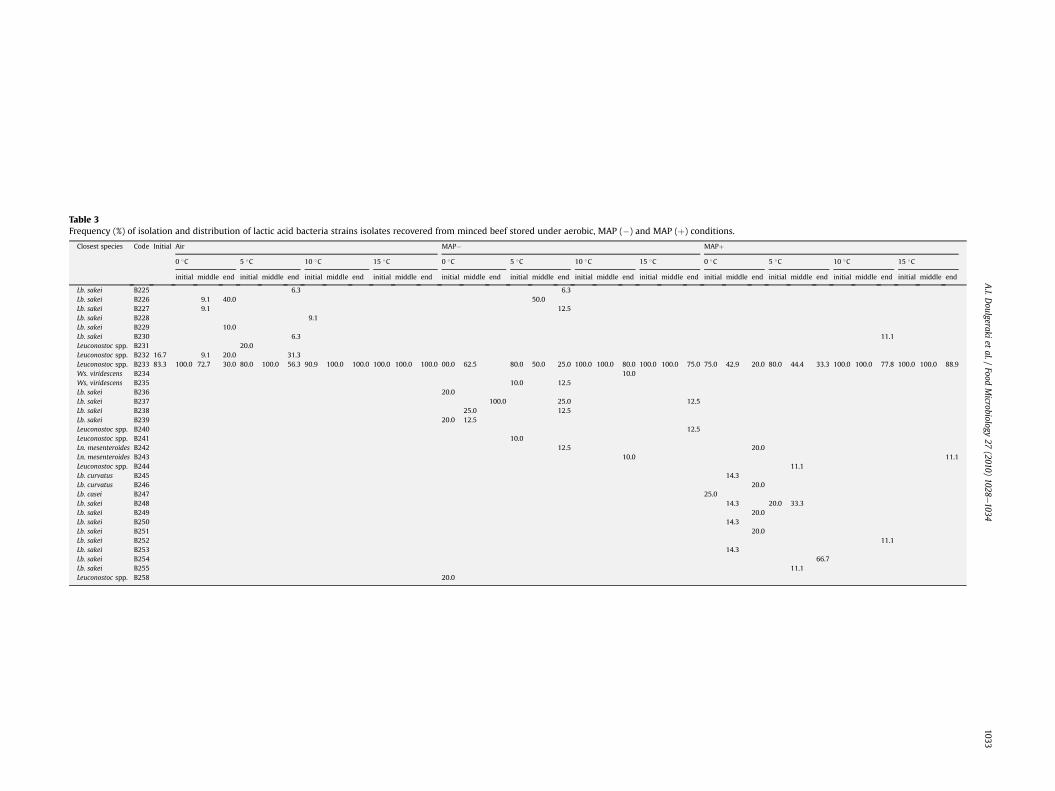
Table 3Frequency (%) of isolation and distribution of lactic acid bacteria strains isolates recovered from minced beef stored under aerobic, MAP (�) and MAP (þ) conditions.
initial middle end initial middle end initial middle end initial middle end initial middle end initial middle end initial middle end initial middle end initial middle end initial middle end initial middle end initial middle end
counts as an indication of the spoilage process, and had ignored thepossibility that different species or strains would prevail underdifferent storage and/or packaging conditions. The qualitativeinformation derived from the microbiological analyses and thecharacterization of the species or even the strains present were notevaluated previously. It has been shown that storage temperaturecombined with packaging conditions induced the selectivity of thespoilage LAB microbiota. Moreover, the microbiota recovered fromthe initial stage of storage was markedly different from that of thefinal stage of storage at chill temperatures. The above observationsare of great importance and, to our opinion, fundamental inunderstanding the spoilage process and in explaining the presenceof different products or by-products that occur during the differentdynamic storage conditions (Skandamis and Nychas, 2002).
Acknowledgements
This work was partly supported by 2 EU Projects ProSafeBeef(Ref. Food-CT-2006-36241) and SYMBIOSIS-EU (No. 211638 Contract)within the 6th and 7th Framework Programme respectively. Theinformation in this document reflects only the authors’ views, andthe European Community is not liable for any use that may be madeof the information contained therein.
References
Altschul, S.F., Madden, T.L., Schaffer, A.A., Zhang, J., Zhang, Z., Miller, W., Lipman, D.J.,1997. Gapped BLAST and PSI-BLAST: a new generation of protein databasesearch programs. Nucleic Acids Res. 25, 3389e3402.
Argyri, A.A., Doulgeraki, A.I., Blana, V.A., Panagou, E.Z., Nychas G J.E., Potential ofa simple HPLC based approach to quantify spoilage of minced beef stored atdifferent temperatures and packaging systems. Revision FM-S-09e00571R1,submitted for publication.
Axelsson, L.T., 1998. Lactic acid bacteria: classification and physiology. In:Salminen, S., Von Wright, A. (Eds.), Lactic Acid Bacteria Microbiology andFunctional Aspects. Marcel Dekker, New York, pp. 1e72.
Borch, E., Kant-Muemans, M.-L., Blixt, Y., 1996. Bacterial spoilage of meat and curedmeat products. Int. J. Food Microbiol. 33, 103e120.
Bourget, N., Simonet, J.M., Decaris, B., 1993. Analysis of the genome of the fiveBifidobacterium breve strains: plasmid content, pulsed-field gel electrophoresisgenome size estimation and rrn loci number. FEMS Microbiol. Lett. 110, 11e20.
Burt, S., 2004. Essential oils: their antibacterial properties and potential applica-tions in foodsda review. Int. J. Food Microbiol. 94, 223e253.
Champomier-Verges, M.-C., Chaillou, S., Cornet, M., Zagorec, M., 2001. Lactobacillussakei: recent developments and future prospects. Res. Microbiol. 152, 839e848.
Chenoll, E., Macian, M.C., Aznar, R., 2003. Identification of Carnobacterium, Lacto-bacillus, leuconostoc and Pediococcus by rDNA-based techniques. System. Appl.Microbiol. 26, 546e556.
Chenoll, E., Macian, M.C., Elizaquivel, P., Aznar, R., 2007. Lactic acid bacteria asso-ciated with vacuum packed cooked meat product spoilage: population analysisby rDNA based methods. J. Appl. Microbiol. 102, 498e508.
Cocolin, L., Innocente, N., Biasutti, M., Comi, G., 2004a. The late blowing in cheese:a new molecular approach based on PCR and DGGE to study the microbialecology of the alteration process. Int. J. Food Microbiol. 90, 83e91.
Cocolin, L., Rantsiou, K., Iacumin, L., Urso, R., Cantoni, C., Comi, G., 2004b. Study ofthe ecology of fresh sausages and characterization of populations of lactic acidbacteria by molecular methods. Appl. Environ. Microbiol. 70, 1883e1894.
Dainty, R.H., Mackey, B.M., 1992. The relationship between the phenotypic prop-erties of bacteria from chill-stored meat and spoilage processes. J. Appl.Bacterial. Symp. Suppl. 73, 103e114.
Daniel, P., 1995. Sizing of the Lactobacillus plantarum genome and other lactic acidbacteria species by transverse alternating field electrophoresis. Curr. Microbiol.30, 243e246.
Ercolini, D., Russo, F., Torrieri, E., Masi, P., Villani, F., 2006. Changes in the spoilage-related microbiota of beef during refrigerated storage under different packagingconditions. Appl. Environ. Microbiol. 72, 4663e4671.
Ercolini, D., Russo, F., Nasi, A., Ferranti, P., Villani, F., 2009. Mesophilic and psy-chrotrophic bacteria from meat and their spoilage potential in vitro and in beef.Appl. Environ. Microbiol. 75, 1990e2001.
Fontana, C., Cocconcelli, P.S., Vignolo, G., 2006. Direct molecular approach tomonitoring bacterial colonization on vacuum-packaged beef. Appl. Environ.Microbiol. 72, 5618e5622.
Harris, L.J., 1998. The microbiology of vegetable fermentations. In: Wood, B.J.B. (Ed.),Microbiology of Fermented Foods. Blackie Academic Professional, London, pp.45e72.
Hertel, C., Schmidt, G., Fischer, M., Oellers, K., Hammes, W.P., 1998. Oxygen-dependent regulation of the expression of the catalase gene katA of Lactoba-cillus sakei LTH677. Appl. Environ. Microbiol. 64, 1359e1365.
Hielm, S., Bjorkroth, J., Hyytia, E., Korkeala, H., 1998. Genomic analysis of Clostridiumbotulinum group II by pulsed-field gel electrophoresis. Appl. Environ. Microbiol.64, 703e708.
Holzapfel, W.H., 1998. The Gram-positive bacteria associated with meat and meatproducts. In: Board, R.G., Davies, A.R. (Eds.), The Microbiology of Meat andPoultry. Blackie Academic and Professional, London, U.K, pp. 35e84.
Hugas, M., Garriga, M., Aymerich, T., Monfort, J.M., 1993. Biochemical characterizationof lactobacilli from dry fermented sausages. Int. J. Food Microbiol. 18, 107e113.
Jay, J.M., 2000. Modern Food Microbiology, sixth ed. An Aspen Publication, Gai-thersburg, Maryland, pp 288.
Jones, R.J., 2004. Observations on the succession dynamics of lactic acid bacteriapopulations in chill-stored vacuum packaged beef. Int. J. Food Microbiol. 90,273e282.
Kagkli, D.-M., Vancanneyt, M., Hill, C., Vandamme, P., Cogan, T.M., 2007. Entero-coccus and Lactobacillus contamination of raw milk in a farm dairy environ-ment. Int. J. Food Microbiol. 114, 243e251.
Knauf, H.J., Vogel, R.F., Hammes, W.P., 1992. Cloning, sequence, and phenotypicexpression of katA, which encodes the catalase of Lactobacillus sakei LTH677.Appl. Environ. Microbiol. 58, 832e839.
Labadie, J., 1999. Consequences of packaging on bacterial growth. Meat is anecological niche. Meat Sci. 52, 299e305.
Leblond, P., Francou, F.X., Simonet, J.-M., Decaris, B., 1990. Pulsed-field gel electro-phoresis analysis of the genome of Streptomyces ambofaciens strains. FEMSMicrobiol. Lett. 72, 79e88.
McMullen, L., Stiles, M.E., 1993. Microbial ecology of fresh pork stored undermodified atmosphere at e 1, 4.4 and 10 �C. Int. J. Food Microbiol. 18, le14.
Nychas, G.-J.E., Skandamis, P., 2005. Fresh meat spoilage and modified atmospherepackaging (MAP). In: Sofos, J.N. (Ed.), Improving the Safety of Fresh Meat. CRC/Woodhead Publishing Limited, Cambridge, UK, pp. 461e502.
O’Sullivan, D.J., 1999. Methods for analysis of the intestinal microflora. In:Tannock, G.W. (Ed.), Probiotics. A Critical Review. Horizon Scientific Press,Norfolk, UK., pp. 23e44.
Ouattara, B., Simard, R.E., Holley, R.A., Piettte, G.J.P., Begin, A., 1997. Antimicrobialactivity of selected fatty acids and essential oils against six meat spoilageorganisms. Int. J. Food Microbiol. 37, 155e162.
Paramithiotis, S., Kagkli, D.-M., Blana, V.A., Nychas, G.-J.E., Drosinos, E.H., 2008.Identification and characterization of Enterococcus spp. in Greek spontaneoussausage fermentation. J. Food Prot. 71, 1244e1247.
Parente, E., Grieco, S., Crudele, M.A., 2001. Phenotypic diversity of lactic acidbacteria isolated from fermented sausages produced in Basilicata (SouthernItaly). J. Appl. Microbiol. 90, 943e952.
Rantsiou, K., Urso, R., Iacumin, L., Cantoni, C., Cattaneo, P., Comi, G., Cocolin, L., 2005.Culture-dependent and -independent methods to investigate the microbialecology of Italian fermented sausages. Appl. Environ. Microbiol. 71, 1977e1986.
Roussel, Y., Colmin, C., Simonet, J.M., Decaris, B., 1993. Strain characterization,genome size and plasmid content in the Lactobacillus acidophilus group (Hansenand Mocquot). J. Appl. Bacteriol. 74, 549e556.
Rovira, A., Nieto, J.C., Rodriguez, A., Reguera, J.I., Gonzalez, Z., 1997. Characterizationand selection of lactobacilli isolated from Spanish fermented sausages. Micro-biol 13, 201e208.
Sanders, M.E., Walker, D.C., Walker, K.M., Aoyama, K., Klaenhammer, T.R., 1996.Performance of commercial cultures in fluid milk applications. J. Dairy Sci. 79,943e955.
Shaw, B.G., Harding, C.D., 1984. A numerical taxonomic study of lactic acidbacteria from vacuum-packed beef, pork, lamb and bacon. J. Appl. Bacterial56, 25e40.
Singh, S., Goswami, P., Singh, R., Heller, K.J., 2009. Application of molecular iden-tification tools for Lactobacillus, with a focus on discrimination between closelyrelated species: a review. Food Sci. Techn 42, 448e457.
Skandamis, P.N., Nychas, G.-J.E., 2001. Effect of oregano essential oil on microbio-logical and physico-chemical attributes of minced meat stored in air andmodified atmospheres. J. Appl. Microbiol. 91, 1011e1022.
Skandamis, P.N., Nychas, G.-J.E., 2002. Preservation of fresh meat with active andmodified atmosphere packaging conditions. Int. J. Food Microbiol. 79, 35e45.
Stanbridge, L.H., Davies, A.R., 1998. The microbiology of chill stored meat. In:Davies, A., Board, R. (Eds.), The Microbiology of Meat and Poultry. BlackieAademic & Professional, London, UK, pp. 174e219.
Tanskanen, E.I., Tulloch, D.L., Hillier, A., Davidson, B.E., 1990. Pulsed-field gel elec-trophoresis of SmaI digests of lactococcal genomic DNA, a novel method ofstrain identification. Appl. Environ. Microbiol. 56, 3105e3111.
Vasilopoulos, C., De Maere, H., De Mey, E., Paelinck, H., De Vuyst, L., Leroy, F., 2010.Technology-induced selection towards the spoilage microbiota of artisan-typecooked ham packed under modified atmosphere. Food Microbiol. 27, 77e84.
Yeung, P.S.M., Kitts, C.L., Cano, R., Tong, P.S., Sanders, M.E., 2004. Application ofgenotyping and phenotyping analyses to commercial probiotic strain identityand relatedness. J.Appl. Microbiol. 97, 1095e1104.
Yost, C.K., Nattress, F.M., 2002. Molecular typing techniques to characterize thedevelopment of a lactic acid bacteria community on vacuum-packaged beef. Int.J. Food Microbiol. 72, 97e105.
A.I. Doulgeraki et al. / Food Microbiology 27 (2010) 1028e10341034
Appendix II
The following manuscript has been published in International Journal of Food
Microbiology
Characterization of the Enterobacteriaceae community that developed during storageof minced beef under aerobic or modified atmosphere packaging conditions
Agapi I. Doulgeraki a,c, Spiros Paramithiotis b, George-John E. Nychas a,⁎a Department of Food Science, Technology and Human Nutrition, Laboratory of Microbiology and Biotechnology of Foods, Agricultural University of Athens, Iera Odos 75,Athens 11855, Greeceb Department of Food Science, Technology and Human Nutrition, Laboratory of Food Quality Control and Hygiene, Agricultural University of Athens, Iera Odos 75, Athens 11855, Greecec Applied Mycology Group, Cranfield Health, Cranfield University, Bedford MK43 0AL, UK
a b s t r a c ta r t i c l e i n f o
Article history:Received 4 August 2010Received in revised form 13 November 2010Accepted 17 November 2010Available online 29 November 2010
The whole cell protein and macrorestriction analysis of DNA of Enterobacteriaceae isolates recovered fromminced beef stored at 0, 5, 10 and 15 °C aerobically and under modified atmosphere packaging consisting of40% CO2–30% O2–30% N2 in the presence (MAP+) and absence (MAP−) of oregano essential oil were studied.Sodium dodecyl sulphate polyacrylamide gel electrophoresis (SDS–PAGE) profiles obtained from whole cellprotein analysis of the Enterobacteriaceae isolates revealed seven groups. Moreover, application of a modifiedPFGE protocol with XbaI restriction, resulted into 19 different fingerprints. The Enterobacteriaceae communityof fresh meat consisted of Serratia liquefaciens and Serratia proteamaculans. S. liquefaciens strain VK23 was thedominant isolate of Enterobacteriaceae for the most conditions adopted, except 10 °C and 15 °C under MAP +and 10 °C under MAP−. In the latter cases, Hafnia alvei represented the dominant fingerprint. Citrobacterfreundiiwas recovered fromminced beef stored aerobically, while H. alvei and Proteus vulgariswere recoveredunder MAP. Storage conditions affected the Enterobacteriaceae community; modified atmosphere packagingincreased both species and strain diversity.
The microbial quality of meat depends on the physiological statusof the animal at slaughter, the spread of contamination duringslaughter and processing, the temperature and other conditions ofstorage and distribution. A wide spectrum of Gram-negative bacteria(Pseudomonas spp., Acinetobacter spp., Serratia spp., Enterobacter spp.,Proteus spp. and Vibrio spp.) have been recovered fromhides andworksurfaces within abattoirs, from carcasses, butchered meat as well asfrom environmental samples in meat processing plants (von Holy etal., 1992; Gill, 2005; Nychas et al., 2008). Members of the familyEnterobacteriaceae are successful colonizers of wet environments inthe structural and work surfaces within abattoirs (Newton and Gill,1978). This group is very common in fresh and frozen beef, pork andrelated meats (Jay, 2000), while cold tolerant Enterobacteriaceae alsooccur on chilledmeat stored aerobically but in lownumbers (Nychas etal., 1998, 2008). Although more attention is generally paid to thepathogenic properties of particular genera of Enterobacteriaceae (e.g.Salmonella), some members of the family constitute an importantspoilage group when conditions favour their growth (Stanbridge and
Davies, 1998; Nychas et al., 2008).Hafnia alvei and Serratia liquefaciensproduce malodorous diamines (putrescine and cadaverine), while agreen discoloration of the meat was associated with the growth ofthese two organisms (Stanbridge and Davies, 1998). The presence ofthese members in large numbers in meat is, therefore, of commercialimportance.
Different treatments such as addition of preservatives, vacuum andmodified atmosphere packaging affect the microbial association, suchas Ephemeral Spoilage Organisms (Stanbridge and Davies, 1998;Nychas et al., 2008; Vasilopoulos et al., 2010). Product storedunpacked or packed in air permeable films tends to develop aspoilage biota dominated by Pseudomonas spp. (chill temperatures) orenvironmental Enterobacteriaceae (higher temperatures) (Stanbridgeand Davies, 1998). In the case of meat stored in vacuum or modifiedatmospheres at abused temperatures Enterobacteriaceae may becomea significant portion of the spoilage microbiota (Penney and Bell,1993).
Differences in the microbial association, at species level, were alsoobserved during storage of meat under different conditions. However,research only recently has taken into consideration the specificcharacteristics of microbiota and its contribution to the deteriorationof the product. Changes in the spoilage related microbiota (Ercoliniet al., 2006, 2010a), in specific microbiota such as lactic acid bacteria(Doulgeraki et al., 2010) and Pseudomonas fragi (Ercolini et al., 2010b)during storage of meat under different conditions have been
International Journal of Food Microbiology 145 (2011) 77–83
j ourna l homepage: www.e lsev ie r.com/ locate / i j foodmicro
monitored. Moreover, the microbial ecology of fresh sausages(Cocolin et al., 2004), Italian fermented sausages (Rantsiou et al.,2005) and artisan-type cooked ham packed under modified atmo-sphere (Vasilopoulos et al., 2010) has been also studied. Regarding,the diversity of Enterobacteriaceae community the data availableindicated that, S. liquefaciens was the most common member of theEnterobacteriaceae family on meat stored in atmospheres of differentcomposition, while H. alveiwas dominant in vacuum packed pork andbeef steaks stored in modified atmospheres (Stanbridge and Davies,1998). Although the effect of natural preservatives such as oreganoessential oil on the microbial population has already been exhibited(Skandamis and Nychas, 2002), limited information is currentlyavailable regarding the effect of antimicrobial compounds on themicrobial diversity of the Enterobacteriaceae on meat at the speciesand strain level.
Thus the aim of the present studywas to determine the diversity ofEnterobacteriaceae that were isolated from minced beef stored underdifferent packaging and temperature conditions. The comparison ofwhole-cell protein patterns obtained by SDS–PAGE as well asmacrorestriction analysis of DNA by a PFGE modified protocol havebeen used for classification at species and strain level.
2. Materials and methods
2.1. Bacterial cultures and growth
Two hundred and thirty two Enterobacteriaceaewere isolated fromminced beef during storage according to Doulgeraki et al. (2010). Inbrief, isolates were recovered from minced beef stored at 0, 5, 10 and15 °C aerobically and under modified atmosphere packaging consist-ing of 40% CO2–30% O2–30% N2 in the presence (MAP+) and absence(MAP−) of oregano essential oil. Minced beef was sampled atappropriate time intervals, depending on storage temperature; the
incubation lasted 650, 482, 386 and 220 hours at 0, 5, 10 and 15 °C,respectively. Colonies (approximately 10) were selected randomly(Harrigan, 1998) from the highest dilution of Violet Red Bile Glucoseagar (VRBG, Biolife, Italiana S.r.l., Milano, Italy) from different timepoints (fresh meat, middle and final stage of storage). Pure culturesincluded in this study (Table 1), were stored at −80 °C in Brain HeartInfusion Broth (BHI, Merck, Darmstadt, Germany) supplemented with20% (v/v) glycerol (Serva, Heidelberg, Germany). Before experimentaluse each isolate was subcultured twice in BHI at 37 °C for 16 h and 6 hrespectively.
2.2. Whole cell protein profiling
The whole cell proteins were analysed by SDS–PAGE in 12%polyacrylamide gel according to Paramithiotis et al. (2000). Briefly,cells were collected and washed with sodium phosphate buffer (perliter bidistilled water: 40.5 mL 0.2 M Na2HPO4.12H2O, 9.5 mL 0.2 MNaH2PO4.H2O, 8 g NaCl, pH 7.3). Cell extracts were prepared bysonicating (3 min, 50 W) 5 mL of bacterial culture resuspended in800 μL sample treatment buffer (62.5 mM Tris–HCl pH 6.8, 20%glycerol, 2% sodium dodecyl sulphate, 5% β-mercaptoethanol, 0.025%bromophenol blue). The lysate was heated at 95 ° C for 10 min andcentrifuged for 10 min at 14,000 rpm. The supernatant (proteinextract) was stored at −20 °C until SDS PAGE analysis. Proteinbands were visualized by using brilliant blue R-250 staining beforebeing photographed using a Model GS-800 Calibrated ImagingDensitometer (Biorad Hercules, CA, USA). All chemicals were of highpurity grade and obtained from Sigma-Aldrich (Sigma, Chemical Co.,St. Louis, Mo. USA). Conversion, normalization and further analysiswere performed using the Pearson coefficient and UPGMA clusteranalysis with Gel compare software, version 4.0 (Applied Maths, Sint-Martens-Latem, Belgium.
Table 1Distribution of Enterobacteriaceae isolates according to their whole-cell protein profiling and the specific storage conditions of minced beef.
Source Temperature(°C)
SDS–PAGE profile Totalisolates
A B C D E F G
Fresh meat 6a 6 12Meat stored aerobically 0 6
(3,3)b4(2,2)
70
5 16(8,8)
4(2,2)
10 18(8,10)
2(2,–c)
15 14(4,10)
2(2,–)
4(4,–)
Meat stored under MAP−d 0 14(10,4)
2(–,2)
4(–,4)
74
5 12(2,10)
4(4,–)
10 10(10,–)
2(–,2)
8(–,8)
15 14(6,8)
2(2,–)
2(–,2)
Meat stored under MAP+e 0 16(8,8)
2(–,2)
2(2,–)
2(2,–)
76
5 18(10,8)
2(–,2)
10 20(10,10)
15 2(2,–)
12(2,10)
Total isolates 144 10 4 18 2 44 10 232
a Number of isolates.b Number of isolates from different time points (middle, final).c None isolated.d Modified atmosphere packaging (40% CO2/30% O2/30% N2).e Volatile compounds of 2% v/w oregano essential oil.
78 A.I. Doulgeraki et al. / International Journal of Food Microbiology 145 (2011) 77–83
2.3. PFGE
PFGE was performed by a modification of the method proposed byHerschleb et al. (2007). Briefly, cells were harvested by centrifugationat 10,000×g for 5 min and washed with 10 mM Tris–HCl (pH 7.6)containing 1 M NaCl; resuspended in 100 μL of the same solution,heated at 37 °C for 10 min andmixedwith an equal volume of 2% (w/v)low melting-point agarose (Biorad) in 0.125 M EDTA pH 7.6 (Appli-chem, GmbH, Darmstadt, Germany) before leaving them to solidify inmoulds (Biorad). The cells were lysed in situ in a solution containing10 mg mL−1 of lysozyme (Applichem) in EC buffer (6 mM Tris–HCl,1 MNaCl, 100 mMEDTA, 1% (w/v) Sarkosyl, pH 7.6) for 16 h at 37 °C. Atreatment with proteinase K (Sigma) (0.5Μ EDTA containing 1%Sarkosyl, pH 8) for 24 h at 55 °C followed the lytic treatment. After theproteinase K treatment, the plugs were incubated for 1 h at roomtemperature in TE solution, containing 50 μM thiourea (Applichem),with gentle agitation. They were subsequently washed with 500 μL ofTE solution with gentle agitation for 30 min. This step was repeated 3times. The restriction enzyme XbaI (10U) (New England Biolabs,Ipswich, MA, USA) was applied according to the manufacturer'srecommendation for 16 h. Restriction fragments were separated in1% PFGE grade agarose gel (Biorad) in 0.5 mM Tris–Borate buffercontaining 100 μΜ thiourea on CHEF-DRII equipment (Biorad) withthe following running parameters: 6 V/cm, 2.2 s initial switching time,54.2 s final switching time and a 20 h of total run at 14 ° (Ferris et al.,2004). Gels were then stained with ethidium bromide (0.5 μg/mL)(Sigma) in water for 1 h and destained for 2 h before beingphotographed using a GelDoc system (Biorad). Conversion, normali-zation and further analysis were performed using the Pearsoncoefficient and UPGMA cluster analysis with Gel compare software,version 4.0 (Applied Maths, Sint-Martens-Latem, Belgium.
2.4. DNA extraction and species identification
DNA was extracted with a modification of the enzymatic methodaccording to Ercolini et al. (2001). One milliliter of overnight culturewas centrifuged at 14,000 rpm for 5 min at 4 °C. The pellet wasresuspended in 0.5 mL buffer solution (1 M sorbitol, 0.1 M EDTA, pH7.5) containing 25 mg/mL lysozyme, incubated for 2 h at 37 °C andcentrifuged at 14,000 rpm for 10 min at 4 °C. After centrifugation, thepellet was resuspended in 0.5 mL of buffer (50 mM Tris–HCl, 20 mMEDTA, pH 7.4) and incubated for 30 min at 65 °C after the addition of50 μL 10% SDS solution. Then, the sample was mixed with 0.2 mLpotassium acetate (5Μ) (Merck), placed on ice for 30 min andcentrifuged at 14,000 rpm for 10 min at 4 °C. The supernatant wasprecipitated with 1 mL ice-cold isopropanol (Applichem) and centri-fuged 14,000 rpm for 10 min at 4 °C. Finally the pellet was dried andresuspended in 50 μL sterile ddH20.
A representative number of isolates per distinct PFGE cluster wereselected and subjected to species identification by sequencing the V1–V3 variable region of the 16S rRNA gene as described previously(Doulgeraki et al., 2010). PCR products were purified using theQIAquick® PCR Purification Kit (Qiagen, Hilden, Germany) accordingto the manufacturer's instructions and directly sequenced with an ABI3730 XL automatic DNA sequencer by Macrogen (http://www.macrogen.com). The results were aligned with those in GenBankusing the BLASTN programme in order to determine their closestknown relatives of the partial 16S rRNA gene sequence (Altschul et al.,1997). The GenBank/EMBL/DDBJ accession numbers for the 16S rRNAgene sequences are exhibited in Table 2.
3. Results
A total of 232 Enterobacteriaceae isolates recovered from differentstorage times and conditions according to Doulgeraki et al. (2010)
were subjected to SDS–PAGE of whole-cell proteins and PFGE in orderto determine the species and strain diversity, respectively.
3.1. Whole cell protein profiling
Enterobacteriaceae isolates were clustered into seven groups onthe basis of their SDS–PAGE profile obtained fromwhole-cell proteins.The protein profile of each group is shown in Fig. 1 whereas thenumber of the isolates as well as the storage condition and time pointsof isolation is presented in Table 1. Profile A was common for allpackaging and temperature conditions, except for 10 °C and 15 °Cunder MAP +.
In fresh meat, two different profiles (A and B) were detected,which were equally recovered. Moreover, profile A was the mostcommon during aerobic storage, representing the dominant biota ofEnterobacteriaceae community in all time points adopted, except forthe middle stage of storage at 15 °C, where profiles C and D were alsorecovered. Furthermore, profiles D and E were detected at 0, 5 and10 °C respectively.
Five profiles were detected during storage of minced beef underMAP−, with profile A dominating the Enterobacteriaceae communityat the middle stage of storage at 0, 10 and 15 °C. The same groupdominated the aforementioned population at the final stage of storageat 5 and 15 °C. On the other hand, profile G and F dominated theEnterobacteriaceae community at the middle stage of storage at 5 °Cand the final stage of storage at 10 °C, respectively. At the final stage ofstorage at 0 °C, profiles A and G were equally contributed, whileprofile F was also detected. Moreover, profiles C and D were obtainedfrom 15 and 10 °C.
In the middle and final stages of storage of minced beef storedunder MAP+, profile A dominated the Enterobacteriaceae communityat 0 and 5 °C, while profiles B, D and G were also recovered. On theother hand, profile F dominated the aforementioned community at 10and 15 °C, except for middle stage of storage at 15 °C where profile Bwas equally represented.
3.2. Genotypic analysis
In the present study, macrorestriction analysis by PFGE was usedfor strain differentiation of Enterobacteriaceae isolates. However, mostof the Enterobacteriaceae isolates (groups A, B, D, E, and G based onSDS–PAGE analysis) could not be analysed by PFGE when isolation ofthe intact chromosomal DNA was performed according to Herschleb
Table 2Species identification after sequencing of the variable V1–V3 region of the 16S rRNAgenes.
Strain Closest relative GenBank accessionnumber of closestrelative
Identity(%)
GenBank accessionnumber of sequence
VK5 S. proteamaculans AJ508694 99 HM242268VK6 S. proteamaculans AJ508694 100 HM242269VK17 S. liquefaciens FJ811866 99 HM242270VK19 Citrobacter freundii AB548826 100 HM242271VK20 Hafnia alvei AJ508360 100 HM242272VK23 S. liquefaciens EU880537 100 HM242273VK25 S. proteamaculans EU627690 100 HM242274VK27 H. alvei AJ508360 99 HM242275VK32 S. proteamaculans EU627690 99 HM242276VK40 S. liquefaciens EU880537 99 HM242277VK53 H. alvei AJ508360 100 HM242278VK60 H. alvei AB244473 99 HM242279VK74 S. liquefaciens AJ306725 100 HM242280VK75 S. liquefaciens AJ306725 99 HM242281VK90 Serratia spp. AJ545753 99 HM242282VK101 Proteus vulgaris AY870320 99 HM242283VK103 P. vulgaris GQ292550 99 HM242284VK108 Serratia spp. EF491959 99 HM242285VK113 S. proteamaculans AJ508694 100 HM242286
79A.I. Doulgeraki et al. / International Journal of Food Microbiology 145 (2011) 77–83
et al. (2007) as a continuous smear of DNA rather than well separatedfragments was produced. Similar results were observed when sodiumdodecyl sulphate was added in the solution used to make the agaroseplugs (Hunter et al., 2005) and when the incubation time forproteinase K treatment was increased from 24 to 48 h (Herschlebet al., 2007) (data not shown). Neither thiourea addition into runningbuffer (Romling and Tummler, 2000; Silbert et al., 2003; Lee et al.,2006; Liesegang and Tschape, 2002) nor the use of HEPES buffer as arunning buffer (Koort et al., 2002; Ray et al., 1992) were able toprevent DNA degradation (data not shown). On the other hand, allisolates yielded well-separated DNA fragments with a modification ofprotocol i.e. the addition of 50 μΜ thiourea after the proteinase Ktreatment described in this study. Even better results were observedwhen the addition of thiourea (50 μM) after the proteinase treatmentwas combined with the addition of thiourea (100 μM) to the runningbuffer. Then, macrorestriction profiles with no background smearingwere produced and at the same time, the pattern of the control strain,which was typeable without thiourea addition and was clustered toprofile C (based on SDS–PAGE analysis), was not affected. Therefore,the modified protocol was chosen to analyse the 232 isolates.
The dendrogram obtained after image analysis of the differentPFGE patterns from XbaI restriction, resulted in 19 different profiles(Fig. 2). Each fingerprint present in Fig. 2 was subjected to 16S rRNAgene sequencing. Five fingerprints (VK17, VK23, VK40, VK74 and
VK75), which belonged to SDS–PAGE profile A, were assigned toS. liquefaciens (Table 2). SDS–PAGE profile B was represented by 4different PFGE fingerprints, namely VK6, VK25, VK113 and VK5; all ofthembeing assigned to Serratia proteamaculans. Similarly, fingerprintsVK90 and VK108, members of SDS–PAGE profiles group D, wereassigned to Serratia spp. Fingerprints VK19 and VK32, belonged toSDS–PAGE profiles C and E and were attributed to Citrobacter freundiiand S. proteamaculans, respectively. The four fingerprints, namelyVK20, VK27, VK53 and VK60 (SDS–PAGE profile F) were identified asH. alvei, while the two fingerprints, namely VK101 and VK103 (SDS–PAGE profile G) were assigned to Proteus vulgaris.
In Table 3 the prevalence of the different Enterobacteriaceae PFGEfingerprints related to the different storage conditions is summarized.S. proteamaculans (VK5, VK6) and S. liquefaciens (VK17, VK23)constituted the Enterobacteriaceae community of fresh meat. Regard-ing the five different PFGE fingerprints assigned to S. liquefaciens,VK17 and VK23 were the most common, whereas the rest (VK40,VK74 and VK75) were only sporadically recovered. S. liquefaciensVK17 was the dominant isolate in the middle stage of storage at 5 and10 °C under aerobic conditions, at 15 °C under MAP− and in the finalstage of storage at 0 °C under aerobic conditions. On the other hand,S. liquefaciens VK23 was the dominant isolate for the rest of thestorage conditions, except for 10 and 15 °C under MAP+.S. proteamaculans VK113 and VK25 were recovered during storage
Fig. 1. Cluster analysis of SDS–PAGE whole-cell protein profiles of representative strains. The distance between the pattern of each strain is indicated by the mean correlationcoefficient (r%) and clustering was performed by UPGMA analysis. The letters (A to G) indicate the respective profile; in the parenthesis the specific strains are given.
Fig. 2. Cluster analysis of PFGE XbaI digestion fragments of the Enterobacteriaceae isolates calculated by the unweighted average pair grouping method. The distance between thepattern of each strain is indicated by the mean correlation coefficient (r%).
80 A.I. Doulgeraki et al. / International Journal of Food Microbiology 145 (2011) 77–83
under MAP + at 0 and 15 °C respectively, while VK32 was recoveredin the middle stage of storage under aerobic conditions at 10 °C. PFGEfingerprint VK20was themost common among the 42 H. alvei isolatesand represented the dominant fingerprint under MAP− at 10 °C (finalstage of storage), under MAP+ at 10 °C (middle stage of storage) andat 15 °C (middle and final stage of storage). Additionally, P. vulgarisVK101 and VK103were both isolated during storage underMAP− at 0and 5 °C, with VK101 also isolated during storage under MAP + at0 °C.
4. Discussion
The diversity of the microbial population has been reported innumerous studies dealing with meat stored under various conditions(Ercolini et al., 2006, 2010a; Stanbridge and Davies, 1998). This wasevident with Enterobacteriaceae where different genera and speciesare reported in different studies. This wide range of findings can beattributed to the different material for the enumeration of this group,to different methods or to different preservation conditions such aspackaging and storage temperature used in these studies. Thedifferent methods that have been applied to monitor the microbiotaof meat and meat products, under different storage conditions, werebased on culture dependent and independent approaches. In the caseof culture dependent approaches, a bias is inserted due to thelimitations of themethods that could have an effect on the descriptionof the microbial community present (Doulgeraki et al., 2010).Nowadays, culture independent approaches such as DGGE (Cocolinet al., 2004; Ercolini et al., 2001, 2006, 2010a) and pyrosequencing(Ronaghi, 2001; Roesch et al., 2007) were found to be efficient formonitoring and profiling microbial populations. These new technol-ogies are very promising and might one day be widely applied in thisfield (King et al., 2008). Nevertheless, all the above mentionedapproaches have certain drawbacks that could be overcome byapplying both approaches (Doulgeraki et al., 2010). This investigationlies beyond the scope of the present study which was actually focusedon determining the diversity of Enterobacteriaceae that were isolatedfrom minced beef stored under different packaging and temperatureconditions. To our knowledge this is the first report of the diversity ofthe Enterobacteriaceae community present during storage of minced
beef under different temperatures (0, 5, 10 and 15 °C) and packagingconditions (air, MAP− and MAP+) with a systematic approach.
In the present study, the development of the Enterobacteriaceaecommunity during storage of minced beef under aerobic and modifiedatmosphere packaging conditionswas assessed by a culture-dependentapproach using SDS–PAGE analysis, PFGEfingerprinting and sequencingof the V1–V3 variable region of the 16S rRNA gene.
Comparison of whole-cell protein patterns obtained by highlystandardised SDS–PAGE has been successfully applied for microbialidentification at species or subspecies level, even of closely relatedspecies (Pot et al., 1994). The high taxonomic resolution of thistechnique, regarding inter- and intra-species divergence, that is oftenthe case in the Enterobacteriaceae family, has already been exhibited(Hantula et al., 1990; Holmes et al., 1991; Coenye et al., 2001). In thepresent study, an effective differentiation between C. freundii, H. alveiand P. vulgaris has been achieved. Regarding the differentiationbetween S. proteamaculans and S. liquefaciens and having in mind thescientific debate over their distinction (Grimont et al., 1978), SDS–PAGE of whole-cell proteins proved adequate to supply cleardiscrimination. Moreover, S. proteamaculans was further subdividedinto 2 sub-clusters, namely B and E demonstrating the remarkableintra-species differentiation capacity of this technique. As far asdifferentiation of profile D was concerned, further study is requiredfor accurate classification and determination of a possible intra- orinter-species variability.
On the other hand, Pulsed-field gel electrophoresis is consideredthe “gold standard” for strain differentiation since it is very precise,reproducible, and reliable. Current PFGE protocols for typing of Gram-positive or-negative microorganisms require embedding intact cellsin agarose, cell lysis, restriction digestion of DNA and gel electropho-resis. However, when the protocols for inter- and intra-speciesdifferentiation of Salmonella Enterica (Liesegang and Tschape, 2002),Escherichia coli (Izumiya et al., 1997), Pseudomonas aeruginosa(Romling and Tummler, 2000), and Leptospira spp. (Ribeiro et al.,2009) were applied, the majority of the Enterobacteriaceae isolatesunder study exhibited smeared bands or a smear of high molecularweight DNA entering the gel matrix. Similar results were observedwhen thiourea was added into Tris-based running buffer (Izumiyaet al., 1997; Romling and Tummler, 2000; Liesegang and Tschape,
Table 3Distribution of Enterobacteriaceae isolates according to their whole-cell protein profiling, PFGE fingerprinting and the specific storage conditions of minced beef.
Source Fresh meat Meat stored aerobically Meat stored under MAP−a Meat stored under MAP +b
Closest species PFGE fingerprint(SDS-PAGE profile)
a Modified atmosphere packaging (40% CO2/30% O2/30% N2).b Volatile compounds of 2% v/w oregano essential oil.c Storage temperature.d Percentage (%) of isolates from different time points (middle/final stage of storage).
81A.I. Doulgeraki et al. / International Journal of Food Microbiology 145 (2011) 77–83
2002; Silbert et al., 2003; Alonso et al., 2005) or when Tris buffer wasreplaced by HEPES (Ray et al., 1992; Koort et al., 2002). Moreover, ithas been reported that proper control must be performed, and allcomponents of the digestion mixture (including the slices of plugs),must be checked for the presence of endogenous nuclease activity(Herschleb et al., 2007). Thus, a number of protocols have beendeveloped in order to prevent this DNA degradation. More particu-larly, culture time should be controlled as some microorganismsdisplay delayed nuclease production (Porschen and Sonntag, 1974),sodium dodecyl sulphate should be added to the solution used tomake the agarose plugs (Hunter et al., 2005) or the incubation time ofproteinase K treatment should be increased (Herschleb et al., 2007).However, none of these methods were successful in obtaining a finePFGE pattern for the isolates in the present study due to DNAdegradation. In contrast, a complete PFGE pattern was achieved whenthe addition of thiourea took place after proteinase K treatment. Thesefindings suggest that a putative nuclease might be present in theagarose blocks and might be responsible for the observed DNAdegradation. After this modification it was possible to differentiate theisolates that were obtained by PFGE fingerprinting after cleavage withthe restriction enzyme XbaI.
In the present study, the Enterobacteriaceae community of freshminced beef consisted of 2 S. liquefaciens and 2 S. proteamaculansstrains but during storage S. liquefaciens prevailed over S. proteama-culans. The Enterobacteriaceae community of minced beef storedunder aerobic conditions was dominated by those S. liquefaciensstrains that were initially detected in fresh minced beef with anoccasional presence of Serratia spp. and C. freundii strains. Thisdominance can be attributed partly to the relatively favourablegrowth conditions. S. liquefaciens represented the dominant isolate ofEnterobacteriaceae for most conditions adopted, except 10 and 15 °Cunder MAP+ and 10 °C under MAP− (final stage of storage). In thelatter cases, H. alvei represented the dominant fingerprint. Thissuggests that different strains of Enterobacteriaceae occur at differenttemperatures, possibly because of temperature-induced differences inadaptation and competitiveness, inherent in the total population ofthese species. S. liquefaciens has been found to be the most commonmember of this family on meat stored in atmospheres of differentconditions (Stanbridge and Davies, 1998). H. alvei has been found tobe one of the major spoilage enterobacteria found in meat, inparticular due to its ability to grow at low temperatures which givesan adaptation advantage over other microbial members (Borch et al.,1996). H. alvei was also the dominant member of Enterobacteriaceaeon beef steaks stored in modified atmospheres at 5 °C (Stanbridge andDavies, 1998). Stanbridge and Davies (1998) also show that H. alveidid not compete well in a high oxygen atmosphere, while it wasinhibited by modified atmosphere more at 0 than at 5 °C.
Packaging under modified atmosphere led to the development of adifferent Enterobacteriaceae consortium since strains of P. vulgariswere detected at 0 and 5 °C and strains ofH. alveiwere recovered at 10and 15 °C. Regarding growth preferences of P. vulgaris, only scarceliterature is currently available (Lucia et al., 1993). As far as H. alvei isconcerned, it is very frequently encountered in minced beef storedunder modified atmospheres or in vacuum packages (Borch et al.,1996; Nychas et al., 1998; Drosinos and Board, 1995). Thus, storageunder MAP without the addition of oregano essential oil wascharacterised by dominance of S. liquefaciens at 5 and 15 °C, a co-existence with P. vulgaris at 0 °C and dominance by H. alvei at 10 °C.The increased diversity can be attributed to the favourable packagingconditions for Enterobacteriaceae growth, due to their facultativelyanaerobic character. When oregano essential oil was applied,S. liquefaciens and P. vulgaris strains seemed to be negatively affectedwhereas H. alvei strain diversity increased. Thus, S. liquefaciens strainsdominated at 0 and 5 °C whereas Enterobacteriaceae microbiotaconsisted almost exclusively by H. alvei strains at 10 and 15 °C.These results suggest a temperature-dependent effect on the specific
species. The negative effect of the essential oil on S. liquefaciens andP. vulgaris diversity and the advance in H. alvei diversity could not beexplained as limited studies are available for antibacterial activity ofessential oils against meat spoilage microorganisms. The onlyavailable information showed that S. liquefaciens overcomes theinhibitory effect of essential oils after 24 h of exposure (Outtara et al.,1997).
Among the species recovered throughout the storage of beef,several meat associated ones were identified. It has been reported thatmany members of the Enterobacteriaceae, belonging to the generaSerratia, Enterobacter, Pantoea, Proteus and Hafnia, often contribute tomeat spoilage (Borch et al., 1996; Nychas et al., 1998), while highcorrelations between cadaverine and Enterobacteriaceae counts havebeen observed (Dainty and Mackey, 1992). S. liquefaciens, H. alvei,Rahnella aquatilis and C. freundii were frequently encountered inminced beef and some of themwere found to harbour toxin-encodinggenes and other putative virulence factors (Lindberg et al., 1998).Moreover, different members of Enterobacteriaceae have beenrecovered from beef, while Rahnella spp. has been shown to play animportant role in the spoilage of meat and has been shown to be thedominant bacterium in the late phases of refrigerated storage(Ercolini et al., 2006). On the other hand, S. grimesii was beenshown to be the dominant Gram negative species at the later stage ofstorage of meat with andwithout the use of active packaging (Ercoliniet al., 2010a). Serratia and Proteus were the genera most commonlypresent on working surfaces in the meat processing industry (Stilesand Ng, 1981). S. liquefaciens has been also found by manyinvestigators to be the most common member of this family onmeat taken from abattoirs (Stanbridge and Davies, 1998). Stiles andNg (1981) reported that Enterobacter agglomerans and S. liquefacienswere predominant Enterobacteriaceae at the retail level, but they hadlimited indicator potential for sanitation and hygiene. It has beenreported that the psychrotrophic nature and simple nutritionalrequirements of these genera enable them to persist and/or multiplyin/on water, condensate, soil, equipment surfaces, brine solutions andmoist floors (von Holy et al., 1992). However, in meats, phychro-trophic Enterobacteriaceae can multiply during refrigerated storage sotheir levels can therefore increase so their hygiene significance mustbe interpreted accordingly (Baylis, 2006).
In this study, two different methods (SDS–PAGE and PFGE) havebeen applied to provide insight into the Enterobacteriaceae commu-nity of meat in relation to the temperature, packaging conditions andthe presence or absence of oregano essential oil. A modification of thecurrent PFGE protocols has been developed that enable typeabilitywithin the assayed isolates while maintaining both a high degree ofdiscrimination and reproducibility of the technique. Both techniqueswere found to be able to ascribe the succession of the communitystudied at species and strain level. It was found that storage conditionsinduced the selectivity of the Enterobacteriaceae community, whilemodified atmosphere packaging increased both species and straindiversity. The overall outcome of the present study was that certainspecies and/or strains are present or dominate only under certainconditions. These observations are of great importance and arefundamental in widening the knowledge of spoilage related bacterialsuccession and consequently understanding the meat spoilageprocess.
Acknowledgments
The present study was funded by the European Commission underthe 6th Framework Programme for RTD (contract no. FOOD-CT-2006-016264) and partly the SYMBIOSIS-EU Project No. 211638 of the 7thFP. The information in this document reflects only the authors' views,and the European Community is not liable for any use that may bemade of the information contained therein.
82 A.I. Doulgeraki et al. / International Journal of Food Microbiology 145 (2011) 77–83
References
Alonso, R., Martin, A., Pelaez, T., Marin, M., Rodriguez–Creixems, M., Bouza, E., 2005. Animproved protocol for pulsed-field gel electrophoresis typing of Clostridium difficile.Journal of Medical Microbiology 54, 155–157.
Altschul, S.F., Madden, T.L., Schaffer, A.A., Zhang, J., Zhang, Z., Miller, W., Lipman, D.J.,1997. Gapped BLAST and PSI-BLAST: a new generation of protein database searchprograms. Nucleic Acids Research 25, 3389–3402.
Baylis, C.L., 2006. Enterobacteriaceae. In: Blackburn, C.W. (Ed.), Food SpoilageMicroorganisms. Woodhead Publishing In Food Science, Technology And Nutrition,New York, pp. 624–667.
Borch, E., Kant-Muemans, M.-L., Blixt, Y., 1996. Bacterial spoilage of meat and curedmeat products. International Journal of Food Microbiology 33, 103–120.
Cocolin, L., Rantsiou, K., Iacumin, L., Urso, R., Cantoni, C., Comi, G., 2004. Study of theecology of fresh sausages and characterization of populations of lactic acid bacteriaby molecular methods. Applied and Environmental Microbiology 70, 1883–1894.
Coenye, T., Vandamme, P., Govan, J.R.W., LiPuma, J.J., 2001. Taxonomy and identificationof the Burkholderia cepacia complex. Journal of Clinical Microbiology 39,3427–3436.
Dainty, R.H., MacKey, B.M., 1992. The relationship between the phenotypic propertiesof bacteria from chill-stored meat and spoilage processes. In: Board, R.G., Jones, D.,Kroll, R.G., Pettipher, G.L. (Eds.), Ecosystems: Microbes: Food: Society for AppliedBacteriology Symposium Series, vol. 73, pp. 103–114.
Doulgeraki, A.I., Paramithiotis, S., Kagkli, D.-M., Nychas, G.-J.E., 2010. Lactic acid bacteriapopulation dynamics during minced beef storage under aerobic or modifiedatmosphere packaging conditions. Food Microbiology 27, 1028–1034.
Drosinos, E.H., Board, R.G., 1995. Microbial and physicochemical attributes of mincedlamb: Sources of contamination with pseudomonads. Food Microbiology 12,189–197.
Ercolini, D., Moschetti, G., Blaiotta, G., Coppola, S., 2001. The potential of a polyphasicPCR-DGGE approach in evaluating microbial diversity of natural whey cultures forwater-buffalo mozzarella cheese production: Bias of culture-dependent andculture-independent analyses. Systematic and Applied Microbiology 24, 610–617.
Ercolini, D., Russo, F., Torrieri, E., Masi, P., Villani, F., 2006. Changes in the spoilage-related microbiota of beef during refrigerated storage under different packagingconditions. Applied and Environmental Microbiology 72, 4663–4671.
Ercolini, D., Ferrocino, I., La Storia, A., Mauriello, G., Gigli, S., Masi, P., Villani, F., 2010a.Development of spoilage microbiota in beef stored in nisin activated packaging.Food Microbiology 27, 137–143.
Ercolini, D., Casaburi, A., Nasi, A., Ferrocino, I., Di Monaco, R., Ferranti, P., Mauriello, G.,Villani, F., 2010b. Different molecular types of Pseudomonas fragi have the overallbehaviour as meat spoilers. International Journal of Food Microbiology 142,120–131.
Ferris, M.M., Yan, X., Habbersett, R.C., Shou, Y., Lemanski, C.L., Jett, J.H., Yoshida, T.M.,Marrone, B.L., 2004. Performance assessment of DNA fragment sizing by highsensitivity flow cytometry and pulsed field gel electrophoresis. Journal of ClinicalMicrobiology 42, 1965–1976.
Gill, C.O., 2005. Sources of bacterial contamination at slaughtering plants. In: Sofos, J.N.(Ed.), Improving the Safety of Fresh Meat. CRC/Woodhead Publishing Limited,Cambridge, pp. 231–243.
Grimont, P.A.D., Grimont, F., Starr, M.P., 1978. Serratia proteamaculans (Paine andStansfield) comb. nov., a senior heterotypic synonym of Serratia liquefaciens(Grimes and Hennerty) Bascomb et al. International Journal of SystematicBacteriology 28, 503–510.
Hantula, J., Korhonen, T.K., Bamford, D.H., 1990. Determination of taxonomic resolutioncapacity of conventional one-dimensional SDS–polyacrylamide gel electrophoresisof whole-cell proteins using Enterobacteriaceae. FEMS Microbiology Letters 70,325–330.
Harrigan, W.F., 1998. Laboratory Methods in Food Microbiology. Elsevier Science andTechnology, California, pp. 89–91.
Herschleb, J., Anaviev, G., Schwartz, D., 2007. Pulsed-field gel electrophoresis. NatureProtocols 2, 677–684.
Holmes, B., Costas, M., Wood, A.C., 1991. Typing of Proteus mirabilis from clinical sourcesby computerized analysis of electrophoretic protein patterns. The Journal ofApplied Bacteriology 71, 46–76.
Hunter, S.B., Vauterin, P., Lambert-Fair, M.A., Van Duyne, M.S., Kubota, K., Graves, L.,Wrigley, D., Barrett, T., Ribot, E., 2005. Establishment of a universal size standardstrain for use with the PulseNet standardized Pulsed-field gel electrophoresisprotocols: converting the national databases to the new size standard. Journal ofClinical Microbiology 43, 1045–1050.
Izumiya, H., Terajima, J., Wada, A., Inagaki, Y., Itoh, K.I., Tamura, K., Watanabe, H., 1997.Molecular typing of enterohemorrhagic Escherichia coli O157:H7 isolates in Japanby using pulsed-field gel electrophoresis. Journal of Clinical Microbiology 35,1675–1680.
Jay, J.M., 2000. Modern Food Microbiology, Sixth edition. An Aspen Publication,Gaithersburg, Maryland, p. 288.
King, R.A., Read, D.S., Traugott, M., Symondson, W.O.C., 2008. Molecular analysis ofpredation: a review of best practice for DNA-based approaches. Molecular Ecology17, 947–963.
Koort, J.M., Lukinmaa, S., Rantala, M., Unkila, E., Siitonen, A., 2002. Technicalimprovement to prevent DNA degradation of enteric pathogens in pulsed-fieldgel electrophoresis. Journal of Clinical Microbiology 40, 3497–3498.
Lee, Y.-A., Chen, K.-P., Hsu, Y.-W., 2006. Characterization of Erwinia chrysanthemi, thesoft-rot pathogen of white-flowered calla lily, based on pathogenicity and PCR-RFLP and PFGE analyses. Plant Pathology 55, 530–536.
Liesegang, A., Tschape, H., 2002. Modified pulsed-field gel electrophoresis method forDNA degradation-sensitive Salmonella enterica and Escherichia coli strains.International Journal of Medical Microbiology 291, 645–648.
Lindberg, A.-M., Ljunghb, A., Ahrne, S., Lofdahlc, S., Molin, G., 1998. Enterobacteriaceaefound in high numbers in fish, minced meat and pasteurised milk or cream and thepresence of toxin encoding genes. International Journal of Food Microbiology 39,11–17.
Lucia, L.M., Vanderzant, C., Acuff, G.R., 1993. Potential role of Proteus–Providencia–Morganella species in quality deterioration of beef. Meat Science 35, 321–330.
Newton, K.G., Gill, C.O., 1978. The development of anaerobic spoilage flora of meatstored at chill temperatures. Journal of Applied Microbiology 44, 91–95.
Nychas, G.J.E., Drosinos, E., Board, R.G., 1998. Chemical changes in stored meat. In:Davies, A.R., Board, R.G. (Eds.), The Microbiology of Meat and Poultry. BlackieAcademic and Professional, London, pp. 288–326.
Outtara, B., Simard, R.E., Holley, R.A., Piettte, G.J.P., Begin, A., 1997. Antimicrobial activityof selected fatty acids and essential oils against six meat spoilage organisms.International Journal of Food Microbiology 37, 155–162.
Paramithiotis, S., Mueller, M.R.A., Ehrmann, M.A., Tsakalidou, E., Seiler, H., Vogel, R.,Kalantzopoulos, G., 2000. Polyphasic identification of wild yeast strains isolatedfrom Greek sourdoughs. Systematic and Applied Microbiology 23, 156–164.
Penney, N., Bell, R.G., 1993. Effect of residual oxygen on the colour, odour and taste ofcarbon dioxide-packaged beef, lamb and pork during short term storage at chilltemperatures. Meat Science 33, 245–252.
Porschen, R.K., Sonntag, S., 1974. Extracellular Dnase production by anaerobic bacteria.Applied Microbiology 27, 1031–1033.
Pot, B., Vandamme, P., Kersters, K., 1994. Analysis of the electrophoretic whole-organism protein fingerprints. In: Goodfellow, M., O'Donnell, A.G.N. (Eds.),Chemical Methods in Prokaryotic Systematics. Wiley, New York, pp. 493–521.
Rantsiou, K., Urso, R., Iacumin, L., Cantoni, C., Cattaneo, P., Comi, G., Cocolin, L., 2005.Culture-dependent and -independent methods to investigate themicrobial ecologyof Italian fermented sausages. Applied and Environmental Microbiology 71,1977–1986.
Ray, T., Weaden, J., Dyson, P., 1992. Tris-dependent site specific cleavage ofStreptomyces lividans DNA. FEMS Microbiology Letters 96, 247–252.
Ribeiro, R.L., Machry, L., Brazil, J.M.V., Ramos, T.M.V., Avelar, K.E.S., Pereira, M.M., 2009.Technical improvement to prevent DNA degradation of Leptospira spp. in pulsedfield gel electrophoresis. Letters in Applied Microbiology 49, 289–291.
Roesch, L.F.W., Fulthorpe, R.R., Riva, A., Casella, G., Hadwin, A.K.M., Kent, A.D., Daroub, S.H.,Camargo, F.A.O., Farmerie,W.G., Triplett, E.W., 2007. Pyrosequencing enumerates andcontrasts soil microbial diversity. International Society for Microbial Ecology 1,283–290.
Romling, U., Tummler, B., 2000. Achieving 100% typeability of Pseudomonas aeruginosa bythe pulsed-field gel electrophoresis. Journal of Clinical Microbiology 38, 464–465.
Ronaghi, M., 2001. Pyrosequencing sheds light on DNA sequencing. Genome Research11, 3–11.
Silbert, S., Boyken, L., Hollis, R.J., Pfaller, M.A., 2003. Improving typeability of multiplebacterial species using pulsed-field gel electrophoresis and thiourea. DiagnosticMicrobiology and Infectious Disease 47, 619–621.
Skandamis, P.N., Nychas, G.-J.E., 2002. Preservation of fresh meat with active andmodified atmosphere packaging conditions. International Journal of Food Micro-biology 79, 35–45.
Stanbridge, L.H., Davies, A.R., 1998. The Microbiology of Chill Stored Meat. In: Davies, A.R., Board, R.G. (Eds.), The microbiology of meat and poultry. Blackie Academic andProfessional, London, pp. 174–219.
Stiles, M.E., Ng, L.-K., 1981. Enterobacteriaceae associated with meats and meathandling. Applied and Environmental Microbiology 41, 867–872.
Vasilopoulos, C., De Maere, H., De Mey, E., Paelinck, H., De Vuyst, L., Leroy, F., 2010.Technology-induced selection towards the spoilage microbiota of artisan-typecooked ham packed under modified atmosphere. Food Microbiology 27, 77–84.
von Holy, A., Holzapfel, W.H., Dykes, G.A., 1992. Bacterial populations associated withVienna sausage packaging. Food Microbiology 9, 45–53.
83A.I. Doulgeraki et al. / International Journal of Food Microbiology 145 (2011) 77–83
Appendix III
The following work has been submitted in International Journal of Food Microbiology
Reviewing status: Revised and resubmitted
Potential of a simple HPLC-based approach to quantify spoilage of minced beef stored
in different temperatures and packaging systems
Anthoula A. Argyri a,b
, Agapi I. Doulgeraki a,b
, Vasiliki A. Blana a,b
, Efstathios Z. Panagou a,
George-John E. Nychas a,*
a Laboratory of Microbiology and Biotechnology of Foods, Department of Food Science and Technology,
Agricultural University of Athens, Iera Odos 75, Athens, Greece, GR-11855
The shelf life of minced beef stored (i) aerobically, (ii) under modified atmosphere packaging
(MAP), and (iii) under MAP with oregano essential oil (MAP/OEO) at 0, 5, 10, and 15 ºC
was investigated. The microbial associations of meat and the temporal biochemical changes
were monitored. Total viable counts (TVC), Pseudomonas spp., Brochothrix thermosphacta,
lactic acid bacteria, Enterobacteriaceae and yeasts/moulds were quantified, in parallel with
sensory assessment, pH measurement and HPLC analysis of the organic acid profiles.
Spectral data collected by HPLC were subjected to statistical analysis, including Principal
Components Analysis (PCA) and Factorial Discriminant Analysis (FDA). This allowed
qualitative discrimination of the samples based on their spoilage status. Partial Least Square
Regression (PLS-R) was used to evaluate quantitative predictions of TVC, Pseudomonas
spp., Br. thermosphacta, lactic acid bacteria, Enterobacteriaceae and yeasts/moulds. Overall,
the metabolic profile of organic acids, determined by HPLC analysis, was found to be a
reliable method to evaluate the spoilage and microbial status of a meat sample regardless of
storage conditions. This could be a very useful tool for monitoring quality of meat batches
during distribution and storage in the meat food chain.
Appendix IV
The following work has been published in Journal of Food Protection
Research Note
Autoinducer-2–Like Activity in Lactic Acid Bacteria Isolated fromMinced Beef Packaged under Modified Atmospheres
VASILIKI A. BLANA,1,2 AGAPI I. DOULGERAKI,1,2 AND GEORGE-JOHN E. NYCHAS1*
1Laboratory of Microbiology and Biotechnology of Foods, Department of Food Science, Technology and Human Nutrition, Agricultural University of
Athens, Iera Odos 75, Athens 11855, Greece; and 2Applied Mycology Group, Cranfield Health, Cranfield University, College Road, Bedford MK43 0AL,United Kingdom
MS 10-276: Received 2 July 2010/Accepted 2 January 2011
ABSTRACT
Fifteen;< fingerprints (assigned to Leuconostoc spp., Leuconostoc mesenteroides, Weissella viridescens, Leuconostoc citreum,and Lactobacillus sakei) of 89 lactic acid bacteria (LAB) isolated from minced beef stored under modified atmospheres at various
temperatures were screened for their ability to exhibit autoinducer-2 (AI-2)–like activity under certain growth conditions. Cell-
free meat extracts (CFME) were collected at the same time as the LAB isolates and tested for the presence of AI-2–like
molecules. All bioassays were conducted using the Vibrio harveyi BAA-1117 (sensor 12, sensor 2z) biosensor strain. The
possible inhibitory effect of meat extracts on the activity of the biosensor strain also was evaluated. AI-2–like activity was
observed for Leuconostoc spp. isolates, but none of the L. sakei strains produced detectable AI-2–like activity. The AI-2–like
activity was evident mainly associated with the Leuconostoc sp. B 233 strain, which was the dominant isolate recovered from
storage at 10 and 15uC and at the initial and middle stages of storage at chill temperatures (0 and 5uC). The tested CFME samples
displayed low AI-2–like activity and inhibited AI-2 activity regardless of the indigenous bacterial populations. The LAB isolated
during meat spoilage exhibited AI-2–like activity, whereas the LAB strains retrieved depended on storage time and temperature.
The production of AI-2–like molecules may affect the dominance of different bacterial strains during storage. The results provide
a basis for further research concerning the effect of storage temperature on the expression of genes encoding AI-2 activity and on
the diversity of the ephemeral bacterial population.
Quorum sensing is a cell-to-cell signaling mechanism
that allows bacterial populations to sense their environment
and coordinate gene expression (33). Various bacterial
behaviors are regulated by quorum sensing, including
are produced and used by gram-negative bacteria primarily
for intraspecies communication. AI-2 signaling molecules
(furanosyl borate diesters) are produced by both gram-
positive and gram-negative bacteria and are thought to serve
as a universal signal for both intra- and interspecies
communication (1). Gram-positive bacteria produce and
use autoinducing peptides (18). Other molecules chemically
similar to N-acyl homoserine lactones have been described,
e.g., 2(5H)-furanones, which were released by Lactobacillushelveticus that was exposed to oxidative and heat stresses
(21). The 2(5H)-furanones were released during different
growth phases by gram-positive bacteria such as Lactoba-cillus plantarum, Lactobacillus paraplantarum, Lactobacil-lus sanfranciscensis, and Enterococcus faecalis (30).
AI-1 and AI-2 signaling compounds are present and/or
increase their concentrations in various food ecosystems
such as meat, milk, and vegetables as the number of
spoilage bacteria increases (4, 16, 17, 22, 24). These
compounds may be produced by the specific spoilage
organisms or a smaller fraction of them, called ephemeral
spoilage organisms (1). However, no direct correlations
have been found between the presence of signaling
compounds and the presence of specific or ephemeral
spoilage organisms (mainly gram-negative bacteria), which
represent most of the microbial community generally
associated with these food products when stored under
aerobic conditions (23). The bacterial strains isolated from
these products have been tested for the production of these
signaling compounds (8, 12, 14, 16). Similar studies have
not been conducted with lactic acid bacteria (LAB), which
are the specific spoilage organisms on meat stored under
modified atmospheres (23).The objective of the present study was to determine
whether the ephemeral LAB isolated throughout spoilage of
minced beef stored under modified atmospheres at various
Journal of Food Protection, Vol. 000, No. 000, 0000, Pages 000–000doi:10.4315/0362-028X.JFP-10-276Copyright G, International Association for Food Protection
activity that could be correlated with the indigenous
microbial population.
MATERIALS AND METHODS
Bacterial strains and culture conditions. From the 89
strains of LAB used in this study, 15 fingerprints were obtained.
These strains were isolated from minced beef stored under modified
atmospheres (40% CO2, 30% O2, 30% N2) at 0, 5, 10, and 15uC (2).The strains were identified using pulsed-field gel electrophoresis
(PFGE) and 16S rRNA gene sequence analysis according to the
methods of Doulgeraki et al. (9). Throughout the storage period,
relevant petri dish cultures from the highest dilution of the minced
beef samples were kept. At the end of the storage period, LAB
strains were isolated from three time points (initial, middle, and final
stages of storage) considering the growth kinetic parameters related
to LAB populations, i.e., LAB were recovered from the lag phase
(initial stage), the middle of the exponential growth phase (middle
stage), and the early stationary phase (final stage of storage). Isolated
LAB were purified by successive subculture in deMan Rogosa
Sharpe (MRS) agar (Biolife, Milano, Italy) and stored at 280uC in
MRS broth (Biolife) supplemented with 20% (vol/vol) glycerol
(Merck, Darmstadt, Germany). Before experimental use, each strain
was grown twice in quarter-strength brain heart infusion (BHI) broth
(Lab M, Bury, UK) at 30uC with agitation (160 rpm).
The Vibrio harveyi BAA-1117 (luxN::Tn5 sensor 12 sensor
2z) biosensor strain, which only senses the AI-2 molecule, and the
AI-2–producing V. harveyi BAA-1119 (luxL::Tn5 AI-12 AI-2z)
strain were used for the AI-2 activity bioassay; both strains were
purchased from LGC Promochem (Teddington, Middlesex, UK)
(3). The V. harveyi strains were stored at 280uC in cryovials (Lab
M). The working stock cultures were streaked onto autoinducer
bioassay (AB) plates, and cells from a single colony were grown
for 16 h at 30uC with agitation (160 rpm) in AB medium. The AB
medium was prepared as described by Lu et al. (17).
As exogenous source of AI-2–like molecules in the inhibition
assays was used. This cell-free culture supernatant (CFCS) from
Germany). Ten microliters of sterile growth medium (quarter-
strength BHI) was used as the negative control (15) when
screening CFCS and 10 ml of CFME of the 0-h minced beef
sample was used as the negative control when screening CFME.
The CFCS (10 ml) of V. harveyi BAA-1119 was used as the
positive control to verify the bioassays.
To identify inhibition of luminescence caused by the CFME
in the biosensor strain V. harveyi BAA-1117, an equal volume
(50 ml) of meat extract and CFCS of an AI-2 producer (SalmonellaTyphimurium) were mixed, and the AI-2 activity bioassay was
performed (17). The CFCSST was used as a positive control (50 ml
of CFCSST and 50 ml of AB medium).
The microplates were incubated at 30uC, and luminescence
was measured every 30 min with a Synergy HT multi-mode
microplate reader (BioTek, Winooski, VT) until the negative
control exhibited an increase in luminescence (8). AI-2–like
activity is expressed as relative AI-2–like activity, which was
calculated as the ratio of luminescence of the test sample
(CFCSLAB or CFME) to that of the control (negative) sample.
The inhibition of the AI-2–like activity was expressed as a
percentage of luminescence relative to the corresponding positive
control: (100 2 [(relative light unit of sample/relative light unit of
positive control) | 100] (17). All bioassays were conducted in
triplicate.
Statistical analysis. Statistical analysis was performed with a
nonparametric one-way analysis of variance. Differences among
replicates were considered nonsignificant (P . 0.05).
RESULTS AND DISCUSSION
To our knowledge, no researchers have documented
AI-2 production in LAB isolated from meat and/or meat
products. In a few studies, the production of AI-2 signaling
molecules was found in LAB isolated from milk, dairy
products, and human or animal gastrointestinal tract. These
LAB were probiotic strains of Lactobacillus (L. rhamnosusGG, L. salivarious UCC118, L. acidophilus NCFM, and L.johnsonii NCC533) isolated from human intestine or human
feces (20). Several strains of L. rhamnosus and Lactobacil-lus casei and strains L. plantarum NCIMB 8826 Int-1, L.johnsonii VPI 11088, and Lactococcus lactis MG1363
originally isolated from human gastrointestinal tract and/or
dairy products also produce AI-2 molecules (8). AI-2
signals also were produced by the pathogen Streptococcussuis serotype 2, which is commonly associated with disease
in pigs and humans (15).Recent reports have associated meat spoilage with
quorum sensing compounds (1). Because LAB are consid-
ered the ephemeral and specific spoilage organisms that
contribute to spoilage of modified-atmosphere-packaged
meat products, the AI-2 signals have been proposed as
potential compounds that may be involved directly or
indirectly with spoilage. In this study, 89 CFCSLAB and 13
CFME samples were tested for the production of AI-2–like
activity and the presence of the AI-2–like signaling
molecules, respectively. The AI-2 activity bioassay used
relies on the ability of the V. harveyi BAA-1117 biosensor
strain to produce light in response to AI-2. The tested
CFCSLAB were collected from equal numbers of isolates
(Leuconostoc spp., Leuconostoc mesenteroides, Weissellaviridescens, Leuconostoc citreum, and Lactobacillus sakei)recovered from initial, middle, and final stage of minced
beef storage. From those isolates, 15 fingerprints were
obtained. Identical isolates were tested and verified for
presence or absence of relative AI-2–like activity. The
isolates exhibiting AI-2–like activity are shown in Table 1.
The CFCSLAB extracted from the Leuconostoc sp. type B
233 isolate expressed AI-2–like activity ranging from 12.41-
to 26.84-fold compared with the negative control. No
significant differences (P . 0.05) in AI-2–like activity were
found among these identical strains regardless of the stage
of storage (initial, middle, and final) and the storage
temperature of the minced meat. This AI-2–like activity
may explain why these bacteria can survive at the last stages
of storage. The Leuconostoc spp. (B 232 and B 240) and L.mesenteroides (B243) strains also expressed AI-2–like
activity (Table 1). Quantification of AI-2 signaling mole-
cules was not possible because there is no linear relationship
between luminescence values and AI-2 signaling molecule
concentrations (31). Eleven fingerprints assigned to L. sakei(B 222, B 227, B 236, B 237, B 238, B 239), W. viridescens(B 234 and B 235), Leuconostoc sp. (B 241), L. citreum (B
258), and L. mesenteroides (B 242) did not express
detectable AI-2–like activity under standard growth condi-
tions. The isolates were propagated under certain growth
conditions to promote growth and the ability of the
biosensor strain to detect AI-2. AI-2 production is affected
by the growth medium and external environmental factors
such as temperature (7, 29), and components of the culture
medium may promote false-negative or false-positive results
(8). The luxS genes are subject to catabolic repression by
glucose; consequently, AI-2 activity cannot be detected
when cells with these genes are grown in the presence of
glucose (1). The luxS gene is responsible for the production
of AI-2 signaling molecules and is present in the genomes of
a wide variety of gram-negative and gram-positive bacteria
(13, 33). Various LAB, such as L. mesenteroides,Lactobacillus gasseri, L. plantarum, Lactococcus lactis,and Leuconostoc oenos, possess a luxS gene (11). However,
many gram-positive bacteria communicate via quorum
sensing autoinducing peptides, which are not detected by
the AI-2 biosensor strain (27). Among LAB, some strains of
L. sakei produce this category of signaling molecules, which
induce bacteriocin (sakacin P) production (5, 10, 19). The
absence of an AI-2 production mechanism and/or the
presence of autoinducing peptides in the tested isolates
would explain the results reported in this study.
All the tested CFME samples had low AI-2–like
activity ranging between 0.47 and 2.24 compared with the
control (negative) sample (Table 2). The control sample was
CFME from the 0-h minced beef sample, which had AI-2–
like activity similar to that of CFME from a ‘‘clean’’ meat
sample (obtained as previously described by Nychas et al.
(22)) and sterile growth medium (data not shown). Similar
results, i.e., very low levels of AI-2 activity (less than
onefold induction of luminescence compared with the
negative control), have been reported in a recent study with
beefsteak, beef patties, chicken breast, and turkey patties,
although the indigenous population loads in that study were
high (6.4 to 8.0 log CFU/ml) (17). The low AI-2 activity
found in CFME in comparison with those from the LAB
raises questions concerning the contribution of these
compounds to growth of the specific LAB during meat
storage and to the spoilage process. No evidence indicates
that the LAB populations were related to AI-2 activity, a
possible inhibitory effect of CFME should be considered.
The CFME could have inhibited the ability of the biosensor
strain to react to AI-2 activity, which was determined by
mixing equal volumes of the CFCS of the AI-2-producing
Salmonella Typhimurium strain with the CFME and
performing the AI-2 activity bioassay. In this study, the
inhibitory effect ranged from approximately 51.11 to
91.09% (Table 2). Comparable results also were reported
previously, when meat matrices were tested for inhibition of
AI-2–like activity. Beefsteak and beef patties produced high
levels of inhibition, 90.6 and 84.4%, when indigenous
bacterial populations were 7.4 and 6.4 log CFU/ml,
respectively (17). Various compounds from food matrices
may lead to incorrect results and false conclusions (17, 25).Previous findings suggest that the presence of fatty acids
(linoleic acid, oleic acid, palmitic acid, and stearic acid)
isolated from ground beef and poultry meat can inhibit AI-2
activity (25, 32). Food additives such as sodium propionate,
sodium benzoate, sodium acetate, and sodium nitrate also
may influence AI-2 production (17).In this study, the majority of the LAB produced AI-2
activity. Among the 89 isolated LAB with 15 different
fingerprints, e.g., B 232, B 233, B 240, and B 243, obtained
by PFGE analysis (9), 76.4% (68) of the isolates produced
AI-2–like activity. Although the LAB isolated at the same
storage times and temperatures were identical and displayed
similar activity patterns, the hypothesis that these signal
compounds affect the dominance of these particular strains
cannot be supported with confidence, and further data are
needed. At chill temperatures (0 and 5uC), isolates with 11
different fingerprints were recovered (9), whereas at relative
high temperatures (10 and 15uC) the strain diversity was
reduced to 5 different fingerprints (9). Two fingerprints, B
233 assigned to Leuconostoc sp. and B 237 assigned to L.sakei, were common among those isolates obtained at both
chill and relative high temperatures. At the initial stage of
storage (day 0), two Leuconostoc spp. strains (B 232 and B
233) were recovered, and both exhibited AI-2–like activity
(Table 1). At 10 and 15uC, Leuconostoc sp. B 233 was the
dominant strain, whereas at 0 and 5uC the same strain was
prevalent in the initial and middle stages of storage. Forty-
four (95.7%) of the tested LAB isolated at 10 and 15uCexhibited AI-2–like activity, whereas only 18 (48.6%) of the
LAB isolated at 0 and 5uC displayed AI-2–like activity.
Twenty-three (95.8%) and 21 (95.5%) isolates recovered
Final 8 Leuconostoc spp. (B 233) 6 26.84 ¡ 13.12 A
Leuconostoc spp. (B 240) 1 3.01 ¡ 1.14 B
Total 89 68
a AI-2–like activity was calculated as the ratio of the luminescence of the test sample (CFCSLAB) to that of the control (negative) sample
and is presented as the mean ¡ standard deviation (n ~ 3). Values with the same letter are not significantly different (P . 0.05).
TABLE 2. Relative CFME AI-2–like activity, bacterial counts, and inhibition of AI-2–like activity at each storage period
Temp (uC) Storage period
Relative AI-2–like
activity of CFMEaBacterial counts
(log CFU/g)
% inhibition of
AI-2–like activityb
Day 0, initial flora 5.26 ¡ 0.13 89.50 ¡ 0.37
0 Initial 1.07 ¡ 0.43 5.10 ¡ 0.11 84.70 ¡ 0.04
Middle 1.21 ¡ 0.30 6.31 ¡ 0.24 82.92 ¡ 4.47
Final 1.24 ¡ 0.13 7.54 ¡ 0.11 85.35 ¡ 3.30
5 Initial 1.78 ¡ 1.23 5.60 ¡ 0.39 75.76 ¡ 2.03
Middle 1.49 ¡ 0.12 6.74 ¡ 0.37 81.30 ¡ 2.88
Final 1.00 ¡ 0.53 7.24 ¡ 0.08 91.09 ¡ 0.49
10 Initial 0.59 ¡ 0.12 5.97 ¡ 0.42 83.87 ¡ 4.31
Middle 0.56 ¡ 0.18 7.02 ¡ 0.17 81.62 ¡ 4.89
Final 0.47 ¡ 0.17 8.56 ¡ 0.15 51.11 ¡ 4.89
15 Initial 2.24 ¡ 1.22 6.86 ¡ 0.08 83.55 ¡ 1.48
Middle 1.01 ¡ 0.54 7.17 ¡ 0.04 85.61 ¡ 2.98
Final 1.69 ¡ 0.91 8.44 ¡ 0.01 78.45 ¡ 1.07
a Relative AI-2–like activity was calculated as the ratio of the luminescence of the test sample (CFME) to the control (negative) sample and
is presented as mean ¡ standard deviation (n ~ 3).b Inhibition of AI-2–like activity was expressed as a percentage relative to the activity of the corresponding positive control.
0 BLANA ET AL. J. Food Prot., Vol. 000, No. 000
spoilage are scarce (1). Further studies are needed to explore
the possible effect of these molecules produced by the
ephemeral spoilage organisms on the dominance of different
bacterial strains during food storage and the probability that
temperature strongly affects the expression of genes
encoding molecules that produce AI-2 activity and thus
affects the diversity of the LAB population.
ACKNOWLEDGMENT
This work was supported by the European Union project ProSafeBeef
(Food-CT-2006-36241) within the 6th Framework Programme.
REFERENCES
1. Ammor, M. S., C. Michaelidis, and G.-J. E. Nychas. 2008. Insights
into the role of quorum sensing in food spoilage. J. Food Prot. 71:
1510–1525.
2. Argyri, A., A. I. Doulgeraki, V. A. Blana, E. Z. Panagou, and G.-J. E.
Nychas. Potential of a simple HPLC based approach to quantify
spoilage of minced beef stored at different temperatures and
packaging systems. Revision FM-S-09-00571R1. Submitted for
publication.
3. Bassler, B. L., E. P. Greenberg, and A. M. Stevens. 1997. Cross-
species induction of luminescence in the quorum-sensing bacterium
Vibrio harveyi. J. Bacteriol. 179:4043–4045.
4. Bruhn, J. B., A. B. Christensen, L. R. Flodgaard, K. F. Nielsen, T. O.
Larsen, M. Givskov, and L. Gram. 2004. Presence of acylated
homoserine lactones (AHLs) and AHL-producing bacteria in meat
and potential role of AHL in spoilage of meat. Appl. Environ.
Microbiol. 70:4293–4302.
5. Brurberg, M. B., I. F. Nes, and V. G. H. Eijsink. 1997. Pheromone-
induced production of antimicrobial peptides in Lactobacillus. Mol.
Microbiol. 26:347–360.
6. Chorianopoulos, N. G., E. D. Giaouris, Y. Kourkoutas, and G.-J. E.
Nychas. 2010. Early stage of biofilm development by SalmonellaEnteritidis on stainless steel is inhibited by the cell-free culture
supernatant of Hafnia alvei. Appl. Environ. Microbiol. 76:2018–
2022.
7. Cloak, O. M., B. T. Solow, C. E. Briggs, C. Y. Chen, and P. M.
Fratamico. 2002. Quorum sensing and production of autoinducer-2 in
Campylobacter spp., Escherichia coli O157:H7, and Salmonella
enterica serovar Typhimurium in foods. Appl. Environ. Microbiol.68:4666–4671.
8. De Keersmaecker, S. C. J., and J. Vanderleyden. 2003. Constraints on
detection of autoinducer-2 (AI-2) signalling molecules using Vibrio
harveyi as a reporter. Microbiology 149:1953–1956. (Comment.)
9. Doulgeraki, A. I., S. Paramithiotis, D.-M. Kagkli, and G.-J. E.
Nychas. 2010. Lactic acid bacteria population dynamics during
minced beef storage under aerobic or modified atmosphere
packaging. Food Microbiol. 27:1028–1034.
10. Eijsink, V. G. H., M. B. Brurberg, P. H. Middelhoven, and I. F. Nes.
1996. Induction of bacteriocin production in Lactobacillus sakei by a
secreted peptide. J. Bacteriol. 178:2232–2237.
11. Federle, M. J., and B. L. Bassler. 2003. Interspecies communication
in bacteria. J. Clin. Invest. 112:1291–1299.
12. Ferrocino, I., D. Ercolini, F. Villani, S. M. Moorhead, and M. W.
Griffiths. 2009. Pseudomonas fragi strains isolated from meat do not
produce N-acyl homoserine lactones as signal molecules. J. Food
Prot. 72:2597–2601.
13. Gobbetti, M., M. De Angelis, R. Di Cagno, F. Minervini, and A.
Limitone. 2007. Cell-cell communication in food related bacteria. Int.
J. Food Microbiol. 120:34–45.
14. Gram, L., A. B. Christensen, L. Ravn, S. Molin, and M. Givskov.
1999. Production of acylated homoserine lactones by psychrotrophic
members of the Enterobacteriaceae isolated from foods. Appl.
Environ. Microbiol. 65:3458–3463.
15. Han, X. G., and C. P. Lu. 2009. Detection of autoinducer-2 and
analysis of the profile of luxS and pfs transcription in Streptococcus
suis serotype 2. Curr. Microbiol. 58:146–152.
16. Liu, M., J. M. Gray, and M. W. Griffiths. 2006. Occurrence of
proteolytic activity and N-acyl-homoserine lactone signals in the
spoilage of aerobically chill-stored proteinaceous raw foods. J. Food
Prot. 69:2729–2737.
17. Lu, L., M. E. Hume, and S. D. Pillai. 2004. Autoinducer-2–like
activity associated with foods and its interaction with food additives.
J. Food Prot. 67:1457–1462.
18. Miller, M. B., and B. L. Bassler. 2001. Quorum sensing in bacteria.
Annu. Rev. Microbiol. 55:165–199.
19. Møretrø, T., K. Naterstad, E. Wang, I. M. Aasen, S. Chaillou, M.
Zagorec, and L. Axelsson. 2005. Sakacin P non-producing Lactoba-
cillus sakei strains contain homologues of the sakacin P gene cluster.
Res. Microbiol. 156:949–960.
20. Moslehi-Jenabian, S., K. Gori, and L. Jespersen. 2009. AI-2
signalling is induced by acidic shock in probiotic strains of
Lactobacillus spp. Int. J. Food Microbiol. 135:295–302.
21. Ndagijimana, M., M. Vallicelli, P. S. Cocconcelli, F. Cappa, F.
Patrignani, R. Lanciotti, and M. E. Guerzoni. 2006. Two 2[5H]-
furanones as possible signalling molecules in Lactobacillus helveti-
cus. Appl. Environ. Microbiol. 72:6053–6061.
22. Nychas, G.-J. E., D. Dourou, P. Skandamis, K. Koutsoumanis, J.
Baranyi, and J. Sofos. 2009. Effect of microbial cell-free meat extract
on the growth of spoilage bacteria. J. Appl. Microbiol. 107:1819–
1829.
23. Nychas, G.-J. E., P. N. Skandamis, C. C. Tassou, and K.
Koutsoumanis. 2008. Meat spoilage during distribution. Meat Sci.
78:77–89.
24. Pinto, U. M., E. S. Viana, M. L. Martins, and M. C. D. Vanetti. 2007.
Detection of acylated homoserine lactones in gram-negative proteo-
lytic psychrotrophic bacteria isolated from cooled raw milk. Food
Control 18:1322–1327.
25. Soni, K. A., P. Jesudhasan, M. Cepeda, K. Widmer, G. K.
Jayaprakasha, B. S. Patil, M. E. Hume, and S. D. Pillai. 2008.
Identification of ground beef–derived fatty acid inhibitors of
autoinducer-2–based cell signaling. J. Food Prot. 71:134–138.
26. Soni, K. A., P. Jesudhasan, M. E. Hume, and S. D. Pillai. 2008.
Influence of autoinducer-2 (AI-2) and beef sample extracts on E. coli
O157:H7 survival and gene expression of virulence genes yadK and
hhA. J. Food Sci. 73:136–139.
27. Sturme, M. H. J., M. Kleerebezem, J. Nakayama, A. D. L.
Akkermans, E. E. Vaughan, and W. M. de Vos. 2002. Cell to cell
communication by autoinducing peptides in gram-positive bacteria.
Antoine Leeuwenhoek 81:233–243.
28. Surette, M. G., and B. L. Bassler. 1998. Quorum sensing in
Escherichia coli and Salmonella typhimurium. Proc. Natl. Acad. Sci.
USA 95:7046–7050.
29. Surette, M. G., and B. L. Bassler. 1999. Quorum sensing in
Escherichia coli, Salmonella typhimurium and Vibrio harveyi: a new
family of genes responsible for autoinducer production. Proc. Natl.Acad. Sci. USA 96:1639–1644.
30. Vannini, L., M. Ndagijimana, P. Saracino, P. Vernocchi, A. Corsetti,
M. Vallicelli, F. Cappa, P. S. Cocconcelli, and M. E. Guerzoni. 2007.
New signaling molecules in some gram-positive and gram-negative
bacteria. Int. J. Food Microbiol. 120:25–33.
31. Vilchez, R., A. Lemme, V. Thiel, S. Schulz, H. Sztajer, and I.
Wagner-Dobler. 2007. Analysing traces of autoinducer-2 requires
standardization of the Vibrio harveyi bioassay. Anal. Bioanal. Chem.
387:489–496.
32. Widmer, K. W., K. A. Soni, M. E. Hume, R. C. Beier, P. Jesudhasan,
and S. D. Pillai. 2007. Identification of poultry meat–derived fatty
acids functioning as quorum sensing signal inhibitors to autoinducer-
2 (AI-2). J. Food Sci. 72:M363–M368.
33. Xavier, K. B., and B. L. Bassler. 2003. LuxS quorum sensing: more
than just a numbers game. Curr. Opin. Microbiol. 6:191–197.
Journal: Journal of Food ProtectionPaper: food-74-04-21Title: Autoinducer-2–Like Activity in Lactic Acid Bacteria Isolated from Minced Beef Packaged underModified Atmospheres
Dear Author
During the preparation of your manuscript for publication, the questions listed below have arisen. Please attend to
these matters and return this form with your proof. Many thanks for your assistance
QueryReference
Query Remarks
1 Author: This article has beenedited for grammar, style, andusage. Please compare it withyour original document and makecorrections on these pages.Please limit your corrections tosubstantive changes that affectmeaning. If no change is requiredin response to a question, pleasewrite ‘‘OK as set’’ in the margin.Copy editor
2 Author: Key words: Autoinducer-2–like activity, lactic acid bacteria,meat, quorum sensing, spoilage.Key words are used for indexing;please approve or edit as needed.Copy editor