CRANFIELD UNIVERSITY
ABTISAM YARBOA
Screening of Novel Compounds for Inhibiting BacteriaInvolved in Dental Caries
Cranfield Health
MSc by Research
Academic Year: 2011 - 2013
Supervisor: Professor Naresh Magan
January, 2013
CRANFIELD UNIVERSITY
Cranfield Health
MSc by Research
Academic Year 2011 - 2012
ABTISAM YARBOA
Screening of Novel Compounds for Inhibiting Bacteria Involvedin Dental Caries
Supervisor: Professor Naresh Magan
January, 2013
© Cranfield University 2013. All rights reserved. No part of this
publication may be reproduced without the written permission of the
copyright owner.
iii
ABSTRACT
Dental caries is the most common infectious disease affecting humans. The
main causative agents of this disease are bacteria especially group of
streptococcal species. This study has examined the potential of using essential
oils/antioxidants to control the growth of Streptococcus mutans and
Streptococcus oralis in vitro in tryptone soya broth and a saliva medium in the
presence and absence of sodium fluoride. A total of 9 essential oils and three
antioxidants were screened using a clearing zone method. This showed that
two essential oils (clove leaf, and cinnamon oils) and two antioxidants (butyl
hydroxy anisole and propyl gallate) were the most effective. These were then
tested at up to 10% concentration for inhibition of cell viability of both S.mutans
and S.oralis. The ED50 and ED90 concentrations of these four compounds were
determined. Studies were then carried out to examine the potential of
integrating these compounds with sodium fluoride for inhibition of these two oral
bacteria. Concentrations of 0.5 and 1% of the essential oils/antioxidants +
2000ppm sodium fluoride was able to completely inhibit growth of populations of
both bacteria in TSB medium. Studies in an artificial saliva medium showed that
these mixture were still effective resulting in >75% decrease in populations after
treatment for 24 hrs. Ecological studies using the Bioscreen system showed
that S.mutans and S.oralis could grow well at pH 5-7, but much more slowly at
pH 4 as indicated by the times to detection and the increase in lag times prior to
growth initiation. An increase in NaCl concentration from 1 to 6% (=0.999 to
0.97 water activity) showed a similar result for both bacteria.
Keywords: Essential oils, antioxidants, Streptococcus mutans, S.oralis, dental
caries, control of growth, sodium fluoride, ecology, pH, water activity
iv
ACKNOWLEDGEMENTS
I would first of all like to thank Allah Almighty for giving me the opportunity,
strength, courage and patience to complete this project.
I feel lucky for being a student of Cranfield University and meeting very
interesting people. It was wonderful experience, which has really broadened my
mind.
I would like to express my greatest and sincere thanks to my supervisor Prof.
Naresh Magan for his guidance and continuous patiently support, throughout
the course of the investigation. Without his consultation and assistance, I would
have hardly completed successfully this work. Also, I would like to thank all of
the staff at Cranfield Health School in particular Mrs Esther Baxter for her
support and assistance.
I am extremely grateful to my father and mother, sisters and brothers. Without
their support, love and constant encouragement throughout so many years it
would not have been possible for me to go this far with my studies. I also wish
to express my gratitude to my dear husband Wanis for his love, support and
tolerance through the many difficult times of this project.
I would like to thank all the families who live in Cranfield University campus for
their help and support.
Finally, I dedicated this work to my beloved children Ahmed, Asala, and Amjed
who always encouraging, inspiring and supporting me during this work.
v
TABLE OF CONTENTS
ABSTRACT ........................................................................................................ iii
ACKNOWLEDGEMENTS...................................................................................iv
LIST OF FIGURES............................................................................................vii
LIST OF TABLES...............................................................................................ix
LIST OF ABBREVIATIONS.............................................................................. 11
Chapter one ....................................................................................................... 12
1 General introduction and Literature Review .................................................. 12
1.1 General Introduction ............................................................................... 12
1.2 The important dental caries causing bacteria ......................................... 12
1.3 Ecology of the dental caries causing bacteria......................................... 16
1.1.1 Streptococcus mutans...................................................................... 18
1.1.2 Streptococcus oralis ......................................................................... 20
1.4 Use of the Bioscreen approach to study microbial ecology..................... 21
1.1.3 Bioscreen Microbiological Analyser.................................................. 23
1.5 Control strategies.................................................................................... 24
1.6 Aim and Objectives ................................................................................. 28
1.6.1 The main objectives of this work were:........................................... 29
2 MATERIALS AND METHODS....................................................................... 30
2.1 Bacterial strains used in this study ............................................................. 30
2.2 Media, essential oils and antioxidants..................................................... 30
2.3 Essential oils and antioxidants used in this study ................................... 30
2.4 Initial screening of essential oils and antioxidants .................................. 31
2.5 Testing of best treatments for determining ED50 and ED90 values for
control of S.mutans and S.oralis .................................................................. 32
2.6 Ecological studies using the Bioscreen system ...................................... 33
2.6.2 Culture preparation and growth studies in the Bioscreen................. 34
2.6.3 Effect of pH and water activity on growth of S.mutans and
S.oralis using the Bioscreen...................................................................... 35
Chapter three ..................................................................................................... 37
3 RESULTS...................................................................................................... 37
3.1 Screening of essential oils ...................................................................... 37
3.2 Screening of antioxidants........................................................................ 38
3.3 Determination of efficacy of best compounds and ED50 and ED90
values ........................................................................................................... 40
3.4 Effect of sodium fluoride concentrations on growth of S.mutans and
S.oralis.......................................................................................................... 42
3.5 Effect of mixtures of antioxidants and essential oils with sodium
fluoride on control of the two bacteria in TSB medium.................................. 44
3.6 Effect of treatments with sodium fluoride on efficacy in an artificial
saliva medium............................................................................................... 45
vi
3.7 Effect of mixtures of antioxidants and essential oils with sodium
fluoride on control of the two bacteria in artificial saliva medium .................. 45
3.8 Effect of pH and NaCl concentrations on growth of S.mutans and
S.oralis using the Bioscreen system ............................................................. 47
4 Discussion..................................................................................................... 51
5 Conclusion and future work ........................................................................... 56
REFERENCES................................................................................................. 57
vii
LIST OF FIGURES
Figure 1-1: Dental caries is a disease where bacterial processes damage hardtooth structure (enamel, dentin and cementum) (Taken from zubari.rs 2009-2012)......................................................................................................... 15
Figure 1-2: The ecological niche where oral bacteria can develop (taken fromluckydentalny.com). .................................................................................. 15
Figure 1-3: Diagrammatic representation of a bacterial growth curve with thedifferent phases (Salih, 2010). .................................................................. 23
Figure 1-4: Effect of inoculums size on Time to Detection (TTD) of Aeromonashydrophila at 30oC (Salih, 2010) ............................................................... 24
Figure 3-1. The effect of 9 essential oils on growth of Streptococcus oralis after24 and 48 hrs. using two types of media TSA and TSB, and theconcentration solution of each one 10%. .................................................. 37
Figure 3-2The effect of 9 essential oils on growth of Streptococcus mutans after24 and 48 hrs. using two types of media TSA and TSB, and theconcentration solution of each one 10%. .................................................. 38
Figure 3-3 Effect of three antioxidant treatments on the mean clearing zones forcontrol of S.oralis at 37oC after 24 hrs. Key to antioxidants: BHA, Butylatedhydroxy anisole; BHT, Butylated hydroxy toluene; PG, Propyl gallate. usingtwo types of media TSA and TSB, and the concentration solution of eachone 10%.................................................................................................... 39
Figure 3-4 Effect of three antioxidants on inhibition of growth of S.mutans after24hrs incubation at 37oC. Key to antioxidants: BHA, butyl hydroxy anisole;BHT, butylhydroxytoluene; PG, propyl gallate. using two types of mediaTSA and TSB, and the concentration solution of each one 10%............... 39
Figure 3-5 Effect of the four treatments on the relative viability of S.oralis cells48 hrs after treatment by plating onto TSA. These are the means of threereplicates................................................................................................... 40
Figure 3-6 The effect of the four treatments on the relative viability of cells ofS.mutans 48 hrs. after treatment by plating on TSA. The data are means ofthree replicates per treatment. .................................................................. 41
Figure 3-7 Effect sodium fluoride concentrations on populations of S.mutans inrelation to concentrations of sodium fluoride at 37oC for 24 hrs. Data aremeans of three replicates per treatment.................................................... 43
Figure 3-8 Effect sodium fluoride concentrations on populations of S.oralisgrown at 37oC for 24 hrs. Data are means of three replicates per treatment................................................................................................................... 43
viii
Figure 3-9 Growth S.mutans and S.oralis in 0.5% of antioxidants and essentialoils + sodium fluoride in an artificial saliva medium after 24 hrs incubation.Key to treatments: BHA, butyl hydroxyanisole; PG, propyl gallate. ........... 46
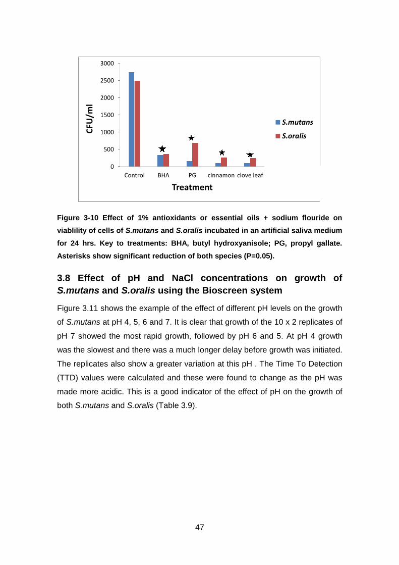
Figure 3-10 Effect of 1% antioxidants or essential oils + sodium flouride onviablility of cells of S.mutans and S.oralis incubated in an artificial salivamedium for 24 hrs. Key to treatments: BHA, butyl hydroxyanisole; PG,propyl gallate............................................................................................. 47
Figure 3-11Effect of pH 4-7 on relative growth of S.mutans over periods of 3000mins on a TSB medium at 37oC. Lines represent wells for each replicate ofeach pH treatment..................................................................................... 48
Figure 3-12 Effect of different NaCl concentrations (%) on relative growth ofS.mutans (8 x 2 replicates) in a TSB broth at 37oC over periods of 2000mins. ......................................................................................................... 49
Figure 3-13 Effect of different NaCl concentrations (%) on relative growth ofS.oralis (8 x 2 replicates) in a TSB broth at 37oC over a period of 2000mins. ......................................................................................................... 49
ix
LIST OF TABLES
Table 1-1: Summary of the available ecological characteristics of these twobacteria ..................................................................................................... 21
Table 3-1. Calculated ED50 ( % ) and ED90 values based on colony viability indifferent concentrations of the treatments against S.oralis........................ 41
Table 3-2. Calculated ED50 and ED90 ( % ) values based on colony viability indifferent concentrations of the treatments against S.mutans .................... 42
Table 3-3. Effect of combinations of antioxidants or essential oils at 0.5%concentration when combined with 2000 ppm of sodium fluoride (NaF).Mean of three replicates per treatment. Key to treatments: BHA, butylhydroxyanisole; PG, propyl gallate............................................................ 44
Table 3-4. Effect of combinations of antioxidants or essential oils at 1%concentration when combined with 2000 ppm of sodium fluoride (NaF) inTSB medium. Means of three replicates per treatment. Key to treatments:BHA, butyl hydroxyanisole; PG, propyl gallate. ......................................... 44
Table 3-5. Efficacy of sodium fluoride on populations of S.mutans and S.oralisafter 24 hrs. in artificial saliva medium treatment at 37oC. Means are ofthree replicates per treatment. *, significant difference from the control atP=0.05....................................................................................................... 45
Table 3-9 The relative Time To Detection (mins) for the effect of pH onS.mutans and S.oralis based on time required to reach 0.2 optical density................................................................................................................... 48
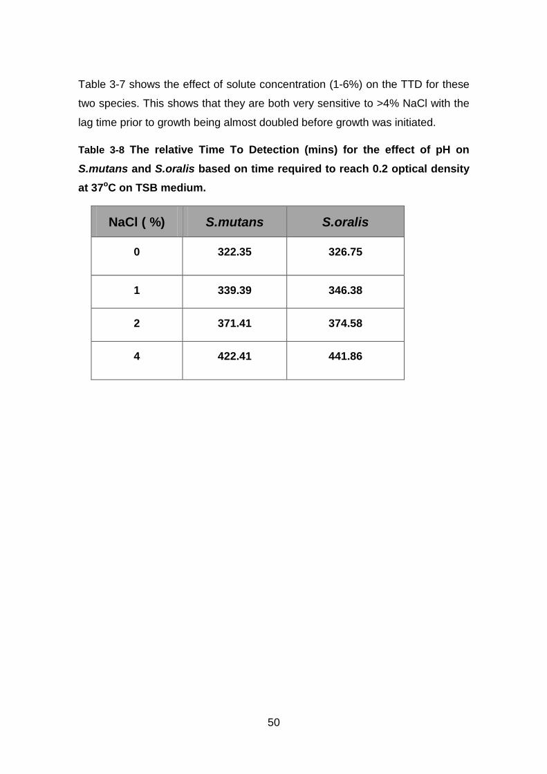
Table 3-10 shows the effect of solute concentration (1-6%) on the TTD for thesetwo species. This shows that they are both very sensitive to >4% NaCl withthe lag time prior to growth being almost doubled before growth wasinitiated...................................................................................................... 50
Table 3-10 The relative Time To Detection (mins) for the effect of pH onS.mutans and S.oralis based on time required to reach 0.2 optical densityat 37oC on TSB medium............................................................................ 50
11
LIST OF ABBREVIATIONS
TSB Tryptone Soya Broth
TSA Tryptone Soya Agar
CFUs Colony forming units
S Streptococcus
ECC Early Childhood Caries
OD optical density
TTD Time To Detection
GTF glycosyltransferases
BHT Butylated hydroxy toluene
BHA Butylated hydroxy anisole
PG Propyl gallate
ppm Parts per million
ul microliter
NaF Sodium fluoride
NaCl Sodium chloride
KCl Potassium chloride
CaCl2.2H2O Calcium chloride dihydrate
NaH2PO4 2H2O Sodium phosphate dihydrate
NH2CONH2Urea
awWater activity
ED50Effective Dose , 50 concentration need to give inhibit availability by 50%
ED90Effective Dose , 90 concentration need to give inhibit availability by 90%
12
Chapter one
1 General introduction and Literature Review
1.1 General Introduction
Tooth decay is caused by poor nutrition associated with a deficiency of
vitamins, minerals, and other nutrients that the body needs together with
development of oral microorganisms. It also results from eating, drinking, or
exposure to high sugar foods which can stimulate microbial colonisation of the
tooth surfaces. A cavity occurs when a tooth decays and the barrier between
the saliva, and the tooth root or pulp, is breached. The inner part of the tooth
contains blood vessels and a nerve. The nerve registers pain and the person
feels a toothache as a result. This is caused by bacterial infections, so called
dental caries.
1.2 The important dental caries causing bacteria
The human mouth contains around 500 to 1000 species of bacteria that have
various functions. There are four main species within the Streptococci: these
are S.mutans, S.salivarius, S.anginosus, and S.mitis groups. S.mutans makes
up a large majority of the bacteria that affects our mouths (Marsh and Martin,
1999). Some oral bacteria act positively by producing organic acids which can
help to inhibit the disease-producing microorganisms that enter via the mouth.
These bacteria work with our immune system to keep our bodies relatively
disease free.
The important bacterial species which are responsible for dental caries are
S.mutans and S.oralis. Other species present in the mouth include Lactobacilli,
Actinomyces and Veillonella species. S.mutans and S.sobrinus are most
commonly found in humans. S.sobrinus is generally found in association with
13
S.mutans and is thought to be principally responsible for the development of
smooth surface caries (Mayooran et al., 2000)
The microbial composition of dental plaque is diverse and remains relatively
stable over time (microbial homeostasis). Microbial homeostasis can break
down, and a major shift in the composition of the microflora can occur. For
example, the frequent consumption of fermentable dietary carbohydrates is
associated with an increased risk of dental caries (Marsh, 1994). Thus sugar
rich diets can lead to a rise in the proportions of caries causing bacteria
including S.mutans and Lactobacilli, with a concomitant decrease in the
populations of other Streptococci, including S.sanguis, S.oralis and S.mitis
(Marsh, 1994). The main location of caries is in pits and fissures and more likely
to develop when food is trapped between the teeth. Thus, poor tooth hygiene,
especially in terms of cleaning teeth and dental flossing on a regular basis will
result in the promotion of bacteria that cause biofilms on the tooth surface and
dental caries.
Dental caries is one of the most common chronic infectious diseases in the
world (Anusavice, 2002; World Health Organization, 2002.). There are three
major hypotheses for the etiology of dental caries: (a) the specific plaque
hypothesis, (b) the non-specific plaque hypothesis, and (c) the ecological
plaque hypothesis (Loesche, 1992; Marsh, 1994;Theilade, 1986). The specific
plaque hypothesis proposed that only a few specific species, such as S.mutans
and S.sobrinus are actively involved in the disease. On the other hand, the non-
specific plaque hypothesis maintains that caries is the outcome of the overall
activity of the total plaque microflora, which is comprised of many bacterial
species (Theilade, 1986). The ecological plaque hypothesis suggests that
caries is a result of a shift in the balance of the resident microflora which may
be driven by changes in local environmental conditions (Marsh, 1994).
However, many studies indicate strongly: (1) the central role of the mutans
streptococci in initiation of caries of smooth surfaces and fissures of crowns of
teeth and suggests their potent role in induction of root surface caries; and (2)
14
that lactobacilli are implicated as important contributory bacteria in tooth decay,
but their role in induction of lesions is not well supported. There have been
studies to determine the source of infection by cariogenic bacteria.
Molecular/genetic studies of the implicated bacteria isolated from humans,
using randomized-blinded-interventional, and longitudinal studies indicate that
mutans streptococci are spread vertically among humans, mostly from mothers
to their children. Implications of these conclusions are briefly discussed. The
most significant problems of literature interpretation include the
benefits/shortcomings of salivary and plaque monitoring of the flora, the role of
sugar(s) in decay as it influences the flora, and modelling strategies to predict
lesion score increments as distinct from determination of the etiological role of
specific bacteria. Future directions for microbiological clinical caries research
are suggested, and the use of the term "caries" to describe the disease, not its
lesions, has been encouraged (Tanzer et al., 2001).
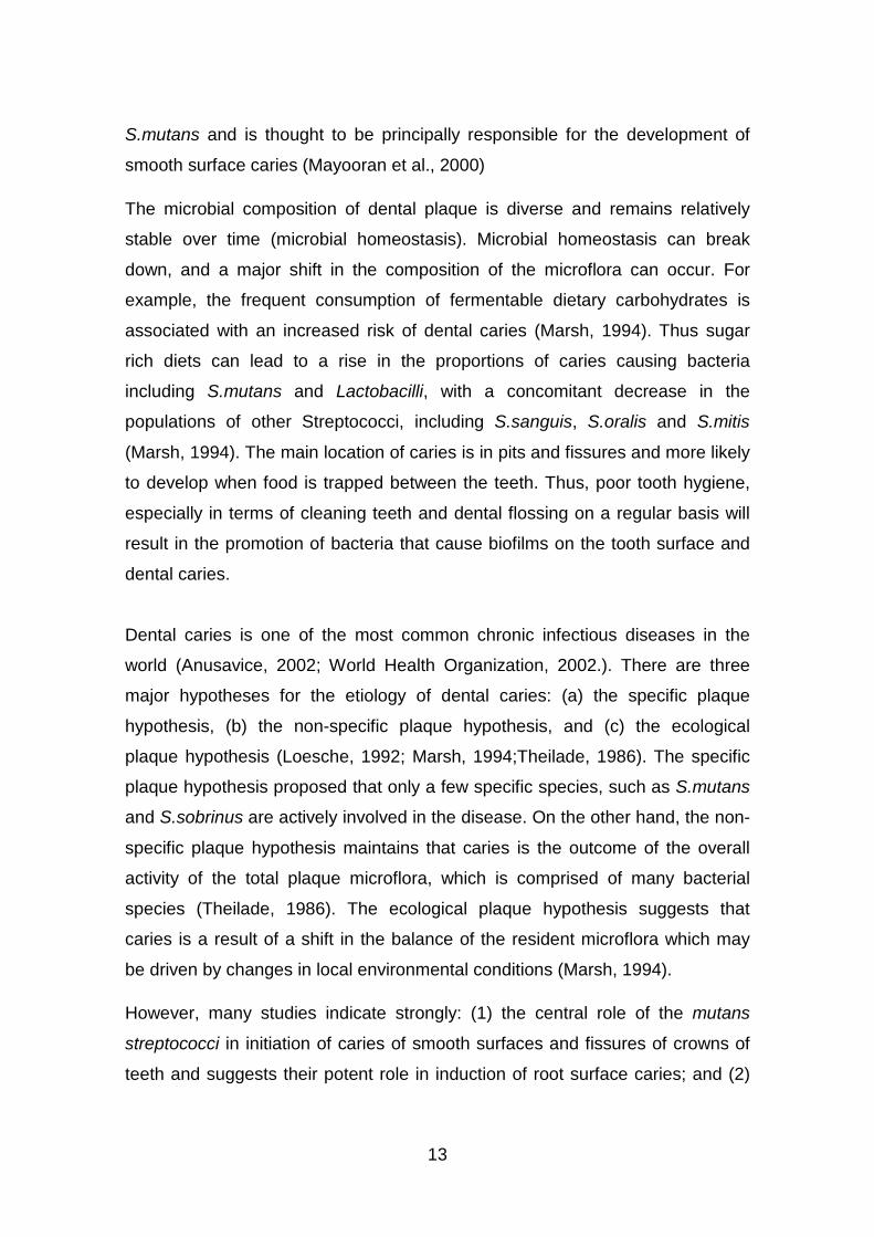
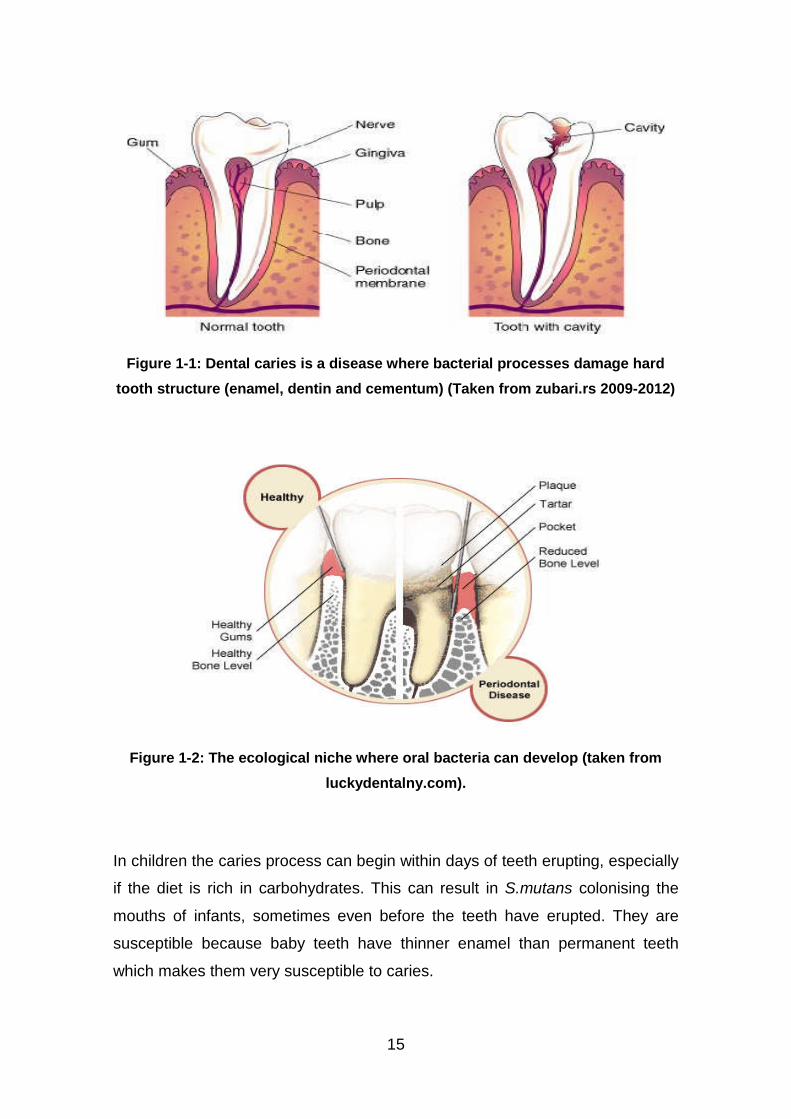
Figures 1.1 and 1.2 show diagrammatically the routes and the ecological niches
in which bacteria can flourish. Under normal conditions the teeth are
continuously exposed and coated with saliva. Saliva is saturated with calcium
and phosphate ions and capable of remineralizing the very early stages of
caries formation, particularly when the fluoride ion is present. Thus, fluoride is
able to slow down the progress of caries. When salivary flow is reduced or
absent, there is an increase in food retention. Since the salivary buffering
capacity can be lost, an acid environment is encouraged and persists for longer.
This in turn encourages acidic bacteria which are able to rapidly grow under
such conducive conditions and metabolize carbohydrates in the low-pH
environment (Edwina, 2005).
15
Figure 1-1: Dental caries is a disease where bacterial processes damage hard
tooth structure (enamel, dentin and cementum) (Taken from zubari.rs 2009-2012)
Figure 1-2: The ecological niche where oral bacteria can develop (taken from
luckydentalny.com).
In children the caries process can begin within days of teeth erupting, especially
if the diet is rich in carbohydrates. This can result in S.mutans colonising the
mouths of infants, sometimes even before the teeth have erupted. They are
susceptible because baby teeth have thinner enamel than permanent teeth
which makes them very susceptible to caries.
16
Often the transmission of S.mutans bacteria in infants is the result of
transmission from the mother (Grönroos et al., 1998). S.mutans also appears
capable of horizontal transmission. For example, children in the same nursery
school class can often have identical strains of the bacteria in their saliva
(Berkowitz, 2003). Also, children who have no detectable S.mutans isolated
until after the age of five often share strains with both mother and father when
the bacteria was finally acquired (Loveren et al., 2000). Generally, the disease
process is hastened by the presence of fructose, sucrose and glucose sugars
from food left on and between teeth. This is converted by the bacteria to acid
and this destroys the tooth enamel, dentine and cement layers. This can result
in demineralisation where enough mineral content is lost resulting in a
disintegration of organic material forming a cavity in the teeth (Michael and
John, 2006). They are classified by location, etiology, rate of progression, and
the type of hard tissues affected.
1.3 Ecology of the dental caries causing bacteria
Ecology describes the interaction between bacteria and the structural, physical,
chemical and biological components of their habitats. Infectious diseases
provide examples of the impact of ecology of specific organisms on their host
populations of plants or humans and other animals. Moreover, disease
promotes responses from the host, changing the ecology balance between the
host and the resident bacteria, influencing the well-being and activities of the
host population (Locker et al., 2000).
The oral cavity provides an excellent environment for the growth and survival of
bacteria. Although saliva is not a complete nutrient for all oral bacteria, some
species or consortia of species utilize it as a substrate (Bowden and Li, 1997).
Other oral nutrients arise from gingival crevicular fluid and desquamated
mucosal cells. Also, in addition to these physiologically based nutrients, oral
bacteria also have access to variable substrates from the hosts’ diet, an
important factor in the relative caries risk (Helderman et al., 1996). Apart from
the nutritional components of saliva there are also molecules that enhance
colonization and those that have an adverse effect on oral bacteria
17
(Scannapieco, 1994). Saliva also acts as a buffer, modifying plaque pH and
reduced salivary flow. Variation in salivary flow over different tooth surfaces can
influence the formation of a caries lesion (Dawes and Macpherson, 1993).
Bacteria decalcify enamel and the tooth root and may follow protein in the
enamel, and invade dentine via the tubules (Thylstrup and Fejerskov, 1996). It
is well accepted that the microflora of lesions in teeth and tooth roots are
extremely complex and may vary at different sites (Schupbach et al., 1995).
Although decalcification is a major factor involved in the initiation of enamel and
root caries, degradation of dentine probably involves proteolysis (Tjaderhane et
al., 1998). Also, specific receptors allow Lactobacillus to localize to exposed
dentine, via collagen receptors (McGrady et al., 1995) and similar molecules
may be present in other bacteria. These bacteria are opportunistic pathogens,
found commonly as members of the resident flora of persons without caries and
expressing their pathogenicity only under specific environmental conditions.
S.mutans and S.sobrinus, two species of the ‘mutans streptococci’ are the most
significant in human caries (Bowden, 1991) and studies of the microbial ecology
of caries have been directed principally at these species (Milnes and Bowden,
1985). There is also a strong association between Lactobacillus spp. and caries
but little is known of the relative significance of the different species. In
particular, although S.mutans and S.sobrinus are the principal agents of enamel
caries, a wider range of organisms is proposed as opportunist pathogens in root
surface caries. Generally, the organisms other than mutans streptococci and
Lactobacillus associated with caries fall into Streptococcus and Actinomyces.
The ecology of the mouth does not just involve interactions among
microorganisms themselves but also the host environment and teeth maturity
and age of the individual. Of course, the host plays a large role in maintaining a
uniform ecosystem, especially through the saliva. Saliva is a complex mineral-
and protein-rich solution that delivers nutrients to the many bacterial species
within the mouth while also protecting host surfaces. During mastication,
increased saliva flow prevents changes in oral pH, because the buffer
bicarbonate is present in saliva and acts as an acid sink at a time when acidic
18
products are being introduced into the mouth. Urea and the peptide saline are
both also present in low concentrations in saliva and produce ammonia when
hydrolyzed, a basic product capable of raising pH (Loesche, 1986). This
buffering counteracts the lactic acid produced by anaerobic bacteria in the
mouth during the fermentation that occurs when nutrients are introduced,
offsetting decay of the teeth caused by this acid. Saliva also contains
glycoproteins that are known to be antibacterial (Loesche, 1986).
1.1.1 Streptococcus mutans
Generally this bacterium inhabits the human oral cavity. It produces plaque and
acids that break down tooth enamel and cause dental caries. S.mutans is a
gram positive bacterium and is a member of the human oral flora which is
widely recognized as the main etiological agent of dental caries. It has a good
ability for adhesion to the tooth surface as biofilms, and it utilises glucose,
fructose and lactose to produce lactic acid. The bacterium grows rapidly forming
a biofilm on and around the teeth which makes them more difficult to destroy.
When these dental biofilms remain on the teeth surfaces these and other
acidogenic bacteria will cause the formation of cavities by the release of a range
of organic acids (Lin Zhu et al., 2006). S.mutans can thrive in temperatures
ranging from 18-40oC (European Bioinformatics Institute, 2011). This species
and other oral bacteria have an optimum pH in the range 6.5-7.5. Acidophilic
bacteria can grow at lower pH levels (Whiley and Beighton, 1998).
It is an important bacterial species to study as it has been associated with many
symptoms including tooth destruction, impaired speech, difficulty in chewing,
multiple infections and has also been implicated in the pathogenesis of certain
cardiovascular diseases (Nakano et al., 2006). Thus methods of control are
required to minimise the ability of this bacterial species to grow.
S.mutans is one of a few specialized organisms equipped with receptors that
improve adhesion to the surface of teeth. Sucrose is used by S.mutans to
produce a sticky, extracellular, dextran-based polysaccharide that allows them
to cohere, forming plaque. Molar teeth are more heavily colonized than anterior
19
teeth and fissures in these teeth are more susceptible to colonization than
proximal, buccal or lingual surfaces.
It has over time developed strategies to successfully colonize and maintain a
dominant presence in the oral cavity. It has been able to evolve from nutrition-
limiting conditions to protect itself in extreme stress conditions. Streptococci
represent 20% of the oral bacteria and actually can determine the development
of oral biofilms. Although S.mutans can be antagonized by pioneer colonizers,
once they become dominant in oral biofilms, dental caries can develop and
thrive.
Transmission of S.mutans: Like any other infectious pathogen, S.mutans
depends on transmission routes to propagate itself among many human hosts.
It favours hard, non-shedding surfaces for the establishment of permanent
colonies. This led to the assumption that levels of S.mutans were undetectable
in infants until the eruption of the primary teeth. Some studies have revealed
that S.mutans can colonize the furrows of the tongue in pre-dentate infants
(Berkowitz, 2003). When the teeth erupt, typically between the ages of one and
two, and S.mutans can establish thriving colonies on the teeth that eventually
lead to cavities, most notably Early Childhood Caries (ECC). It is the
appearance of detectable levels of the bacteria on the teeth that indicate that
cavity formation is possible. Detection of S.mutans in the furrows of the tongue
reinforces the conclusion that the most common transmission route for the
bacteria is vertical, from mother to child, most likely shortly after birth. Studies of
saliva samples from two to five year-old children and their mothers by Caufield
and Ratanapridakul (1988) and Caufield et al. (1993) revealed a high fidelity in
the genetic makeup of each host's S.mutans population. The same study also
concluded that plasmid DNA similarities correlate to different races, also
implying primarily vertical transmission. As a result, mothers with high titres of
the bacteria or who have suffered from dental caries themselves are likely to
pass the same virulence and associated problems on to their children. In fact,
mothers whose salivary S.mutans levels exceeded 100+ colony forming units
20
(CFUs) were about nine times more likely to pass the bacteria on to their
children (Berkowitz, 2003).
1.1.2 Streptococcus oralis
This is a gram positive bacterium that grows characteristically in chains. It is
found as an early colonizing microorganism in the oral cavity of humans and
can be present in high numbers in the oral cavity. S.oralis grows optimally at
37oC, in both liquid films and on solid substrates. It is also able to grow under
conditions of low pH, cultured at pH 5.2 or 7.
Most bacteria have an optimum pH for growth in the range 6.5 – 7.5 with limits
somewhere between 5 and 9 (Whiley and Beighton, 1998).
S.oralis causes platelet aggregation and oxidation of iron in haemoglobin when
it enters the blood stream via open wounds such as those created during oral
surgery. And it is common cause of endocarditis and it is implicated in dental
plaque formation (Marsh and Martin, 1999). S.oralis is the most predominant
acidic non-S.mutans streptococcus causing significant dental caries.
S.oralis, one of commensal bacteria inhabiting the oral cavity, belongs to the
oral viridans group of streptococci. It has been implicated as a potential
causative organism of human cardiovascular diseases including infective
endocarditis and atherosclerosis. S.oralis is frequently isolated from infective
endocarditis (Douglas et al., 1993).
Various studies have shown that certain strains of enterococci and other oral
streptococcal species including S.sanguis, S.oralis, S.mitis and S.salivarius are
capable of causing caries development in rats. Formation of fissure caries,
rather than smooth surface lesions, was most evident and the severity of
disease was mild compared with that induced by Mutans streptococci (van
Houte, 1980; Willcox et al., 1987). On the basis of these findings, the
contribution of non-Mutans streptococci to the aetiology of dental caries
appears minimal. The accumulated evidence from animal experiments and
human epidemiological studies overwhelmingly indicates Mutans streptococci
21
are the principal aetiological agents of both enamel and root caries (Mayooran
et al., 2000).
S.oralis is a numerically important member of the commensal oral microbiota,
isolated from all intra-oral surfaces and a pioneer organism involved in the
primary colonization of the dentition (Nyvad and Kilian, 1990). Genotyping
studies using repetitive extragenic palindromic (REP)-PCR have shown that S.
oralis is usually present as multiple genotypes in the same individual and that it
is rare for unrelated individuals to share the same genotypes (Alam et al., 1999;
O'Neill et al., 1999). Extensive sequencing of gdh alleles of members of the
‘oralis-pneumonia-mitis’ group in samples from two subjects found that the
sequences clustered with the previously described species (Bek-Thomsen et
al., 2008). Table 1.1 summarises the available information on effect of
environmental factors on these two bacteria.
Table 1-1: Summary of the available ecological characteristics of these two
bacteria
Species Temperature range pH Water activity Reference
S.mutans Max: 30-37 6.5-7.5 0.999-0.91 EBI, 2011
S.oralis Max: 30-37 5.2-7.0 0.999-0.91 Wiley/Beighton, 1998
In addition, the oxygen levels in the mouth may change throughout the day.
These fluctuations in oral conditions can cause the populations of the biofilm
within the mouth to also change because it will affect the bacteria-bacteria inter
and intra-interactions.
1.4 Use of the Bioscreen approach to study microbial ecology
The rate of growth of oral bacteria will depend on the relative population present
in the mouth and the availability of nutrients. The growth curves can be
determined as the algorithm of the relative population size [y = in (N/N0) as a
22
function of time (t) (Zwietering et al., 1994)). The growth of a microbial culture at
a specific temperature and under a set of environmental conditions can be
followed using traditional plating methods: at specified times an amount of the
test culture is transferred from the growth medium, diluted and spread plated
onto a relevant nutrient agar, and incubated at an appropriate temperature (e.g.
optimal for growth) for 24-48 hours or until a viable count of the colonies can be
done. Plotting the resulting log numbers against the incubation time gives the
standardised microbial growth curve (Figure 1.3). There are four distinct phases
which can be recognised in this microbial growth curve:
Lag phase: where there is no apparent increase in the bacterial population, but
the cells may be becoming conditioned and metabolically active in preparation
for the next phase. The length of the lag time depends on many factors
including the inoculum size, the time needed by cells to recover from any
previous physical stress, e.g., environmental stresses such as low temperature,
low or high pH and modified solute concentration.
Exponential (log) phase: this is when the division of the cells starts and
continues at a constant rate (generation or doubling time) of the population
depending on the incubation conditions (media, temperature and time) and the
microbial species.
Stationary phase: when the nutrients are becoming exhausted or a lack of
biological space or/and the accumulation of inhibitory metabolites; the bacterial
population growth will slow down and not increase anymore. This is especially
so in a batch culture with a limited nutrient content.
Death phase: during this phase the viable cell population declines and the
number of viable cells decreases quite quickly (in a reverse order to the
exponential (log) phase) when incubation continues beyond the stationary
phase. When using a turbidimetric measurement such as the Bioscreen
analyser this phase cannot be detected because the optical density and
turbidity of the growth broth remains unchanged in this phase, although the
number of viable cells decreases.
23
Figure 1-3: Diagrammatic representation of a bacterial growth curve with the
different phases (Salih, 2010).
1.1.3 Bioscreen Microbiological Analyser
Using the traditional method for microbial growth curves is time consuming and
labour intensive due to the need for multiple serial dilutions and plating of each
sampling taken. Thus, to obtain one growth curve may require several days’
work to obtain the data. The optical density or turbidity of a cell suspension is a
non-destructive technique to determine or measure the amount of light
scattered by the bacterial suspension and it is normally related to the number of
cells or the mass of cells. This approach has been automated in the Bioscreen
Microbiological Analyser which can be used to examine bacterial growth rates
by using optical density (OD) in 100 well titre plates (100 x 2) and in which it is
possible to control both temperature and agitation rates. Additionally, it is
possible to measure the growth rate automatically every few seconds or
minutes as required.
The optical density of microbial cells suspension increases with time as the
organism grows in the medium. A plot of the log10 initial inoculum (cfus/ml)
against time is shows in (Figure 1.4). Usually these data sets are used to
determine the Time To Detection (TTD) which is usually set at 0.2 optical
density value. This helps to determine the lag phase length and the relative
growth rates by reference to plots of the log CFUs/ml against the TTD of
different inoculum sizes.
24
Figure 1-4: Effect of inoculums size on Time to Detection (TTD) of Aeromonas
hydrophila at 30oC (Salih, 2010)
From this plot the time to detect an OD=0.2 for each well (curve) can be
obtained and this plot of TTD against Log inoculum size can be constructed.
Lambert and Pearson (2000) developed have used this approach for bacterial
susceptibility testing to novel compounds. However, this depends on the
medium being relatively clear as opaque media will not allow measurements to
be made.
The time to detection (TTD) is defined as “the time to produce an optical density
of 0.2 (Lambert and Bidlas, 2007), the assumption made that at an OD=0.2
each well in the Bioscreen plate has approx. identical numbers of
microorganisms.
1.5 Control strategies
The best methods of protection from bacterial caries are the following:
Good Oral hygiene
The use of dental sealants as a means of prevention. A sealant is a thin
plastic-like coating applied to the chewing surfaces of the molars to
prevent food from being trapped inside pits and fissures
Calcium, found in foods such as milk and green vegetables, is often
recommended to protect against dental caries. It has been demonstrated
25
that calcium and fluoride supplements decrease the incidence of dental
caries. Fluoride helps prevent decay of a tooth by binding to the
hydroxyapatite crystals in enamel.
The problem is that education has often been lacking resulting in poor teeth
hygiene. Thus knowledge of the fact that bacterial fermentation of dietary
carbohydrates producing organic acids capable of attacking the enamel causes
decay is sometimes not recognised, especially in school children. The role of
the dental plaque, the sticky deposit which accumulates and adheres
tenaciously to the surfaces of teeth not subjected to cleansing by mastication
and the activity of the oral muscles is thus not addressed effectively.
Dental caries can be controlled first of all by personal oral hygiene care, which
consist of proper brushing and flossing daily at least two times a day. The
purpose of oral hygiene is to minimize any pathologic agents in the mouth. The
brushing and flossing facilitates the removal and prevention of the accumulation
of plaque around the teeth. Plaque consists of a mixture of bacteria and thus
the plaque causing bacteria can increase, with the teeth becoming more
susceptible to dental caries. The objective of regular brushing is to remove or
reduce this accumulating plaque on accessible surfaces, especially proximal
caries. Usually, X-rays are taken on a regular basis to monitor the potential
development of cavities in high risk areas of the mouth. Chewy, sticky foods
(such as dried fruit or candy) are best if eaten as part of a meal rather than as a
snack. If possible, the teeth need to be brushed and rinsed with water after
eating such foods. By Minimizing snacking, which creates a constant supply of
acid in the mouth can be prevented the development of these dental caries
causing bacteria.
Dental sealants can be used to try and prevent some cavities. Sealants are thin
plastic-like coatings applied to the chewing surfaces of the molars. This coating
prevents the accumulation of plaque in the deep grooves on these vulnerable
surfaces. Sealants are usually applied on the teeth of children, shortly after the
molars erupt. Older people may also benefit from the use of tooth sealants. It
has been suggested that the chewing of xylitol-containing gum can help to
26
decrease bacterial growth. The bacteria cannot use the xylitol as a food source,
like sugar. Other products may also reduce the acid level in the mouth.
Increased tooth resistance to caries development may be achieved by the use
of fluorides. Indeed, the use of fluoride in toothpaste and other oral products is
believed to be the major reason for the substantial decline in caries incidence in
many developed countries (Ten Cate, 1998). Fluoride helps to prevent dental
caries by binding to the hydroxyapatite crystal in enamel. Topical fluoride is also
recommended to protect the surface of the teeth. This may include a fluoride
toothpaste or mouthwash. Phosphates have been used as food additives to
prevent dental caries. It was reported that the addition of sodium
trimetaphosphates to chewing gum and calcium sucrose phosphate to the diet
can prevent dental caries (Mayooran et al., 2000) and regular visits to the
dentist should control the decay in the mouth.
Several previous studies found that chlorhexidine is a very effective compound
with very good anti-plaque properties. In a supragingival biofilm model,
chlorhexidine was shown to inhibit bacterial growth and biofilm formation
(Guggenheim et al., 2001; Shapiro et al., 2002). Because chlorhexidine is
positively charged, it binds to various surfaces including enamel pellicle,
hydroxyapatite and mucous membranes. A major part of the effectiveness of
chlorhexidine is due to this (Balakrishnan et al., 2000). However, the retention
of chlorhexidine on tooth surface also leads to an undesirable side-effect which
is tooth staining and calculus formation (Moshref, 2002; Yates et al., 1993). To
address this problem, an oral hygiene composition comprising chlorhexidine
gluconate with an anionic anticalculus agent has been suggested (Barton and
Galley, 1997).
Recent advances in caries prevention using plant extracts are more focused in
finding novel active extracts (Mezine. et al., 2009). They found a formulation
derived from water soluble components of the Labiate family of plant extracts.
This formulation was able to prevent dental plaque accumulation through
inhibition of GTF enzyme activity, reduce caries-associated inflammation in the
oral cavity by cyclooxygenase inhibition, and provide a strong anti-oxidative
27
capacity. A non-food anti-microbial-adhesion and aggregation composition
comprising of a suitable carrier and an effective amount of an adhesion
inhibitory fraction isolated from berry juice of the Vaccinium plant genus was
found to be effective by Ofek et al. (2005). This adhesion inhibitory fraction was
characterized as being polymeric and having a molecular weight of 14,000; an
elemental analysis of carbon 43-51%, hydrogen 4-5%, no nitrogen, sulphur or
chlorine. This composition was able to inhibit bacteria-bacteria interaction and
interactions between bacteria and the pellicle layer on tooth surface. A possible
mechanism for this inhibitory effect might be the interruption of lectin-
carbohydrate interaction whereby the sugar residues on one bacterial pair
interact with a lectin on the surface of the other bacterial pair (Majeed and
Prakash, 2003). They also found an essential oil composition derived from
Coleus forskohlii which showed significant inhibitory action against S.mutans
which represents a novel natural essential oil for prevention and treatment of
dental caries. There has thus been interest in finding different novel essential
oils or alternative such as antioxidants which could be used to try and inhibit the
growth of species such as S.mutans and S.oralis.
1.5.1 Antioxidants and essential oils
An antioxidant is a molecule that inhibits the oxidation of other molecules.
Oxidation is a chemical reaction that transfers electrons or hydrogen from a
substance to an oxidizing agent. Oxidation reactions can produce free radicals.
In turn, these radicals can start chain reactions. When the chain reaction occurs
in a cell, it can cause damage or death to the cell. Antioxidants terminate these
chain reactions by removing free radical intermediates, and inhibit other
oxidation reactions. They do this by being oxidized themselves, so antioxidants
are often reducing agents such as thiols, ascorbic acid, or polyphenols (Helmut
(1997).
Antioxidants as control compounds: Antioxidants can help to maintain the
balance between oxidative stress and other oxidation reactions. There are
several thousand antioxidants, including enzymes, vitamins, minerals and other
nutrients and compounds. Some antioxidants are produced within the body;
28
others, such as vitamins A and C, must be provided by external sources. A
healthy, varied diet rich in fruits and vegetables, whole grains and nuts is an
excellent source of antioxidants. Antioxidants may be supplied by other external
means as well.
Antioxidants have been commonly examined for efficacy against
microorganisms involved in disease as well as in food applications. The esters
of p-hydroxy benzoic acid (paraben) were found to be very effective in inhibiting
growth of spoilage bacteria and fungi. There mode of action may be at the cell
membrane level eliminating the pH-related component of the protomotive force
and affecting energy transduction and substrate transport. BHA has also been
shown to have a direct effect on the mitochondrial electron chain of
trypanosomes, thus inhibiting respiration. Antioxidants have also been found to
be effective in treatment of disorders associated with gingival tissues and other
supporting structures of the teeth (San Miguel et al., 2011).
Essential oil: these are natural extracts which have been examined as new
natural antimicrobial therapeutic agents for control of microbial diseases. They
have been investigated for the control of many bacterial species including
dental caries causing bacteria such as S. mutans. They are complex, volatile,
natural compounds formed by aromatic plants as secondary metabolites. They
are known for their bactericidal, virucidal, fungicidal, sedative, anti-inflammatory,
analgesic, spasmolytic, and locally anesthetic properties. The presence of
complex chemical structures constituted of several groups, such as terpenes
and terpenoids, aromatic and aliphatic constituents, all characterized by low
molecular weight, may explain their successful bacteriostatic and bactericidal
action. Detailed studies of essential oils for efficacy against the two Streptococci
examined in this thesis are however limited (Lıvia Camara et al., 2012).
1.6 Aims and Objectives
The aims of this study was to examine two dental caries causing bacteria,
S.mutans and S.oralis, as model systems to examine the efficacy of using
antioxidants and essential oil components to inhibit their growth. The second
29
aim was to examine whether the best ones could be combined with fluoride for
better efficacy. The third aim was to provide more ecological data on the activity
of these two bacteria under different environmental conditions (temperature,
water activity).
1.6.1 The main objectives of this work were:
1. An initial screening of nine essential oils and three antioxidants on
growth of S.mutans and S.oralis was done using a clearing zone assay
at 37oC.
2. Examination of the effect of different concentrations of the best essential
oil and antioxidant treatments on the numbers of viable CFUs of
S.mutans and S.oralis after incubation at 37oC for 24-48hrs.
3. Determination of the ED50 and ED90 concentrations of the best
compounds for inhibition of these two dental caries bacteria
4. To examine the potential of combining the best compounds in
combination with fluoride for improved control of these two bacterial
carries causing organisms in defined media and in an artificial saliva
medium
5. Ecological studies to examine the effect of pH and water activity on
growth of these two Streptococci species using the Bioscreen
instrument.
30
Chapter Two
2 MATERIALS AND METHODS
2.1 Bacterial strains used in this study
A type culture of S.mutans (11516) and S.oralis (702680) were obtained from
the UK National Culture Collection in Scotland. .These were cultured as per
instructions and sub-cultured regularly on Tryptone Soya Broth (TSB) and on
Tryptone Soya Agar 9 cm Petri plates and kept at 4oC until required.
2.2 Media, essential oils and antioxidants
Two types of media were used in this study. These included TSA (tryptone soya
agar) and TSB (tryptone soya broth).
For the preparation of TSA, 40 grams of TSA was weighed into 1 L of water and
the mixture shaken well before autoclaving at 120oC for 25-20 mins. The molten
cooled agar was poured into 9 cm Petri plates (approx. 15 ml per plate). These
were stored at 4oC until used.
TSB was prepared by weighing 30 g of the medium in 1 L of water. This was
well mixed and heated. The medium was dispensed into 20 ml Universal bottles
(10 mls) and autoclaved as detailed previously. These were also stored at 4oC
until used in experiments.
Overall, initial studies showed that both S.mutans and S.oralis grew well on
both TSA and TSB when incubated at 37oC for 24-96 hrs.
2.3 Essential oils and antioxidants used in this studyAn initial screening was done using the following list of essential oils obtained
from (F.D. Copeland & Sons, Ltd., London):
Oil of clove leaf
Oil of spearmint
Oil of thyme
31
Oil of lemongrass
Oil of mandarin
Oil of sweet fennel
Oil of ginger
Oil of clove bud
Oil of cinnamon leaf
These essential oils (Sigma Aldrich, UK) were diluted in methanol (1g of
essential oil to 10ml of methanol).
The antioxidants examined in this study were:
Butylated hydroxy toluene ( BHT)
Butylated hydroxy anisole (BHA)
Propyl gallate (PG)
1 gram of the antioxidant was added to 10 ml of methanol, thus making up a
10% concentration solution of each one.
2.4 Initial screening of essential oils and antioxidants
A traditional clearing zone screening assay was used to compare the efficacy of
the 10% concentrations of the essential oils and antioxidants for obtaining the
most inhibitory treatments.
The agar media were inoculated with a 200 ul of each bacterial species
(S.mutans; S.oralis) and spread using a sterile glass spreader over the whole
agar plate. Then, three holes (5 mm diam, with a sterile cork borer) were made
equidistant from each other on replicate Petri plate treatment. The experiments
were carried out in duplicate. In each of these holes a 25 ul of the diluted
essential oil and one hole was filled with methanol as a control. The treatments
and replicates were incubated at 37oC for 48 hrs. After 24 and 48 hrs, the zones
of clearing around the essential oil treatments were examined and the diameter
(mms) measured. These experiments were repeated twice to confirm the
results.
32
Similar methods were used for the testing of the antioxidants. The bacteria were
spread plated onto the media and then the 25 µl of the test antioxidants were
added to the wells made in the agar plates as detailed previously. The
treatments and replicates were again incubated at 37oC and the clearing zones
measured after 24 and 48 hrs.
2.5 Testing of best treatments for determining ED50 and ED90
values for control of S.mutans and S.oralis
Screening of essential oils and antioxidants: For these experiments clove leaf,
cinnamon oils and BHA, PG antioxidants were tested. For essential oils the
following concentrations were tested: 0.1, 1, 5 and 10%. These were added in
methanol to the TSB media.
Similarly BHA and PG were also added to the liquid broth media.
Three replicates of each treatment including controls were incubated at 37oC for
24 hrs. Then 100 µl of each treatment and replicate were spread plated onto
TSA agar plates (3 replicates per dilution) and the numbers of viable colonies
counted at each concentration for each treatment and replicate. The viable
populations were compared with the controls. This enabled the ED50 and ED90
concentrations to be quantified by reference to the control populations..
Fluoride solutions and ED50 and ED90 concentrations: For these experiments
sodium fluoride stock solutions in sterile water was initially made up
(10.000ppm). Concentrations of 100, 500 and 1000 ppm were used as the
treatments concentrations. The effect of treatments was investigated as
described previously and after incubation at 37oC for 24 hrs. An inoculum of 100
µl of a 104 CFUs/ml concentration was used in these studies.
Experiment of antioxidants or essential oils + sodium fluoride: For these
experiments 0.5% and 1% of essential oils and antioxidants were used in
combination with 1000 ppm of NaF (filter sterilised through a 0.22 micron sterile
filter. In this case the essential oils/antioxidants were dissolved in methanol and
then added to 10 ml TSB media in combination with 1000 ppm NaF in 25 ml
Universal bottles.
33
The TSB treatments and replicates were inoculated with 100 µl of a 24 hrs
culture (104 CFUs/ml) of either S.mutans or S.oralis. These were incubated for
24hrs at 37oC. Subsequently, 12ul of each treatment for each species was
spread plated onto three replicates TSA plates including the controls and
incubated for 48 hrs at 37oC to examine the viability of the treatments and
replicates.
2.5.1 Artificial saliva media and efficacy of the best treatments
Studies were subsequently carried out with an artificial saliva medium to
examine under more realistic conditions the effect of the best treatments in the
presence of NaF. The artificial saliva medium consisted of NaCl, 0.4 g; KCl, 0.4
g; CaCl2.2H2O, 0.795 g; NaH2PO4 2H2O, 0.78 g; NH2CONH2, 1.0 g; distilled
water, 1000 mL The pH of the medium was 3.5.
For efficacy of NaF on growth of the two bacteria the concentrations of 100, 500
and 1000 ppm were tested as described previously using the initial stock
solution of 10,000 ppm.
Studies were then carried out with the antioxidants and essential oils (0.5 and
1% concentration) and sodium fluoride (1000 ppm) on the viability of S.mutans
and S.oralis by incubation in the artificial saliva medium for 24 and 48 hrs and
then checking viability by plating on TSA medium. In all cases the experiments
were carried out in duplicate and repeated twice.
2.6 Ecological studies using the Bioscreen system
There is little detailed information on the effect of pH and water activity on the
growth of S.mutans and S.oralis. This study utilised the Bioscreen method to
examine the effect of different pH values (4-7) and ionic solute concentrations
(1-6% NaCl= 0.999, 0.99, 0.98, 0.97 water activity) on the growth of these two
bacteria by comparing the TTD under different ecological conditions in TSB.
The Bioscreen uses the automated optical density (OD) measurements to
effectively monitor and measure the growth of bacteria in real time (Begot et al.,
1996).
34
2.6.1 Bioscreen system
The Bioscreen machine is an automated turbidity reader which uses 2 x 100
well micro titre plates, linked to an integrated PC (Lab systems, Helsinki,
Finland). The temperature can be accurately controlled and provides growth
curves from each well directly and the data sets based on monitoring on a very
regular basis (5-10 secs to 5-10 mins) can be downloaded and analysed using
other software (in this case Excel was used).
2.6.2 Culture preparation and growth studies in the Bioscreen
Bacteria were grown overnight in conical flasks containing 80 ml TSB in shaken
cultures at 37oC. The cells were harvested, centrifuged at 3000 rpm (10 min)
and the resulting cell pellets resuspended in 2 ml TSB. The inoculum was
standardised by diluting to an approximate OD = 0.5 at 600 nm giving
approximately 2x10⁵ cfu/ml. This standardised culture was subject to either ten
decimal or ten half –fold dilutions in TSB.
Each well in the Bioscreen microarray plates was filled as follow:
200ul of TSB was decanted into each of the wells except column 10.
The wells of column 10 were filled with 400μl of the appropriate serial
dilutions (decimal or half fold), with the highest inoculum (the zero
dilution) in well 100
Using a multi-pipette, 200μl were removed from each well of column 10
and transferred into the wells of column 9, mixed by repeated syringing,
and then 200μl were removed (using new tips) from the wells of column
9 and transferred to column 8 etc. This was repeated across the line
finishing with column 1 (discarding 200μl after final mixing). There was
no need for a negative control as this is the background OD of the broth
Extra care was taken when performing repeated syringing to get the right
dilution and to avoid carrying extra cells between dilutions. This was done by
changing the tips of the multi-pipette for each column and also by placing the
35
tips in at the right depth of each well (if the tips are not placed far enough into
the well, bubbles form which impaired the performance of the experiment.
Plating and colony counting: From the tubes labelled -5 and -6 decimal
dilution, 0.1 ml of each was transferred and spread onto previously prepared
TSA plates in triplicate and incubated at 37°C for 1-2 days. Plates with <300
CFUs were counted and the approximate log number of the initial (zero dilution)
culture were calculated. The following calculation is an example of this method:
Plates counts for the -6 dilution: 102, 123 and 107 colonies
Average counts: 111 colonies
Due to the plating dilution the number of colonies are multiplied by 10
(111x10)
To get the approximate colony number in the Zero dilution multiply by
106 (6 serial dilutions from -6 to 0)
The initial inoculum was: 1.11x10⁹ cfus/ml.
An example of the temporal effect of different concentrations of initial inoculum
on growth rates and optical density of S.mutans and S.oralis is shown in
Appendix I.
2.6.3 Effect of pH and water activity on growth of S.mutans and
S.oralis using the Bioscreen
The pH of the media was modified using buffers as described below using
phosphate/citrate buffers. The amounts of each component are detailed below
to obtain the target pH values.
1. pH 4: 19.3ml (Na2HPO4) + 30.7ml (citrate)
2. pH 5: 25.7ml (Na2HPO4) + 24.3ml (citrate)
3. pH 6: 32.1ml (Na2HPO4) +17.9 ml (citrate)
4. pH 7: 43.6ml (Na2HPO4) + 6.5ml (citrate)
36
pH narrow range indicator strips were used to check the accuracy of the
treatments. These were confirmed by using a pH meter and shown to be
accurate.
In the experiments the wells were filled with 150ul of pH treatment to a column
of 8 wells with two replicates per treatment. This gave a total of 16 wells per
treatment. To this 50 ul of the bacterial suspension (at the same pH, x CFUs/
ml) added. The last two wells in each column were filled with the control TSB
solution (200 µl). This was repeated for each of the pH levels tested with two x 8
column of wells for each treatment. The plates were incubated in the Bioscreen
machine at 37oC for 24-48 hrs.
The effect of water activity was determined by examining the effect of different
ionic solute concentrations on growth of S.mutans and S.oralis. The
concentrations used in TSB were 1, 2, 4 and 6% NaCl (=0.999, 0.99, 0.98 and
0.97 water activity).
150 µl of NaCl treatment was added to each well in two columns of 8 wells. To
this 50 ul of the bacterial suspension (at the same NaCl, x CFUs/ml) added. The
first two wells in each column were filled with the control TSB solution (200 µl).
This was repeated for each treatment. The plates were incubated in the
Bioscreen machine at 37oC for 24-48 hrs.
The data was plotted and the time to detection (TTD) for each treatment was
compared with a standard initial inoculum in the ecological studies.
2.6.4 Statistical treatment of results
The means of three replicates were made in the screening assays, Analysis of
variance was used to examine the relationship between treatments in the
assays examining viability assay of individual and treatments combined with
fluoride and the treatments which were significant at P=0.05 identified.
37
Chapter three
3 RESULTS
3.1 Screening of essential oils
S.oralis:
Figure 3.1 show the effects of the 9 essential oils on growth of S. oralis after 24
and 48 hrs. The clove leaf, thyme and cinnamon oils were found to be effective
after 24hrs. For the others, including clove leaf, thyme, cinnamon, ginger,
mandarina and clove bud they were only effective after 48hrs incubation.
Figure 3-1. The effect of 9 essential oils on growth of Streptococcus oralis after
24 and 48 hrs using two types of media (TSA, TSB) and the concentration of each
treatment was 10%.
0
2
4
6
8
10
12
14
16
Me
an
zon
eo
fc
lea
rin
g(m
m)
Essential oils
S.oralis 24hrs
S.oralis 48hrs
38
S.mutans:
Figures 3.2 the effect of the 9 essential oils on growth of S.mutans after both 24
and 48 hrs incubation. For this species, clove leaf, cinnamon and clove bud oils
were found to be effective in inhibiting growth after 24 hrs. The other treatments
were ineffective after 24 hrs incubation. Lemongrass, mandarin and ginger
essential oils only had some effect after 48 hrs.
Figure 3-2The effect of 9 essential oils on growth of Streptococcus mutans after
24 and 48 hrs using two types of media (TSA, TSB) and the concentration
solution of each was 10%. Bars indicate SE of the means.
3.2 Screening of antioxidants
Figures 3.3 shows the effect of 3 antioxidants tested on control of S.oralis
colony growth after 24 and 48 hrs. The BHA and PG were found to be the most
effective treatments. Figure 3.4 show that for S.mutans similar effects were
observed, with the same two antioxidants being more effective than BHT.
0
2
4
6
8
10
12
14
16
Mea
nzo
ne
of
clea
rin
g(m
m)
Essential oils
s.mutans 24hrs
s.mutans 48hrs
39
Figure 3-3 Effect of three antioxidant treatments on the mean clearing zones for
control of S.oralis at 37oC after 24 hrs. Key to antioxidants: BHA, Butylated
hydroxy anisole; BHT, Butylated hydroxy toluene; PG, Propyl gallate using two
types of media (TSA, TSB) using a concentration of 10%. Bars are SE of the
means.
S.mutans:
Figure 3-4 Effect of three antioxidants on inhibition of growth of S.mutans after
24hrs incubation at 37oC. Key to antioxidants: BHA, butyl hydroxy anisole; BHT,
butylhydroxytoluene; PG, propyl gallate using two types of media (TSA, TSB)
and a concentration of 10%. Bars indicate SE of the mean.
0
5
10
15
20
25
30
35
40
BHA BHT PG
Mea
nzo
ne
of
clea
rin
g(m
m)
Antioxidants
s.oralis 24hrs
s.oralis 48hrs
0
5
10
15
20
25
30
35
40
BHA BHT PGMea
nzo
ne
of
clea
rin
g(m
m)
Antioxidants
s.mutans 24hrs
s.mutans 48hrs
40
Overall, based on the initial screening experiments, it was demonstrated that
clove leaf and cinnamon oils both gave the best results in terms of inhibition of
the two bacterial species. Of the antioxidants tested, BHA and PG appeared to
be the best treatments for further testing.
3.3 Determination of efficacy of best compounds and ED50 and ED90 values
Figure 3.5 and 3.6 show the effect of different concentrations of best two
essential oils and two antioxidants on the viability of cells of both S.oralis and
S.mutans. This shows that for the former species the propyl gallate was the
most effective with complete inhibition by 5% concentration.
For S.mutans, only the propyl gallate treatment at 10% was able to completely
inhibit growth of this dental caries species. Against this species clove oil was
effective but did not inhibit viability completely at any of the concentrations
tested.
Based on these results the approx. concentrations of the treatments required for
50 and 90% inhibition of viability were calculated and are shown in Table 3.1
and 3.2.
Figure 3-5 Effect of the four treatments on the relative viability of S.oralis cells 48
hrs after treatment by plating onto TSA. These are the means of three replicates.
Bar indicates Least Significant Difference (P=0.05).
Concentration (%)
Via
bili
ty(L
og 1
0)
CFU
sm
l
S.oralis
41
Figure 3-6 The effect of the four treatments on the relative viability of cells of
S.mutans 48 hrs after treatment by plating on TSA. The data are means of three
replicates per treatment. Bar indicates Least Significant Difference (P=0.05).
Table 3-1. Calculated ED50 ( % ) and ED90 values based on colony viability
in different concentrations of the treatments against S.oralis
ED50 ED90
Clove leaf 4.2 <10
Cinnamon oil >10 >10
BHA 6 9.8
PG 2 4.6
0
1
2
3
4
5
6
0 2 4 6 8 10 12
Clove
Cinnamon
BHA
PG
Concentration (%)
Via
bili
ty(L
og 1
0)
CFU
sm
l
S.mutans
42
Table 3-2. Calculated ED50 and ED90 ( % ) values based on colony viability
in different concentrations of the treatments against S.mutans
ED50 ED90
Clove leaf 4 >10
Cinnamon oil >10 >10
BHA >10 >10
PG 6.9 9.6
3.4 Effect of sodium fluoride concentrations on growth of
S.mutans and S.oralis
(a) S.mutans
Figure 3.7 shows the growth of S.mutans in sodium fluoride at different
concentrations at 37oC for 24hrs. This shows that at 500 and 1000 ppm there
was a >75% reduction in colonies of this bacteria.
43
Figure 3-7 Effect sodium fluoride concentrations on populations of S.mutans in
relation to concentrations of sodium fluoride at 37oC for 24 hrs. Data are means
of three replicates per treatment.
(b) S.oralis
Figure 3.8 shows the effect of sodium fluoride on populations of S.oralis at 37oC
for 24hrs. The efficacy of sodium fluoride against this bacterium was not as
effective with only approx. 25-30% reduction in populations.
Figure 3-8 Effect sodium fluoride concentrations on populations of S.oralis
grown at 37oC for 24 hrs. Data are means of three replicates per treatment.
0
1000
2000
3000
4000
5000
6000
control 100 500 1000
CFU
s/m
l
sodium fluoride
S.mutans
0
500
1000
1500
2000
2500
3000
Control 100 500 1000
CFU
s/m
l
Sodium Fluoride
S.oralis
44
3.5 Effect of mixtures of antioxidants and essential oils with
sodium fluoride on control of the two bacteria in TSB medium
Table 3.3 and 3.4 shows the effect of mixtures of anti-oxidants/essential oils
(0.5%, 1% concentration) with sodium fluoride (2000 ppm) on the growth of the
two bacteria. This shows that all treatment completely inhibited growth when
compared with the untreated controls regardless of the combinations used.
Table 3-3. Effect of combinations of antioxidants or essential oils at 0.5%
concentration when combined with 2000 ppm of sodium fluoride (NaF). Mean of
three replicates per treatment. Key to treatments: BHA, butyl hydroxyanisole;
PG, propyl gallate In TSB medium. All treatments significantly reduced viability
of cells.
CFUs/ml_____________________
Treatment S.mutans S.oralis
Control 3.96 3.92
BHA + NaF 1 1
PG + NaF 1 1
Cinnamon + NaF 1 1
Clove leaf + NaF 1 1
Table 3-4. Effect of combinations of antioxidants or essential oils at 1%
concentration on viable populations (Log10 CFUs/ml) when combined with 2000
ppm of sodium fluoride (NaF) in TSB medium. Means of three replicates per
treatment. Key to treatments: BHA, butyl hydroxyanisole; PG, propyl gallate.
CFUs/ml_____________________
Treatment S.mutans S.oralis
Control 3.48 3.86
BHA + NaF 1 1
PG + NaF 1 1
Cinnamon + NaF 1 1
Clove leaf + NaF 1 1
45
3.6 Effect of treatments with sodium fluoride on efficacy in an
artificial saliva medium
Effect of sodium fluoride on growth of the two bacteria: Table 3.5 shows the
effect of different concentrations of sodium fluoride alone on populations of the
two bacteria after 24 hrs. in an artificial saliva medium at 37oC. This shows that
the treatment was only effective against both bacteria at 1000 ppm. However,
at this concentration both bacteria could still grow. Thus a higher concentration
would be required to inhibit growth effectively.
Table 3-5. Efficacy of sodium fluoride on Log10 CFUS/ml populations of S.mutans
and S.oralis after 24 hrs. in artificial saliva medium treatment at 37oC. Means are
of three replicates per treatment. *, significant difference from the control at
P=0.05.
CFUs/ml_____________________
Treatment S.mutans S.oralis
Control 3.39 3.53
100ppm 3.30 3.21
500 ppm 3.27 3.04*
1000 ppm 3.20* 2.96*
3.7 Effect of mixtures of antioxidants and essential oils with
sodium fluoride on control of the two bacteria in artificial saliva
medium
Figure 3.9 shows the effect of 0.5% antioxidants and essential oils + sodium
fluoride in artificial saliva on growth S.mutans and S.oralis at 37oC for 24hrs.
This shows that for both species a >95% reduction in viable colonies was
obtained by combining the best treatments with sodium fluoride.
46
Figure 3-9 Growth S.mutans and S.oralis in 0.5% of antioxidants and essential
oils + sodium fluoride in an artificial saliva medium after 24 hrs incubation. Key
to treatments: BHA, butyl hydroxyanisole; PG, propyl gallate. Asterisk indicates
significant inhibition of both species at P=0.05.
Figure 3.10 shows the efficacy of 1% of antioxidants or essential oils + sodium
fluoride in artificial saliva medium on the viable populations of S.mutans and
S.oralis after 24 hrs incubation at 37oC and then plated onto TSB medium.
There was a significant reduction in the viable populations especially of
S.mutans. The efficacy of combined treatments was less effective against
S.oralis. All combined treatments significantly inhibited the two species.
0
500
1000
1500
2000
2500
3000
3500
4000
Control BHA PG cinnamon clove leaf
CFU
s/m
l
Treatment
S.mutans
S.oralis
47
Figure 3-10 Effect of 1% antioxidants or essential oils + sodium flouride on
viablility of cells of S.mutans and S.oralis incubated in an artificial saliva medium
for 24 hrs. Key to treatments: BHA, butyl hydroxyanisole; PG, propyl gallate.
Asterisks show significant reduction of both species (P=0.05).
3.8 Effect of pH and NaCl concentrations on growth of
S.mutans and S.oralis using the Bioscreen system
Figure 3.11 shows the example of the effect of different pH levels on the growth
of S.mutans at pH 4, 5, 6 and 7. It is clear that growth of the 10 x 2 replicates of
pH 7 showed the most rapid growth, followed by pH 6 and 5. At pH 4 growth
was the slowest and there was a much longer delay before growth was initiated.
The replicates also show a greater variation at this pH . The Time To Detection
(TTD) values were calculated and these were found to change as the pH was
made more acidic. This is a good indicator of the effect of pH on the growth of
both S.mutans and S.oralis (Table 3.9).
0
500
1000
1500
2000
2500
3000
Control BHA PG cinnamon clove leaf
CFU
/ml
Treatment
S.mutans
S.oralis
48
Figure 3-11Effect of pH 4-7 on relative growth of S.mutans over periods of 3000
mins on a TSB medium at 37oC. Lines represent wells for each replicate of each
pH treatment.
Table 3-6 The relative Time To Detection (mins) for the effect of pH on S.mutans
and S.oralis based on time required to reach 0.2 optical density.
pH S.mutans S.oralis
7 666.5 695.61
6 719.31 703.45
5 749.05 812.36
4 >1000 >1000
Figure 3-12 and Figure 3-13 show the effects of different NaCl concentrations
on the growth of S.mutans and S.oralis on TSB at 37oC. This shows that as the
percentage NaCl was increased from 1-2% to 6% (0.999 to 0.97 water activity)
49
the growth was delayed and the rate of growth was slower over the
experimental period. The lag times prior to growth were also increased.
Figure 3-14 Effect of different NaCl concentrations (%) on relative growth of
S.mutans (8 x 2 replicates) in a TSB broth at 37oC over periods of 2000 mins.
Figure 3-15 Effect of different NaCl concentrations (%) on relative growth of
S.oralis (8 x 2 replicates) in a TSB broth at 37oC over a period of 2000 mins.
50
Table 3-7 shows the effect of solute concentration (1-6%) on the TTD for these
two species. This shows that they are both very sensitive to >4% NaCl with the
lag time prior to growth being almost doubled before growth was initiated.
Table 3-8 The relative Time To Detection (mins) for the effect of pH on
S.mutans and S.oralis based on time required to reach 0.2 optical density
at 37oC on TSB medium.
NaCl ( %) S.mutans S.oralis
0 322.35 326.75
1 339.39 346.38
2 371.41 374.58
4 422.41 441.86
51
Chapter four
4 Discussion
In this study we examined the effect of antioxidants and essential oils on growth
of two types of bacteria which related to dental caries (S.mutans; S.oralis). A
total of 9 essential oils and 3 antioxidants were screened in vitro using TSB
media at 37oC. Based on zone of inhibition it was found that clove leaf and
cinnamon essential oils and BHA and PG antioxidants had the best efficacy
against S.mutans and S.oralis after both 24 and 48 hrs.
Studies by Chaudhari et al. (2012) examined the effect of different ESOs
against S.mutans. They used 9 commercially available ones in in vitro studies
using a blood agar and 37oC. Based on zone of inhibition they found that
cinnamon, lemon grass, peppermint, clove and eucalyptus oil had best efficacy
against S.mutans. They also suggest that they could be effective against oral
yeast infections as well. Effects against S.oralis were not determined. There
appears to be many more studies with S.mutans when compared to that on
S.oralis. The present study suggests that the latter species may have slightly
more resistance to the essential oils and antioxidants tested when compared to
S.mutans.
Yengopal (2009) discussed the potential viability of using essential oils for
caries prevention. He suggested that the key to controlling dental caries by
Streptococcus species, at least one component needs to be effectively
controlled, i.e., intervention in relation to the teeth, substrate, or the flora. Thus
mouthwashes have been found to effectively reduce microbial counts by simple
intervention. However, they predominantly control gram negative microflora,
although some, e.g. gingivitis mouthwash, has been shown to control gram
positive microorganisms including S.mutans.
Takarada et al. (2004) compared the effect of manuka oil, tea tree oil,
eucalyptus oil, lavender and romarinus oils against a range of oral
microorganisms including S.mutans. They found that 0.2% manuka or tea tree
52
oils were most effective and they also inhibited adhesion by S.mutans.
Interestingly, it was found to have little effect against cultured human cell lines.
Thus they could have potential to be used in formulations of toothpaste.
Propolis is a resinous beehive product which is used by bees as a glue to close
the opening of hives. The ethanolic extract of propolis was examined for
antimicrobial efficacy against oral microorganisms by Park et al. (1998). They
found that this extract inhibited glucosyltransferase activity by S.mutans as well
as inhibiting growth. They found that the extracts varied from different regions of
Brazil and that the components of the extract varied. However, they suggested
good potential for controlling such oral microorganisms.
Lee et al. (2011) examined an extract from Curcuma longa, a common spice
used in oriental foods to control biofilm plaque formation by S.mutans. They
found that essential oil from this spice inhibited growth of S.mutans at 0.5-4
mg/ml. Significant inhibition of biofilm formation on saliva-coated hydroxyapatite
beads was also observed using scanning electron microscopy at >0.5 mg/ml
concentrations. The key components appeared to be α-turmerone and
germacrone (approx. 50%). However, effects against S.oralis were not
evaluated.
Some studies have concentrated on the inhibition of biofilm formation by
bacteria such as S.mutans. These studies have often concentrated on inhibition
of glycosyltransferases and on the membrane integrity of the bacterium. Thus
studies by Koo et al. (2003) examined the effect of apigen and tt-farnesol for
these two inhibitory effects respectively. They found that at 1.33 mm apigenin
and farnesol the dry weights of biofilms were significantly reduced (by 30-50%)
than when treated with control saline solutions. The ability of S. mutans to
produce polysaccharides was reduced as well as effects on viability in such
biofilms, suggesting potential use in formulations.
Recent studies Hamoud et al. (2012) examined the effect of a complex
essential oil distillate (Olbas© Tropfen) against the individual essential oil
components (peppermint, eucalyptus, jumiper berry and wintergreen oils)
against different microorganisms including S.oralis but not S.mutans. Against
53
S.oralis the complex Olbas compound had a MIC of 1.25 mg/ml. However, the
individual essential oils had MICs at 10-40 mg/ml. This suggested that
interactions between mixtures may affect the final effect in controlling such
bacteria. Comparison of different aqueous and organic solvent extracts of teas
(oolong, green and black tea) showed that those of oolong and green tea were
most effective at inhibiting S.mutans. Interestingly, the extracts were more
effective than chlorhexidine (Subramaniam et al., 2012). Previously, it was
suggested that mixed compounds from green tea combined with indole was
very effective against S.mutans (Muroi and Kubo, 1993). They suggested that
there was a synergistic effect of sesquiterpene hydrocarbons such as cadinene
and caryophyllene + indole resulting in a 128 and 256-fold increase in effect on
S.mutans.
A very recent study by Subramaniam et al. (2012) also examined the effect of
pomegranate and aloe vera extract on S.mutans. They again used
hydroalcoholic extracts of pulp from both Punica granatum (pomegranate) and
from Aloe barbadensis (aloe vera) at 5-100%. There was a significantly better
effect of the pomegranate extract on growth of S.mutans than the aloe vera
extracts. They suggested that this extract is a significant antibacterial agent with
potential for control of such oral bacteria.
Polyphenols have been examined for the antimicrobial effects against oral
bacteria, especially S.mutans (Sendamangalam et al., 2011). They examined
natural gallic acid and tannic acid, and salicylic acid and compared this with
ascorbic acid, a common antimicrobial compound for comparison. Overall,
salicylic acid was the weakest with a MIC of 3.8 mg/l while tannic acid was the
best with a MIC of 0.4 mg/ml. They also suggested that the antioxidant
properties may contribute to the antimicrobial effects.
In the present study it was found that sodium fluoride at different concentrations
(100, 500, 1000ppm) at 37oC had better effect on S.mutans than on S.oralis
populations. The relative efficacy was >75% for S.mutans, but only about 25-
30% for S.oralis at 1000 ppm. This suggests that higher concentrations of
sodium fluoride are required. For this reason it was decided to use 2000 ppm
54
sodium fluoride in subsequent studies on TSB and the artificial saliva medium
when testing combinations of the best essential oils or antioxidants + sodium
fluoride.
The effect of mixtures of anti-oxidants/essential oils (0.5%, 1% concentration)
with sodium fluoride (2000 ppm) on the growth of the two bacteria were
interesting. It was found that on TSB the combination treatments completely
inhibited growth when compared with the untreated controls regardless of the
mixtures used. In contrast, in the artificial saliva medium at 37oC the mixtures
were not as effective against both bacteria. Thus, on artificial saliva medium at
1000 ppm some growth of the two bacteria still occurred. The effect of
combinations of 0.5% of antioxidants and essential oils + 2000 ppm sodium
fluoride were quite effective against both bacteria, significantly reducing the
remaining populations after 24 hrs treatment. At 1% the antioxidants/essential
oils + sodium fluoride control was much more effective against both S.mutans
and S.oralis after 24 hrs treatment at 37oC in the artificial saliva medium.
The artificial saliva experiments were useful as they showed the potentially
more realistic effect of the combined treatments. Saliva is important in as it
keeps the ecosystem of the mouth in balance. It contains its own bacterial
enzymes that are beneficial in minimising caries causing bacteria. It contains
phosphate and calcium ions that help repair teeth. The major organic
constituents of saliva are proteins and glycoproteins. Proteins in saliva influence
the oral ecosystem. Some may be used as nutrients by bacteria and of course
can help wash out caries causing bacteria as far as is possible. Of course,
depending on the nutritional balance, especially presence of sugars, this will
influence the attachment of caries causing bacteria and the formation of
biofilms.
The ecology of both bacteria from the results the growth of the 10 x 2 replicates
of pH 7 showed the most rapid growth, followed by pH 6 and 5. At pH 4 growth
was the slowest and there was a much longer delay before growth was initiated.
The replicates also show a greater variation at this pH . The Time To Detection
(TTD) values were calculated and these were found to change as the pH was
55
made more acidic. This is a good indicator of the effect of pH on the growth of
both S.mutans and S.oralis. and different NaCl concentrations on the growth of
S.mutans and S.oralis on TSB at 37oC. This shows that as the percentage NaCl
was increased from 1-2% to 6% the growth was delayed and the rate of growth
was slower over the experimental period.
Chen et al. (2012) examined the effect of pH on S.mutans growth on denture
adhesives in vitro on Polident cream, Protefix cream and Protefix powder. The
pH values were measured immediately after preparation and after 1-24 hr
intervals. Bacterial growth was observed by measuring absorption at 600 nm
every 1 h for 12 h using a spectrophotometer. The tested adhesives generally
remained relatively pH-stable over 24 hrs ranging from 5.5 to 7.0. There were
no statistically significant differences in S.mutans growth rates between the
extract-treated and control cultures (p > 0.05). However, it has been suggested
that S.mutans may be better adapted to lowered pH levels than some other oral
bacteria. Thus dominance of S.mutans and S.sobrinus in resting biofilms at low
pH has been suggested because of their ability to survive and remain viable in
such a niche. Some have proposed a succession of bacteria in caries that takes
into account carbohydrate intake, low pH groups of bacteria and the mutans
streptococci (Van Ruyven, 2000). This could mean that a succession of bacteria
in a suitable medium can lead to dominance by species such as S.mutans
which can then lead to caries lesions.
The ability to tolerate a range of ionic salt concentrations may also facilitate the
survival and growth of oral caries causing bacteria such as S.mutans and
S.oralis, although less information is available on the latter species (Bowden,
2000). However, water activity of solutions used for oral hygiene can be
important in attempts to break up the biofilms and inhibit the activity of these
oral caries causing bacteria. The present study suggests that quite high ionic
solutions are required to delay or inhibit the growth of these bacteria.
56
Chapter five
5 Conclusion and future work
Some essential oils (2 of 9) and antioxidants (2 of 3) have good efficacy
to control the growth of S.mutans and S.oralis
This study showed that clove leaf and cinnamon oils and BHA and PG
were the most effective at 0.5 and 1% concentrations in a TSB medium
Combinations of 0.5% and 1% of these essential oil/antioxidant
treatments with sodium fluoride (2000 ppm) completely inhibited growth
of S.mutans and S.oralis in TSB medium.
In an artificial saliva medium efficacy was not as effective as on TSB,
however the populations of the two oral caries bacteria were significantly
reduced by combined treatments
This suggests that potential exists for using such combinations in
formulations of toothpaste or liquid treatments to reduce the growth of
such bacteria to improve oral hygiene
Ecological studies showed the effect of pH (4-7) and NaCl (1-6%)
concentration on growth of these two bacteria
This showed the optimum pH for these two bacteria was 6-7
The optimum water activity was at 0.99-0.99 (1-2% NaCl concentration).
Efficacy when biofilms are formed by dental caries bacteria needs to be
examined
Potential for the penetration of biofilms of Streptococcus species or
mixed inoculate needs to be examined
Differential efficacy against mixed populations needs to be quantified.
57
REFERENCES
Alam. S., Brailsford. S, R., Whiley. R, A. and Beighton. D. ((1999).), " PCR-based methods for genotyping viridans group streptococci. J Clin Microbiol", vol. 37, no. 2772–2776.
Anusavice, K. J. (2002.), " Dental caries: risk assessment and treatmentsolutions for an elderly population. Compend. Contin. Educ. Dent", vol. 23,pp. 12–20.
Balakrishnan M, , Simmonds RS, and Tagg JR. (2000 Dec), " Dental caries is apreventable infectious disease. ", Aust Dent J., vol. 45, no. 4, pp. 235–45.
Barton. S. and Galley. E. (1997), " Oral hygiene composition.", US5695745, .
Begot, C., Isabelle Desnier, Jean D. Daudin, Jean C. Labadie and AndreLebert. (1996), "Recomendation for calculating growth parameters by optical densitymeasurements", Journal of Microbiological Methods, vol. 25, pp. 225-232.
Bek-Thomsen.M., Tettelin. H., Hance. I., Nelson. K, E. and Kilian. M. ((2008).), "Population diversity and dynamics of Streptococcus mitis, Streptococcusoralis, and Streptococcus infantis in the upper respiratory tracts of adults,determined by a nonculture strategy. ", Infect Immun, vol. 76, pp. 1889–1896.
Berkowitz, R. J. (2003), "Causes, Treatment and Prevention of Early ChildhoodCaries: A Microbiologic Perspective", vol. 69(5), no. 304-307.
Bowden. G, H.,W. (1991), " Which bacteria are cariogenic in humans? In:Johnson NW, ed. Risk Markers for Oral Diseases ", Dental Caries.Cambridge: Cambridge University Press, vol. 1, pp. 266–86.
Bowden. G, H.,W and Li. Y-H. (1997), "Nutritional influences on biofilmdevelopment. ", Adv Dent Res, vol. 11, pp. 81–99.
Bowden, G.H.W. (2000). The microbial ecology of dental caries. MicrobialEcology in Health and Disease Vol 12, pp 138-148.
Caufield. P, W. and Ratanapridakul.K. (1988), " "Plasmid-Containing Strains ofStreptococcus mutans Cluster within Family and Racial Cohorts:Implications for Natural Transmission." Infection and Immunity ", vol. 56, no.12, pp. 3216-3220.
Caufield.P, W., Cutter.G, R. and Dasanayake.A, P. (1993), "Initial acquisition ofmutans streptococci by infants:. evidence for a discrete window ofinfectivity. ", J Dent Res, vol. 72, pp. 37–45.
58
Chaudhari. L, K., Jawale. B, A., Sharma. H., Kumar. CD. and Kulkarni P.A.(2012), "Antimicrobial activity of commercially available essential oilsagainst S.mutans.", .
Cuppers. H,G,A,M. and Smelt. J,P,P,M. (1993), "Time to turbidity measurement as atool for modelling spoilage byLactobacillus. ", Journal of Industrial Microbiology, vol. 12, no. 3-5, pp. 168-171.
Dawes .C and Macpherson. L, M.,D. (1993), "The distribution of saliva andsucrose around the mouth during use of chewing gum and the implicationsfor the site-specificty of caries and calculus deposition. ", J Dent Res, vol.72, pp. 852–7.
Douglas. C, W., Heath. J., Hampton. K, K. and Preston. F. E. ((1993).), "Identityof viridans streptococci isolated from cases of infective endocarditis. ", JMed Microbiol, vol. 39, no. 179–182.
Edwina. A, M., Kidd. (2005.), Essentials of Dental Caries
Essentials of Dental Caries 3rd Ed. Edwina A. M. Kidd, 2005. 3rd Ed ed, .
European Bioinformatics Institute (2011), available at:http://www.ebi.ac.uk/2can/genomes/bacteria/Streptococcus_mutans.html(accessed 10/2011).
Grönroos. L., Saarela. M., Mättö. J., Tanner-Salo. U., Vuorela. A. andAlaluusua. S. ((1998).), "Mutacin production by Streptococcus mutans maybe promote transmission of bacteria mother to child.", Infect. Immun., vol. 66, no. 6, pp. 2595-2600.
Guggenheim B., Giertsen E. and Schupbach P. Shapiro S. (2001 Jan), "Validation of an in vitro biofilm model of supragingival plaque", J Dent Res.,vol. 80, no. 1, pp. 363–70.
Hamoud. R., Sporer. F., Reachling. J. and Wink. M. (2012), " Antimicrobialactivity of a traditionally used complex essential oil stidtillate (Olbas Trofen)in comparison to its individual essential oil ingredients. Phytomedicine ",vol. 19, pp. 969-976.
Koo. HK., Hayacibara. MF., Schobel. BD., Cury. JA., Rosalen. PL., Park. YK.and Vacca-Smith, A. M. &. B., (2003), " WH Inhibition of Streptomycesmutans biofilm accumulation and polysaccharide production by apigen andtt-farnesol. ", Journal of Antimicorbial Chemotherapy, vol. 52, pp. 782-789.
Lambert. R, J. and Pearson. J. (2000), " Susceptibility testing: accurate andreproducible minimum inhibitory concentration (MIC) and non-inhibitory
59
concentration (NIC) values,", Journal of Applied Microbiology, vol. 88, pp.784–790.
Lambert. R, J. and Bidlas. Eva. (2007), "A study of the Gamma hypothesis: Predictive modelling of the growth andinhibition of Enterobacter sakazakii", International Journal of FoodMicrobiology, vol. 115, pp. 204–213.
Lee. K, H., Keum. K, S., Kim. Y, H., Chang. B, S., Ra. Moon.JY., HD.Seo., BR.,Choi., NY . and You, Y. (2011), " Essential oil of Corcuma longa inhibitsStreptomyces mutans biofilm formation. ", Journal of Food Science, vol. 76,pp. 226-230.
Lin. Zhu., Jens, K., Sarah, E., . Cross, J., K and . Gimzewski, Wenyuan Shi, andFengxia Qi (2006), "Functional characterization of cell-wall-associatedprotein WapA in Streptococcus mutans", vol. 152, no. 2395–2404.
Locker. D., Clarke. M. and Payne. B. (2000), "Self-perceived oral health status,psychological well-being and life satisfaction in an older population. ", JDent Res, vol. 79, pp. 970–5.
Loesche, W. J. (1992.), " The specific plaque hypothesis and the antimicrobialtreatment of periodontal disease. Dent. Update ", vol. 19, pp. 68–74.
Loesche., W., J. (1986), "Role of Streptococcus mutans in Human DentalDecay." Microbiological Reviews ", vol. 50, no. 4, pp. 353-380.
Loveren. C, V., Buijs. and J. F. (2000), ""Similarity of Bacteriocin ActivityProfiles of Mutans streptococci within the Family When the ChildrenAcquire the Strains After the age of 5." Caries Research", vol. 34, no. (6):,pp. 481-5.
Magdi A. M. Salih. (2009 - 2010),Growth of Listeria monocytogenes under Non-isothermal Conditions (MScthesis), Cranfield Health Food Chain Systems, CRANFIELD UNIVERSITY.
Majeed , M., and Prakash , S. (2003.), " Composition and methods containingan antimicrobial essential oil extended from Coleus forskohlii. ",US6607712, .
Marsh, P. D. (1994), "Microbial ecology of dental plaque and its significance inhealth and disease. Adv. Dent. Res.", vol. 8, pp. 263–271.
Marsh, P. and Michael V. Martin (1999), "Oral microbiology", , no. 0-7236-1051-7.
Marsh, P. and Michael V. Martin. (1999), Oral microbiology, fifth ed ed, , Oxford[England].
60
Mayooran, B., Robin. S, S. and John , R.,Tagg. (2000), "Dental caries is apreventable infectious disease", Australian Dental Journal, vol. 45(4), no.235-245.
McGrady. J, A., Butcher. W, G., Beighton. D and Switalski .L, M. (1995),"Specific and charge interactions mediate collagen recognition by orallactobacilli. ", J Dent Res, vol. 74, pp. 649–57.
Mezine. I., Zhang. HZ. and Petteruti. M. (2009), " Oral care compositionsderived from the Labiatae family. ", US7517541 ., .
Michael. T, M. and John. M, M. (2006), Brock Biology of Microorganisms, 11thed, Prentice Hall, USA.
Milnes. A, R. and Bowden. G, H. (1985), "The microflora associated with thedeveloping lesions of nursing caries.", Caries Res, vol. 19, pp. 289–97.
miniscience.com , pH indicator sticks (Standard and Special Range),available at:http://catalog.miniscience.com/catalog/PH_Papers/pH_indicator_Sticks.html(accessed 04/12/2012).
Moshrefi .A. (2002), "Chlorhexidine Priodontol Periodontal Abstr.J.West Soc",vol. 50, no. 1, pp. 5–9.
Muroi, H. and Kubo, I. (1993). Combination effects of antibacterial compoundsin green tea flavor against Streptomyces mutans. J. Agric. Food Chem. 41,1102-1105/
Nakano.K., Inaba H, Nomura R, Nemoto. H, Takeda .M, Yoshioka .H,Matsue.H,Takahashi.T, Taniguchi . and Amano A, O. T. (2006), "Detection ofcariogenic Streptococcus mutans in extirpated heart valve andatheromatous plaque specimens.", J Clin Microbio, vol. 44(9):, no. 3313-7,pp. 1-8.
Nyvad. B. and Kilian. M. ((1990).), " Microflora associated with experimentalroot surface caries in humans. Infect Immun ", vol. 58, pp. 1628–1633.
Ofek. I., Weiss .E. and Kashman , Y. (2005.), " Anti-microbial-adhesion fractionderived from vaccinium. ", US6843993, .
O'Neill. A, M., Gillespie. S, H. and Whiting. G, C. ((1999).), " Detection ofpenicillin susceptibility in Streptococcus pneumoniae by pbp2b PCR-restriction fragment length polymorphism analysis. J Clin Microbiol ", vol.37, pp. 157–160.
P.D. Marsh (Jul 1, 1994), "Microbial Ecology of Dental Plaque and itsSignificance in Health and Disease", SAGE, .
61
Park., YK., Koo., MH. Abreu., Ikegaki. M.., Cury. JA. and Rosalen. PL (1998),"Antimicorbial activity of propolis against oral microorgnaims. ", CurrentMicrobiology, vol. 36, pp. 24-28.
Scannapieco. F, A. (1994), "Saliva-bacterium interactions in oral microbialecology. ", Crit Rev Oral Biol Med, vol. 5, pp. 203–48.
Schu¨pbach. P, Osterwalder. V and Guggenheim. B. (1995), " Human rootcaries:microbiota in plaque covering sound, carious andarrested carious root caries. ", Caries Res, vol. 29, pp. 382–95.
Sendamangalam, V., Choi, OK, Seo, Y. and Kim, DS. (2011). Antimicrobial andantioxidant activities of polyphenols against Streptococcus mutans. FreeRadicals and Antioxidants Vol.
Shapiro. S., Giertsen. E. and Guggenheim. B. (2002 Mar-Apr), " An in vitro oralbiofilm model for comparing the efficacy of antimicrobial mouthrinses",Caries Res, vol. 36, no. 2, pp. 93–100.
Stevens. Jane, E. and Jack. Desrocher. , Oral ecology.Technology Review(00401692) 100.1 (Jan. 1997): 48. Academic Search Premier. EBSCO.Brigham Young University, Provo, UT. , available at:ebscohost.com/login.aspx?direct=true&db=aph&AN=9701222410&site=ehost-live&scope=site (accessed 10 Oct. 2012).
Subramaniam, P., Uma, E. and Reddy, KRM. (2012). Effect of different types oftea on Steptomyces mutans: an in vitro study. Idian Journal of DentalResearch 23, ni 1, 43-48.
Subramaniam, P., Dwivedi, S., Uma, E. and Babu, GKL (2012). Effect ofpomegraniate and aloe vera extract on Streptococcus mutans: an invitrostudy. Dental Hypohteses, Vol 3, no 3., 99105.
Takarada. K., Kimizuka. N., Takahashi. K., Honma. K., Okuda. K. and Kato. T.(2004), " comparison of the antibacterial efficacies of essential oil againstoral pathogens.", Oral Microbiology and Immunology, vol. 19, pp. 61-64.
Tanzer. JM., Livingston. J. and Thompson. AM. (2001Oct), " The microbiologyof primary dental caries in humans.", Dent Educ., vol. 65, no. (10), pp.1028-1037.
Ten Cate, A. R. (1998), Oral Histology: Development, Structure, and Function,5th Edition ed, Mosby Inc, St. Louis, Missouri.
Theilade, E. (1986), " The non-specific theory in microbial etiology ofinflammatory periodontal diseases. J. Clin. Periodontol. ", vol. 13, pp. 905–911.
62
Thylstrup. A and Fejerskov. O. (1996), Thylstrup A, Fejerskov O. Clinical andpathological features of dental caries. In: Thylstrup A, Fejerskov O, eds.Textbook of Clinical Cariology, 111–48 2nd ed, Munksgaard, Copenhagen,Copenhagen.
Tja¨derhane. L, Larjava. H, Sorsa.T, Uitto. V-J and Larmas. M. (1998), "Theactivation and function of host matrix metalloproteinases in dentine matrixbreakdown in caries lesions. ", J Dent Res, vol. 77, pp. 1622–9.
van Houte. J. (1980), " Bacterial specificity in the etiology of dental caries.", Int Dent J, vol. 30, pp. 305-326.
van Palenstein. Helderman, W.,H, Matee. MI and van der Hoeven. (1996),"Cariogenicity depends more on diet than the prevailing streptococcusspecies. ", J Dent Res, vol. 75, pp. 535–45.
van Ruyven, F.O.J., Linstrom, P., van Houte,J. and Kent, R. (2000).Relationship between mutans streptococci, low pH bactyeria and iodophilicpolysacharide-producing bacteria in dental plaque and early enamel cariesin humans. J Dent Res, Vol 79, pp 778-784.
Whiley.R, A. and Beighton. D. (1998), "Current Classification of the OralStreptococci", Oral Microbiol. Immunol., vol. 13, pp. 195-216.
Willcox. MDP., Drucker .DB. and Green .RM. (1987), "Relative cariogenicity andin vivo plaque-forming ability of the bacterium Streptococcus", oralis ingnotibiotic WAG/RIJ rats. Arch Oral Biol, vol. 32.
World Health Organization (2002.), " The world health report. Reducing risks,promoting healthy life. http://www.who.int/whr/2002/en/.", .
Yates .R., Jenkins. S. and Newcombe. R. (1993 Feb), "A 6-month home usagetrial of a 1% chlorhexidine toothpaste . Effects on plaque, gingivitis, calculusand toothstaining. J Clin Periodontol", vol. 20, no. 2, pp. 130–8.
Yengopal. V. (2009), " Essential oils for caries prevention: a viable alternative",Jounal of Minimum Intervention in Dentistry, vol. 2, pp. 190-196.
Zwietering, M. H., De Wit, J. C. and Cuppers, H. G. A. M. AND Van tT Riet, K(1994), " Modeling of Bacterial Growth with Shifts in Temperature, Appliedand Environmental Microbiology, Jan. ", , pp. p. 204-213.
63
Appendix I
Figure I.1 shows the effect of different concentrations of initial populations ofS.mutans on growth profiles in relation to optical density using theBioscreen system. The range used was 1.4 x 10⁷ to 1.4 x 10² CFUs/ml.
Figure I.2 shows the effect of different concentrations of initial populations ofS.oralis on growth profiles in relation to optical density using the BIoScreen overthe range of initial concentrations of 5.24 x 10⁷ to 3.3 x 10² CFUs/ml.