Creating an Automated Blood Vessel Diameter Tracking Tool Peter McLachlan Department of Medical Biophysics The University of Western Ontario Supervisor: Dr. Graham Fraser Co-supervisor: Dr. Dwayne Jackson
Transcript
Creating an Automated Blood Vessel Diameter Tracking Tool
Peter McLachlanDepartment of Medical BiophysicsThe University of Western Ontario
Supervisor: Dr. Graham FraserCo-supervisor: Dr. Dwayne Jackson
Introduction• Blood flow is modulated to meet metabolic demands • Blood flow, Q, described by fluid flow equations
• Ohm’s law:
• Poiseuille’s law:
• Small change in radius large change in flow
• Need to measure vessel diameters
• Currently, diameters are measured in ImageJ
Motivation
In vivo video stills
• A graduate student may perform:• 1 experiments / week• ~10 000 images / experiment (conservatively!)• ~5 seconds per measurement with ImageJ• = 700 person hours per year
• Very time consuming!• This is twice the time to run the experiment
• This process needs to be automated
Motivation
Current Attempts• Sarelius:
• purely horizontal vessels and sub-regions
• successful but limited to pre-aligned vessels
• Our goal:
• vessels at any orientation
• more general sub-regions:
• can vary the position wrt the input points
1. Image Registration• In vivo microscopic sequences of blood flow• Minimize motion in sequence frames
2. Vessel Diameter Measurement• Automated over video sequence
Project Objectives
Quick Overview
In vivo microvessel
video
User inputs initial diameter seed
points
Image Registration
Track input points over sequence
Output diameter measurements
• Outline of programming tasks:
Why Image Registration?
• Consecutive frames experience tissue motion
• Breathing
• Response to experimental intervention
Raw Video
Image Registration
• Measure shift of a frame wrt reference frame
• measure similarity to a reference frame
• 2D normalized cross-correlation: similarity of frames
•
• outputs correlation amplitude as function of x,y
Methods• Correlation amplitude plotted versus position (x,y)
• Best overlap: at the position of maximum similarity• Calculate offset of frame to reference from this• Repeat for every frame
Registered Video
Image Registration• Correlation Amplitude: how good is the match
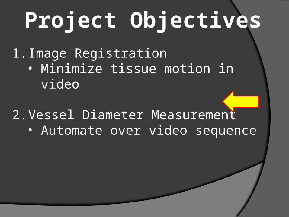
1. Image Registration• Minimize tissue motion in video
2. Vessel Diameter Measurement• Automate over video sequence
Project Objectives
Diameter Measurement• User inputs two seed points in first image• Diameter is distance between two points
𝑑2=(𝑥¿¿2−𝑥1)2+(𝑦2− 𝑦1)
2 ¿
Diameter Measurement• Program creates sub-regions around seed points• Compute similarity of current frame sub-region to reference frame sub-region
Diameter Measurement• Peak cross-correlation amplitude how far the regions have moved• Shift seed points by offset and re-calculate diameter
𝑑2=(𝑥¿¿2−𝑥1)2+(𝑦2− 𝑦1)
2 ¿
d
Feature TrackingFirst frame with
seed pointsfrom user
Create sub-regions based on input
points from previous frame
Final frame?
Go to next frame
Calculate new points (and
diameter) from peak cross-
correlation offset
End
Yes
No
Model Validation• Obtained expert manual diameter measurements
• The gold standard
• Compare these to diameters generated by the program with the same initial seed points
Results• Expert manual measurement
Results• Expert manual measurement
Results• Expert manual measurement
Conclusions• Successfully stabilized tissue motion in sequences
• Software is capable of making automated diameter measurements
• Resulting diameter measurements are on average within 1.5 microns of the gold standard
• Some post-hoc analysis and selection of results may be necessary (to identify periods of poor measurements)
Future Work• Test software on other sequences and imaging techniques
• Test with other similarity metrics
• Expand functionality to measure multiple vessels and ROIs along a single vessel
• Dr. Graham Fraser• Dr. Dwayne Jackson• Nicole Novielli
Acknowledgements
• Lee, J., Jirapatnakul, A., Reeves, A., Crowe, W., Sarelius, I. Vessel Diameter Measurement from Intravital Microscopy Annals of Biomedical Engineering, Vol. 37, No. 5, May 2009 (2009) pp. 913–926 • Brown, L. G. A survey of image registration techniques. ACM Comput. Surv. 24(4):325–376, 1992.
• J. P. Lewis. Fast Normalized Cross-Correlation. Industrial Light & Magic
References
Future Work• Optimize correlation amplitude
• Test software on other sequences and imaging techniques
• Test various size and position of sub-regions• Test with other similarity metrics
• Expand functionality to measure multiple vessels and ROIs along a single vessel
Results• Non-expert Measurement
Tracking• Video of tracked diameters:
•
Diameter Measurement• Automated method
J. Lee et al., Annals of Biomedical Eng., V. 37. No. 5:913–926, 2009
Image Registration• Pick sub-regions in an image• Compare relative positions with respect to reference• Best position: where regions have the highest similarity


![I Love You - Sarah McLachlan[1]](https://static.documents.pub/doc/80x56/577cdcba1a28ab9e78ab405f/i-love-you-sarah-mclachlan1.jpg)


