Case ReportNutcracker Cuboid Fracture: A Case Report and Review
Alan Lucerna ,1 James Espinosa ,1 Nicholas Butler,2
AshleyWenke,3 and Nicole Caltabiano3
1Department of Emergency Medicine, Rowan University SOM and Jefferson Health, Stratford, NJ, USA2Department of Podiatric Medicine, Jefferson Health, Stratford, NJ, USA3Rowan University SOM, Stratford, NJ, USA
Correspondence should be addressed to Alan Lucerna; [email protected]
Received 26 November 2017; Accepted 3 January 2018; Published 3 April 2018
Here we report the case of a 20-year-old female restrained driver who presented to the emergency department (ED) after a motorvehicle accident. She sustained an isolated fracture of her left cuboid, consistent with a nutcracker cuboid fracture. A cuboid fractureis considered rare. It is even more uncommon for a cuboid fracture to occur in isolation, without other associated injuries to thefoot. We discuss the mechanism, relevant anatomy, diagnosis, and principles of treatment of the nutcracker cuboid fracture.
1. Introduction
The anatomy of the cuboid is complex, with six articularsurfaces involvement in all of the intrinsic movements ofthe midfoot and hindfoot [1]. Due to this anatomy, a cuboidfracture is considered rare. It is even more uncommon for acuboid fracture to occur in isolation, without other associatedinjuries to the foot [2]. We report the case of a 20-year-oldfemale restrained driver who presented to the emergencydepartment (ED) after a motor vehicle accident.
2. Case Report
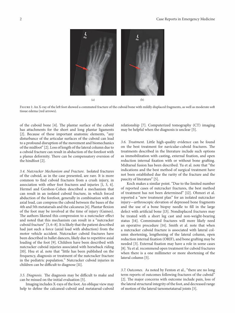
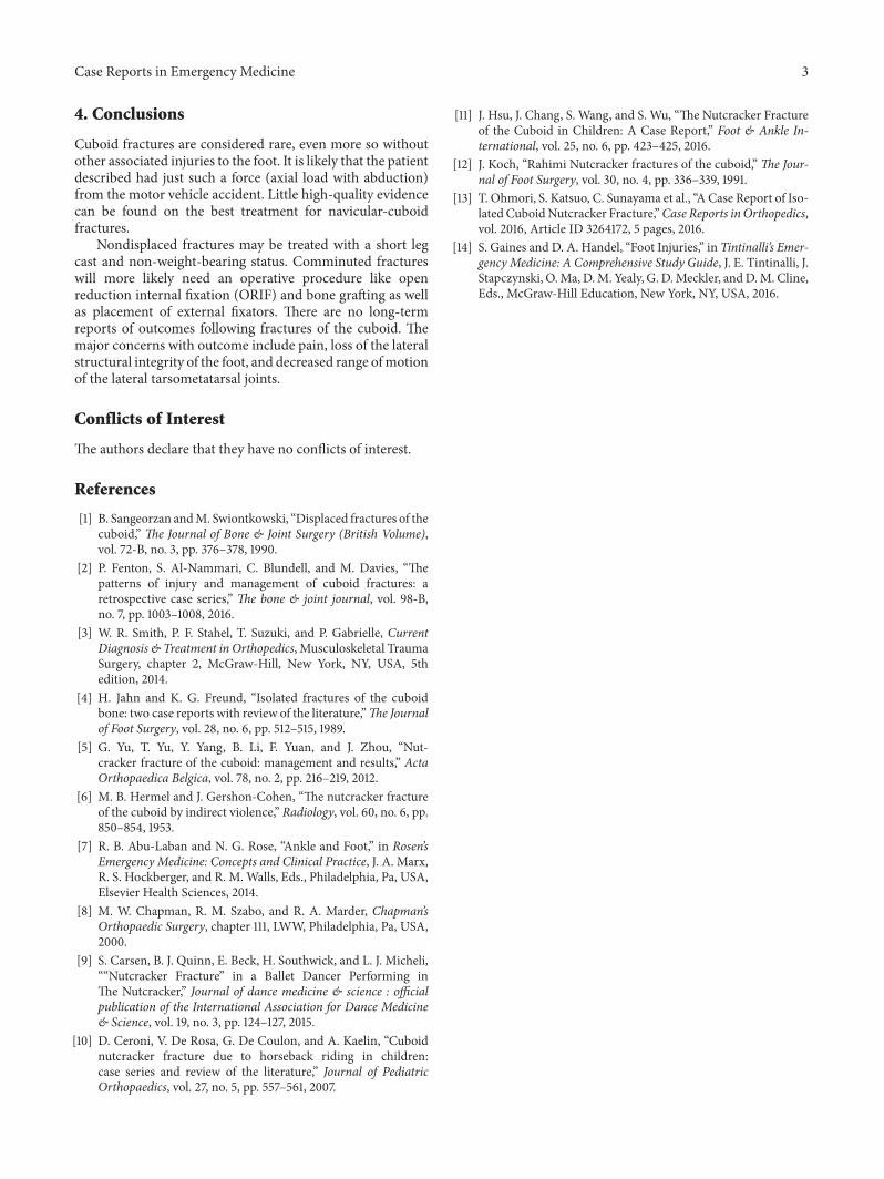
A 20-year-old female restrained driver presented to theemergency department (ED) after motor vehicle accident.She complained of left clavicular and left foot pain. A leftclavicular fracture was found on exam and on the imagingstudies. AnX-ray of the left foot showed a commuted fractureof the cuboid bone with mildly displaced fragments, as wellas moderate soft tissue edema (Figure 1). Clinical exam didnot show evidence of other foot or leg injuries. The restof her exam was unremarkable. An orthopedic consult wasobtained in the emergency department. A posterior fiberglasssplintwas recommended aswell as non-weight bearing on the
affected foot. The patient was to follow up on an outpatientbasis with orthopedic surgery. The fracture was consistentwith a nutcracker cuboid fracture.
3. Discussion
3.1. Incidence of Cuboid Fractures. A cuboid fracture is con-sidered rare. It is even more uncommon for a cuboid fractureto occur in isolation, without other associated injuries to thefoot [2]. This is said to be because of a relatively protectedposition in the midfoot [3].
3.2. Classification. According to Smith et al., who performeda large retrospective study of cuboid fractures, there is nocurrent widely accepted classification for cuboid fracturesand there are no long-term outcome studies [3].
3.3. Anatomy. The anatomy of the cuboid is complex, withsix articular surfaces’ involvement in all of the intrinsicmovements of the midfoot and hindfoot [1]. The cuboidconstitutes part of the lateral longitudinal arch of the foot[4]. It is the only bony structural support for the lateralcolumn of the midfoot [5]. Of note, the tendon of theperoneus longus courses in a groove on the inferior surface
HindawiCase Reports in Emergency MedicineVolume 2018, Article ID 3804642, 3 pageshttps://doi.org/10.1155/2018/3804642
Figure 1: An X-ray of the left foot showed a commuted fracture of the cuboid bone with mildly displaced fragments, as well as moderate softtissue edema (red arrows).
of the cuboid bone [4]. The plantar surface of the cuboidhas attachments for the short and long plantar ligaments[2]. Because of these important anatomic elements, “anydisturbance of the articular surfaces of the cuboid can leadto a profound disruption of the movement and biomechanicsof themidfoot” [2]. Loss of length of the lateral column due toa cuboid fracture can result in abduction of the forefoot witha planus deformity. There can be compensatory eversion ofthe hindfoot [2].
3.4. Nutcracker Mechanism and Fracture. Isolated fracturesof the cuboid, as in the case presented, are rare. It is morecommon to find cuboid fractures from a crush injury, inassociation with other foot fractures and injuries [1, 3, 4].Hermel and Gershon-Cohen described a mechanism thatcan result in an isolated cuboid fracture, in which forcedabduction of the forefoot, generally in combination with anaxial load, can compress the cuboid between the bases of the4th and 5th metatarsals and the calcaneus [6]. Plantar flexionof the foot may be involved at the time of injury (Gaines).The authors likened this compression to a nutcracker effectand noted that this mechanism can result in a “nutcrackercuboid fracture” [3, 6–8]. It is likely that the patient describedhad just such a force (axial load with abduction) from themotor vehicle accident. Nutcracker cuboid fractures havebeen described in ballet dancers, likely due to repetitive axialloading of the foot [9]. Children have been described withnutcracker cuboid injuries associated with horseback riding[10]. Hsu et al. note that “little has been published on thefrequency, diagnosis or treatment of the nutcracker fracturein the pediatric population.” Nutcracker cuboid injuries inchildren can be difficult to diagnose [11].
3.5. Diagnosis. The diagnosis may be difficult to make andcan be missed on the initial evaluation [5].
Imaging includes X-rays of the foot. An oblique viewmayhelp to define the calcaneal-cuboid and metatarsal-cuboid
relationship [7]. Computerized tomography (CT) imagingmay be helpful when the diagnosis is unclear [5].
3.6. Treatment. Little high-quality evidence can be foundon the best treatment for navicular-cuboid fractures. Thetreatments described in the literature include such optionsas immobilization with casting, external fixation, and openreduction internal fixation with or without bone grafting.Midtarsal fusion has been described. Yu et al. note that “theindications and the best method of surgical treatment havenot been established due the rarity of the fracture and thepaucity of literature” [5].
Koch makes a similar point. “Due to the limited numberof reported cases of nutcracker fractures, the best methodof treatment has not been determined” [12]. Ohmori et al.reported a “new treatment plan” for an isolated nutcrackerinjury—arthroscopic elevation of depressed bone fragmentsand the use of a bone biopsy needle to fill in the largedefect with artificial bone [13]. Nondisplaced fractures maybe treated with a short leg cast and non-weight-bearingstatus [14]. Comminuted fractures will more likely needan operative procedure [14]. Smith et al. note that whena nutcracker cuboid fracture is associated with lateral col-umn shortening, lengthening of the lateral column, openreduction internal fixation (ORIF), and bone grafting may beneeded [3]. External fixation may have a role in some cases[8]. Yu et al. recommend open treatment for cuboid fractureswhen there is a one millimeter or more shortening of thelateral column [5].
3.7. Outcomes. As noted by Fenton et al., “there are no longterm reports of outcomes following fractures of the cuboid”[2]. The major concerns with outcome include pain, loss ofthe lateral structural integrity of the foot, and decreased rangeof motion of the lateral tarsometatarsal joints [5].
Case Reports in Emergency Medicine 3
4. Conclusions
Cuboid fractures are considered rare, even more so withoutother associated injuries to the foot. It is likely that the patientdescribed had just such a force (axial load with abduction)from the motor vehicle accident. Little high-quality evidencecan be found on the best treatment for navicular-cuboidfractures.
Nondisplaced fractures may be treated with a short legcast and non-weight-bearing status. Comminuted fractureswill more likely need an operative procedure like openreduction internal fixation (ORIF) and bone grafting as wellas placement of external fixators. There are no long-termreports of outcomes following fractures of the cuboid. Themajor concerns with outcome include pain, loss of the lateralstructural integrity of the foot, and decreased range ofmotionof the lateral tarsometatarsal joints.
Conflicts of Interest
The authors declare that they have no conflicts of interest.
References
[1] B. Sangeorzan andM. Swiontkowski, “Displaced fractures of thecuboid,” The Journal of Bone & Joint Surgery (British Volume),vol. 72-B, no. 3, pp. 376–378, 1990.
[2] P. Fenton, S. Al-Nammari, C. Blundell, and M. Davies, “Thepatterns of injury and management of cuboid fractures: aretrospective case series,” The bone & joint journal, vol. 98-B,no. 7, pp. 1003–1008, 2016.
[3] W. R. Smith, P. F. Stahel, T. Suzuki, and P. Gabrielle, CurrentDiagnosis & Treatment in Orthopedics, Musculoskeletal TraumaSurgery, chapter 2, McGraw-Hill, New York, NY, USA, 5thedition, 2014.
[4] H. Jahn and K. G. Freund, “Isolated fractures of the cuboidbone: two case reports with review of the literature,”The Journalof Foot Surgery, vol. 28, no. 6, pp. 512–515, 1989.
[5] G. Yu, T. Yu, Y. Yang, B. Li, F. Yuan, and J. Zhou, “Nut-cracker fracture of the cuboid: management and results,” ActaOrthopaedica Belgica, vol. 78, no. 2, pp. 216–219, 2012.
[6] M. B. Hermel and J. Gershon-Cohen, “The nutcracker fractureof the cuboid by indirect violence,” Radiology, vol. 60, no. 6, pp.850–854, 1953.
[7] R. B. Abu-Laban and N. G. Rose, “Ankle and Foot,” in Rosen’sEmergency Medicine: Concepts and Clinical Practice, J. A. Marx,R. S. Hockberger, and R. M. Walls, Eds., Philadelphia, Pa, USA,Elsevier Health Sciences, 2014.
[8] M. W. Chapman, R. M. Szabo, and R. A. Marder, Chapman’sOrthopaedic Surgery, chapter 111, LWW, Philadelphia, Pa, USA,2000.
[9] S. Carsen, B. J. Quinn, E. Beck, H. Southwick, and L. J. Micheli,““Nutcracker Fracture” in a Ballet Dancer Performing inThe Nutcracker,” Journal of dance medicine & science : officialpublication of the International Association for Dance Medicine& Science, vol. 19, no. 3, pp. 124–127, 2015.
[10] D. Ceroni, V. De Rosa, G. De Coulon, and A. Kaelin, “Cuboidnutcracker fracture due to horseback riding in children:case series and review of the literature,” Journal of PediatricOrthopaedics, vol. 27, no. 5, pp. 557–561, 2007.
[11] J. Hsu, J. Chang, S. Wang, and S. Wu, “The Nutcracker Fractureof the Cuboid in Children: A Case Report,” Foot & Ankle In-ternational, vol. 25, no. 6, pp. 423–425, 2016.
[12] J. Koch, “Rahimi Nutcracker fractures of the cuboid,”The Jour-nal of Foot Surgery, vol. 30, no. 4, pp. 336–339, 1991.
[13] T. Ohmori, S. Katsuo, C. Sunayama et al., “A Case Report of Iso-lated CuboidNutcracker Fracture,”Case Reports in Orthopedics,vol. 2016, Article ID 3264172, 5 pages, 2016.
[14] S. Gaines and D. A. Handel, “Foot Injuries,” in Tintinalli’s Emer-gency Medicine: A Comprehensive Study Guide, J. E. Tintinalli, J.Stapczynski, O.Ma, D.M. Yealy, G. D.Meckler, andD.M. Cline,Eds., McGraw-Hill Education, New York, NY, USA, 2016.