1)Laboratory of Veterinary Pathology, School of Veterinary Medicine, Azabu University, Sagamihara, Kanagawa 252–5201, Japan2)Hamamatsu Zoo, Hamamatsu, Shizuoka 431–1209, Japan
(Received 28 February 2013/Accepted 28 June 2013/Published online in J-STAGE 12 July 2013)
ABSTRACT. Spontaneous crescentic glomerulonephritis (CrGN) in animals has only been reported in dog and sheep. We report the pathological features of CrGN in a 17-year-old male polar bear that died due to renal failure. Histologically, the lesions were characterized by fibrocel-lular crescents, adhesion between Bowman’s capsule and the glomerular capillary tuft and an increase in the mesangial matrix in glomeruli. The proliferating cells in the crescent were partly immunopositive for cytokeratin and intensely positive for vimentin, WT-1 and α-smooth muscle actin, suggesting they originated from parietal epithelial cells. Ultrastructually, thickening of the glomerular basement membrane and loss of epithelial cell foot processes were observed with electron-dense deposits.KEY WORDS: crescentic glomerulonephritis, immune-complex type, immunohistochemistry, polar bear, spontaneous.
doi: 10.1292/jvms.13-0108; J. Vet. Med. Sci. 75(11): 1535–1538, 2013
Primary glomerular disease, based on the WHO clas-sification for humans [3], is classified into the following three categories: 1) minor glomerular abnormalities, 2) focal/segmental lesions and 3) diffuse glomerulonephritis. Of these, crescentic glomerulonephritis (CrGN) is classi-fied as diffuse glomerulonephritis. A simple classification system that consists of membranous, mesangioproliferative, membranoproliferative and glomerulosclerosis is currently used for glomerulonephritis in domestic animals, because, compared with that in humans, knowledge about glomerular disease in animals is insufficient. CrGN is very rare and poorly understood in animals; therefore, it is not included in this classification [6].
In animals, glomerular crescent formation produced in as-sociation with various glomerular diseases, namely, familial renal disease in dog [20], sodium chloride poisoning in chick [15] and induced CrGN, has been reported in laboratory ani-mals including mouse [8], goat [12], sheep [17] and rabbit [5]. However, reports of naturally occurring CrGN as diffuse glomerulonephritis with poor prognosis are extremely rare. In livestock, it has been reported only in sheep [4]. Even though dogs with glomerular disease have been studied rela-tively well, CrGN has been documented in only 2 articles [11, 19]. Reports on renal disease in Ursidae are rare, and only one study has examined the effects of organic halogen concentration on renal tissue in 75 polar bears (Ursus mariti-mus), but there are no reports on primary glomerular disease [16]. This report contains the first description of CrGN in Ursidae.
The animal was a 17-year-old male polar bear born at the zoo. From January 30, 2010, diarrhea and vomiting were ob-
served. Severe renal failure (BUN: 305 mg/dl and creatinine: 9.95 mg/dl; normal ranges: 5–43 mg/dl and 0.4–2.4 mg/dl, respectively [14]) and liver failure (AST: 340 IU/l; normal range: 24–197 IU/l [14]) were revealed by blood biochemi-cal tests on January 30, 2010. Urine was not examined. The animal died on February 8, 2010 and was necropsied. Body weight was 300 kg at the time of death. The bear had not been given food or drugs with renal toxicity or inoculated with any vaccines. Grossly, all reniculi of both kidneys were discolored, with a lightly tanned color, in association with moderate sclerosis. Brown spots of 1–2 mm were diffusely scattered throughout the parenchyma. On the cut surface of kidney cortex, pinhead-sized white nodules were densely distributed, and there were a lot of petechiae. The liver was enlarged and yellow-brown in color with multiple dark-brown patches of 2–7 mm.
Partial specimens from visceral organs, including the kidneys, were fixed in 10% neutral buffered formalin and provided for histopathological examination. Three-microm-eter-thick sections were prepared from paraffin-embedded tissues and stained by HE, periodic acid-Schiff (PAS), Azan-Mallory (AZAN), periodic acid methenamine silver (PAM) and phosphotungstic acid-hematoxylin (PTAH). Acid fuchsin orange G (AFOG) stain was used to demon-strate immune deposits. The ratio of affected glomeruli was calculated using the HE sections. For investigation of the crescent components, immunostaining was performed on kidney sections with the following antibodies: cytokeratin (clone AE1/AE3, × 400, Dako, Japan Co., Kyoto, Japan), vimentin (clone V9, × 200, Dako), Wilm’s tumor 1 (WT-1, clone 6F-H2,× 100, Dako), α-smooth muscle actin (α-SMA, clone 1A4, × 20,000, Dako), lysozyme EC 3.2.1.17 (Mu-ramidase) (lysozyme, × 500, Dako) and myeloid /histiocyte antigen (MAC 387, clone MAC 387, × 300, Dako). To confirm the specificity of these antibodies, the tissues of this case were used as internal positive controls. In addition, for ultrastructural examination, a portion of the kidney tissue af-ter formalin fixation was refixed with 2.5% glutaraldehyde,
*CorrespondenCe to: Une, Y., Laboratory of Veterinary Pathology, Azabu University, 1–17–71 Fuchinobe, Chuo-ku, Sagamihara, Kanagawa 252–5201, Japan.
post-fixed in osmium tetroxide and embedded in epon resin. Ultra-thin sections (90–150 nm) were stained with uranyl acetate and lead citrate and then examined under a Hitachi H300 electron microscope.
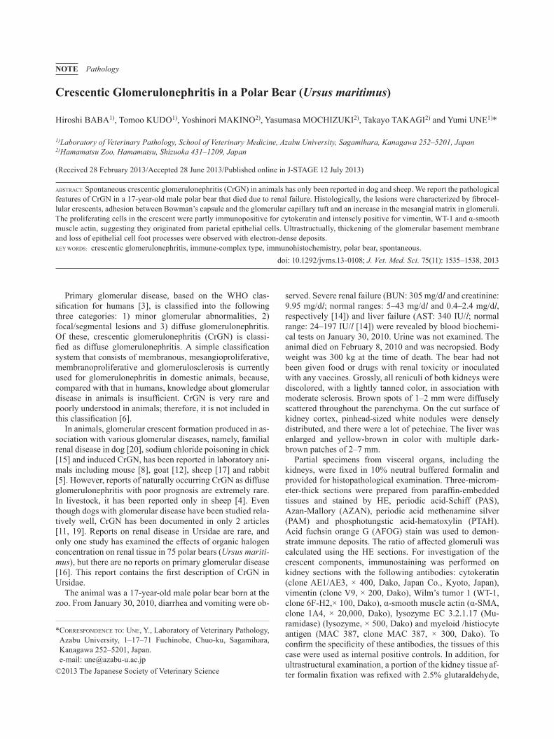
Histopathological examination of both kidneys revealed extensive interstitial fibrosis and multifocal tubular atrophy with hyaline casts. Seventy-two percent of the glomeruli
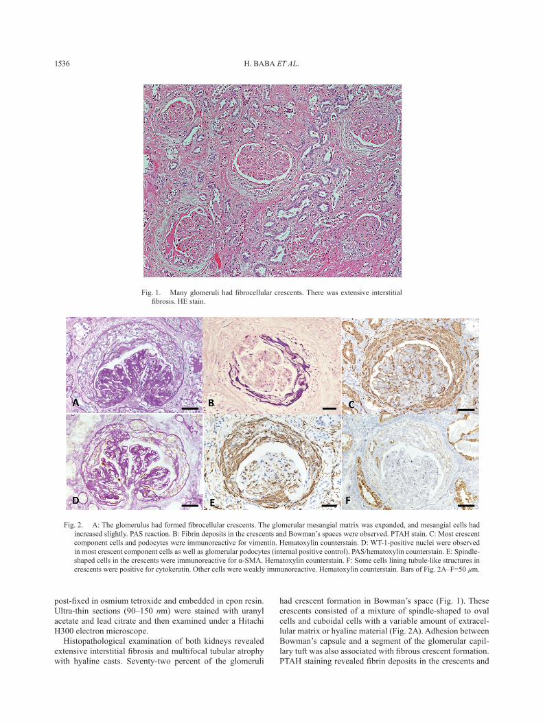
had crescent formation in Bowman’s space (Fig. 1). These crescents consisted of a mixture of spindle-shaped to oval cells and cuboidal cells with a variable amount of extracel-lular matrix or hyaline material (Fig. 2A). Adhesion between Bowman’s capsule and a segment of the glomerular capil-lary tuft was also associated with fibrous crescent formation. PTAH staining revealed fibrin deposits in the crescents and
Fig. 1. Many glomeruli had fibrocellular crescents. There was extensive interstitial fibrosis. HE stain.
Fig. 2. A: The glomerulus had formed fibrocellular crescents. The glomerular mesangial matrix was expanded, and mesangial cells had increased slightly. PAS reaction. B: Fibrin deposits in the crescents and Bowman’s spaces were observed. PTAH stain. C: Most crescent component cells and podocytes were immunoreactive for vimentin. Hematoxylin counterstain. D: WT-1-positive nuclei were observed in most crescent component cells as well as glomerular podocytes (internal positive control). PAS/hematoxylin counterstain. E: Spindle-shaped cells in the crescents were immunoreactive for α-SMA. Hematoxylin counterstain. F: Some cells lining tubule-like structures in crescents were positive for cytokeratin. Other cells were weakly immunoreactive. Hematoxylin counterstain. Bars of Fig. 2A–F=50 µm.
CRESCENTIC GLOMERULONEPHRITIS IN A POLAR BEAR 1537
Bowman’s spaces in about half of the affected glomeruli (Fig.2B). As features of the glomeruli, segmental duplica-tions of the glomerular basement membrane, mesangial ex-pansion, which exhibited a mild increase of mesangial cellularity, and glomerular fibrosis were generally observed. Focally, segmental or global glomerulosclerosis was ob-served. Deposits of bright red material were visible by AFOG staining in a small part of the mesangial matrix and in the capillary walls, which gave a positive reaction for fi-brin on PTAH staining. The grade of the lesion did not differ according to the location or reniculus. Diffuse degeneration and multifocal necrosis of cardiac muscle and hepatocytes were observed.
On immunostaining, cellular components of crescents stained diffusely positive for vimentin (Fig. 2C). WT-1-pos-
itive nuclei were observed in most crescent component cells as well as glomerular podocytes (internal positive control) (Fig. 2D). These crescentic cells were mainly composed of α-SMA-positive spindle-shaped cells (Fig. 2E). Variable cytokeratin expression was also found in crescentic cells (Fig. 2F). Lysozyme and MAC387 immunostaining did not reveal monocytic infiltration into the crescents, despite the existence of a positive reaction of macrophages of lung and liver as internal positive controls (Table 1).
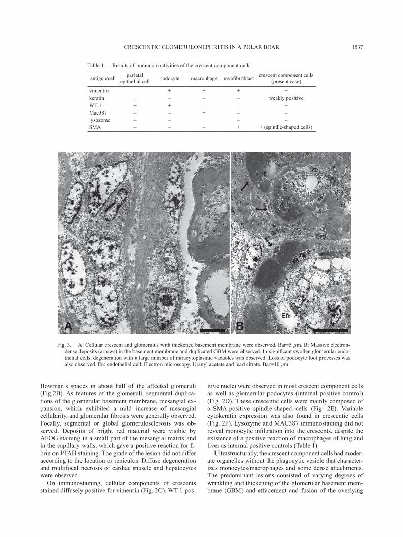
Ultrastructurally, the crescent component cells had moder-ate organelles without the phagocytic vesicle that character-izes monocytes/macrophages and some dense attachments. The predominant lesions consisted of varying degrees of wrinkling and thickening of the glomerular basement mem-brane (GBM) and effacement and fusion of the overlying
Table 1. Results of immunoreactivities of the crescent component cells
Fig. 3. A: Cellular crescent and glomerulus with thickened basement membrane were observed. Bar=5 µm. B: Massive electron-dense deposits (arrows) in the basement membrane and duplicated GBM were observed. In significant swollen glomerular endo-thelial cells, degeneration with a large number of intracytoplasmic vacuoles was observed. Loss of podocyte foot processes was also observed. En: endothelial cell. Electron microscopy. Uranyl acetate and lead citrate. Bar=10 µm.
H. BABA ET AL.1538
foot processes (Fig. 3A). Massive electron-dense deposits in the duplicated GBM were occasionally observed (Fig. 3B).
CrGN is a typical pathological finding that characterizes the pathology of rapidly progressive glomerulonephritis in humans, and crescent formation is observed in>50% of the glomeruli in human CrGN patients [9, 13]. Since fre-quent crescent formation observed in more than 70% of the glomeruli and the presence of fibrin exudation indicating rapid progression are consistent with CrGN of fibro-cellular crescents in humans and animal-induced models, the present case was diagnosed as CrGN in a polar bear. To our knowl-edge, this is the first reported case of CrGN in Ursidae.
The pathogenesis of CrGN has not been fully elucidated, but it has been considered to involve an immune-mediated mechanism [8, 12]. On the basis of the immunological findings, CrGN in humans is classified into the following three types: 1) anti-glomerular basement membrane (GBM) type, 2) pauci-immune type and 3) immune-complex type [1]. Spontaneous CrGN in animals has not been studied sufficiently, because CrGN is rarely seen. In this case, the involvement of an immune-complex mechanism was sus-pected, because the electron-dense deposits in GBM were observed by transmission electron microscopy and there is no glomerular disease associated with glomerular crescent formation. However, in order to draw a definitive conclu-sion, further examination by immunostaining is required.
In humans, monocytes/macrophages [2, 7, 10, 18] and parietal epithelial cells (PEC) [13] have been considered as the origin of crescent component cells. In addition, a positive reaction for α-SMA of crescent component cells has been interpreted as an epithelial-mesenchymal transition [12]. From these findings of ultrastructural examination and immunostaining of crescent component cells in this case, showing a similar immune phenotype to human PEC [3], the crescents themselves seemed to originate from the prolifera-tion of PEC.
REFERENCES
1. Alpers, C. E. 2010. The kidney. pp. 903–969. In: Robbins & Cotran Pathologic Basis of Disease, 8th ed. (Vinay, K., Abul, K. A., Nelson, F. and Jon, A. eds.), Saunders, Philadelphia.
2. Atkins, R. C., Holdsworth, S. R., Glasgow, E. F. and Matthews, F. E. 1976. The macrophagen in human rapidly progressive glo-merulonephritis. Lancet 1: 830–832. [Medline] [CrossRef]
3. Churg, J., Bernstein, J. and Glassock, R. J. 1995. Renal Disease: Classification and Atlas of Glomerular Diseases, 2nd ed., Igaku-Shoin, Tokyo.
4. Frelier, P. F., Armstrong, D. L. and Pritchard, J. 1990. Ovine mesangiocapillary glomerulonephritis type I and crescent for-mation. Vet. Pathol. 27: 26–34. [Medline] [CrossRef]
5. Germuth, F. G. Jr., Taylor, J. J., Siddiqui, S. Y. and Rodriguez, E. 1977. Immune complex disease. VI. Some determinants of the varieties of glomerular lesions in the chronic bovine serum
albumin-rabbit system. Lab. Invest. 37: 162–169. [Medline] 6. Grant Maxie, M. and Newman, S. J. 2007. pp.425–522. Pathol-
ogy of Domestic Animals, Chapter 4 Urinary System. 5th ed. (Grant Maxie ed.), W. B. Saunders, Philadelphia.
7. Hancock, W. W. and Atkins, R. C. 1984. Cellular composition of crescents in human rapidly progressive glomerulonephritis iden-tified using monoclonal antibodies. Am. J. Nephrol. 4: 177–181. [Medline] [CrossRef]
8. Iskandar, S. S., Jennette, J. C., Wilkman, A. S. and Becker, R. L. 1982. Interstrain variations in nephritogenicity of heterologous protein in mice. Lab. Invest. 46: 344–351. [Medline]
9. Jennette, J. C. 2003. Rapidly progressive crescentic glomerulo-nephritis. Kidney Int. 63: 1164–1177. [Medline] [CrossRef]
10. Lan, H. Y., Nikolic-Paterson, D. J., Mu, W. and Atkins, R. C. 1997. Local macrophage proliferation in the pathogenesis of glomerular crescent formation in rat anti-glomerular basement membrane (GBM) glomerulonephritis. Clin. Exp. Immunol. 110: 233–240. [Medline] [CrossRef]
11. Macdougall, D. F., Cook, T., Steward, A. P. and Cattell, V. 1986. Canine chronic renal disease: prevalence and types of glomeru-lonephritis in the dog. Kidney Int. 29: 1144–1151. [Medline] [CrossRef]
12. Ohnuki, T. 1975. Crescentic glomerulonephritis induced in the goat by immunization with homologous or heterologous glomerular basement membrane antigen. Acta Pathol. Jpn. 25: 319–331. [Medline]
13. Pusey, C. D. and Lockwood, C. M. 1989. Autoimmunity in rap-idly progressive glomerulonephritis. Kidney Int. 35: 929–937. [Medline] [CrossRef]
14. Ramsay, E. C. 2003. Ursidae and Hyaenidae. pp. 523–538. In: Zoo and Wild Animal Medicine, 5th ed. (Fowler, M. E. and Miller, R. E. eds.), W. B. Saunders, Philadelphia.
15. Sokkar, S. M., Hussein, B. M. and Mohammed, M. A. 1983. Renal lesions in baby chicks due to sodium chloride poisoning. Avian Pathol. 12: 277–285. [Medline] [CrossRef]
16. Sonne, C., Dietz, R., Leifsson, P. S., Born, E. W., Kirkegaard, M., Letcher, R. J., Muir, D. C., Riget, F. E. and Hyldstrup, L. 2006. Are organohalogen contaminants a cofactor in the devel-opment of renal lesions in east Greenland polar bears (Ursus maritimus)? Environ. Toxicol. Chem. 25: 1551–1557. [Medline] [CrossRef]
17. Steblay, R. W. 1962. Glomerulonephritis induced in sheep by injections of heterologous glomerular basement membrane and Freund’s complete adjuvant. J. Exp. Med. 116: 253–272. [Med-line] [CrossRef]
18. Thomson, N. M., Holdsworth, S. R., Glasgow, E. F. and Atkins, R. C. 1979. The macrophage in the development of experimental crescentic glomerulonephritis. Studies using tissue culture and electron microscopy. Am. J. Pathol. 94: 223–240. [Medline]
19. Vilafranca, M., Wohlsein, P., Trautwein, G., Leopold-Temmler, B. and Nolte, I. 1994. Histological and immunohistological clas-sification of canine glomerular disease. Zentralbl. Veterinarmed. B A. 41: 599–610. [Medline]
20. Wakamatsu, N., Surdyk, K., Carmichael, K. P. and Brown, C. A. 2007. Histologic and ultrastructural studies of juvenile onset renal disease in four Rottweiler dogs. Vet. Pathol. 44: 96–100. [Medline] [CrossRef]