Critical role of myofascial reeducation in pediatric sleep-disordered breathing C. Guilleminault a,⇑ , Y.S. Huang b , P.J. Monteyrol d , R. Sato a , S. Quo e , C.H. Lin c a Stanford University, Sleep Medicine Division, United States b Pediatric Sleep Medicine, Taiwan c Cranio-Facial Center, Chang-Gung University and Memorial, Taiwan d Clinique Oto-Laryngologique, Bordeaux, France e Orthodontic Department, University of California, San Francisco Dental School, United States article info Article history: Received 25 September 2012 Received in revised form 16 December 2012 Accepted 25 January 2013 Available online xxxx Keywords: Pediatric sleep-disordered breathing Adenotonsillectomy Orthodontics Myofunctional therapy Retrospective study Long-term recurrence abstract Background: Limited studies suggest that pubertal development may lead to a recurrence of sleep- disordered breathing (SDB) despite previous curative surgery. Our study evaluates the impact of myofunctional reeducation in children with SDB referred for adenotonsillectomy, orthodontia, and myofunctional treatment in three different geographic areas. Methods: A retrospective investigation of children with polysomnographic analysis following adenoton- sillectomy were referred for orthodontic treatment and were considered for myofunctional therapy. Clinical information was obtained during pediatric and orthodontic follow-up. Polysomnography (PSG) at the time of diagnosis, following adenotonsillectomy, and at long-term follow-up, were compared. The PSG obtained at long-term follow-up was scored by a single-blinded investigator. Results: Complete charts providing the necessary medical information for long-term follow-up were lim- ited. A subgroup of 24 subjects (14 boys) with normal PSG following adenotonsillectomy and orthodontia were referred for myofunctional therapy, with only 11 subjects receiving treatment. Follow-up evalua- tion was performed between the 22nd and 50th month after termination of myofunctional reeducation or orthodontic treatment if reeducation was not received. Thirteen out of 24 subjects who did not receive myofunctional reeducation developed recurrence of symptoms with a mean apnea–hypopnea index (AHI) = 5.3 ± 1.5 and mean minimum oxygen saturation = 91 ± 1.8%. All 11 subjects who completed myo- functional reeducation for 24 months revealed healthy results. Conclusion: Despite experimental and orthodontic data supporting the connection between orofacial muscle activity and oropharyngeal development as well as the demonstration of abnormal muscle con- traction of upper airway muscles during sleep in patients with SDB, myofunctional therapy rarely is con- sidered in the treatment of pediatric SDB. Absence of myofascial treatment is associated with a recurrence of SDB. Ó 2013 Elsevier B.V. All rights reserved. 1. Introduction Obstructive sleep apnea (OSA) has become increasingly recog- nized as a notable health concern in children given its conse- quences on behavior, function, and quality of life. The importance of early recognition and treatment in children is paramount to maximizing resolution of symptoms and potential avoidance of OSA syndrome during adulthood. Adenotonsillectomy and palatal expansion have established their roles in the treatment of OSA after demonstrating considerable improvement related to adenoid or tonsillar hypertrophy, maxillary or mandibular deficiency, and orthodontic or craniofacial abnormalities. However, the imple- mentation of other treatment modalities such as myofascial reeducation also may play a role in the optimization of sleep- disordered breathing (SDB). Functional myofascial reeducation in children has been well- established in the treatment of abnormal orofacial development for more than 40 years [1]. However, few studies have been pub- lished supporting the benefits of orofacial reeducation compared to the numerous studies reinforcing the utility of surgical and orthodontic treatments in SDB [2]. Although the role of orofacial education remains largely variable between institutions, the most notable results have been described when myofunctional thera- pists and orthodontists worked in collaboration to manage orofa- cial weakness. Although promising, the efficacy of myofunctional therapy in combination with surgical and orthodontic treatment is unclear. The purpose of our study was to evaluate the impact of myofunctional reeducation protocols on orofacial muscle weak- ness and the treatment of SDB in children following surgical and orthodontic optimization. 1389-9457/$ - see front matter Ó 2013 Elsevier B.V. All rights reserved. http://dx.doi.org/10.1016/j.sleep.2013.01.013 ⇑ Corresponding author. Tel.: +1 650 723 6601; fax: +1 650 725 8910. E-mail address: [email protected](C. Guilleminault). Sleep Medicine xxx (2013) xxx–xxx Contents lists available at SciVerse ScienceDirect Sleep Medicine journal homepage: www.elsevier.com/locate/sleep Please cite this article in press as: Guilleminault C et al. Critical role of myofascial reeducation in pediatric sleep-disordered breathing. Sleep Med (2013), http://dx.doi.org/10.1016/j.sleep.2013.01.013
Transcript
Critical role of myofascial reeducation in pediatric sleep-disordered breathing
C. Guilleminault a,!, Y.S. Huang b, P.J. Monteyrol d, R. Sato a, S. Quo e, C.H. Lin c
a Stanford University, Sleep Medicine Division, United Statesb Pediatric Sleep Medicine, TaiwancCranio-Facial Center, Chang-Gung University and Memorial, TaiwandClinique Oto-Laryngologique, Bordeaux, FranceeOrthodontic Department, University of California, San Francisco Dental School, United States
a r t i c l e i n f o
Article history:Received 25 September 2012Received in revised form 16 December 2012Accepted 25 January 2013Available online xxxx
Background: Limited studies suggest that pubertal development may lead to a recurrence of sleep-disordered breathing (SDB) despite previous curative surgery. Our study evaluates the impact ofmyofunctional reeducation in children with SDB referred for adenotonsillectomy, orthodontia, andmyofunctional treatment in three different geographic areas.Methods: A retrospective investigation of children with polysomnographic analysis following adenoton-sillectomy were referred for orthodontic treatment and were considered for myofunctional therapy.Clinical information was obtained during pediatric and orthodontic follow-up. Polysomnography (PSG)at the time of diagnosis, following adenotonsillectomy, and at long-term follow-up, were compared.The PSG obtained at long-term follow-up was scored by a single-blinded investigator.Results: Complete charts providing the necessary medical information for long-term follow-up were lim-ited. A subgroup of 24 subjects (14 boys) with normal PSG following adenotonsillectomy and orthodontiawere referred for myofunctional therapy, with only 11 subjects receiving treatment. Follow-up evalua-tion was performed between the 22nd and 50th month after termination of myofunctional reeducationor orthodontic treatment if reeducation was not received. Thirteen out of 24 subjects who did not receivemyofunctional reeducation developed recurrence of symptoms with a mean apnea–hypopnea index(AHI) = 5.3 ± 1.5 and mean minimum oxygen saturation = 91 ± 1.8%. All 11 subjects who completed myo-functional reeducation for 24 months revealed healthy results.Conclusion: Despite experimental and orthodontic data supporting the connection between orofacialmuscle activity and oropharyngeal development as well as the demonstration of abnormal muscle con-traction of upper airway muscles during sleep in patients with SDB, myofunctional therapy rarely is con-sidered in the treatment of pediatric SDB. Absence of myofascial treatment is associated with arecurrence of SDB.
! 2013 Elsevier B.V. All rights reserved.
1. Introduction
Obstructive sleep apnea (OSA) has become increasingly recog-nized as a notable health concern in children given its conse-quences on behavior, function, and quality of life. The importanceof early recognition and treatment in children is paramount tomaximizing resolution of symptoms and potential avoidance ofOSA syndrome during adulthood. Adenotonsillectomy and palatalexpansion have established their roles in the treatment of OSA afterdemonstrating considerable improvement related to adenoid ortonsillar hypertrophy, maxillary or mandibular deficiency, andorthodontic or craniofacial abnormalities. However, the imple-mentation of other treatment modalities such as myofascial
reeducation also may play a role in the optimization of sleep-disordered breathing (SDB).
Functional myofascial reeducation in children has been well-established in the treatment of abnormal orofacial developmentfor more than 40 years [1]. However, few studies have been pub-lished supporting the benefits of orofacial reeducation comparedto the numerous studies reinforcing the utility of surgical andorthodontic treatments in SDB [2]. Although the role of orofacialeducation remains largely variable between institutions, the mostnotable results have been described when myofunctional thera-pists and orthodontists worked in collaboration to manage orofa-cial weakness. Although promising, the efficacy of myofunctionaltherapy in combination with surgical and orthodontic treatmentis unclear. The purpose of our study was to evaluate the impactof myofunctional reeducation protocols on orofacial muscle weak-ness and the treatment of SDB in children following surgical andorthodontic optimization.
1389-9457/$ - see front matter ! 2013 Elsevier B.V. All rights reserved.http://dx.doi.org/10.1016/j.sleep.2013.01.013
Please cite this article in press as: Guilleminault C et al. Critical role of myofascial reeducation in pediatric sleep-disordered breathing. Sleep Med (2013),http://dx.doi.org/10.1016/j.sleep.2013.01.013
Our retrospective analysis involving prepubertal children diag-nosed with OSA, who were referred for orthodontic treatment afterpresenting with residual symptoms of abnormal breathing follow-ing adenotonsillectomy, couldonlydrawa small numberof subjects.
Data collection was performed in three different regions of theworld, including the San Francisco Bay area, Taiwan, and France.Our analysis involved three different pediatric sleep centers work-ing with otolaryngologists, orthodontists, and functional thera-pists. The three sleep centers performed all sleep monitoring andwere referral centers for large geographic areas. The participatingsleep clinics and the orthodontic practices had a collaborativeworking relationship spanning from 6 to 14 years.
Retrieval of health information for children was variable buttargeted those initially seen between the ages of 3 and 6 years pre-ceding confirmation of SBD by nocturnal polysomnography (PSG).If a child was confirmed to have OSA by PSG, the second stepwas to determine the presence of adequate follow-up and appro-priate documentation including subsequent PSGs and documenta-tion from other specialists. Most charts did not fulfill these criteriaand were excluded from our study. Charts that had systematic PSGat different phases of follow-up were those of children seen byorthodontists either postadenotonsillectomy or without otolaryng-ologic intervention. Children often were referred to both a func-tional reeducation specialist and to an orthodontist in an effortto perform the investigation where myofunctional therapy waspracticed. Children were followed in sleep medicine and orthodon-tic clinics with variable schedules.
Despite being followed in these clinics, postorthodontic treat-ment PSG records often were unavailable and complete documen-tation often was absent, excluding a large number of cases. Oncethe necessary clinical data and PSG reports were confirmed, iden-tifiers were removed and data were extracted (Fig. 1). Anonymousanalyses of clinical and polysomnographic data were performed.Retrospective analyses of unidentified PSG and of clinical informa-tion were approved by the internal review boards.
All surveyed subjects were prepubertal children between theages of 3.6 and 6.6 years at the time of their initial visit. Initialassessment of each child included clinical interview, pediatric andsleep clinical evaluation, completion of the pediatric sleep ques-tionnaire (PSQ), a questionnaire validated in different languages[3,4], and nocturnal PSG. Following clinical and PSG evaluation,all children diagnosed with OSA were referred to otolaryngologyfor surgical evaluation. All subjects except for one had adenotonsil-lectomy performed and all were followed up after surgery with re-peat clinical evaluation and PSG. Subjects with residual OSAdetected on postsurgery PSG were sent for orthodontic evaluation[5]. Once the decision regarding orthodontic treatment was made(i.e., rapid maxillary expansion or bimaxillary expansion), recom-mended myofunctional reeducation also was performed [1].
Subjects were followed at an orthodontic practice during theapplication of orthodontic treatment and also were followed attheir sleep clinics 6 to10 months following initiation of their ortho-dontic treatment. Concomitant use of myofunctional reeducationwas documented as being implemented or as recommendationnot followed. Repeat PSG was performed following orthodontictreatment with or without functional reeducation. Data from myo-functional reeducation clinics were used solely to monitor compli-ance with follow-up appointments and to monitor duration oftreatment. Subjects were most often seen during their scheduledorthodontic follow-up. Less frequently they were seen severalyears after initiation of orthodontic treatment due to planned fol-low-up visits or due to recurrence of sleep-related symptoms; inthis case, they were referred back to sleep clinics. During long-termfollow-up visits, the reassessment always involved clinical inter-
views, PSQ, clinical pediatric evaluation and sleep evaluation,determination of height and weight based on body mass index,sleep medicine examination, myofunctional orofacial status, andnocturnal PSG.
All long-term follow-up PSGs (i.e., last investigation performed)were transferred to new compact discs with recordings formattedin European Data Format. This transfer allowed analysis of all PSGsperformed on various sleep programs to be anonymously rescoredby a single scorer. PSG rescoring could not be performed on the ini-tial PSGs in the same fashion. However, all centers used the sameatlases and guidelines for scoring sleep and breathing variables.
All subjects were evaluated by full-night PSG performed in asleep laboratory and included the following electrophysiologicparameters, electroencephalogram (EEG) (three channels), electro-oculogram (two channels), electrocardiogram, chin electromyo-gram (EMG), leg EMG (one channel), nasal pressure cannula, oralthermistor, thoracic and abdominal belts, snoring sensor, pulseoximetry, position sensor, and video recording. Variations to themontage included an additional second leg EMG, a fourth EEG,transcutaneous CO2 or end-tidal CO2, and the thoracic and abdom-inal belts were either piezoelectric or inductive plethysmography.All recordings lasted a minimum of 7.5 hours. Individuals were as-signed corresponding identification numbers and their data werecompiled using the Microsoft Excel program to perform statisticalanalyses of the results.
Myofunctional reeducation specialists were trained in variouscountries and were divided into two categories of either speechtherapists or specialists in muscle reeducation. Speech therapistswere trained in the United States, whereas muscle reeducationspecialists were trained outside of the United States. Myofunc-tional specialists obtained university degrees in functional reedu-cation with a subspecialty in myofunctional reeducation andpracticed validated therapeutic protocols. Treatment protocolsare similar in different countries [1]. In the United States, if notsanctioned by a diploma, courses are administered (particularlyin California) by trained individuals often trained in other coun-tries. The myofunctional re-educators involved in the three partic-ipating sleep centers had similar myofunctional reeducationtraining, including several years of experience with treatmentmodalities and use of the same type of report forms. Similar exer-cise regimens and daily durations of treatment were recommendedto parents. Frequency of visits varied not with the sleep center butwith the individual and were based on the needs of each case. Vis-its were more frequent at the initiation of treatment and less fre-quent as time passed. Daily exercise performance was recordedby parents in a log and reviewed by re-educators at visits. Reedu-cation programs were completed after 2 years.
3. Reeducation
Myofunctional reeducation involves strengthening of the ton-gue and orofacial muscles by teaching individuals how to reposi-tion muscles to the appropriate position. The tongue should bekept in a high position during sleep with its dorsal-terminal endin constant contact with the palatine striae located on the anterioraspect of the palate. Reeducation typically is easier in children ages6 years and older, but it is largely related to the degree of effortparents make in reinforcing a subject to perform his or her exer-cises. Exercises are initially repeated several times per day with aquick initial increase in frequency during the earliest phase oftreatment. This phase requires the subject and one parent to fre-quently follow-up with a specialist during the first 6 months andless frequently thereafter. The amount of follow-up depends onthe duration of therapy needed, but once the subject has gainedthe desired tongue position along with appropriate strength the
2 C. Guilleminault et al. / Sleep Medicine xxx (2013) xxx–xxx
Please cite this article in press as: Guilleminault C et al. Critical role of myofascial reeducation in pediatric sleep-disordered breathing. Sleep Med (2013),http://dx.doi.org/10.1016/j.sleep.2013.01.013
frequency of follow-up can be extended. The subject is then pri-marily monitored to insure continued appropriate developmentuntil the completion of treatment. The investigation took place be-tween the 22nd and 50th month following termination of the myo-functional treatment program, independent of the amount of timespent in the program. If the program was never implemented, sub-jects were seen for follow-up between the 28th and 34th monthfollowing termination of orthodontic treatment.
4. Analysis
The information collected for our study included gender, age attime of each treatment phase and testing, clinical concerns andsymptoms, PSQ results, and results of clinical orofacial evaluation.Description of the nasofacial and orofacial examination included
Friedman classification tonsil size [6]; modified Mallampati score[7,8]; calculated overjet (mm); evaluation of the hard palate, whichwas categorized as high and narrow, low lying, or normal; and thepresence of enlarged inferior nasal turbinates categorized as occupy-ing less than 50% or 50% or more of the space inside the nostrils. Thepresence or absence of nasal valve collapse, deviated septum, smallmandible, overbite, and awake-mouth breathing also were docu-mented. Absence of anterior short frenulumwas affirmed. Head pos-ture was noted in lateral position [9,10] but nuchal Solow angle wasnot calculated. Clinical information thatwas recorded during follow-up evaluation but was unavailable at initial presentation includedpreferential chewing to one side, presence of visualized facial asym-metry, presence of palpable asymmetry of masseteric muscles atmaximum clinching, and results of myofunctional evaluation per-formed by a reeducation specialist. Such information was retrievedfrom orthodontic and myofunctional reeducation charts. Individual
Fig. 1. Graph of initial charts retrieved for study. The graph documents the poor follow-up that occurred for a long time in the past after diagnosis and initial treatment ofobstructive sleep apnea (OSA). Many children had no sleep follow-up and absence of sleep recordings postadenotonsillectomy. Seventy one children were seen againposttreatment and had systematic investigation by both a sleep specialist and an orthodontist. Evaluation indicated validity of performing orthodontic treatment in 46children. Rapid maxillary expansion and bimaxillary treatments were recommended (32 rapid maxillary expansion; 14 bimaxillary treatments). Only 26 children had gooddocumentation of treatment at follow-up. Myofunctional therapy had been recommended in 24 of them in association with orthodontic treatment. However, myofunctionaltherapy was only performed in 11 children and 13 either did not follow the recommendations or quickly dropped out of the study.
C. Guilleminault et al. / Sleep Medicine xxx (2013) xxx–xxx 3
Please cite this article in press as: Guilleminault C et al. Critical role of myofascial reeducation in pediatric sleep-disordered breathing. Sleep Med (2013),http://dx.doi.org/10.1016/j.sleep.2013.01.013
orthodontists and myofunctional re-educators worked as a teamwith one orthodontist working with one preferred re-educator.
The long-term follow-up PSG recording was scored blindly by asingle investigator, whereas all other PSG results were reportedwithout access to the actual recording. The scoring was based onthe manual for sleep scoring by Rechtschaffen and Kale [11], theAmerican Sleep Disorders Association recommendation for EEGarousal scoring [12], and the American Academy of Sleep Medicinecriteria for scoring hypopneas and apneas [13]. Hypopneas weredefined by a 50% reduction in nasal cannula curve amplitude andan associated drop of 3% or more in oxygen saturation. The usualsubdivision of obstructive, mixed, and central events was followed.Events defined as postarousal central apneas were eliminated fromthe apnea–hypopnea index (AHI) score. There was additionalscoring of flow limitation based on the definition of Hosselet et al.[14]. The nasal cannula curve was compared to published patternsinvolving flattening or truncation of the curve during inspiration[15]. The percentage of flow limitation was determined by thenumber of 30-second epochs containing the presence of flow limita-tion [16]. An epochwas scoredwith flow limitation if it was presentfor more than 15 seconds (i.e., more than 50% of the scored PSGepoch). The percentage of flow limitationwas calculated by dividingthe total flow limited sleep time (i.e., number of 30-s epochs scoredwith flow limitationmultiplied by two) by the total sleep time [16].The score of flow limitation was not available for the initial record-ings in many subjects and was only systematically obtained in thereports from the postorthodontic treatment and the rescoredrecordings.
Presence or absence of mouth breathing was noted in the resultsof each PSG based on the mouth thermistor tracing but was notquantified. As previously mentioned the long-term follow-up PSGswere scored by a single-blinded scorer. Comparisons of PSG resultsbetween the subjects with and without myofunctional treatmentwere performed using Wilcoxon signed rank tests and v2 tests.
5. Results
5.1. Subjects involved in retrospective survey
An initial database of 408 pediatric cases diagnosed with OSA byPSG was established and was evaluated by an otolaryngologist whoperformed surgery and who subsequently had a postsurgical PSG.As previously mentioned many charts were incomplete when look-ing for further follow-up andwere excluded. From this database, 71subjects with documented visits to an orthodontist postadenoton-sillectomy were retrieved. Children seen by orthodontists for eval-uation had better documentation than those seen in other places,reflecting the higher representation of this subgroup in the fol-low-up survey. Children lacking the syndromic presentation butwho had close orthodontic follow-up led to closer evaluation andanatomic findings observed in subjects recognized with SDB. Docu-mented charts revealed that 46 of these subjects were consideredfor orthodontic treatment and simultaneous myofascial reeduca-tion due to persistent OSA at PSG, even if improvement was notedpostadenotonsillectomy. Of these 46 subjects, 24 had retrievablefollow-up documentation including myofunctional treatmentinformation (Fig. 1 [graph]). These 24 nonoverweight subjects (14boys) (17% of initial database) formed the study group of thosewho satisfied the inclusion parameters being evaluated.
5.2. Evaluation at entry
The results at entry are presented in Tables 1 and 2. All subjectspresented with clinical concerns, symptoms, and anatomic findingsconsistent with OSA, with the PSG confirming the diagnosis. Ana-tomic investigation at entry showed that out of the 24 cases, 23
had a tonsil-size scale of three or four. Twenty-three subjectshad a modified Mallampati scale score of three or four, and onesubject had a modifiedMallampati scale score of two. Inferior nasalturbinates were scored with occupying nasal space >50% in 13cases. Nasal septum deviation was found in 14 cases, and all ofthem had a narrow palatal vault. Fourteen subjects had been re-ferred and treated for nasal allergies with treatment consisting ofnasal steroids, allergic desensitization, or both. Of the 401 initialnonoverweight subjects being evaluated, 90% had a tonsil scoreof three or four, 73% had a modified Mallampati scale score of threeor four, and 48% were mentioned to have a high and narrow palatalvault. Statistics revealed from the v2 test indicated that the 24studied subjects were significantly different in Mallampati scalescore three and four (P = .01) and presence of high and narrow pal-atal vault (P = .001), but the initial anatomic description was simi-lar in 46% of the cases.
5.3. Initial treatment
Twenty-three subjects had adenotonsillectomy (T&A). Addi-tionally, five subjects had radiofrequency ablation of the inferiornasal turbinates performed at the time of T&A. One girl was feltto have small tonsils that would not benefit from surgery butwas directly referred for orthodontia, given her high and narrowarched hard palate. None of the subjects had an abnormally placedanterior frenulum.
The data retrieved postadenotonsillectomy and postorthodontiatreatments are presented in Tables 1 and 2, including the one girlsubject who was sent directly for orthodontia treatment. Althoughsymptoms were reportedly improved in all 23 cases following sur-gery, clinical concerns and symptoms were not completely elimi-nated, and the persistence of abnormal breathing was confirmedwith PSG analysis. The presence of mouth breathing was noted inall postsurgical cases but was not quantified. All 24 subjects weresent to orthodontists and in all cases were expected to benefit fromorthodontic treatment.
Following orthodontic evaluation rapid maxillary or bimaxillaryexpansion was performed, and orthodontic equipment was kept inplace for 8 to12 months. Follow-up evaluation in the sleep clinicwith follow-up PSG was performed near the time of orthodonticequipment removal (Tables 1 and 2). Clinical concerns and symp-toms related to SDB were absent with the exception of one subjectwith persistence of attention deficit and hyperactivity disorderthat may not have been related to SDB. Despite noted improve-ment in symptoms following treatment, persistence of intermit-tent agitated sleep with teeth clenching was reported, for whichthe subject was referred back to the orthodontist. PSG showed anormal AHI and oxygen saturation. However, in seven cases pres-ence of mouth breathing without indication of frequency was iden-tified in the PSG. Parents also had been referred to myofunctionalre-educators. Review of charts indicated that parents regularly fol-lowed up for orthodontic treatments. Of the 24 subjects, 10 did notgo to myofunctional reeducation and three children missed routineappointments and training sessions, did not adhere to the re-quested exercise regimen, or did not participate in long-termfollow-up with re-educators. Conversely 11 subjects were adher-ent to myofunctional treatment and were compliant with routinefollow-up with their orthodontists. None of the subjects had begunpuberty throughout the follow-up period and all remained Tannerstage 1. Children at end of treatment were told to have a yearlyorthodontic follow-up to assure persistence of healthy oraldevelopment. As part of this follow-up, as subjects were growingorthodontists recommended a follow-up evaluation at the sleepclinic; the timing of this postorthodontic treatment reevaluationranged between 38 and 50 months postorthodontic treatment.
4 C. Guilleminault et al. / Sleep Medicine xxx (2013) xxx–xxx
Please cite this article in press as: Guilleminault C et al. Critical role of myofascial reeducation in pediatric sleep-disordered breathing. Sleep Med (2013),http://dx.doi.org/10.1016/j.sleep.2013.01.013
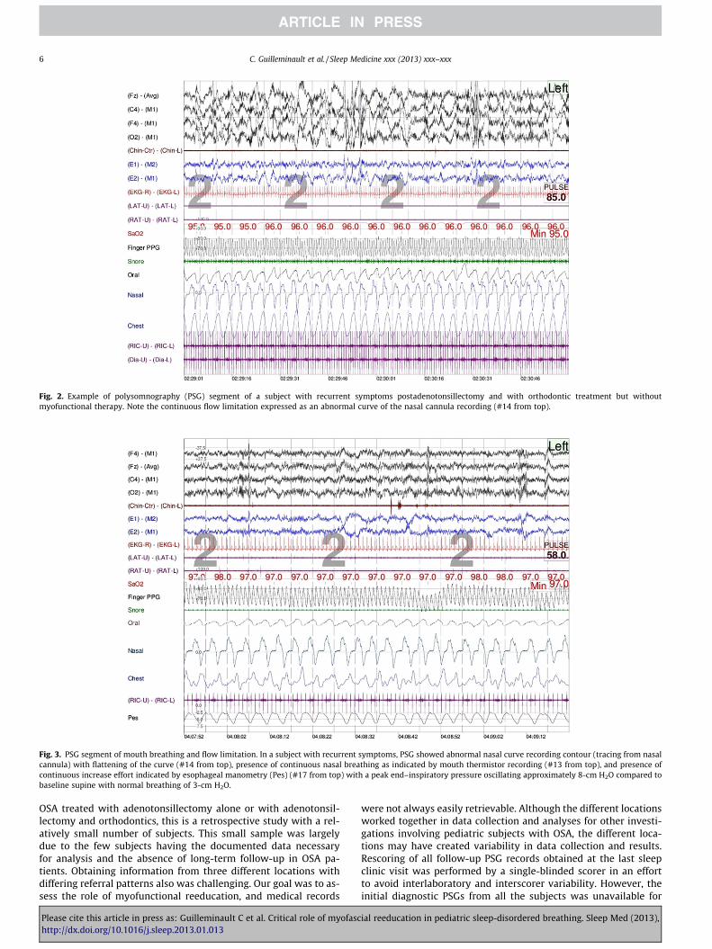
Results are presented in Tables 1 and 2. There was a clear differ-ence between the subjects who had valid myofunctional reeduca-tion and those who did not. Clinically, none of the 11 subjects withreeducation reported clinical concerns related to sleep disorders,and their PSG showed no evidence of breathing abnormalities dur-ing sleep. All subjects had continuous nasal breathing noted onPSG. Wilcoxon rank sum test showed that the AHI was significantlydifferent between the two groups (P = .001) and v2 test statisticsshowed that the percentage of lowest oxygen saturation and thepercentage of flow limitation also were significantly different(P = .01 and P = .0001, respectively) At the long-term follow-up,the 13 other subjects reported persistent daytime concerns; par-ents indicated presence of school difficulties including inattentionin school and in some degree attributed these to fatigue in the sub-ject. Interviews and questionnaires also indicated that specificsleep concerns persisted in some subjects, including the presenceof snoring, agitated sleep, symptoms of sleep-phase delay, andmorning headaches (Table 1). Children with the highest amountof flow limitation (Fig. 2) and the highest AHI scores reported morefrequent concerns. All 13 subjects in this subgroup displayedmouth breathing during sleep, as demonstrated by analysis ofthe mouth thermistor curve on the PSG (Fig. 3). Clinical evaluationreported abnormal head posture in four of the subjects during thedaytime (Fig. 4), and the previous improvements noted at the time
of removal of the orthodontic equipment were lost after develop-ment of a counter-clockwise rotation of the maxilla and high andnarrow palatal vault. Such findings were confirmed on evaluationby an orthodontist. These anatomic presentations were not foundin the 11 subjects with normal breathing during sleep.
Myofunctional evaluation of the orofacial region showed thatsubjects had an abnormally low tongue position in the mouthwhile awake. Among the subjects, 12 were unable to performappropriate clicking sounds with the tongue, 10 were unable toprotrude their tongue upward when asked to try to touch theirnose with the tip of their tongue, four had difficulties holding abutton between their lips, and one had difficulties swallowingwhile drinking quickly. All subjects acknowledged having a prefer-ential side for mastication, and nine subjects presented with slightasymmetry of masseteric muscles when evaluated during activecontraction. At the end of the evaluation by re-educators, all sub-jects were scored with abnormal orofacial muscle tone whileawake. All subjects without clinical concerns had been scored asnormal at myofunctional testing.
6. Discussion
Our retrospective study has typical limitations associated withretrospective studies, particularly when evaluating subjects diag-nosed with OSA years ago. First despite the many subjects with
Abbreviations: AT, adenotonsillectomy; n, number of children; y, years; EDS, excessive daytime sleepiness. One child never had adenotonsillectomy (see text).The number in each column represents the number of children of which the clinical concern was mentioned by parents.Parents did not report of school performance concerns in younger children, but they did report concerns of attention and hyperactivity, children often were considered tohave possible attention-deficit/hyperactivity disorder.As previously reported, parental concerns associated with obstructive sleep apnea vary with age [36].
Table 2Sleep-disordered breathing documented with polysomnography.
Abbreviations: AT, adenotonsillectomy; n, number of individuals affected; y, years; AHI, apnea–hypopnea-index; TST, total sleep time.Percent of flow limitation was determined based on number of 30-second epochs of sleep with abnormal nasal cannula contour not responding to definition of hypopnea withflattening of curve.If abnormal pattern was present for more than 50% of sleep epoch, epoch was scored as flow limited. The percentage was calculated by number of flow-limited epochs ! 2(i.e., number of min) divided by TST in minute. The percentage was extracted from this calculation. Flow limitation was unavailable in most of the initial reports and initialposttreatment recordings. Flow limitation was introduced in polysomnography scoring later on, and the scoring criteria and definition used were those of the Stanford center,which had trained scorers [33].Myofunctional treatment: significant differences between treated and untreated children Wilcoxon signed rank test.* Significant difference (p = .001).** v2 test (p = .01).*** v2 test (p = .0001).
C. Guilleminault et al. / Sleep Medicine xxx (2013) xxx–xxx 5
Please cite this article in press as: Guilleminault C et al. Critical role of myofascial reeducation in pediatric sleep-disordered breathing. Sleep Med (2013),http://dx.doi.org/10.1016/j.sleep.2013.01.013
OSA treated with adenotonsillectomy alone or with adenotonsil-lectomy and orthodontics, this is a retrospective study with a rel-atively small number of subjects. This small sample was largelydue to the few subjects having the documented data necessaryfor analysis and the absence of long-term follow-up in OSA pa-tients. Obtaining information from three different locations withdiffering referral patterns also was challenging. Our goal was to as-sess the role of myofunctional reeducation, and medical records
were not always easily retrievable. Although the different locationsworked together in data collection and analyses for other investi-gations involving pediatric subjects with OSA, the different loca-tions may have created variability in data collection and results.Rescoring of all follow-up PSG records obtained at the last sleepclinic visit was performed by a single-blinded scorer in an effortto avoid interlaboratory and interscorer variability. However, theinitial diagnostic PSGs from all the subjects was unavailable for
Fig. 2. Example of polysomnography (PSG) segment of a subject with recurrent symptoms postadenotonsillectomy and with orthodontic treatment but withoutmyofunctional therapy. Note the continuous flow limitation expressed as an abnormal curve of the nasal cannula recording (#14 from top).
Fig. 3. PSG segment of mouth breathing and flow limitation. In a subject with recurrent symptoms, PSG showed abnormal nasal curve recording contour (tracing from nasalcannula) with flattening of the curve (#14 from top), presence of continuous nasal breathing as indicated by mouth thermistor recording (#13 from top), and presence ofcontinuous increase effort indicated by esophageal manometry (Pes) (#17 from top) with a peak end–inspiratory pressure oscillating approximately 8-cm H2O compared tobaseline supine with normal breathing of 3-cm H2O.
6 C. Guilleminault et al. / Sleep Medicine xxx (2013) xxx–xxx
Please cite this article in press as: Guilleminault C et al. Critical role of myofascial reeducation in pediatric sleep-disordered breathing. Sleep Med (2013),http://dx.doi.org/10.1016/j.sleep.2013.01.013
review and results solely relied on the PSG reports kept on file.Finally the different laboratories were affiliated with differenthealthcare systems that may have influenced subjects’ ability toaccept or adhere to treatment recommendations. The cost of myo-functional reeducation could have been free, covered by medicalinsurance, or paid entirely out of pocket. Numbers may have beenhigher if this type of study had been performed in Brazil wheresuch treatment is routinely included in the management of pa-tients with OSA [2]. Additionally most of the data obtained werefrom children seen at orthodontic clinics, likely adding anotherbias. We do not claim that all children with OSA should have myo-functional reeducation, and our study does not show the role ofmyofunctional treatment performed without orthodontic treat-ment postadenotonsillectomy; however, clearly more studies areneeded. Despite these biases, our study is the first retrospectivestudy investigating myofunctional reeducation and underliningits benefits in the treatment of SDB in the pediatric population.Approximately 46% of nonoverweight children initially diagnosedwith OSA had similar anatomic risk factors as in our 24 children.It is possible that adenotonsillectomy itself led back to nasalbreathing during sleep, but such changes should be objectivelydocumented several months’ postsurgery. The intricate relation-ship between nasal breathing and orofacial growth has been stud-ied for several years [17–28], and myofunctional reeducationprograms were established with the understanding and intentionof optimizing orofacial development and breathing in children.Mouth breathing is associated with malposition of the tongue,which further reinforces impaired development and growth ofthe maxilla and mandible. The intricate relationship betweenbreathing and orofacial growth was studied for many years,supported by experimental animal models that were extensivelystudied in the 1970s. Harvold et al. [17], Miller et al. [18], andVargervik et al. [19] showed that abnormal nasal breathingleads to abnormal EMG discharges in tongue and orofacial muscleswith secondary impact on the facial skeleton and dentition.Impairment of nasal breathing also has been investigated inchildren and has demonstrated an impact on facial growth, headposture, and general medical consequences [20–29]. Swedishresearchers also have suggested that early mouth breathingwithout appropriate humidification of air through the nose leadsto repetitive tonsillar trauma [29]. Such trauma may lead to aninflammatory response of the tonsils, previously histologicallydemonstrated and also may lead to progressive enlargement ofan already narrow airway.
With the understanding of these implications, treatment pro-grams were established with the goal of optimizing orofacial devel-opment to improve breathing in children. The benefits of thecombination of orthodontic and myofunctional reeducation onbreathing, speech, swallowing, orofacial growth, and the elimina-tion of abnormal head-neck posture, with a focus on eliminatingtongue and orofacialmuscle hypotonia, have been published partic-ularly in the orthodontic literature [2]. This movement also has ledto the development ofmyofunctional reeducation specialistswhoseexpertise is sanctioned in many countries. There is no systematicprospective study involving myofunctional therapy in childrenwith OSA, but there has been an abundance of literature on the ben-efits of myofunctional treatment on growth and orthodontic devel-opment for more than 20 years [1]. This literature emphasizes theimportance of nasal breathing and obtaining good orofacial muscletone to maintain orthodontic gains in children. It also stressesmaintenance of obtained gains during pubertal years. However,none of these studies involved systematic PSG evaluation, and thereports emphasized orthodontic development rather than noctur-nal breathing. SDB invariably involves abnormal nasal breathingand impaired facial growth associated with mouth breathing.Unfortunately, this concern often has been ignored. Our report indi-cates that a combined treatment approach including adenotonsil-lectomy and orthodontia with myofunctional reeducation can becrucial in the elimination of OSA. This finding is especially critical,as the failure to eliminate oral breathing will lead to the reappear-ance of the OSA syndrome in children. A recent prospective follow-up study [30] lasting 36 months that included clinical and PSG datahad followed 67% of an initial OSA children cohort and showed that68% of the children still involved in the study had either worseningabnormal breathing if adenotonsillectomy had not fully resolvedthe concern (with complete OSA resolution defined as AHI <1) orhad reappearance of OSA, even if complete resolution had been ob-tained postadenotonsillectomy. Mean AHI of the cohort wasapproximately six events per hour and none of the children hadbeen monitored for mouth breathing or received myofunctionaltherapy. Treatment achieving a normal upper airway in childrendoes not guarantee normal tongue position or normal tongue andorofacial muscle strength during sleep. This in turn affects thedevelopment of the airway as demonstrated in monkey models[17–19]. Persistence of oral breathing during sleep directly affectstongue position and strength as well as that of the orofacial mus-cles, leading to abnormal airway development unless myofunc-tional reeducation is performed to avoid this evolution.
Fig. 4. Abnormal head position during wakefulness with abnormal breathing during sleep. Note the progressive change in head position over time associated with thedevelopment of an abnormal nuchal angle. The angle was normal in 2007 with progressive development of abnormal head position associated with abnormal breathing andOSA despite absence of snoring but the presence of mouth breathing during sleep. The last photo clearly shows the abnormal head-neck posture related to the sleep-disordered breathing.
C. Guilleminault et al. / Sleep Medicine xxx (2013) xxx–xxx 7
Please cite this article in press as: Guilleminault C et al. Critical role of myofascial reeducation in pediatric sleep-disordered breathing. Sleep Med (2013),http://dx.doi.org/10.1016/j.sleep.2013.01.013
Despite its deficiencies our study highlights the importance ofinfluencing normal facial growth by using the available tools andresources to optimize orofacial development in children withabnormal breathing during sleep. As previously mentioned,although integrated care between myofunctional re-educatorsand orthodontists may be routine in some countries, such asFrance, Belgium, Brazil, or Taiwan, this is not the case in all partsof the world. The lack of understanding of these interactions leadsto the misconception that pediatric OSA is an upper airway syn-drome and not a facial growth dysfunction with secondary impacton the upper airway.
Finally, our study shows that scoring only apneas and hypopne-as is not sufficient to recognize abnormal breathing during sleep.Flow limitation [14] is a much more adequate indicator of abnor-mal breathing in our patients. Previous studies have shown theinvolvement of flow limitation in parasomnias and in abnormallyhigh amounts of cyclic alternating pattern phases A2 and A3[31]. Chervin et al. [32] showed that abnormal breaths that donot meet criteria for defined apneas and hypopneas still may havea disrupting effect on the sleep EEG. Recently it was shown thatyoung women with an abnormal amount of flow limitation and alow or normal AHI had the same clinical presentation as womenwith pathologic AHIs [33]. It also is imperative that we recognizeabnormal breathing in children during sleep and have the knowl-edge to select the appropriate indices to detect SDB [33]. Thisknowledge requires increased attentiveness for abnormal breath-ing, as many of our children had no snoring likely due to priortreatment, yet clearly displayed flow limitation disrupts their sleepalong with persistent symptoms. We must treat children to opti-mize and insure normal development of the airway, orofacial mus-cle strength, and positioning, and in turn normal breathing duringsleep. Myofunctional reeducation may be considered to treat adultpatients with OSA; however, as demonstrated by Guimaraes et al.[2] even if it is effective, it has a limited impact in adulthood. Thisfinding further reinforces the importance of early identificationand intervention during childhood development to optimize nor-mal growth of the airway and to insure a lasting impact in thetreatment of SDB. With the help of orthodontists and myofunc-tional therapists and appropriate testing of nasal resistance inchildren, we may be able to recognize and treat children at riskfor SDB early in life [1,34].
Conflict of interest
The ICMJE Uniform Disclosure Form for Potential Conflicts ofInterest associated with this article can be viewed by clicking onthe following link: http://dx.doi.org/10.1016/j.sleep.2013.01.013.
Acknowledgments
Some of the data presented here are part of the data collectedfor the PhD thesis of Dr. Y.S. Huang. These data were orally pre-sented at the European Sleep Research Society 21st CongressSeptember 2012.
We thank Marion Girard-Gervais (D.U. reeducation maxillo-faciale) for her help in France and Dr. Shannon Sullivan for her helpin editing the manuscript. Examples of myofunctional exercisescan be seen at www.myofunctionaltherapy.blogspot.com [35].
References
[1] Chauvois A, Fournier M, Girardin F. Rééducation des fonctions dans lathérapeutique orthodontique. Paris (France): SID Publish; 1991. p. 1–231.
[2] Guimaraes KC, Drager LR, Genta PR, Marcondes BF, Lorenzi-Filho G. Effects oforopharyngeal exercises on patients with moderate obstructive sleep apneasyndrome. Am J Respir Crit Care Med 2009;179:962–9.
[3] Chervin RD, Hedger K, Dillon JE, Pituch KJ. Pediatric sleep questionnaire (PSQ):validity and reliability of scales for sleep disordered breathing, snoring,sleepiness, and behavioural problems. Sleep Med 2000;1:21–32.
[4] Wang CH, Yang CM, Huang YS. The validation and reliability of Chinese versionof the pediatric sleep questionnaire for patient with sleep-breathing problems.Taiwan J Psychiatry TJP 2012;6:177–86.
[5] Pirelli P, Saponara M, Guilleminault C. Rapid maxillary expansion in childrenwith obstructive sleep apnea syndrome. Sleep 2004;27:761–6.
[6] Friedman M, Tanyeri H, La Rosa M, Landsberg R, Vaidyanathan K, Pieri S, et al.Clinical predictors of obstructive sleep apnea. Laryngoscope 1999;109:1901–7.
[7] Mallampati SR, Gatt S, Gugino SL, Desai S, Waraksa B, Freiberger D, et al. Aclinical sign to predict difficult tracheal intubation: a prospective study. CanAnaesth Soc J 1985;32:429–34.
[8] Friedman M, Ibrahim H, Bass L. Clinical staging for sleep-disordered-breathing.Otolaryngol Head Neck Surg 2002;127:13–21.
[9] Solow B, Tallgren A. Head posture and craniofacial morphology. Am J PhysAnthropol 1976;44:417–35.
[10] Solow, Siersbaek-Nielsen S, Greve E. Airway adequacy, head postures andcraniofacial morphology. Am J Orthod 1984;86:214–23.
[11] Rechtschaffen A, Kales A, editors. A manual of standardized terminology,techniques and scoring system for sleep stages of human subjects. UCLA(LA): Brain Research Institute; 1968.
[12] EEG arousals: scoring rules and examples: a preliminary report from the SleepDisorders Atlas Task Force of the American Sleep Disorders Sssociation. Sleep1992;15:173–84.
[13] Iber C, Ancoli-Israel S, Chesson AL, Quan SF. The AASM manual for the scoringof sleep and associated events. Westchester (IL): American Academy of SleepMedicine; 2007. p. 45–50.
[14] Hosselet JJ, Norman RG, Ayappa I, Rapoport D. Detection of flow limitationwith nasal cannula/pressure transducer system. Am J Respir Crit Care Med1998;157:1461–7.
[15] Aittokallio T, Saaresranta T, Polo-Kantola P, Nevalainen O, Polo O. Analysis ofinspiratory flow shapes in patients with partial upper-airway obstructionduring sleep. Chest 2001;119:337–44.
[16] Guilleminault C, Li KK, Khramtsov A, Pelayo R, Martinez S. Sleep disorderedbreathing: surgical outcome in prepubertal children. Laryngoscope2004;114:132–7.
[17] Harvold EP, Tomer BS, Vargervik K, Chierici G. Primate experiments on oralrespiration. Am J Orthod 1981;79:359–72.
[18] Miller AJ, Vargervik K, Chierici G. Experimentally induced neuromuscularchanges during and after nasal airway obstruction. Am J Orthod1984;85:385–92.
[19] Vargervik K, Miller AJ, Chierici G, Harvold E, Tomer BS. Morphologic responseto changes in neuromuscular patterns experimentally induced by alteredmodes of respiration. Am J Orthod 1984;85:115–24.
[20] LeechH. A clinical analysis of orofacialmorphology and behavior of 500 patientsattending an upper airway respiratory clinic. Dental Pract 1958;9:57–91.
[21] Linder-Aronson S. Dimensions of face and palate in nose breathers andhabitual mouth breathers. Odont Rev 1959;14:187–200.
[22] Hawkins AC. Mouth breathing as the cause of malocclusion and other facialabnormallities. Tex Dent J 1965;83:10–5.
[23] Timms DJ. Some medical aspects of rapid maxillary expansion. Br J Orthod1974;1:127–32.
[24] Gray LP. Results of 310 cases of rapid maxillary expansion selected for medicalreasons. J Laryngol Otol 1975;89:601–14.
[25] Hershey HG, Stewart BL, Warren DW. Changes in nasal airway resistanceassociated with rapid maxillary expansion. Am J Orthod 1976;69:274–84.
[26] McNamara JA. Influence of respiratory pattern on craniofacial growth. AngleOrthod 1981;51:269–300.
[27] Timms DJ. The reduction of nasal airway resistance by rapid maxillaryexpansion and its effect on respiratory disease. J Laryngol Otol1984;98:357–62.
[28] Rubin RM. Effects of nasal airway obstruction on facial growth. Ear NoseThroat J 1987;66:212–9.
[29] Zettergreen L, Linder-Aronson S, Norlander B, Agren K, Svanborg E.Longitudinal effect on facial growth after tonsillectomy in children withobstructive sleep apnea. World J Orthod 2002;3:67–72.
[30] Chen K, Huang YS, Guilleminault C. Neurocognitive function improvementafter adenotonsillectomy in obstructive sleep apnea. Sleep 2012;35(Suppl.1):S1143.
[31] Guilleminault C, Kirisoglu C, da Rosa AC, Lopes C, Chan A. Sleepwalking, adisorder of NREM sleep instability. Sleep Med 2006;7:163–70.
[32] Chervin RD, Burns JW, Subotic NS, Roussi C, Thelen B, Ruzicka DL. Correlates ofrespiratory cycle-related EEG changes in children with sleep-disorderedbreathing. Sleep 2004;27:116–21.
[33] Tantrakul V, Pack SC, Guilleminault C. Sleep-disordered breathing inpremenopausal women: differences between younger (less than 30 yearsold) and older women. Sleep Med 2012;13:656–62.
8 C. Guilleminault et al. / Sleep Medicine xxx (2013) xxx–xxx
Please cite this article in press as: Guilleminault C et al. Critical role of myofascial reeducation in pediatric sleep-disordered breathing. Sleep Med (2013),http://dx.doi.org/10.1016/j.sleep.2013.01.013


![Current Trends in Medicine - Somato Publications · myofascial pain [28,29]. Myofascial pain is associated with myofascial trigger points (MTPs), muscles in sustained contraction](https://static.documents.pub/doc/80x56/5e43a3f992ffb312756e8245/current-trends-in-medicine-somato-publications-myofascial-pain-2829-myofascial.jpg)


