Citation: Norah Alotaibi., et al. “The Role of Orthodontics in the Management of Obstructive Sleep Apnea OSA in Children: Systematic Review”. EC Dental Science 18.9 (2019):2238-2254. *Corresponding Author: Norah Alotaibi, General Dentist, Graduate of Qassim University, Jubail, Saudi Arabia. Received: August 06, 2019; Published: August 22, 2019 Objective: Review the effectiveness of the use of different orthodontic oral appliance as a primary or adjective treatment of OSAS in children. Methods: Systemic search of online databases (PubMed, Cochrane Central Register of Controlled Trials (CENTRAL), international Registry Platform for ongoing trials and clinical trails.gov, Embase Ovid, Medline complete) for randomized control trail and non- randomized prospective or retrospective clinical studies published in 2000-2019 that uses oral appliances for the treatment of OSA in children. Study selection was done by two reviewers. Results: 9 studies were included in the review. Based on the limited evidence oral appliances improve OSA in children measured by the reduction of AHI. Due to heterogeneity of included study designs and reported data meta-analysis was not possible. Limitation: Limited number of studies and study populations. High risk of bias. Conclusion: Oral appliances show a promising improvement in AHI and should be considered as a treatment option for OSA in children Introduction Cronicon OPEN ACCESS EC DENTAL SCIENCE Research Article The Role of Orthodontics in the Management of Obstructive Sleep Apnea OSA in Children: Systematic Review Norah Alotaibi 1 *, Amani Alkhamees 2 and Rabia Bilal 3 1 General Dentist, Graduate of Qassim University, Jubail, Saudi Arabia 2 Saudi Board Orthodontics Resident, National Guard Hospital, Riyadh, Saudi Arabia 3 Associate Professor, Department of Orthodontics and Pediatric Dentistry, College of Dentistry, Qassim University, Saudi Arabia Abstract Keywords: Obstructive Sleep Apnea; Children; Oral Appliances; Mandibular Advancement Appliance; Maxillary Expansion Abbreviations AHI: Apnea Hypopnea Index; OSA: Obstructive Sleep Apnea; AASM: American Academy of Sleep Medicine; PSG: Polysomnography; AI: Apnea Index; RERAs: Respiratory Event-Related Arousals; RDI: Respiratory Disturbance Index; RME: Rapid Maxillary Expansion; SRME: Semi Rapid Maxillary Expansion; SME: Slow Maxillary Expansion; RCT: Randomized Controlled Trials Obstructive sleep apnea (OSA) is defined by the American academy of sleep medicine AASM as "interrupted airflow despite persis- tent respiratory effort. It occurs several times every hour during sleep. Breathing continues but the airflow is blocked. This is due to the complete or partial collapse, and/or complete or partial obstructions, of the upper airway during sleep but not during wakefulness. With reduced airflow, gaseous exchange is impaired. Sleep is fragmented due to recurrent arousals" [1]. The prevalence of OSA was estimated to be 1.2 - 5.7%. the peak of OSA was observed at 2 periods. The first was in children from the ages of 2 - 8 years most commonly with adenio-tonsilar enlargement, the second peak was during adolescent in relation to obesity [2,3]. In pre pubertal children, the incidence of OSAS is similar in boys and girls. After puberty, OSA is more common in boys [4].
Transcript
Citation: Norah Alotaibi., et al. “The Role of Orthodontics in the Management of Obstructive Sleep Apnea OSA in Children: Systematic Review”. EC Dental Science 18.9 (2019):2238-2254.
*Corresponding Author: Norah Alotaibi, General Dentist, Graduate of Qassim University, Jubail, Saudi Arabia.
Received: August 06, 2019; Published: August 22, 2019
Objective: Review the effectiveness of the use of different orthodontic oral appliance as a primary or adjective treatment of OSAS in children.
Methods: Systemic search of online databases (PubMed, Cochrane Central Register of Controlled Trials (CENTRAL), international Registry Platform for ongoing trials and clinical trails.gov, Embase Ovid, Medline complete) for randomized control trail and non-randomized prospective or retrospective clinical studies published in 2000-2019 that uses oral appliances for the treatment of OSA in children. Study selection was done by two reviewers.
Results: 9 studies were included in the review. Based on the limited evidence oral appliances improve OSA in children measured by the reduction of AHI. Due to heterogeneity of included study designs and reported data meta-analysis was not possible.
Limitation: Limited number of studies and study populations. High risk of bias.
Conclusion: Oral appliances show a promising improvement in AHI and should be considered as a treatment option for OSA in children
Introduction
CroniconO P E N A C C E S S EC DENTAL SCIENCE
Research Article
The Role of Orthodontics in the Management of Obstructive Sleep Apnea OSA in Children: Systematic Review
Norah Alotaibi1*, Amani Alkhamees2 and Rabia Bilal3 1General Dentist, Graduate of Qassim University, Jubail, Saudi Arabia 2Saudi Board Orthodontics Resident, National Guard Hospital, Riyadh, Saudi Arabia3Associate Professor, Department of Orthodontics and Pediatric Dentistry, College of Dentistry, Qassim University, Saudi Arabia
Obstructive sleep apnea (OSA) is defined by the American academy of sleep medicine AASM as "interrupted airflow despite persis-tent respiratory effort. It occurs several times every hour during sleep. Breathing continues but the airflow is blocked. This is due to the complete or partial collapse, and/or complete or partial obstructions, of the upper airway during sleep but not during wakefulness. With reduced airflow, gaseous exchange is impaired. Sleep is fragmented due to recurrent arousals" [1].
The prevalence of OSA was estimated to be 1.2 - 5.7%. the peak of OSA was observed at 2 periods. The first was in children from the ages of 2 - 8 years most commonly with adenio-tonsilar enlargement, the second peak was during adolescent in relation to obesity [2,3]. In pre pubertal children, the incidence of OSAS is similar in boys and girls. After puberty, OSA is more common in boys [4].
The Role of Orthodontics in the Management of Obstructive Sleep Apnea OSA in Children: Systematic Review
Citation: Norah Alotaibi., et al. “The Role of Orthodontics in the Management of Obstructive Sleep Apnea OSA in Children: Systematic Review”. EC Dental Science 18.9 (2019):2238-2254.
OSA occurs when there is an imbalance between the factors that maintain a patent airway. These risk factors can be divided into fac-tors that affect the airway collapsibility, factors that cause anatomical narrowing or a combination of both2. Factors that affect the airway collapsibility include inflammation, neuromuscular disorders, and decrease in upper respiratory tract muscle tone [5]. Factors that causes anatomical narrowing include adenotonsillar hypertrophy [6], craniofacial anomalies (retrognathia, micrognathia, and midface hypopla-sia), and macroglossia. A combination of these factors can be observes in some conditions like Down syndrome [4].
The diagnosis of OSA in children is based on medical history, physical examination and confirmed with polysomnography PSG findings.
Children with OSA display symptoms that are mainly divided into daytime and nocturnal symptoms. Daytime symptoms include mor-ning headache, daytime sleepiness, frequent airway infection. Nighttime symptoms include snoring, sweating, restless sleep [3]. Children with OSA also show some behavioral manifestation that ranges from aggression, hyperactivity, inattentiveness and pathological shyness [7].
Physical examination should begin with general observation of child. The child weight and growth should be noted, other indications like mouth breathing, and nasal voice should be noted as well. In addition, the facial profile should be examined for craniofacial anomalies. Intra-oral examination to detect any abnormality that could lead to an obstruction of the airway including adenotonsillar hypertrophy, size of the tongue [2].
PSG is considered the golden standard test for confirming the diagnosing of sleep apnea. It uses multi physiological parameters to diagnose OSA and other sleep disorders. The most used parameters for the diagnosis of OSA are apnea-hypopnea index (AHI), apnea index (AI), respiratory event-related arousals (RERAs), and respiratory disturbance index (RDI). AHI indicates the number of apneic and hypopneic events per hour of sleep. Apnea is defined as a complete interruption of airflow lasts at least two breaths, whereas hypopnea is defined as a ≥ 50% reduction in airflow with an arousal, awakening, or ≥ 3% desaturation for same durations. According to AASM the diagnosis of OSAS in children is defined as AHI index > 1 [8].
Treatment of OSA depends on the underlying cause and may include one or a combination of: Adenotonsillectomy, continues positive airway pressure, diet and medication, and use of oral appliances [9].
The use of oral appliances for the management of OSA was first introduced in 1902 by Dr. Pierre Robin. He fabricated an appliance by the name of "the monobloc" and prescribed it for children with hypotrophy of the mandible, which aimed to establish a normal spatial relationship of the maxilla and mandible [10].
One of the most commonly used intraoral treatment modality is rapid maxillary expansion RME or slow maxillary expansion SME. RME devices are used for patients with constricted maxilla and posterior cross bite. It is used to increase the transvers width of maxilla increasing the palatal and retroplatal area. It also enlarges the nasal cavities, which in turn leads to less nasal resistance and promote nasal breathing [11].
Stimulating the maxillary and mandibular growth was also used for the treatment of OSA in children with craniofacial anomalies. Forward reposition of the mandible was hypothesized to have a therapeutic effect by enlarging the upper airway (velopharynx) [12].
Aim of the StudyThe aim of this study is to review the effectiveness of the use of different orthodontic oral appliance as a primary or adjective treatment
of OSA in children.
2239
2240
The Role of Orthodontics in the Management of Obstructive Sleep Apnea OSA in Children: Systematic Review
Citation: Norah Alotaibi., et al. “The Role of Orthodontics in the Management of Obstructive Sleep Apnea OSA in Children: Systematic Review”. EC Dental Science 18.9 (2019):2238-2254.
Materials and MethodsPreferred Reporting Items for Systematic review and Meta-Analysis (PRISMA) checklist was used as a template for this review.
Protocol and registration: Not available.
Eligibility criteria
Participants, interventions, comparators, and outcomes (PICO) was used to formulate the review objective and inclusion criteria.
Participant: Children up to 18 years old.
Interventions: Any type of oral appliance as the primary treatment, use of oral appliance in conjunction with other treatment modalities, comparison between different types of oral appliances, comparison between oral appliances and other treatment modalities.
Comparators: Treatment vs control group, treatment vs different type of treatment, before vs after treatment.
Outcome: Primary outcome reduction of AHI, secondary outcomes: significant changes in other sleep parameters (e.g.: oxygen saturation, arousal index), craniodental changes, daytime and nocturnal symptoms.
Included studies: Randomized controlled trials RCT, clinical studies both randomized and nonrandomized either retrospective or pro-spective.
Due to the limited number of published RCT in the literature other types of clinical studies were included. pilot, preliminary and case report studies were not included due to limited sample size and higher risk of bias.
Studies publish in English from the year 2000 to May 2019.
Exclusion criteriaStudies that includes adults in its sample, Studies that evaluate the use of other modalities in the treatment of OSA in children and
Studies including a specific sample group: children with syndromes or systemic disorders.
Information sourcesSystemic research of electronic databases: PubMed, Cochrane Central Register of Controlled Trials (CENTRAL), international Registry
Platform for ongoing trials and clinical trails.gov, Embase Ovid, Medline complete.
RCT, NRCT, prospective and retrospective clinical studies that uses an oral appliance as a primary treatment or in conjunction with other treatment modalities and uses AHI to measure the effectiveness of the intervention were included in this review.
Studies publish in English langue from 2000-May 2019.
The last search was done on 27th of May 2019.
The search was done independently in duplication by 2 reviewers (Norah Alotaibi and Amani Alkhamees) and any disagreement be-tween the reviews was dissolved by the supervisor (Rabia Bilal).
SearchSearch words were a combination of: “oral appliances/ oral device/ orthodontics/mandibular advancement appliance/ maxillary
expansion” and “obstructive sleep apnea or obstructive sleep apnea or OSA” and “children/pediatric”.
For PubMed the following limits were set: study type: controlled clinical trials, randomized control trails.
2241
The Role of Orthodontics in the Management of Obstructive Sleep Apnea OSA in Children: Systematic Review
Citation: Norah Alotaibi., et al. “The Role of Orthodontics in the Management of Obstructive Sleep Apnea OSA in Children: Systematic Review”. EC Dental Science 18.9 (2019):2238-2254.
Custom date range 01-01-2000 to 25-05-2019, language: English, Age: child: birth- 18 years.
For Medline complete the following limits were set: Date (2000 - 2019), language: English, age: all child: 0-18.
For Cochrane Central Register of Controlled Trials (central) the following limits were set: year (2000 - 2019), Trails.
For Embase Ovid the search was done using the multifield feature was used and the following limits were set: publication year (2000-current), Age group (All child 0 to 18), languages (English), publication type (clinical trial), publication types (clinical trials, ALL).
Study selectionDatabase search was done for possible studies, abstracts of the studies were screened for identification of eligible studies, Full text
articles were obtained and assessed and a final list of included studies was made.
This process was done independently and in duplication by 2 reviewers and any disagreement was resolved by the 3rd reviewer.
References were organized and managed using Mendeley desktop (version 1.19.4).
Data collection process
We developed a form based on the Cochrane Consumers and Communication Review Group’s data extraction template and followed it for each study.
Two reviewers collected the data independently collected data was combined and compared for accuracy any discrepancies were solved by a third reviewer.
Data itemsData collected from the selected studies included: study design, study population (sample size, age, diagnosis, method of diagnosis,
pre intervention records), intervention applied (type, duration of treatment and follow up), outcomes (primary outcome: changes in AHI, secondary outcome: changes in oxygen saturation, craniofacial and dental changes, reported symptoms changes).
Risk of bias in individual studiesRisk of bias assessment was done following The Cochrane Risk of Bias criteria. The assessment was done by two reviewers indepen-
dently and in duplication and any discrepancies were resolved by the third reviewer.
Summary measuresprimary outcome reduction of AHI, secondary outcomes: other significant changes in sleep parameters, craniodental changes, daytime
and nocturnal symptoms.
Synthesis of resultsDue to the difference in the included studies: study designs, lack of control groups in some studies, sample size, type of intervention
used, duration of treatment, duration of follow up meta-analysis was not be conducted.
Results and DiscussionStudy selection
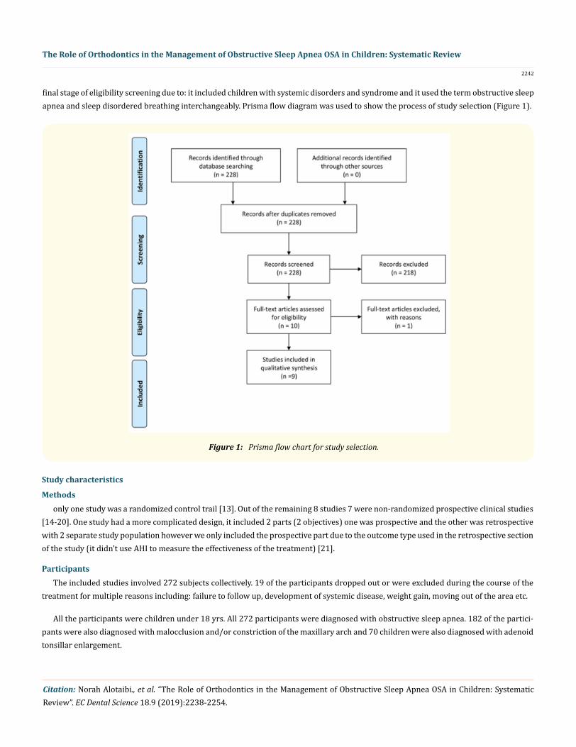
After the electronic search of the prespecified data basis (PubMed, Cochrane Central Register of Controlled Trials (CENTRAL), inter-national Registry Platform for ongoing trials and clinical trails.gov, Embase Ovid, Medline complete) with the preset search terms and limits 228 studies were identified as for the first phase of screening. After screening the abstracts 218 studies were excluded for multiple reasons including but not limited to study type, study population: age, systemic health. Full text articles were obtained for the remaining 10 studies. Full text articles were read through by 2 reviewers 9 of which were included in the final review. 1 study was excluded at the
2242
The Role of Orthodontics in the Management of Obstructive Sleep Apnea OSA in Children: Systematic Review
Citation: Norah Alotaibi., et al. “The Role of Orthodontics in the Management of Obstructive Sleep Apnea OSA in Children: Systematic Review”. EC Dental Science 18.9 (2019):2238-2254.
final stage of eligibility screening due to: it included children with systemic disorders and syndrome and it used the term obstructive sleep apnea and sleep disordered breathing interchangeably. Prisma flow diagram was used to show the process of study selection (Figure 1).
Figure 1: Prisma flow chart for study selection.
Study characteristics
Methodsonly one study was a randomized control trail [13]. Out of the remaining 8 studies 7 were non-randomized prospective clinical studies
[14-20]. One study had a more complicated design, it included 2 parts (2 objectives) one was prospective and the other was retrospective with 2 separate study population however we only included the prospective part due to the outcome type used in the retrospective section of the study (it didn’t use AHI to measure the effectiveness of the treatment) [21].
ParticipantsThe included studies involved 272 subjects collectively. 19 of the participants dropped out or were excluded during the course of the
treatment for multiple reasons including: failure to follow up, development of systemic disease, weight gain, moving out of the area etc.
All the participants were children under 18 yrs. All 272 participants were diagnosed with obstructive sleep apnea. 182 of the partici-pants were also diagnosed with malocclusion and/or constriction of the maxillary arch and 70 children were also diagnosed with adenoid tonsillar enlargement.
2243
The Role of Orthodontics in the Management of Obstructive Sleep Apnea OSA in Children: Systematic Review
Citation: Norah Alotaibi., et al. “The Role of Orthodontics in the Management of Obstructive Sleep Apnea OSA in Children: Systematic Review”. EC Dental Science 18.9 (2019):2238-2254.
One study included 20 healthy children as a control group [18]. Author study published in 2011 was a follow up study for a part of the study population (10 children) of a previously published study in 2007 [16,21].
InterventionTwo of the included studies used oral appliances to reposition the mandible as their intervention. The 1st study used a personalized
oral appliance while the 2nd study used a modified version of the monobloc appliance. Both appliances were used for 6 months duration [13,18].
Six studies reported the use of maxillary expansion as an intervention for obstructive sleep apnea in children as follows:
1- A study in 2004 by Pirelli P., et al. used RME for 10 - 20 days for the active phase of the expansion and then for 6 - 12 months for the retention period [17].
2- In 2007 a study by Villa MP., et al. Used RME (leone Sesto Fiorentino-Florence) the active phase was 10 days or until the paleal cusp of the upper molar made a contact with the buccal cusp of the lower molar and the retention period was 12 months [16].
3- In 2011 a follow up study by Villa MP., et al. included 10 out of 14 children that were included in the previous study and followed up with them after 36 months to evaluate the effectiveness and the stability of the intervention [21].
4- In 2015 a study by Pirelli P., et al. Used RME with a follow up for 12 years. It didn’t give details on RME Duration [14].
5- In 2015 a study by Villa MP., et al. used RME on 40 children the active phase was until the paleal cusp of the upper molar made a contact with the buccal cusp of the lower molar and the retention period was 12 months [20].
6- A study by Hoxha S., et al. in 2018 used a semi rapid maxillary expansion for 5 months [19].
The last study divided its population into 3 group and compared the effectiveness of different interventions: adenotonsillectomy in the first group (cold dissection tonsillectomy and curettage of adenoid vegetation under direct vision via oral access) vs RME in the second group vs both treatments in the third group [15].
Outcome
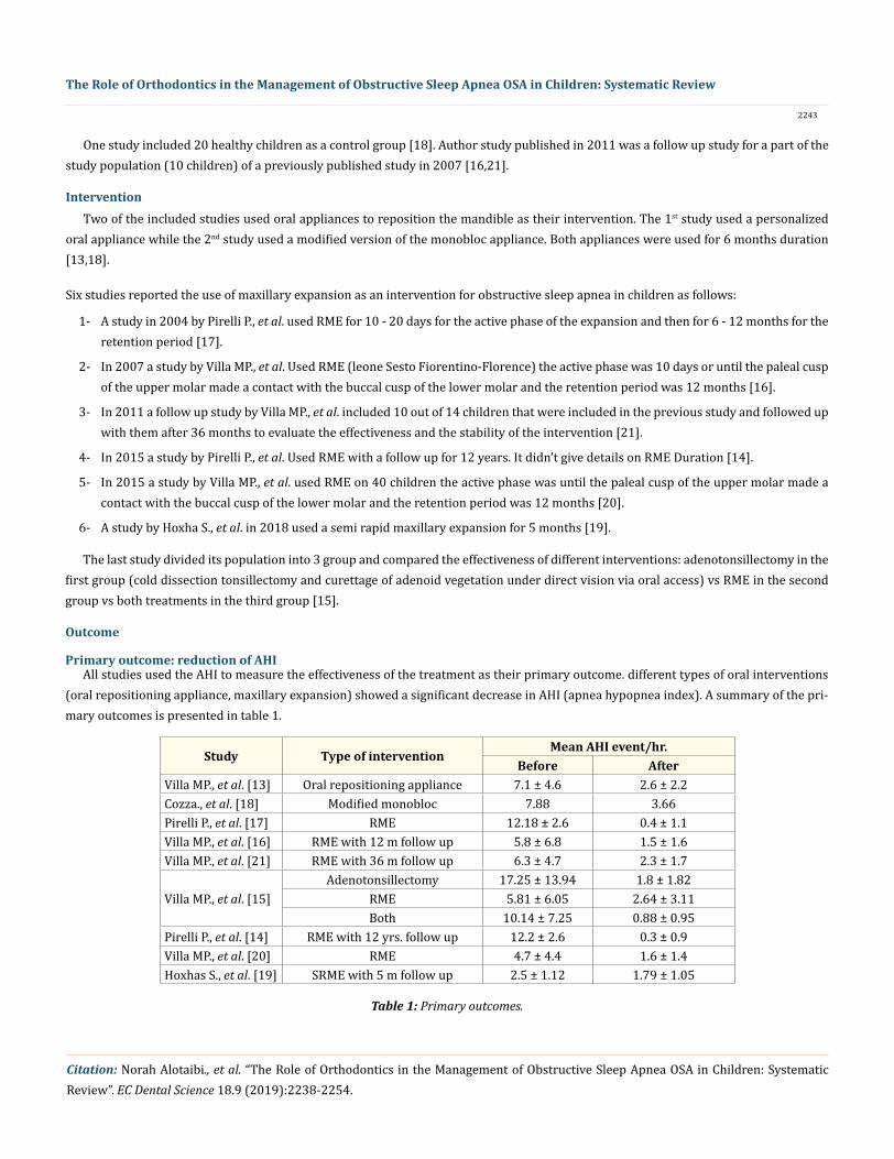
Primary outcome: reduction of AHI All studies used the AHI to measure the effectiveness of the treatment as their primary outcome. different types of oral interventions
(oral repositioning appliance, maxillary expansion) showed a significant decrease in AHI (apnea hypopnea index). A summary of the pri-mary outcomes is presented in table 1.
Study Type of interventionMean AHI event/hr.
Before AfterVilla MP., et al. [13] Oral repositioning appliance 7.1 ± 4.6 2.6 ± 2.2Cozza., et al. [18] Modified monobloc 7.88 3.66Pirelli P., et al. [17] RME 12.18 ± 2.6 0.4 ± 1.1Villa MP., et al. [16] RME with 12 m follow up 5.8 ± 6.8 1.5 ± 1.6Villa MP., et al. [21] RME with 36 m follow up 6.3 ± 4.7 2.3 ± 1.7
Villa MP., et al. [15]Adenotonsillectomy 17.25 ± 13.94 1.8 ± 1.82
Pirelli P., et al. [14] RME with 12 yrs. follow up 12.2 ± 2.6 0.3 ± 0.9Villa MP., et al. [20] RME 4.7 ± 4.4 1.6 ± 1.4Hoxhas S., et al. [19] SRME with 5 m follow up 2.5 ± 1.12 1.79 ± 1.05
Table 1: Primary outcomes.
2244
The Role of Orthodontics in the Management of Obstructive Sleep Apnea OSA in Children: Systematic Review
Citation: Norah Alotaibi., et al. “The Role of Orthodontics in the Management of Obstructive Sleep Apnea OSA in Children: Systematic Review”. EC Dental Science 18.9 (2019):2238-2254.
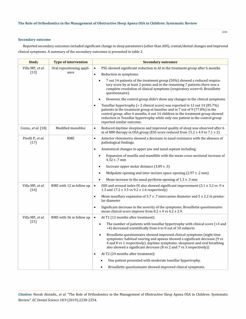
Reported secondary outcomes included significant change in sleep parameters (other than AHI), cranial/dental changes and improved clinical symptoms. A summary of the secondary outcomes is presented in table 2.
Secondary outcome
Study Type of intervention Secondary outcomesVilla MP., et al.
[13]Oral repositioning appli-
ance• PSG showed significant reduction in AI in the treatment group after 6 months
• Reduction in symptoms:
• 7 out 14 patients of the treatment group (50%) showed a reduced respira-tory score by at least 2 points and in the remaining 7 patients there was a complete resolution of clinical symptoms (respiratory score=0, Brouillette questionnaire).
• However, the control group didn’t show any changes in the clinical symptoms.
• Tonsillar hypertrophy (> 2 clinical score) was reported in 12 out 14 (85.7%) patients in the treatment group at baseline and in 7 out of 9 (77.8%) in the control group. after 6 months, 6 out 14 children in the treatment group showed reduction in Tonsillar hypertrophy while only one patient in the control group reported similar outcome.
Cozza., et al. [18] Modified monobloc • Reduced daytime sleepiness and improved quality of sleep was observed after 6 m of MM therapy in OSA group (ESS score reduced from 15.2 ± 4.9 to 7.1 ± 2)
Pirelli P., et al. [17]
RME • Anterior rhinometry showed a decrease in nasal resistance with the absence of pathological findings.
• Anatomical changes in upper jaw and nasal septum including:
• Expansion of maxilla and mandible with the mean cross-sectional increase of 4.32 ± .7 mm
• Increase upper molar distance (3.89 ± .3)
• Midpalate opening and inter incisive space opening (2.97 ± .2 mm)
• Mean increase in the nasal pyriform opening of 1.3 ± .3 mmVilla MP., et al.
[16]RME with 12 m follow up • OHI and arousal index OI also showed significant improvement (3.1 ± 3.2 vs .9 ±
1.3 and 17.2 ± 3.5 vs 9.2 ± 1.6 respectively)
• Mean maxillary expansion of 3.7 ± .7 intercanine diameter and 5 ± 2.2 in premo-lar diameter
• Significant decrease in the severity of the symptoms. Brouillette questionnaire: mean clinical score improve from 8.2 ± 4 vs 4.2 ± 2.9.
Villa MP., et al. [21]
RME with 36 m follow up • At T1 (12 months after treatment):
• The number of patients with tonsillar hypertrophy with clinical score (+3 and +4) decreased scientifically from 6 to 0 out of 10 subjects.
• Brouillette questionnaire showed improved clinical symptoms (night time symptoms: habitual snoring and apneas showed a significant decrease (9 vs 4 and 8 vs 1 respectively), daytime symptoms: sleepiness and oral breathing also showed a significant decrease (8 vs 2 and 7 vs 3 respectively))
• At T2 (24 months after treatment):
• One patient presented with moderate tonsillar hypertrophy.
The Role of Orthodontics in the Management of Obstructive Sleep Apnea OSA in Children: Systematic Review
Citation: Norah Alotaibi., et al. “The Role of Orthodontics in the Management of Obstructive Sleep Apnea OSA in Children: Systematic Review”. EC Dental Science 18.9 (2019): 2238-2254.
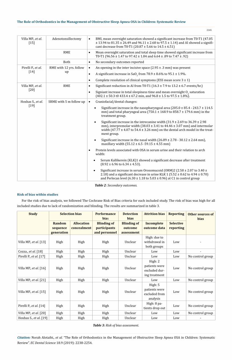
Villa MP., et al. [15]
Adenotonsillectomy • BMI, mean overnight saturation showed a significant increase from T0-T1 (47.05 ± 13.94 to 81.35 ± 26.49 and 96.11 ± 2.68 to 97.5 ± 1.14) and AI showed a signifi-cant decrease from T0-T1 (20.87 ± 5.66 to 14.5 ± 6.51)
RME • Mean overnight saturation and total sleep time showed significant increase from T0-T1 (96.56 ± 1.47 to 97.42 ± 1.84 and 6.64 ± .89 to 7.47 ± .92)
Both • No secondary outcomes reportedPirelli P., et al.
[14]RME with 12 yrs. follow
up• An opening in the inter incisive space (2.95 ± .3 mm) was present
• A significant increase in SaO2 from 78.9 ± 8.6% to 95.1 ± 1.9%.
• Complete resolution of clinical symptoms (ESS mean score 3 ± 1)Villa MP., et al.
[20]RME • Significant reduction in AI from T0-T1 (16.3 ± 7.9 to 13.2 ± 6.7 events/hr.)
• Signiant increase in total sleepiness time and mean overnight O2 saturation (402.1 ± 50.3 t0 433.4 ± 67.2 min, and 96.8 ± 1.5 to 97.5 ± 1.8%).
Hoxhas S., et al. [19]
SRME with 5 m follow up • Craniofacial/dental changes:
• Significant increase in the nasopharyngeal area (205.0 ± 85.4 - 243.7 ± 114.5 mm) and total pharyngeal area (750.1 ± 168.9 to 858.7 ± 179.6 mm) in the treatment group.
• Significant increase in the intrecanine width (31.9 ± 2.69 to 36.39 ± 2.98 mm), interpremolar width (38.03 ± 3.41 to 44.46 ± 3.07 mm) and intermolar width (47.77 ± 4.07 to 54.4 ± 3.26 mm) on the dental arch model in the treat-ment group.
• Protein levels associated with OSA in serum urine and their relation to arch width:
• Serum Kallikerein (KLK)1 showed a significant decrease after treatment (8.92 ± 6.96 to 6.34 ± 4.53).
• Significant increase in serum Orosomucoid (ORM)2 (2.58 ± 2.07 to 3.40 ± 2.18) and a significant decrease in urine KLK 1 (5.52 ± 0.62 to 4.94 ± 0.70) and Perlecan level (6.30 ± 1.18 to 5.03 ± 0.96) at C1 in control group
Table 2: Secondary outcomes.
For the risk of bias analysis, we followed The Cochrane Risk of Bias criteria for each included study. The risk of bias was high for all included studies due to lack of randomization and blinding. The results are summarized in table 3.
Risk of bias within studies
Study Selection bias Performance bias
Detection bias
Attrition bias Reporting Other sources of bias
Random sequence
generation
Allocation concealment
Blinding of participants
and personnel
Blinding of outcome
assessment
Incomplete outcome data
Selective reporting
Villa MP., et al. [13] High High High UnclearHigh: due to
withdrawal in both groups
Low -
Cozza., et al. [18] High High High Unclear Low Low -Pirelli P., et al. [17] High High High Unclear Low Low No control group
Villa MP., et al. [16] High High High Unclear
High: 2 patients were excluded dur-ing treatment
Low No control group
Villa MP., et al. [21] High High High Unclear Low Low No control group
Villa MP., et al. [15] High High High Unclear
High: 5 patients were excluded from
analysis
Low No control group
Pirelli P., et al. [14] High High High Unclear High: 8 pa-tients drop out Low No control group
Villa MP., et al. [20] High High High Unclear Low Low No control groupHoxhas S., et al. [19] High High High Unclear Low Low -
Table 3: Risk of bias assessment.
2246
The Role of Orthodontics in the Management of Obstructive Sleep Apnea OSA in Children: Systematic Review
Citation: Norah Alotaibi., et al. “The Role of Orthodontics in the Management of Obstructive Sleep Apnea OSA in Children: Systematic Review”. EC Dental Science 18.9 (2019):2238-2254.
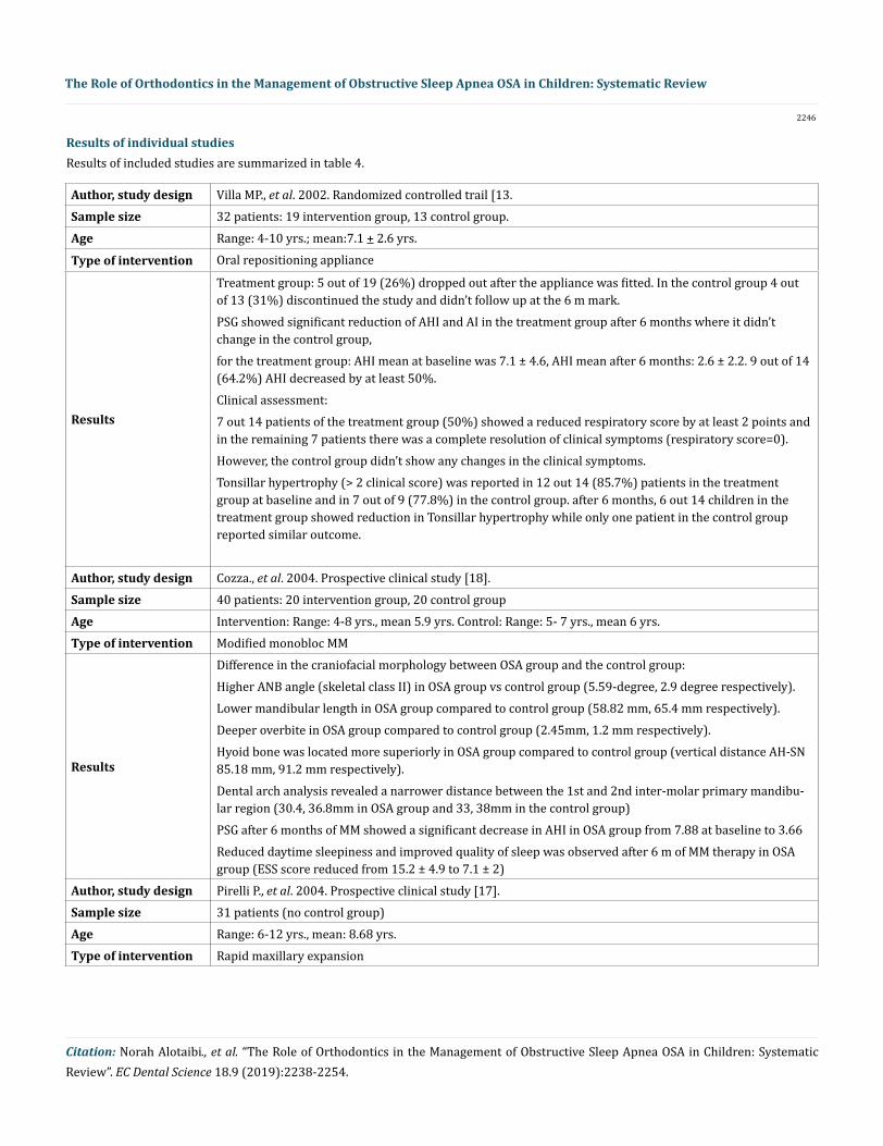
Author, study design Villa MP., et al. 2002. Randomized controlled trail [13. Sample size 32 patients: 19 intervention group, 13 control group.Age Range: 4-10 yrs.; mean:7.1 + 2.6 yrs.
Type of intervention Oral repositioning appliance
Results
Treatment group: 5 out of 19 (26%) dropped out after the appliance was fitted. In the control group 4 out of 13 (31%) discontinued the study and didn’t follow up at the 6 m mark. PSG showed significant reduction of AHI and AI in the treatment group after 6 months where it didn’t change in the control group, for the treatment group: AHI mean at baseline was 7.1 ± 4.6, AHI mean after 6 months: 2.6 ± 2.2. 9 out of 14 (64.2%) AHI decreased by at least 50%.Clinical assessment: 7 out 14 patients of the treatment group (50%) showed a reduced respiratory score by at least 2 points and in the remaining 7 patients there was a complete resolution of clinical symptoms (respiratory score=0). However, the control group didn’t show any changes in the clinical symptoms. Tonsillar hypertrophy (> 2 clinical score) was reported in 12 out 14 (85.7%) patients in the treatment group at baseline and in 7 out of 9 (77.8%) in the control group. after 6 months, 6 out 14 children in the treatment group showed reduction in Tonsillar hypertrophy while only one patient in the control group reported similar outcome.
Author, study design Cozza., et al. 2004. Prospective clinical study [18].Sample size 40 patients: 20 intervention group, 20 control groupAge Intervention: Range: 4-8 yrs., mean 5.9 yrs. Control: Range: 5- 7 yrs., mean 6 yrs. Type of intervention Modified monobloc MM
Results
Difference in the craniofacial morphology between OSA group and the control group: Higher ANB angle (skeletal class II) in OSA group vs control group (5.59-degree, 2.9 degree respectively). Lower mandibular length in OSA group compared to control group (58.82 mm, 65.4 mm respectively). Deeper overbite in OSA group compared to control group (2.45mm, 1.2 mm respectively).Hyoid bone was located more superiorly in OSA group compared to control group (vertical distance AH-SN 85.18 mm, 91.2 mm respectively). Dental arch analysis revealed a narrower distance between the 1st and 2nd inter-molar primary mandibu-lar region (30.4, 36.8mm in OSA group and 33, 38mm in the control group) PSG after 6 months of MM showed a significant decrease in AHI in OSA group from 7.88 at baseline to 3.66 Reduced daytime sleepiness and improved quality of sleep was observed after 6 m of MM therapy in OSA group (ESS score reduced from 15.2 ± 4.9 to 7.1 ± 2)
Author, study design Pirelli P., et al. 2004. Prospective clinical study [17].Sample size 31 patients (no control group)Age Range: 6-12 yrs., mean: 8.68 yrs.Type of intervention Rapid maxillary expansion
Results of included studies are summarized in table 4.Results of individual studies
2247
The Role of Orthodontics in the Management of Obstructive Sleep Apnea OSA in Children: Systematic Review
Citation: Norah Alotaibi., et al. “The Role of Orthodontics in the Management of Obstructive Sleep Apnea OSA in Children: Systematic Review”. EC Dental Science 18.9 (2019):2238-2254.
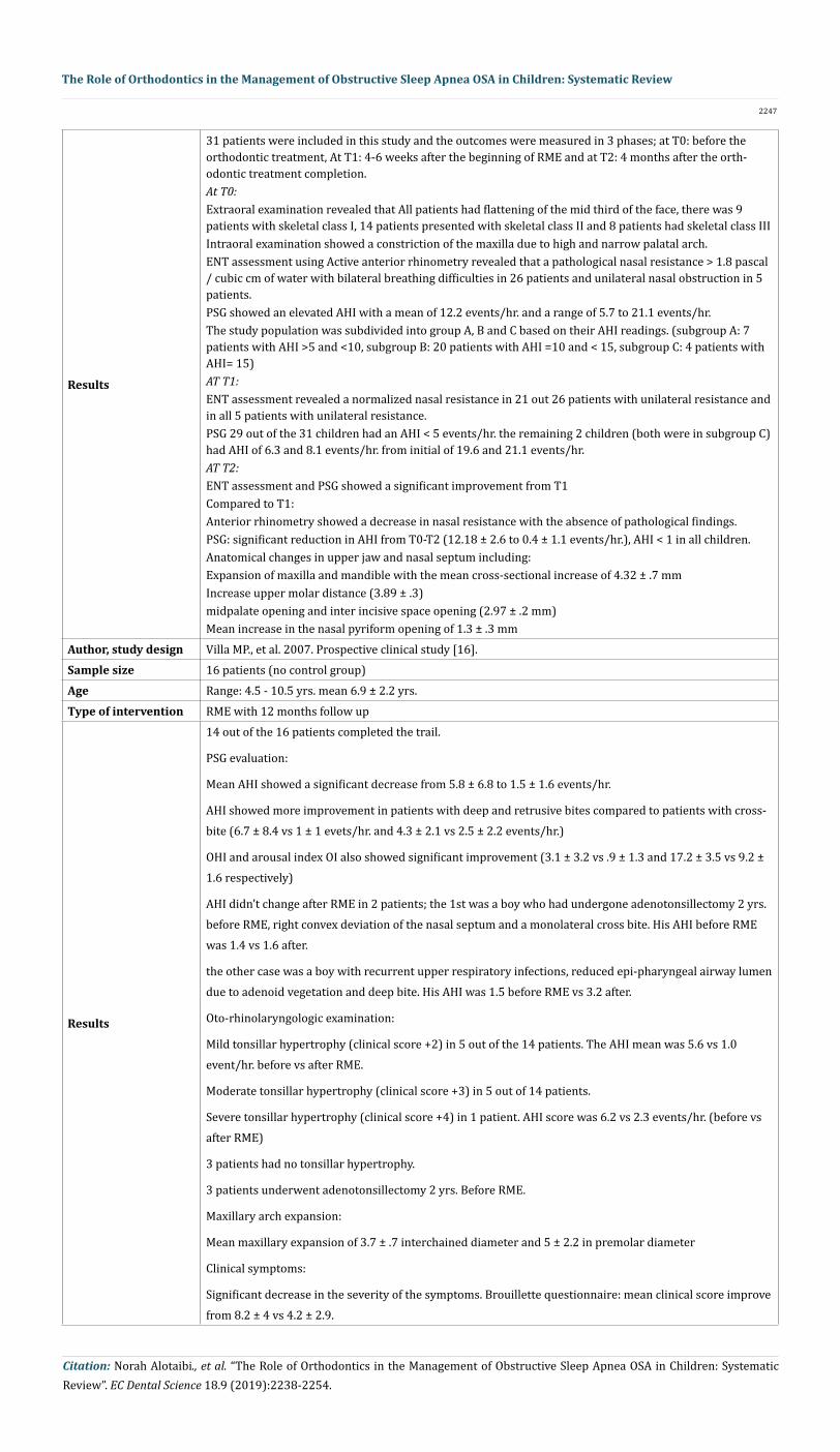
Results
31 patients were included in this study and the outcomes were measured in 3 phases; at T0: before the orthodontic treatment, At T1: 4-6 weeks after the beginning of RME and at T2: 4 months after the orth-odontic treatment completion. At T0: Extraoral examination revealed that All patients had flattening of the mid third of the face, there was 9 patients with skeletal class I, 14 patients presented with skeletal class II and 8 patients had skeletal class III Intraoral examination showed a constriction of the maxilla due to high and narrow palatal arch. ENT assessment using Active anterior rhinometry revealed that a pathological nasal resistance > 1.8 pascal / cubic cm of water with bilateral breathing difficulties in 26 patients and unilateral nasal obstruction in 5 patients.PSG showed an elevated AHI with a mean of 12.2 events/hr. and a range of 5.7 to 21.1 events/hr. The study population was subdivided into group A, B and C based on their AHI readings. (subgroup A: 7 patients with AHI >5 and <10, subgroup B: 20 patients with AHI =10 and < 15, subgroup C: 4 patients with AHI= 15) AT T1: ENT assessment revealed a normalized nasal resistance in 21 out 26 patients with unilateral resistance and in all 5 patients with unilateral resistance. PSG 29 out of the 31 children had an AHI < 5 events/hr. the remaining 2 children (both were in subgroup C) had AHI of 6.3 and 8.1 events/hr. from initial of 19.6 and 21.1 events/hr. AT T2: ENT assessment and PSG showed a significant improvement from T1 Compared to T1: Anterior rhinometry showed a decrease in nasal resistance with the absence of pathological findings. PSG: significant reduction in AHI from T0-T2 (12.18 ± 2.6 to 0.4 ± 1.1 events/hr.), AHI < 1 in all children. Anatomical changes in upper jaw and nasal septum including: Expansion of maxilla and mandible with the mean cross-sectional increase of 4.32 ± .7 mm Increase upper molar distance (3.89 ± .3) midpalate opening and inter incisive space opening (2.97 ± .2 mm) Mean increase in the nasal pyriform opening of 1.3 ± .3 mm
Author, study design Villa MP., et al. 2007. Prospective clinical study [16].Sample size 16 patients (no control group)Age Range: 4.5 - 10.5 yrs. mean 6.9 ± 2.2 yrs.Type of intervention RME with 12 months follow up
Results
14 out of the 16 patients completed the trail.
PSG evaluation:
Mean AHI showed a significant decrease from 5.8 ± 6.8 to 1.5 ± 1.6 events/hr.
AHI showed more improvement in patients with deep and retrusive bites compared to patients with cross-bite (6.7 ± 8.4 vs 1 ± 1 evets/hr. and 4.3 ± 2.1 vs 2.5 ± 2.2 events/hr.)
OHI and arousal index OI also showed significant improvement (3.1 ± 3.2 vs .9 ± 1.3 and 17.2 ± 3.5 vs 9.2 ± 1.6 respectively)
AHI didn’t change after RME in 2 patients; the 1st was a boy who had undergone adenotonsillectomy 2 yrs. before RME, right convex deviation of the nasal septum and a monolateral cross bite. His AHI before RME was 1.4 vs 1.6 after.
the other case was a boy with recurrent upper respiratory infections, reduced epi-pharyngeal airway lumen due to adenoid vegetation and deep bite. His AHI was 1.5 before RME vs 3.2 after.
Oto-rhinolaryngologic examination:
Mild tonsillar hypertrophy (clinical score +2) in 5 out of the 14 patients. The AHI mean was 5.6 vs 1.0 event/hr. before vs after RME.
Moderate tonsillar hypertrophy (clinical score +3) in 5 out of 14 patients.
Severe tonsillar hypertrophy (clinical score +4) in 1 patient. AHI score was 6.2 vs 2.3 events/hr. (before vs after RME)
3 patients had no tonsillar hypertrophy.
3 patients underwent adenotonsillectomy 2 yrs. Before RME.
Maxillary arch expansion:
Mean maxillary expansion of 3.7 ± .7 interchained diameter and 5 ± 2.2 in premolar diameter
Clinical symptoms:
Significant decrease in the severity of the symptoms. Brouillette questionnaire: mean clinical score improve from 8.2 ± 4 vs 4.2 ± 2.9.
2248
The Role of Orthodontics in the Management of Obstructive Sleep Apnea OSA in Children: Systematic Review
Citation: Norah Alotaibi., et al. “The Role of Orthodontics in the Management of Obstructive Sleep Apnea OSA in Children: Systematic Review”. EC Dental Science 18.9 (2019):2238-2254.
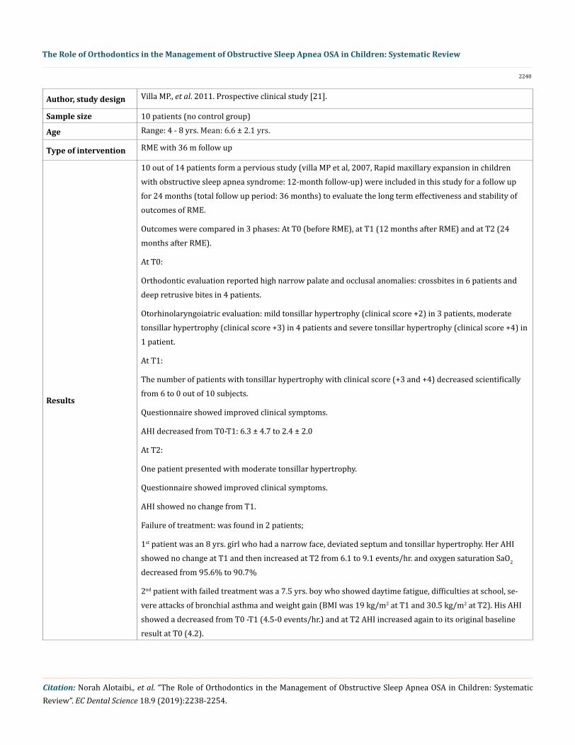
Author, study design Villa MP., et al. 2011. Prospective clinical study [21].
Sample size 10 patients (no control group)
Age Range: 4 - 8 yrs. Mean: 6.6 ± 2.1 yrs.
Type of intervention RME with 36 m follow up
Results
10 out of 14 patients form a pervious study (villa MP et al, 2007, Rapid maxillary expansion in children with obstructive sleep apnea syndrome: 12-month follow-up) were included in this study for a follow up for 24 months (total follow up period: 36 months) to evaluate the long term effectiveness and stability of outcomes of RME.
Outcomes were compared in 3 phases: At T0 (before RME), at T1 (12 months after RME) and at T2 (24 months after RME).
At T0:
Orthodontic evaluation reported high narrow palate and occlusal anomalies: crossbites in 6 patients and deep retrusive bites in 4 patients.
Otorhinolaryngoiatric evaluation: mild tonsillar hypertrophy (clinical score +2) in 3 patients, moderate tonsillar hypertrophy (clinical score +3) in 4 patients and severe tonsillar hypertrophy (clinical score +4) in 1 patient.
At T1:
The number of patients with tonsillar hypertrophy with clinical score (+3 and +4) decreased scientifically from 6 to 0 out of 10 subjects.
Questionnaire showed improved clinical symptoms.
AHI decreased from T0-T1: 6.3 ± 4.7 to 2.4 ± 2.0
At T2:
One patient presented with moderate tonsillar hypertrophy.
Questionnaire showed improved clinical symptoms.
AHI showed no change from T1.
Failure of treatment: was found in 2 patients;
1st patient was an 8 yrs. girl who had a narrow face, deviated septum and tonsillar hypertrophy. Her AHI showed no change at T1 and then increased at T2 from 6.1 to 9.1 events/hr. and oxygen saturation SaO2 decreased from 95.6% to 90.7%
2nd patient with failed treatment was a 7.5 yrs. boy who showed daytime fatigue, difficulties at school, se-vere attacks of bronchial asthma and weight gain (BMI was 19 kg/m2 at T1 and 30.5 kg/m2 at T2). His AHI showed a decreased from T0 -T1 (4.5-0 events/hr.) and at T2 AHI increased again to its original baseline result at T0 (4.2).
2249
The Role of Orthodontics in the Management of Obstructive Sleep Apnea OSA in Children: Systematic Review
Citation: Norah Alotaibi., et al. “The Role of Orthodontics in the Management of Obstructive Sleep Apnea OSA in Children: Systematic Review”. EC Dental Science 18.9 (2019):2238-2254.
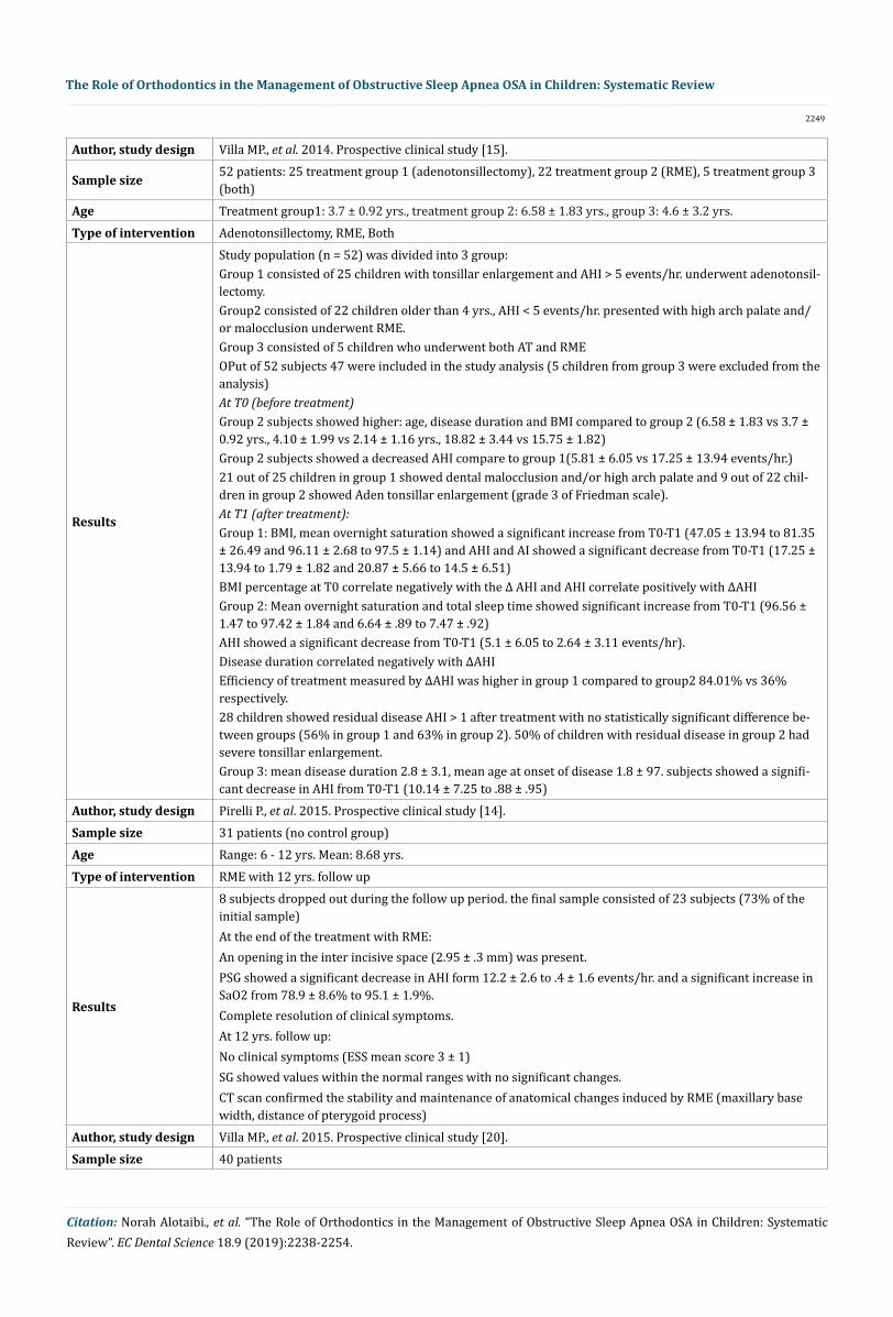
Author, study design Villa MP., et al. 2014. Prospective clinical study [15].
Sample size 52 patients: 25 treatment group 1 (adenotonsillectomy), 22 treatment group 2 (RME), 5 treatment group 3 (both)
Age Treatment group1: 3.7 ± 0.92 yrs., treatment group 2: 6.58 ± 1.83 yrs., group 3: 4.6 ± 3.2 yrs.Type of intervention Adenotonsillectomy, RME, Both
Results
Study population (n = 52) was divided into 3 group:Group 1 consisted of 25 children with tonsillar enlargement and AHI > 5 events/hr. underwent adenotonsil-lectomy.Group2 consisted of 22 children older than 4 yrs., AHI < 5 events/hr. presented with high arch palate and/or malocclusion underwent RME. Group 3 consisted of 5 children who underwent both AT and RME OPut of 52 subjects 47 were included in the study analysis (5 children from group 3 were excluded from the analysis)At T0 (before treatment) Group 2 subjects showed higher: age, disease duration and BMI compared to group 2 (6.58 ± 1.83 vs 3.7 ± 0.92 yrs., 4.10 ± 1.99 vs 2.14 ± 1.16 yrs., 18.82 ± 3.44 vs 15.75 ± 1.82)Group 2 subjects showed a decreased AHI compare to group 1(5.81 ± 6.05 vs 17.25 ± 13.94 events/hr.) 21 out of 25 children in group 1 showed dental malocclusion and/or high arch palate and 9 out of 22 chil-dren in group 2 showed Aden tonsillar enlargement (grade 3 of Friedman scale). At T1 (after treatment):Group 1: BMI, mean overnight saturation showed a significant increase from T0-T1 (47.05 ± 13.94 to 81.35 ± 26.49 and 96.11 ± 2.68 to 97.5 ± 1.14) and AHI and AI showed a significant decrease from T0-T1 (17.25 ± 13.94 to 1.79 ± 1.82 and 20.87 ± 5.66 to 14.5 ± 6.51) BMI percentage at T0 correlate negatively with the Δ AHI and AHI correlate positively with ΔAHIGroup 2: Mean overnight saturation and total sleep time showed significant increase from T0-T1 (96.56 ± 1.47 to 97.42 ± 1.84 and 6.64 ± .89 to 7.47 ± .92)AHI showed a significant decrease from T0-T1 (5.1 ± 6.05 to 2.64 ± 3.11 events/hr).Disease duration correlated negatively with ΔAHIEfficiency of treatment measured by ΔAHI was higher in group 1 compared to group2 84.01% vs 36% respectively.28 children showed residual disease AHI > 1 after treatment with no statistically significant difference be-tween groups (56% in group 1 and 63% in group 2). 50% of children with residual disease in group 2 had severe tonsillar enlargement. Group 3: mean disease duration 2.8 ± 3.1, mean age at onset of disease 1.8 ± 97. subjects showed a signifi-cant decrease in AHI from T0-T1 (10.14 ± 7.25 to .88 ± .95)
Author, study design Pirelli P., et al. 2015. Prospective clinical study [14].Sample size 31 patients (no control group)Age Range: 6 - 12 yrs. Mean: 8.68 yrs.Type of intervention RME with 12 yrs. follow up
Results
8 subjects dropped out during the follow up period. the final sample consisted of 23 subjects (73% of the initial sample) At the end of the treatment with RME: An opening in the inter incisive space (2.95 ± .3 mm) was present. PSG showed a significant decrease in AHI form 12.2 ± 2.6 to .4 ± 1.6 events/hr. and a significant increase in SaO2 from 78.9 ± 8.6% to 95.1 ± 1.9%. Complete resolution of clinical symptoms.At 12 yrs. follow up: No clinical symptoms (ESS mean score 3 ± 1)SG showed values within the normal ranges with no significant changes.CT scan confirmed the stability and maintenance of anatomical changes induced by RME (maxillary base width, distance of pterygoid process)
Author, study design Villa MP., et al. 2015. Prospective clinical study [20].Sample size 40 patients
2250
The Role of Orthodontics in the Management of Obstructive Sleep Apnea OSA in Children: Systematic Review
Citation: Norah Alotaibi., et al. “The Role of Orthodontics in the Management of Obstructive Sleep Apnea OSA in Children: Systematic Review”. EC Dental Science 18.9 (2019):2238-2254.
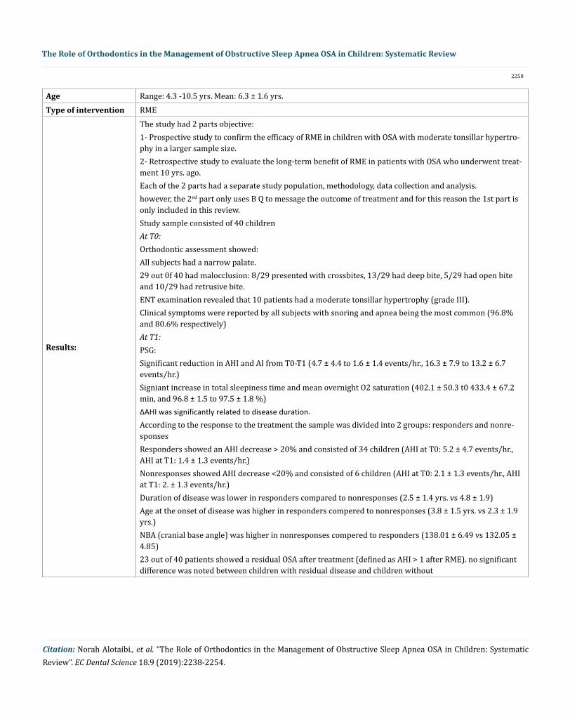
Age Range: 4.3 -10.5 yrs. Mean: 6.3 ± 1.6 yrs.Type of intervention RME
Results:
The study had 2 parts objective: 1- Prospective study to confirm the efficacy of RME in children with OSA with moderate tonsillar hypertro-phy in a larger sample size.2- Retrospective study to evaluate the long-term benefit of RME in patients with OSA who underwent treat-ment 10 yrs. ago. Each of the 2 parts had a separate study population, methodology, data collection and analysis.however, the 2nd part only uses B Q to message the outcome of treatment and for this reason the 1st part is only included in this review. Study sample consisted of 40 children At T0: Orthodontic assessment showed: All subjects had a narrow palate. 29 out 0f 40 had malocclusion: 8/29 presented with crossbites, 13/29 had deep bite, 5/29 had open bite and 10/29 had retrusive bite. ENT examination revealed that 10 patients had a moderate tonsillar hypertrophy (grade III). Clinical symptoms were reported by all subjects with snoring and apnea being the most common (96.8% and 80.6% respectively) At T1: PSG: Significant reduction in AHI and AI from T0-T1 (4.7 ± 4.4 to 1.6 ± 1.4 events/hr., 16.3 ± 7.9 to 13.2 ± 6.7 events/hr.) Signiant increase in total sleepiness time and mean overnight O2 saturation (402.1 ± 50.3 t0 433.4 ± 67.2 min, and 96.8 ± 1.5 to 97.5 ± 1.8 %) ΔAHI was significantly related to disease duration. According to the response to the treatment the sample was divided into 2 groups: responders and nonre-sponses Responders showed an AHI decrease > 20% and consisted of 34 children (AHI at T0: 5.2 ± 4.7 events/hr., AHI at T1: 1.4 ± 1.3 events/hr.)Nonresponses showed AHI decrease <20% and consisted of 6 children (AHI at T0: 2.1 ± 1.3 events/hr., AHI at T1: 2. ± 1.3 events/hr.)Duration of disease was lower in responders compared to nonresponses (2.5 ± 1.4 yrs. vs 4.8 ± 1.9) Age at the onset of disease was higher in responders compered to nonresponses (3.8 ± 1.5 yrs. vs 2.3 ± 1.9 yrs.) NBA (cranial base angle) was higher in nonresponses compered to responders (138.01 ± 6.49 vs 132.05 ± 4.85) 23 out of 40 patients showed a residual OSA after treatment (defined as AHI > 1 after RME). no significant difference was noted between children with residual disease and children without
2251
The Role of Orthodontics in the Management of Obstructive Sleep Apnea OSA in Children: Systematic Review
Citation: Norah Alotaibi., et al. “The Role of Orthodontics in the Management of Obstructive Sleep Apnea OSA in Children: Systematic Review”. EC Dental Science 18.9 (2019):2238-2254.
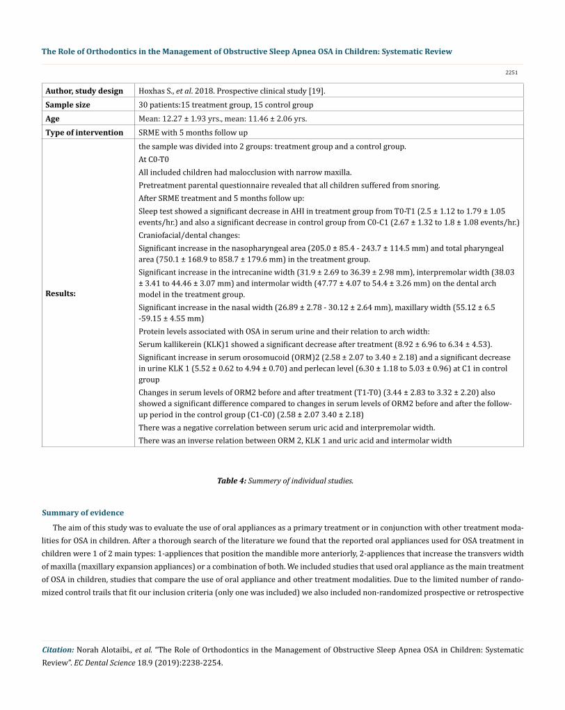
Author, study design Hoxhas S., et al. 2018. Prospective clinical study [19].Sample size 30 patients:15 treatment group, 15 control groupAge Mean: 12.27 ± 1.93 yrs., mean: 11.46 ± 2.06 yrs.Type of intervention SRME with 5 months follow up
Results:
the sample was divided into 2 groups: treatment group and a control group.At C0-T0All included children had malocclusion with narrow maxilla. Pretreatment parental questionnaire revealed that all children suffered from snoring. After SRME treatment and 5 months follow up: Sleep test showed a significant decrease in AHI in treatment group from T0-T1 (2.5 ± 1.12 to 1.79 ± 1.05 events/hr.) and also a significant decrease in control group from C0-C1 (2.67 ± 1.32 to 1.8 ± 1.08 events/hr.) Craniofacial/dental changes:Significant increase in the nasopharyngeal area (205.0 ± 85.4 - 243.7 ± 114.5 mm) and total pharyngeal area (750.1 ± 168.9 to 858.7 ± 179.6 mm) in the treatment group.Significant increase in the intrecanine width (31.9 ± 2.69 to 36.39 ± 2.98 mm), interpremolar width (38.03 ± 3.41 to 44.46 ± 3.07 mm) and intermolar width (47.77 ± 4.07 to 54.4 ± 3.26 mm) on the dental arch model in the treatment group.Significant increase in the nasal width (26.89 ± 2.78 - 30.12 ± 2.64 mm), maxillary width (55.12 ± 6.5 -59.15 ± 4.55 mm)Protein levels associated with OSA in serum urine and their relation to arch width: Serum kallikerein (KLK)1 showed a significant decrease after treatment (8.92 ± 6.96 to 6.34 ± 4.53).Significant increase in serum orosomucoid (ORM)2 (2.58 ± 2.07 to 3.40 ± 2.18) and a significant decrease in urine KLK 1 (5.52 ± 0.62 to 4.94 ± 0.70) and perlecan level (6.30 ± 1.18 to 5.03 ± 0.96) at C1 in control group Changes in serum levels of ORM2 before and after treatment (T1-T0) (3.44 ± 2.83 to 3.32 ± 2.20) also showed a significant difference compared to changes in serum levels of ORM2 before and after the follow-up period in the control group (C1-C0) (2.58 ± 2.07 3.40 ± 2.18)There was a negative correlation between serum uric acid and interpremolar width. There was an inverse relation between ORM 2, KLK 1 and uric acid and intermolar width
Table 4: Summery of individual studies.
Summary of evidence
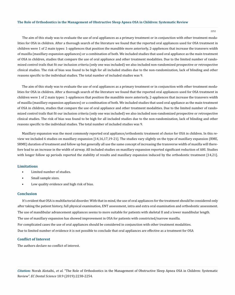
The aim of this study was to evaluate the use of oral appliances as a primary treatment or in conjunction with other treatment moda-lities for OSA in children. After a thorough search of the literature we found that the reported oral appliances used for OSA treatment in children were 1 of 2 main types: 1-appliences that position the mandible more anteriorly, 2-appliences that increase the transvers width of maxilla (maxillary expansion appliances) or a combination of both. We included studies that used oral appliance as the main treatment of OSA in children, studies that compare the use of oral appliance and other treatment modalities. Due to the limited number of rando-mized control trails that fit our inclusion criteria (only one was included) we also included non-randomized prospective or retrospective
2252
The Role of Orthodontics in the Management of Obstructive Sleep Apnea OSA in Children: Systematic Review
Citation: Norah Alotaibi., et al. “The Role of Orthodontics in the Management of Obstructive Sleep Apnea OSA in Children: Systematic Review”. EC Dental Science 18.9 (2019):2238-2254.
The aim of this study was to evaluate the use of oral appliances as a primary treatment or in conjunction with other treatment moda-lities for OSA in children. After a thorough search of the literature we found that the reported oral appliances used for OSA treatment in children were 1 of 2 main types: 1-appliences that position the mandible more anteriorly, 2-appliences that increase the transvers width of maxilla (maxillary expansion appliances) or a combination of both. We included studies that used oral appliance as the main treatment of OSA in children, studies that compare the use of oral appliance and other treatment modalities. Due to the limited number of rando-mized control trails that fit our inclusion criteria (only one was included) we also included non-randomized prospective or retrospective clinical studies. The risk of bias was found to be high for all included studies due to the non-randomization, lack of blinding and other reasons specific to the individual studies. The total number of included studies was 9.
Maxillary expansion was the most commonly reported oral appliance/orthodontic treatment of choice for OSA in children. In this re-view we included 6 studies on maxillary expansion [14,16,17,19-21]. The studies vary slightly on the type of maxillary expansion (RME, SRME) duration of treatment and follow up but generally all use the same concept of increasing the transverse width of maxilla will there-fore lead to an increase in the width of airway. All included studies on maxillary expansion reported significant reduction of AHI. Studies with longer follow up periods reported the stability of results and maxillary expansion induced by the orthodontic treatment [14,21].
• Limited number of studies.
• Small sample sizes.
• Low quality evidence and high risk of bias.
Limitations
Conclusion
It’s evident that OSA is multifactorial disorder. With that in mind, the use of oral appliances for the treatment should be considered only after taking the patient history, full physical examination, ENT assessment, intra and extra oral examination and orthodontic assessment.
The use of mandibular advancement appliances seems to more suitable for patients with skeletal II and a lower mandibular length.
The use of maxillary expansion has showed improvement in OSA for patients with constricted/narrow maxilla.
For complicated cases the use of oral appliances should be considered in conjunction with other treatment modalities.
Due to limited number of evidence it is not possible to conclude that oral appliances are effective as a treatment for OSA
Conflict of InterestThe authors declare no conflict of interest.
The aim of this study was to evaluate the use of oral appliances as a primary treatment or in conjunction with other treatment moda-lities for OSA in children. After a thorough search of the literature we found that the reported oral appliances used for OSA treatment in children were 1 of 2 main types: 1-appliences that position the mandible more anteriorly, 2-appliences that increase the transvers width of maxilla (maxillary expansion appliances) or a combination of both. We included studies that used oral appliance as the main treatment of OSA in children, studies that compare the use of oral appliance and other treatment modalities. Due to the limited number of rando-mized control trails that fit our inclusion criteria (only one was included) we also included non-randomized prospective or retrospective clinical studies. The risk of bias was found to be high for all included studies due to the non-randomization, lack of blinding and other reasons specific to the individual studies. The total number of included studies was 9.
2253
The Role of Orthodontics in the Management of Obstructive Sleep Apnea OSA in Children: Systematic Review
Citation: Norah Alotaibi., et al. “The Role of Orthodontics in the Management of Obstructive Sleep Apnea OSA in Children: Systematic Review”. EC Dental Science 18.9 (2019):2238-2254.
Bibliography
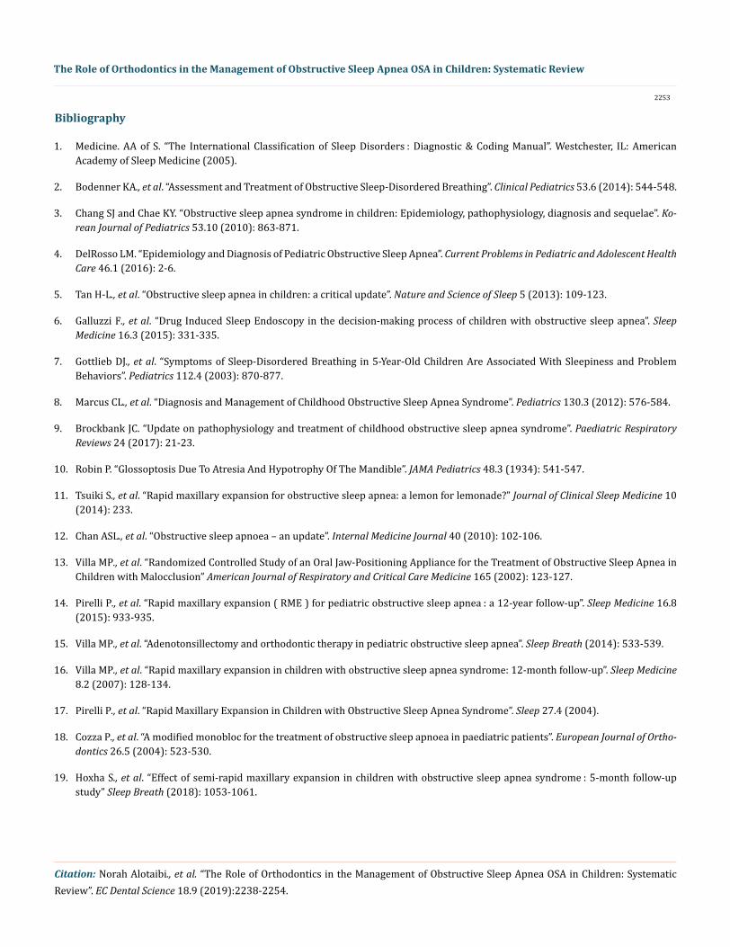
1. Medicine. AA of S. “The International Classification of Sleep Disorders : Diagnostic & Coding Manual”. Westchester, IL: American Academy of Sleep Medicine (2005).
2. Bodenner KA., et al. “Assessment and Treatment of Obstructive Sleep-Disordered Breathing”. Clinical Pediatrics 53.6 (2014): 544-548.
3. Chang SJ and Chae KY. “Obstructive sleep apnea syndrome in children: Epidemiology, pathophysiology, diagnosis and sequelae”. Ko-rean Journal of Pediatrics 53.10 (2010): 863-871.
4. DelRosso LM. “Epidemiology and Diagnosis of Pediatric Obstructive Sleep Apnea”. Current Problems in Pediatric and Adolescent Health Care 46.1 (2016): 2-6.
5. Tan H-L., et al. “Obstructive sleep apnea in children: a critical update”. Nature and Science of Sleep 5 (2013): 109-123.
6. Galluzzi F., et al. “Drug Induced Sleep Endoscopy in the decision-making process of children with obstructive sleep apnea”. Sleep Medicine 16.3 (2015): 331-335.
7. Gottlieb DJ., et al. “Symptoms of Sleep-Disordered Breathing in 5-Year-Old Children Are Associated With Sleepiness and Problem Behaviors”. Pediatrics 112.4 (2003): 870-877.
8. Marcus CL., et al. “Diagnosis and Management of Childhood Obstructive Sleep Apnea Syndrome”. Pediatrics 130.3 (2012): 576-584.
9. Brockbank JC. “Update on pathophysiology and treatment of childhood obstructive sleep apnea syndrome”. Paediatric Respiratory Reviews 24 (2017): 21-23.
10. Robin P. “Glossoptosis Due To Atresia And Hypotrophy Of The Mandible”. JAMA Pediatrics 48.3 (1934): 541-547.
11. Tsuiki S., et al. “Rapid maxillary expansion for obstructive sleep apnea: a lemon for lemonade?” Journal of Clinical Sleep Medicine 10 (2014): 233.
12. Chan ASL., et al. “Obstructive sleep apnoea – an update”. Internal Medicine Journal 40 (2010): 102-106.
13. Villa MP., et al. “Randomized Controlled Study of an Oral Jaw-Positioning Appliance for the Treatment of Obstructive Sleep Apnea in Children with Malocclusion” American Journal of Respiratory and Critical Care Medicine 165 (2002): 123-127.
14. Pirelli P., et al. “Rapid maxillary expansion ( RME ) for pediatric obstructive sleep apnea : a 12-year follow-up”. Sleep Medicine 16.8 (2015): 933-935.
15. Villa MP., et al. “Adenotonsillectomy and orthodontic therapy in pediatric obstructive sleep apnea”. Sleep Breath (2014): 533-539.
16. Villa MP., et al. “Rapid maxillary expansion in children with obstructive sleep apnea syndrome: 12-month follow-up”. Sleep Medicine 8.2 (2007): 128-134.
17. Pirelli P., et al. “Rapid Maxillary Expansion in Children with Obstructive Sleep Apnea Syndrome”. Sleep 27.4 (2004).
18. Cozza P., et al. “A modified monobloc for the treatment of obstructive sleep apnoea in paediatric patients”. European Journal of Ortho-dontics 26.5 (2004): 523-530.
19. Hoxha S., et al. “Effect of semi-rapid maxillary expansion in children with obstructive sleep apnea syndrome : 5-month follow-up study” Sleep Breath (2018): 1053-1061.
The Role of Orthodontics in the Management of Obstructive Sleep Apnea OSA in Children: Systematic Review
Citation: Norah Alotaibi., et al. “The Role of Orthodontics in the Management of Obstructive Sleep Apnea OSA in Children: Systematic Review”. EC Dental Science 18.9 (2019):2238-2254.
20. Villa MP., et al. “Rapid maxillary expansion outcomes in treatment of obstructive sleep apnea in children”. Sleep Medicine 16.6 (2015): 709-716.
21. Villa MP., et al. “Efficacy of rapid maxillary expansion in children with obstructive sleep apnea syndrome : 36 months of follow-up”. Sleep Breath (2011): 179-184.