Cronicon OPEN ACCESS EC OPHTHALMOLOGY Research Article Citation: Gabriela Blanco., et al. “Reconstruction of Ocular Surface with Suture vs. Tissue Adhesive”. EC Ophthalmology 9.7 (2018): 444- 456. Reconstruction of Ocular Surface with Suture vs. Tissue Adhesive Katya Torres 1 , Eduardo Oliva 2 and Gabriela Blanco 3 * 1 Medical Ophthalmologist, Wong Ophthalmological Institute, Lima, Peru 2 Intern Ophthalmology Service Military Hospital Dr. Carlos Arvelo, Caracas, Venezuela 3 Ophthalmologist, Professor Fellowship Cornea and Ocular Surface, Military Hospital Dr. Carlos Arvelo, Caracas, Venezuela *Corresponding Author: Gabriela Blanco, Ophthalmologist, Professor Fellowship Cornea and Ocular Surface, Military Hospital Dr. Carlos Arvelo, Caracas, Venezuela. Received: November 08, 2017; Published: June 19, 2018 Abstract Objective: To analyze comparatively the techniques of Reconstruction of the Ocular Surface with suture and with tissue adhesive in the weather. Design: Prospective, descriptive, comparative, non-experimental, longitudinal study. Methods: Of a total of 50 eyes, 33 surface reconstructions were performed with suture; among them, 25 (25.76%) presented recur- rent pterygium, 2 (6.06%) eyes presented syphilis in addition to pterygium, one (3.03%) eye presented conjunctival carcinoma, 3 (9.09%) eyes presented persistent epithelial defect (DEP), and 2 eyes presented chemical alkaline burn. Of the 50 eyes, 24 surface reconstruction surgeries were performed with fibrin adhesive; among them, 15 (62.50%) eyes presented recurrent pterygium, 4 (16.67%) presented syphilis in addition to pterygium, 2 (8.33%) eyes conjunctival carcinoma, 1 (4.17%) eye presented Persistent epithelial defect (PED), and 2 eyes present alkaline burn. Success is defined: in the pterygium, no recurrence, in the case of PED, re- epithelialization; in case of syphilis, without recurrence and improvement of ocular motility, in case of carcinoma, no recurrence, in case of alkaline burn, corneal epithelization, decreased neovascularization and improvement of visual acuity. Re-epithelialization of the amniotic membrane between 10 and 14 days was also considered successful. Results: The use of fibrin adhesive had a success rate of 100%, while the sutures resulted in a success rate of 90.1%. Conclusions: As pioneers in the country, we can say that surgery performed with fibrin is the highest success rate method in the pathologies of this study. Keywords: Reconstruction; Ocular Surface; Suture; Tissue Adhesive Introduction The ocular surface is formed by the conjunctiva (palpebral, bulbar and cul-de-sac), the cornea (epithelium and underlying stroma), the limbus esclerocorneal (anatomical zone of transition between the conjunctiva and the cornea) and the tear film. All these structures behave like a true functional unit so that a significant alteration in one of them often ends up affecting the rest.
Transcript
CroniconO P E N A C C E S S EC OPHTHALMOLOGY
Research Article
Citation: Gabriela Blanco., et al. “Reconstruction of Ocular Surface with Suture vs. Tissue Adhesive”. EC Ophthalmology 9.7 (2018): 444-456.
Reconstruction of Ocular Surface with Suture vs. Tissue Adhesive
Katya Torres1, Eduardo Oliva2 and Gabriela Blanco3*1Medical Ophthalmologist, Wong Ophthalmological Institute, Lima, Peru2Intern Ophthalmology Service Military Hospital Dr. Carlos Arvelo, Caracas, Venezuela3Ophthalmologist, Professor Fellowship Cornea and Ocular Surface, Military Hospital Dr. Carlos Arvelo, Caracas, Venezuela
*Corresponding Author: Gabriela Blanco, Ophthalmologist, Professor Fellowship Cornea and Ocular Surface, Military Hospital Dr. Carlos Arvelo, Caracas, Venezuela.
Received: November 08, 2017; Published: June 19, 2018
Abstract
Objective: To analyze comparatively the techniques of Reconstruction of the Ocular Surface with suture and with tissue adhesive in the weather.
Methods: Of a total of 50 eyes, 33 surface reconstructions were performed with suture; among them, 25 (25.76%) presented recur-rent pterygium, 2 (6.06%) eyes presented syphilis in addition to pterygium, one (3.03%) eye presented conjunctival carcinoma, 3 (9.09%) eyes presented persistent epithelial defect (DEP), and 2 eyes presented chemical alkaline burn. Of the 50 eyes, 24 surface reconstruction surgeries were performed with fibrin adhesive; among them, 15 (62.50%) eyes presented recurrent pterygium, 4 (16.67%) presented syphilis in addition to pterygium, 2 (8.33%) eyes conjunctival carcinoma, 1 (4.17%) eye presented Persistent epithelial defect (PED), and 2 eyes present alkaline burn. Success is defined: in the pterygium, no recurrence, in the case of PED, re-epithelialization; in case of syphilis, without recurrence and improvement of ocular motility, in case of carcinoma, no recurrence, in case of alkaline burn, corneal epithelization, decreased neovascularization and improvement of visual acuity. Re-epithelialization of the amniotic membrane between 10 and 14 days was also considered successful.
Results: The use of fibrin adhesive had a success rate of 100%, while the sutures resulted in a success rate of 90.1%.
Conclusions: As pioneers in the country, we can say that surgery performed with fibrin is the highest success rate method in the pathologies of this study.
IntroductionThe ocular surface is formed by the conjunctiva (palpebral, bulbar and cul-de-sac), the cornea (epithelium and underlying stroma),
the limbus esclerocorneal (anatomical zone of transition between the conjunctiva and the cornea) and the tear film. All these structures behave like a true functional unit so that a significant alteration in one of them often ends up affecting the rest.
445
Reconstruction of Ocular Surface with Suture vs. Tissue Adhesive
Citation: Gabriela Blanco., et al. “Reconstruction of Ocular Surface with Suture vs. Tissue Adhesive”. EC Ophthalmology 9.7 (2018): 444-456.
The alterations of the conjunctiva and the cornea re-present a very important part in the pathology of the ocular surface and in oph-thalmology.
In recent years there has been a great advance in knowledge of the pathophysiology and therapeutics of diseases of the ocular surface, and this has allowed to improve the results in different pathologies of the conjunctiva and the cornea. However, some of them still repre-sent a real therapeutic challenge.
Different works published in recent years on amniotic membrane transplantation have shown satisfactory results from the clinical point of view in multiple pathological situations of the conjunctiva and the cornea.
The aforementioned groups include pathologies difficult to manage with the treatments existing to date, and in which amniotic mem-brane transplantation represents an effective therapeutic alternative.
At present, the amniotic membrane implant and in particular the cryopreserved by KrioTekTM method is an expanding surgical proce-dure, already approved by the FDA, the majority used in the USA and now approved for early distribution in Europe.
Most ophthalmologists carry out the amniotic membrane transplant by means of suture, which is time-consuming and is associated with some disadvantages. To overcome this drawback, a novel suture less technique with tissue adhesive is promoted in the reconstruc-tion of the ocular surface.
Objectives
General
Evaluate the effectiveness of cryopreserved amniotic membrane transplantation and reconstruction of the ocular surface.
Specific
1. Describe the surgical technique with suture of the posterior plaque of amniotic membrane cryo preserved in ocular surface reconstruction.
2. Describe the suture less surgical technique of the cryopreserved amniotic membrane preserved in ocular surface recon-struction.
3. Compare the surgical technique of cryopreserved amniotic membrane transplant without suture(tissue adhesive) and with suture.
4. To evaluate the efficacy and safety in the short term of the cryopreserved amniotic membrane transplant without suture (with tissue adhesive) in ocular surface reconstruction.
5. To know the stability of the results obtained with the amniotic membrane transplant in the short term, in ocular surface reconstruction.
MethodologyPopulation and Sample
Citation: Gabriela Blanco., et al. “Reconstruction of Ocular Surface with Suture vs. Tissue Adhesive”. EC Ophthalmology 9.7 (2018): 444-456.
Reconstruction of Ocular Surface with Suture vs. Tissue Adhesive
446
The group in this study consisted of all those patients who were undergoing amniotic membrane transplant at the Military Hospital “Dr. Carlos Arvelo “(Caracas, Venezuela), Santa Lucía ophthalmological clinic and the Ophthalmological Surgery Center (CECOF), during the period between January and August.
Description of the Study Group
The study group consisted of 55 patients, consisting of 28 women (49.12% of the operated cases) and 27 men (50.88% of the operated cases), 57 amniotic membrane transplants were performed in 57 eyes of the patients. 55 patients, with different pathologies of the ocular surface. The average age of the group is 45.96 years ± 14.73 years. In the cases in which there was an absence or damage of the tissue (epithelium or stroma) in the conjunctiva, the amniotic membrane was implanted as a graft. In the cases with epithelial defect alone, the membrane was implanted as a coating. The patients were divided into 2 groups according to the surgical technique used. Group 1: was constituted by the group of patients who underwent the amniotic membrane transplant with suture; 33 eyes of 33 patients, (57.90% of the operations), being the average age of the group 44.79 years ± 16.04 years. Group 2: it was constituted by the group of patients who underwent the amniotic membrane transplant without suture, using tissue adhesive; 24 eyes of 22 patients, (42.10% of the operations), being the average age of the group 47.58 years ± 12.89 years. It is important to note that no significant difference was found (p < 0.05) between the ages of both groups.
Description of the methodology
Five alterations of the ocular surface were included in the study, which were recurrent pterygium, symblepharon, persistent corneal de-epithelization, alkali chemical burns and conjunctival CA.
A complete ophthalmological history was performed, evaluating visual acuity, biomicroscopy, intraocular pressure (IOP) and fundus.
Patient follow-up was performed the first postoperative day, on the 10th postoperative day, (between 10 and 14 days suture stitches are removed, and the amniotic membrane epithelization is evaluated); at month and the 3rd postoperative month.
A questionnaire was made to the patients in the Preoperative, 1st postoperative day, 10th postoperative day and the 1st postoperative month; I took into account a series of symptoms and signs, which were evaluated on a scale from 0 to 4 (0 = No, 1 = Mild, 2 = Moderate, 3 = Severe, 4 = Very severe). The symptoms and signs included were: Ocular pain, extra-body sensation, eye irritation, epiphora, pruritus, and conjunctival hyperemia. At the 3rd postoperative month the final evaluation was made determining the success or failure of the surgery.
It was taken as success of the surgery:
• Pterygium: Absence of recurrence during postoperative follow-up, corresponding to the period studied.
• Persistent corneal de-epithelization: Total re-epithelialization of the cornea.
• Symblepharon: No readhesion of symblepharon and improvement of ocular motility.
• Conjunctival CA: Relapse of the lesion.
• Chemical burns: Corneal epithelization, decrease in corneal neovascularization, and improvement in visual acuity.
In general, the re-epithelialization and complete adhesion of the amniotic membrane between 10 and 14 days was also successful.
Failure was considered if any explanation is presented. Recurrence in Pterygium and CA conjunctiva. Re-adhesion in symblepharon. Persistence of corneal desepitalization in persistent epithelial defect. Lack of neovascularization in chemical burns. Presence of granulo-mas in the conjunctiva.
Postoperatively all patients were treated with artificial tears and antibiotics, every 4 hours for 2 to 3 weeks. Steroids were added in
Citation: Gabriela Blanco., et al. “Reconstruction of Ocular Surface with Suture vs. Tissue Adhesive”. EC Ophthalmology 9.7 (2018): 444-456.
Reconstruction of Ocular Surface with Suture vs. Tissue Adhesive
447
the cases that warranted it.
Data collection instrument
A written instrument was applied that gathered the necessary information.
Statistical treatment of the data
For the present study, the basic descriptive statistics (percentages, measures of central tendency and dispersion) were applied, as well as student t-tests (for the qualitative variables) and the chi-square test, the latter to evaluate the relationship existing between the two groups conformed and the evolution of their status through time (preoperative, first day, tenth day and first month).
ResultsAnalysis and interpretation of the results
Following the technique of data collection, the results were classified according to the statistical presentation, and thus the tabulation of the response options established in the open questionnaire applied was continued.
For the presentation and analysis of the results obtained with the application of the technique for the re-collection of the data, the so-called quantitative analysis and the qualitative analysis were used, since this allowed to explain them. That is, the data collected through both instruments were emptied into tables, using the simple statistics of frequency, percentage and measures of central tendency later they are reflected in bar graphs, to which they were appended a brief quantitative and qualitative analysis of the analyzed data. Student t-tests were also applied (for qualitative variables) and the chi-square test, the latter to assess the existing relationship between students two groups formed and the evolution of their status over time (preoperative, first day, tenth day and first month).
In this sense, tables and graphs were made describing the frequency distribution f(X) and percentage distribution (%), with the aver-
Diagnosis No of cases %Recurrent pterygium 40 70.17Simblefaron + pterygium 6 10,53Conjunctive CA 3 5,26Corneal de-epithelization persistent 4 7,02Chemical burn by alkali 4 7,02Total 57 100,00
Table 1: Related percentage distribution with studied group according to ocular surface pathology. Source: Applied instrument
Citation: Gabriela Blanco., et al. “Reconstruction of Ocular Surface with Suture vs. Tissue Adhesive”. EC Ophthalmology 9.7 (2018): 444-456.
Reconstruction of Ocular Surface with Suture vs. Tissue Adhesive
448
Graph 1: Percentage distribution according to pathology ocular surface.
Table 2: Frequency distribution related to the answers according to the distribu-tion of Cases operated on suture vs. tissue adhesive according to diagnosis.
Source: applied instrument
449
Reconstruction of Ocular Surface with Suture vs. Tissue Adhesive
Citation: Gabriela Blanco., et al. “Reconstruction of Ocular Surface with Suture vs. Tissue Adhesive”. EC Ophthalmology 9.7 (2018): 444-456.
Graph 2: Percentage distribution by type of intervention according to diagnosis.
Age Groups No. of Patients No. of Cases %15 - 25 years 6 6 10,5326 - 35 years 7 7 12,2836 - 45 years 15 15 26,3146 - 55 years 12 13 22,8156 - 65 years 9 9 17,5466 years and Above 6 6 10,53Total 57 57 100,00
Table 3: Distribution of frequencies related to responses according to the Grupo Etáreo.
Source: applied instrument
Sex Patients No. of Cases %Female 28 28 49,12Male 27 29 50,88Total 55 57 100,00
Table 4: Distribution of related frequencies with the answers of the distribution according to sex. Source: Applied instrument.
450
Reconstruction of Ocular Surface with Suture vs. Tissue Adhesive
Citation: Gabriela Blanco., et al. “Reconstruction of Ocular Surface with Suture vs. Tissue Adhesive”. EC Ophthalmology 9.7 (2018): 444-456.
First Day Pain Epiphora Hyperemia Conjunction
Irritation Ocular
Pruritus Ocular
Sensation of Strange Body
Tissue Adhesive
It does not refer 12,50 8,33Mild 50,00 25,00 20,83 12,50 45,83 29,17
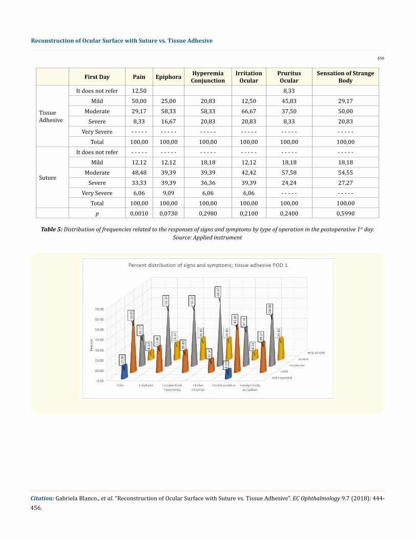
Table 5: Distribution of frequencies related to the responses of signs and symptoms by type of operation in the postoperative 1st day. Source: Applied instrument
Citation: Gabriela Blanco., et al. “Reconstruction of Ocular Surface with Suture vs. Tissue Adhesive”. EC Ophthalmology 9.7 (2018): 444-456.
Reconstruction of Ocular Surface with Suture vs. Tissue Adhesive
451
Graph 3.1 and 3.2: Percentage distribution of signs and symptoms on the 1st postoperative day.
age value of the items, likewise the interpretation was made keeping in mind the Variable Operationalization system.
Next, the option with the highest percentage and the absolute frequency is highlighted, describing the analysis with the indicators, therefore, and to establish the information obtained, 8 tables and ten 6 graphs were analyzed and interpreted. At the end of each of them, an analysis related to them to study the group of cases included in the study.
The results of graphs 3.1 and 3.2, in relation to Signs and Symptoms by type of operation in the postoperative period on the 1st day with tissue adhesive, it is observed that in the Pain category 12.5 percent of the group studied did not present pain, 50 percent had mild pain; 29.17 percent had moderate pain, 8.33 percent had severe pain; compared to cases with suture where a 12.12 percent presented pain mild, 48.48 percent presented moderate pain; 33.33 percent presented severe pain and only 6.06 percent presented very severe pain. A ratio of 0.0010 was obtained.
Regarding the epiphora (tearing) category with tissue adhesive, it is evident that 25 percent were affected by this type of symptom in a way mild, 58.33 percent presented moderate epiphora and 16.67 percent presented it severely; in balance with the suture where the light item presents with 12.12 percent, in the moderate option it presented 39.39 percent; in the severe option also presented 33.33 percent and in the very severe option 9.09 percent. A ratio of 0.0730 was obtained.
Continuing with the analysis, it is observed that in the conjunctival hyperemia (HC) category treated with the technique of tissue ad-hesive in the mild item, 20.83 percent was presented, in the moderate item, 58.33 percent presented this sign. A 20.83 percent presented HC in a severe way; this in comparison with those treated with sutures who presented mild HC in 18.18 percent, 39.39 presented HC moderately and 0.09 percent severely. A ratio of 0.2980 was obtained.
Following the study, in relation to the category Ocular Irritation (OI) treated with the tissue adhesive technique, it was shown that 12.5 percent presented OI in a mild way, 66.67 percent presented moderate OI and 20, 83 percent presented it as severe in comparison with sutured patients who presented mild OI in 12.12%, 42.42% moderate, 39.39 percent in a severe way and only 6.06 percent in a very
Citation: Gabriela Blanco., et al. “Reconstruction of Ocular Surface with Suture vs. Tissue Adhesive”. EC Ophthalmology 9.7 (2018): 444-456.
Reconstruction of Ocular Surface with Suture vs. Tissue Adhesive
452
severe way, having a ratio of 0.2100.
Regarding the ocular pruritus (PO) category with the tissue adhesive technique, it was shown that 8.33% did not report discomfort, 45.83 presented mild PO; 37.50 percent had moderate OP and 8.33 percent presented it severely; compared to those treated with sutures, which presented mild PO in 18.18 percent, a 57.58 percent in a moderate way, and 24.24 percent had PO in a severe way, obtaining a ratio of 0,2400.
Tenth Day Pain Epiphora Hyperemia conjunction
Irritation ocular
Pruritus ocular
Sensation of strange body
Tissue Adhesive
It does not refer 50,00 20,83 20,83 8,33 45,83 29,17
Table 6: Distribution of frequencies related to the responses of Signs and Symptoms by type of operation in the postoperative tenth day. Source: Applied instrument UN MES Postoperatorio
Citation: Gabriela Blanco., et al. “Reconstruction of Ocular Surface with Suture vs. Tissue Adhesive”. EC Ophthalmology 9.7 (2018): 444-456.
Reconstruction of Ocular Surface with Suture vs. Tissue Adhesive
453
Graphs 4.1 and 4.2: Percentage distribution of signs and symptoms on the tenth postoperative day.
Culminating the analysis, in relation to the category of foreign body sensation (SCE) treated with the tissue adhesive technique, it is shown that 29.17 percent presented SCE in a mild way; 50 percent presented moderate SCE, while 20.83 percent presented SCE severely; this compared with the cases treated with suture which presented mild SCE in 18.18 percent, SCE moderate in 54.55 percent and 27.27 percent in a severe way, obtaining a ratio of 0.5990.
A Month Postoperative
Pain Epiphora Hyperemia conjunction
Irritation ocular
Pruritus ocular
Sensation of strange body
Tissue Adhesive
It does not refer 79,17 62,50 87,50 62,50 79,17 83,33Mild 20,83 33,33 12,50 37,50 20,83 16,67
Table 8: Distribution of Frequencies related to final evaluation at the 3rd month.
Graphs 6: Percentage distribution of success and failure according to the type of operation.
By the tenth postoperative day, a statistically significant difference was observed (p < 0.05) between both groups, for all the signs and symptoms.
In general, there was complete epithelialization of the amniotic membrane at 12 days in all the cases studied, corresponding to the 2 groups under study.
There was no statistically significant difference (p < 0.05) between both groups, in relation to the signs and symptoms, during the evolution of the cases at the 1st postoperative month.
Regarding the number of successes and failures of both techniques at the third month after the interventions, it is evident that the tis-sue adhesive had 100% success and zero failures, compared to the suture which presented a 90.9 percent of successes and 9.1 percent of failures, which corresponded to 3 cases, among which there was a recurrence of Pterygium, a granuloma in a post operatory of Pterygium
Citation: Gabriela Blanco., et al. “Reconstruction of Ocular Surface with Suture vs. Tissue Adhesive”. EC Ophthalmology 9.7 (2018): 444-456.
Reconstruction of Ocular Surface with Suture vs. Tissue Adhesive
456
and a readmission of sim- blepharon, which indicates that the technique with tissue adhesive would be the most recommended when performing surgical interventions in the ophthalmologic area, in this group of ocular surface diseases.
Discussion and Conclusion
Amniotic membrane transplantation is an effective and safe technique for the treatment of different ocular surface pathologies, with stable results. After resection of extensive conjunctival lesions, the amniotic membrane graft is currently the treatment of choice. In cases with corneal epithelial defect, amniotic membrane transplantation is an effective therapeutic procedure, and can be considered a useful surgical alternative in those cases in which conservative medical treatment has failed.
The preserved amniotic membrane preserves anti-scarring, anti-inflammatory-anti-antigenic properties, facilitates epithelialization and maintains the normal epithelial phenotype. The success of the amniotic membrane transplant is dependent on the established clinical condition and because of the Suboptimal results in some indications, a strict selection of cases is recommended. The spectrum of clinical indications continues to expand and accompanies a variety of ocular surface pathology ranges.
It is evident that the amniotic membrane transplant has gained an acceptable position in the surgical armamentarium of the ocular surface surgeon. The relative ease of the procedure, and the low rate of intraoperative and postoperative complications makes it an ad-vantageous and attractive surgical option.
Bibliography
1. Kheirkhah A., et al. “Surgical strategies for fornix reconstruction based on symblepharon severity”. American Journal of Ophthalmol-ogy 146.2 (2008): 266-275.
2. Houses V., et al. “Surgical Approach for Scleral Ischemia and Melt”. Cornea 27.2 (2008): 196-201.
3. Li W., et al. “Down-regulation of Pax6 is associated with abnormal differentiation of corneal epithelial cells in severe ocular surface diseases”. Journal of Pathology 214.1 (2008): 114-122.
4. Kim JC and Tseng SCG. “Transplantation of preserved human amniotic membrane for surface reconstruction in severely damaged corneas”. Cornea 14.5 (1995): 473-484.
5. Lee S and Tseng SCG. “Transplantation of preserved human amniotic membrane for persistent epithelial defects with ulceration”. American Journal of Ophthalmology 123.3 (1997): 303-312.
6. Shimazaki J., et al. “Amniotic membrane transplantation for ocular surface reconstruction in patients with chemical and thermal burns”. Ophthalmology 104.12 (1997): 2068-2076.
7. Coroneo MT. “Pterygium as early indicator of ultraviolet insolation: a hypothesis”. British Journal of Ophthalmology 77.11 (1993): 734-739.