Cross-linking of apoproteins in high density lipoprotein by dimethylsuberimidate inhibits specific lipoprotein binding to membranes George K. Chacko, Florence H. Mahlberg,' and William J. Johnson Department of Physiology and Biochemistry, The Medical College of Pennsylvania, 3300 Henry Avenue, Philadelphia, PA 19129 Abstract Apoprotein E-free high density lipoproteins (HDL) bind to various cells and cell membrane preparations with proper- ties typical of ligand-receptor interactions. This specific binding can be inhibited by treatment of HDL with tetranitromethane (TNM). During treatment of HDL with TNM, in addition to the expected nitration of tyrosine residues, cross-linking of lipids to apoproteins and of apoproteins to each other occurs. We have re- cently shown that cross-linking of phospholipids to apoproteins is not responsible for the inhibition of binding (1987. Chacko, G. K., et al. J. Lipid Res. 28: 332-337). To determine the role of cross-linking of apoproteins to each other in the inhibition, we used the bifunctional reagent dimethylsuberimidate (DMS) to cross-link the apoproteins in HDL3. Over 80% of apoproteins in DMS-HDLS were cross-linked, as analyzed by SDS-polyacrylamide gel electrophoresis. DMS-HDL3was similar to control HDL3 in its lipid composition. Gel filtration chromatography did not reveal any significant differencein size between DMS-HDL, and control HDL,. As determined by competitive binding with '251-labeled HDL,, DMS-HDL, was almost completely unable to bind specifically to rat liver plasma membranes and human skin fibro- blasts. It is concluded from these results that TNM inhibits the specific binding of HDLs to membranes by a mechanism that in- volves cross-linking of apoproteins to each other in HDL, parti- cles. This observation implies that the specific binding of HDL, to cells may depend on the native quaternary structure of apoproteins in the HDL particle. Because of its reduced ability to bind to the specific binding sites, DMS-HDL, may be useful for studies related to the functional aspects of HDL binding sites.-Chacko, G. K., F. H. Mahlberg, and W. J. Johnson. Cross-linking of apoproteins in high density lipoprotein by dimethylsuberimidate LDL (apoB/E) receptor (3) or the apoE receptor (4); they are insensitive to proteolytic digestion (5-8), to chemical modification of the lysine or arginine residues of the lipo- protein (4, 9, lo), and to calcium concentration (11, 12). Recently, investigators in several laboratories (13, 14) includ- ing ours (15) have shown that the treatment of HDL with tetranitromethane (TNM) inhibits specific HDL binding. The mechanism of this inhibition is not known. During treatment of HDL with TNM, in addition to the expected nitration of tyrosine residues of apoproteins, cross-linking of lipids to apoproteins and of apoproteins to each other occurs (15). Previously we have shown that cross-linking of phospholipids to apoproteins is not responsible for the inhibition (16). In the present study, we have investigated the role of cross-linking of apoproteins to apoproteins as a possible mechanism of inhibition of HDL binding during the treatment of HDL with TNM. The bifunctional cross- linking reagent, dimethylsuberimidate (DMS), was used to crosslink apoproteins in HDL3, Upon apoprotein cross- linking, QDL3lost its ability to bind to specific HDL bind- ing sites of isolated rat liver membranes and of GM3468 human skin fibroblasts. MATERIALS AND METHODS . .. inhibits specific lipoprotein binding to membranes. J. 1988. 29: 319-324. Supplementary key words apoA-I tetranitromethane Lipid Res. Materials Human HDL, (1.125 < d C 1.21 g/ml) was isolated by differential ultracentrifugation (17) from fresh human plasma that had been treated with 5 mM N-ethylmaleimide Because of the putative role of high density lipoprotein (HDL) as a protective lipoprotein against arteriosclerosis (l), interaction of HDL with various cells and membranes has been under intensive study (2). High affinity saturable binding sites for apoE-free HDL have been detected in a variety of tissues and cells of different species (2). These binding sites have properties different from those of the Abbreviations: HDL, high density lipoproteins; DMS, dimethyl- suberimidate; TNM, tetranitromethane; BSA, bovine serum albumin; SDS, sodium dodecyl sulfate; PAGE, polyacrylamide gel electrophoresis. 'Present address: Laboratoire de Nutrition Humaine, Facult6 de M a e - cine, Xavier Bichat, UniversitC Paris 7, 16 Rue Henri Huchard, 75018 Paris. France. Journal of Lipid Research Volume 29, 1988 319 by guest, on December 25, 2018 www.jlr.org Downloaded from
Transcript
Cross-linking of apoproteins in high density lipoprotein by dimethylsuberimidate inhibits specific lipoprotein binding to membranes
George K. Chacko, Florence H. Mahlberg,' and William J. Johnson Department of Physiology and Biochemistry, The Medical College of Pennsylvania, 3300 Henry Avenue, Philadelphia, PA 19129
Abstract Apoprotein E-free high density lipoproteins (HDL) bind to various cells and cell membrane preparations with proper- ties typical of ligand-receptor interactions. This specific binding can be inhibited by treatment of HDL with tetranitromethane (TNM). During treatment of HDL with TNM, in addition to the expected nitration of tyrosine residues, cross-linking of lipids to apoproteins and of apoproteins to each other occurs. We have re- cently shown that cross-linking of phospholipids to apoproteins is not responsible for the inhibition of binding (1987. Chacko, G. K., et al. J. Lipid Res. 28: 332-337). To determine the role of cross-linking of apoproteins to each other in the inhibition, we used the bifunctional reagent dimethylsuberimidate (DMS) to cross-link the apoproteins in HDL3. Over 80% of apoproteins in DMS-HDLS were cross-linked, as analyzed by SDS-polyacrylamide gel electrophoresis. DMS-HDL3 was similar to control HDL3 in its lipid composition. Gel filtration chromatography did not reveal any significant difference in size between DMS-HDL, and control HDL,. As determined by competitive binding with '251-labeled HDL,, DMS-HDL, was almost completely unable to bind specifically to rat liver plasma membranes and human skin fibro- blasts. It is concluded from these results that TNM inhibits the specific binding of HDLs to membranes by a mechanism that in- volves cross-linking of apoproteins to each other in HDL, parti- cles. This observation implies that the specific binding of HDL, to cells may depend on the native quaternary structure of apoproteins in the HDL particle. Because of its reduced ability to bind to the specific binding sites, DMS-HDL, may be useful for studies related to the functional aspects of HDL binding sites.-Chacko, G. K., F. H. Mahlberg, and W. J. Johnson. Cross-linking of apoproteins in high density lipoprotein by dimethylsuberimidate
LDL (apoB/E) receptor (3) or the apoE receptor (4); they are insensitive to proteolytic digestion (5-8), to chemical modification of the lysine or arginine residues of the lipo- protein (4, 9, lo), and to calcium concentration (11, 12). Recently, investigators in several laboratories (13, 14) includ- ing ours (15) have shown that the treatment of HDL with tetranitromethane (TNM) inhibits specific HDL binding. The mechanism of this inhibition is not known. During treatment of HDL with TNM, in addition to the expected nitration of tyrosine residues of apoproteins, cross-linking of lipids to apoproteins and of apoproteins to each other occurs (15). Previously we have shown that cross-linking of phospholipids to apoproteins is not responsible for the inhibition (16). In the present study, we have investigated the role of cross-linking of apoproteins to apoproteins as a possible mechanism of inhibition of HDL binding during the treatment of HDL with TNM. The bifunctional cross- linking reagent, dimethylsuberimidate (DMS), was used to crosslink apoproteins in HDL3, Upon apoprotein cross- linking, QDL3 lost its ability to bind to specific HDL bind- ing sites of isolated rat liver membranes and of GM3468 human skin fibroblasts.
MATERIALS AND METHODS . . .
inhibits specific lipoprotein binding to membranes. J. 1988. 29: 319-324.
Supplementary key words apoA-I tetranitromethane
Lipid Res. Materials
Human HDL, (1.125 < d C 1.21 g/ml) was isolated by differential ultracentrifugation (17) from fresh human plasma that had been treated with 5 mM N-ethylmaleimide
Because of the putative role of high density lipoprotein (HDL) as a protective lipoprotein against arteriosclerosis (l), interaction of HDL with various cells and membranes has been under intensive study (2). High affinity saturable binding sites for apoE-free HDL have been detected in a variety of tissues and cells of different species (2). These binding sites have properties different from those of the
Abbreviations: HDL, high density lipoproteins; DMS, dimethyl- suberimidate; TNM, tetranitromethane; BSA, bovine serum albumin; SDS, sodium dodecyl sulfate; PAGE, polyacrylamide gel electrophoresis.
'Present address: Laboratoire de Nutrition Humaine, Facult6 de M a e - cine, Xavier Bichat, UniversitC Paris 7, 16 Rue Henri Huchard, 75018 Paris. France.
to inhibit 1ecithin:cholesterol acyltransferase (18). It was further processed by heparin-Sepharose affinity chromatog- raphy (19) to remove traces of apoprotein E. Apoprotein A-I constituted about 70% of the human HDL3 apoproteins as determined by SDS-polyacrylamide gel electrophoresis (PAGE), followed by Coomassie blue staining and densito- metric scanning (20); the other apoprotein components were apoprotein A-I1 and the C apoproteins. HDL3 was labeled with lZ5I using the iodine monochloride procedure (21). The specific activities ranged from 70 to 100 cpm/ng protein. No more than 2 % ofthe label was associated with lipids. Fatty acid-free bovine serum albumin (BSA) and dimethylsuberimidate were from Sigma Chemical Co. (St. Louis, MO). All other chemicals were of reagent grade.
Methods
Isolation o f rat liver plasma mbranes . Sprague-Dawley male rats weighing 100-150 g were used. Liver plasma mem- branes were isolated according to the procedure of Ray (22), as described previously (10). The membranes were sus- pended in 10 mM Tris-HC1 buffer, pH 7.4 (containing 0.15 M NaCl and 0.5 mM caClz) at a protein concentration of 5 mg of protein/ml and used for the binding studies.
Binding of '251-labeled human HDL, to plasma membranes. The binding of 9-labeled human HDL3 to isolated membranes was determined according to the procedure described previ- ously (10). Briefly, aliquots of rat liver plasma membranes (200 pg of protein) were incubated with lZ5I-labeled human HDL, at room temperature for 1 hr in a total volume of 0.2 ml, containing 10 mM Tris-HC1, pH 7.4, 0.15 M NaCl, 0.5 mM CaClZ, and 1% BSA (incubation medium). After incubation, 0.175-ml aliquots of the incubation mixture were centrifuged in a Beckman 42.2 Ti rotor at 30,000 rpm for 15 min to recover the membranes. The membrane pellets were washed once with 0.175 ml of the incubation medium, followed by centrifugation and aspiration of the superna- tant. The tubes containing the membranes were assayed for radioactivity in an NE 1600 gamma counter (Nuclear Enterprises Ltd, Edinburgh, Scotland) with a lZ5I counting efficiency of 75%. The effect of apoprotein cross-linking on the binding of HDL to the membranes was investigated by studying the ability of cross-linked HDL to compete for the binding of lz5I-1abeled HDL3 to membranes; DMS- HDL, and lZ5I-labeled HDL3 were added simultaneously.
Binding of '251-labeled human HDL3 to cells. The binding assay was as described by Oram, Brinton, and Bierman (23). Briefly, cell monolayers were rinsed three times with 2.0 ml of PBS at 37OC. Then 0.5 ml of serum-free culture medium containing 2 mg/ml BSA and '251-labeled HDL3 with or without unlabeled lipoproteins (prewarmed for 1 hr at 37OC) was added to cells, and the plates were covered and incubated at 37OC for 1 hr. To end incubations, the culture plates were chilled on ice, and the medium was removed. The cells were rinsed five times with PBS-BSA (2 mg of BSA/ml) (4°C) and two times with PBS (4OC).
After the final rinse, the cells were digested with 1.0 ml of Markwell Lowry A solution (24) (37OC overnight in hu- midified air) and aliquots were taken for the determination of lZ5I radioactivity and protein (24).
Preparation ofDMS-HDL3. After a series of trials, the fol- lowing reaction conditions, patterned after the published procedures of Davies and Stark (25) and Swaney (26), were used for the preparation of highly cross-linked HDL3 : HDL3 concentration, 5 mg of protein/ml; DMS concentra- tion, 5 mg/ml; incubation medium, 0.09 M triethanolamine -HC1 buffer, pH 9.5; and duration of reaction, 2 hr at room temperature. Freshly made DMS solution was used. After incubation, the reaction mixture was applied to a Bio-Gel P-6 DG column (1 x 30 cm) and components were eluted with saline-EDTA (2 mM) buffer, pH 7.4. Fractions were collected and analyzed. Fractions containing protein were combined. DMS-HDL3 was recovered in almost quantita- tive yield.
Chemical and physical analyses. Protein was determined by the method of Lowry et al. (27). BSA was used as the stan- dard. Polyacrylamide gel electrophoresis of apoproteins was routinely performed on 10% gels containing 0.1% SDS (20). Apoproteins in DMS-HDL were also analyzed by gra- dient (3-27%) polyacrylamide slab gel (28).
Total lipids were extracted from lipoprotein preparations using the procedure of Bligh and Dyer (29). Lipid phos- phorus was measured according to the procedure of Rouser, Siakotos, and Fleischer (30). For the determination of phos- phorus linked to apoproteins, the lipids were extracted from the lipoprotein samples by the methanol-chloroform-ethyl ether extraction procedure of Lux, John, and Brewer (31) and the apoprotein residues were analyzed for phosphorus by the procedure of Rouser et al. (30). Free and total cholesterol were determined by gas-liquid chromatography using coprostanol as an internal standard (32). Esterified cholesterol was the difference between total and free cho- lesterol. Agarose gel electrophoresis of lipoprotein samples was carried out in barbital buffer, pH 8.6, in a Bio-Rad apparatus and according to the direction of Bio-Rad Laboratories (Richmond, CA). After electrophoresis, the lipoproteins were visualized by staining with Fat Red B. Gel filtration of HDL3 and DMS-HDL3 was done in an 85 x 2 cm Sepharose 4B column (Pharmacia Fine Chem- icals, Upsala, Sweden); the column was eluted with 0.15 M NaCl containing 0.02% EDTA, pH 7.4, and 0.02% so- dium azide, at a flow rate of 20 ml/hr. Fractions (2.1 ml) were collected and analyzed for absorbance at 280 nm.
RESULTS
Preparation of apoprotein-cross-linked DMS-HDL3 The present procedure for preparing apoprotein-cross-
linked DMS-HDL3 differs from that of Swaney (26) in two respects: 1) use of high concentrations of HDL3 (5 mg of
proteidml) and dimethylsuberimidate (5 mglml), and 2) the use of a desalting column to isolate the modified HDLS from the excess reagent at the end of the reaction. In Fig. 1 is shown a comparison of the apoprotein pattern of con- trol and DMS-HDL3, as determined by SDS-slab gel elec- trophoresis. The predominant apoprotein in control HDL3 was apoA-I; the minor band corresponded to a mixture of apoA-I1 and the C apoproteins (A). Extensive cross-linking of apoproteins was seen in DMS-HDLS (B); a marked reduction in the concentration of both apoA-I and apoA-II/ apoCs occurred. A diffuse high molecular weight band with a mean molecular weight of 95,000 was seen in DMS- HDL3. Densitometric scanning of the SDS-PAGE revealed that over 80% of the apoproteins were cross-linked. A prolonged incubation period or addition of more reagent did not result in complete cross-linking. In order to detect particle to particle cross-linking during preparation of DMS-HDLS, the DMS-HDL3 was chromatographed on a column of Sepharose 4B and the elution profile was com- pared to that of control HDL3. As is shown in Fig. 2, the elution profiles of control and DMS-HDLS were similar, indicating no appreciable cross-linking between particles in DMS-HDLS. Except for the cross-linking of apoproteins in HDL3 particles, the composition of DMS-HDL3 was similar to that of control HDL3 (Table 1). On agarose gel
AI -
A
- 140
- 70
- 28
B C STD Fig. 1. Sodium dodecyl sulfate polyacrylamide g e l electrophoresis of HDL, and DMS-HDL, on a 3-27% gradient slab. Lanes: A, ca. 100 pg of HDL, protein; R, ca. 100 pg of DMS-HDL, protein; C, ca. 10 pg of DMS-HDL, protein; STD, cross-linked hemocyanins as molecular weight markers. The g e l was stained with Coomassie blue.
A
sn,j H C L ~ + D M S - H D L Q
n * 0.
f t [ 50
Fraction number
Fig. 2. Comparison of the elution profiles obtained upon gel filtration chromatography of control HDL, (5 mg of protein) (A) and mixture of control HDL, (2.5 mg of protein) and DMS-HDL, (2.5 mg of protein) (R) on a Sepharose 4R column (2 x 85 cm) in 0.15 M NaCl containing 0.02% EDTA, pH 7.4, and 0.02% azide. Fractions (2.1 ml) were ana- lyzed for absorbance at 280 nm.
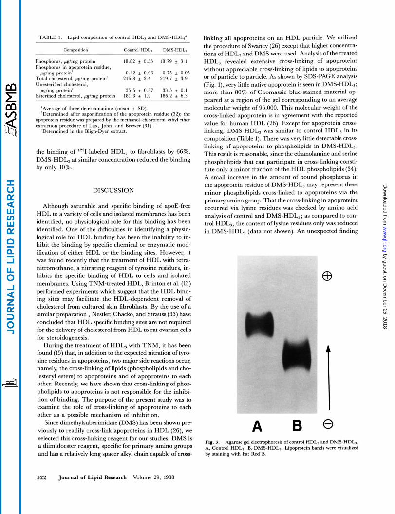
electrophoresis, the DMS-HDL3 moved as a single band (Fig. 3) and it moved less toward the anode than did the control HDL3. The reason for this reduced anodic mobility is not known.
Effect of apoprotein cross-linking on the binding
In order to determine the effect of apoprotein cross- linking on the binding of HDL3, the ability of DMS-HDL3 to compete with '251-labeled HDL3 for the binding sites of rat liver plasma membranes and of human skin GM3468 fibroblasts was studied. The results obtained with rat liver plasma membranes are shown in Fig. 4. At 25-fold excess concentration of lipoprotein, DMS-HDL3 reduced the binding of '251-labeled HDL3 to the membranes by only about 25%, whereas control unlabeled HDLS reduced the binding more than 80%. Thus, cross-linking of apoproteins in HDL3 resulted in particles with reduced ability to com- pete with '251~labeled HDL3 for the binding sites. The ex- tent of cross-linking of HDLS apoproteins could be varied by changing either the concentration of DMS or the length of exposure to DMS. In this case, the ability of the unla- beled HDL3 to inhibit 1251-labeled-HDLS binding cor- related with the amount of non-cross-linked apoA-I remain- ing in the treated HDL (Table 2). Essentially similar results were obtained when the effect of apoprotein cross-linking on the binding of HDL, to human skin fibroblasts was studied (Table 3). As compared to control HDLS, the DMS-HDL3 was much less effective in inhibiting the binding of 1251- labeled HDL to GM3468 human skin fibroblasts. Thus whereas five-fold excess of unlabeled control HDL3 reduced
Chackq Mahlbng, and Johnson Apoprotein cross-linking inhibits HDL binding 321
TABLE 1. Lipid composition of control HDL, and DMS-HDL,'
Composition Control HDLy DMS-HDLy
Phosphorus, p g h g protein 18.82 f 0.35 18.79 f 3.1 Phosphorus in a protein residue,
Total cholesterol, p g h g protein' 216.8 f 2.4 219.7 f 3.9 Unesterified cholesterol,
pg/mg protein' 35.5 f 0.37 33.5 * 0.1 Esterified cholesterol, pglmg protein 181.3 f 1.9 186.2 f 6.3
p" pgImg protein 0.42 f 0.03 0.75 f 0.05
'Average of three determinations (mean + SD). 'Determined after saponification of the apoprotein residue (32); the
apoprotein residue was prepared by the methanol-chloroform-ethyl ether extraction procedure of Lux, John, and Brewer (31).
'Determined in the Bligh-Dyer extract.
the binding of '251-labeled HDL3 to fibroblasts by 6676, DMS-HDL, at similar concentration reduced the binding by only 10%.
DISCUSSION
Although saturable and specific binding of apoE-free HDL to a variety of cells and isolated membranes has been identified, no physiological role for this binding has been identified. One of the difficulties in identifying a physio- logical role for HDL binding has been the inability to in- hibit the binding by specific chemical or enzymatic mod- ification of either HDL or the binding sites. However, it was found recently that the treatment of HDL with tetra- nitromethane, a nitrating reagent of tyrosine residues, in- hibits the specific binding of HDL to cells and isolated membranes. Using TNM-treated HDL, Brinton et al. (13) performed experiments which suggest that the HDL bind- ing sites may facilitate the HDL-dependent removal of cholesterol from cultured skin fibroblasts. By the use of a similar preparation , Nestler, Chacko, and Strauss (33) have concluded that HDL specific binding sites are not required for the delivery of cholesterol from HDL to rat ovarian cells for steroidogenesis.
During the treatment of HDL3 with TNM, it has been found (15) that, in addition to the expected nitration of tyro- sine residues in apoproteins, two major side reactions occur, namely, the cross-linking of lipids (phospholipids and cho- lesteryl esters) to apoproteins and of apoproteins to each other. Recently, we have shown that cross-linking of phos- pholipids to apoproteins is not responsible for the inhibi- tion of binding. The purpose of the present study was to examine the role of cross-linking of apoproteins to each other as a possible mechanism of inhibition.
Since dimethylsuberimidate (DMS) has been shown pre- viously to readily cross-link apoproteins in HDL (26), we selected this cross-linking reagent for our studies. DMS is a diimidoester reagent, specific for primary amino groups and has a relatively long spacer alkyl chain capable of cross-
linking all apoproteins on an HDL particle. We utilized the procedure of Swaney (26) except that higher concentra- tions of HDL, and DMS were used. Analysis of the treated HDL, revealed extensive cross-linking of apoproteins without appreciable cross-linking of lipids to apoproteins or of particle to particle. As shown by SDS-PAGE analysis (Fig. I), very little native apoprotein is seen in DMS-HDL,; more than 80% of Coomassie blue-stained material ap- peared at a region of the gel corresponding to an average molecular weight of 95,000. This molecular weight of the cross-linked apoprotein is in agreement with the reported value for human HDL (26). Except for apoprotein cross- linking, DMS-HDL3 was similar to control HDL, in its composition (Table 1). There was very little detectable cross- linking of apoproteins to phospholipids in DMS-HDL,. This result is reasonable, since the ethanolamine and serine phospholipids that can participate in cross-linking consti- tute only a minor fraction of the HDL phospholipids (34). A small increase in the amount of bound phosphorus in the apoprotein residue of DMS-HDL, may represent these minor phospholipids cross-linked to apoproteins via the primary amino group. That the cross-linking in apoproteins occurred via lysine residues was checked by amino acid analysis of control and DMS-HDL,; as compared to con- trol HDL,, the content of lysine residues only was reduced in DMS-HDL, (data not shown). An unexpected finding
A Fig. 3. Agarose g e l electrophoresis of control HDL, and DMS-HDL,. A, Control HDL,; B, DMS-HDL,. Lipoprotein bands were visualized by staining with Fat Red B.
Fig. 4. Effect of control HDL, and DMS-HDL3 on the binding of '251-labeled HDL, to rat liver membranes. Each incubation mixture con- tained 200 pg of membrane protein, 10 pg of 1251-labeled HDLS proteidml of incubation medium (0.15 M NaCI, 0.5 mM CaCI,, 10 mM Tris-HC1, pH 7.4), 10 mg/ml of BSA, and the indicated concentration of either control HDL, (a) or DMS-HDLI (+). After 1 hr incubation at 22%, the amount of 'Z51-labeled HDL, bound to the membranes was determined. Data in this figure are from one experiment, and are representative of five separate experiments.
in the properties of DMS-HDL3 was its reduced anodic electrophoretic mobility on agarose gel. No difference in electrophoretic mobility was expected, as the amidine group that results from the reaction of imidoester group to primary amine group was thought to have charge charac- teristics similar to the original primary amine. The reason for this reduced mobility is not clear.
TABLE 2. Competitive inhibition of L251-labeled HDL3 binding by DMS-HDL3 : effect of varying degrees of cross-linking
Unlabeled Competitor Binding of 1Z51-labeled HDL,
'70 of Cross-Linking' Concentration (W of uncompetcd binding)
I d m l 0 0 100
50 30 250 13
35 0 100 50 66
250 53 55 0 100
50 86 250 71
80 0 100 50 100
250 89 ~ ~ ~~
The binding of '251-labeled HDLs (1Opg of proteinhl) to the mem-
"The extent of cross-linking was determined by spectrophotometric branes was determined as described in the legend to Fig. 4.
scanning of Coomassie blue-stained gels.
TABLE 3. Effect of control HDL, and DMS-HDLs on the binding of 1251-labeled HDLs to GM3468 human fibroblasts
pg ofproteidml ng HDL prolndmg ccll prolCin Control HDL3 0 81.6 f 1.0 (100%)
25 27.9 f 1.8 (34%) 100 18.4 f 2.3 (22%)
25 69.4 f 4.0 (90%) 100 59.1 f 6.6 (76%)
DMS-HDL, 0 77.2 f 2.4 (100%)
Cells were grown in MEM-Bicarb, containing 10% fetal bovine se- rum and incubated with 5 pg/ml of 1251-labeled HDL3 with the indicated amounts of unlabeled control HDL3 or DMS-HDL,. '251-Labeled HDL3 bound was determined as described in Materials and Methods (mean f SD, n = 3).
The treatment of HDL3 with DMS produced an inhibi- tion of HDL binding which is similar to the inhibition pre- viously obtained with TNM treatment (15). This similarity suggests that TNM treatment inhibits binding by a mech- anism involving the cross-linking of apoproteins and not by nitration of tyrosine residues. This observation also sug- gests that the specific binding of HDL3 to cells depends on the native quaternary structure of apoproteins in the HDL3 particle.
In summary, dimethylsuberimidate can be used to exten- sively cross-link apoproteins in HDL3 without significant changes in composition and without causing HDLS particles to aggregate with each other. As determined by competitive binding assays, the DMS-HDL3 binds poorly to HDL binding sites of rat liver plasma membranes and human fibroblasts. Because of its reduced ability to bind to the high affinity specific binding sites, and because of the absence of lipid cross-linking, DMS-HDL3 may be useful for studies related to the functional aspects of HDL binding sites.
I We gratefully acknowledge the excellent technical assistance of Beth Bailey and Bernard Bury. We are indebted to Dr. John Swaney for the gradient SDS-PAGE slab gel analysis and to Joan Letizia for the gel filtration chromatography. This research was supported by NIH grants HL 37550 and HL 22633. FHM was supported by a grant from the ComitC des Aides i la Recherche Fournier-Dijon-France. Manuscript rcceiucd 10 Jub 1987, in nuked form 26 Augusi 1987, and in re- reuised form 12 October 1987.
REFERENCES
1. Miller, G. J., and N. E. Miller. 1975. Plasma high density lipoprotein concentration and development of ischaemic heart disease. Lancet. I: 16-19.
2. Eisenberg, S. 1984. High density lipoprotein metabolism. J Lipid Rcs. 25: 1017-1058.
3. Brown, M. S., P. T. Kovanen, and J. L. Goldstein. 1981.
Chadq Mahlberg, and Johnson Apoprotein cross-linking inhibits HDL binding 323
Regulation of plasma cholesterol by lipoprotein receptors. Science. 212: 628-635. Hui, D. Y., T. L. Innerarity, and R. W. Mahley. 1981. Lipo- protein binding to canine hepatic membranes. Metabolically distinct apo-E and apo-B,E receptors. J. Biol. Chem. 256:
Gwynne, J. T., and B. Hess. 1980. The role of high density lipoproteins in rat adrenal cholesterol metabolism and steroidogenesis. J. Biol. Chem. 255: 10875-10883. Hwang, J., and K. M. J. Menon. 1983. Characterization of low density and high density lipoprotein receptors in rat corpus luteum and regulation by gonadotropin. J. Biol. Chem. 258: 8020-8027. Wu, J-D., J. Butler, and J. M. Bailey. 1979. Lipid metabo- lism in cultured cells. XVIII. Comparative uptake of low den- sity and high density lipoproteins by normal, hypercholestero- lemic and tumor virus-transformed human fibroblasts. J Lipid Res. 20: 472-480. Miller, N. E., D. B. Weinstein, and D. Steinberg. 1977. Bind- ing, internalization and degradation of high density lipopro- tein by cultured normal human fibroblasts. J Lipid Res. 18: 438-450. Kagami, A., N. Fidge, N. Suzuki, and P. Nestel. 1984. Characteristics of the binding of high-density lipoprotein3 by intact cells and membrane preparations of rat intestinal mucosa. Biochim. Biophys. Acta. 795: 179-190. Chacko, G. K. 1984. Characterization of high-density lipo- protein binding sites in rat liver and testes membranes. Bio- chim. Biophys. Acta. 795: 417-426. Chen, Y. I., F. B. Kraemer, andG. M. Reaven. 1980. Iden- tification of specific high density lipoprotein-binding sites in rat testes and regulation of binding by human chorionic gonadotropin. J. Biol. Chem. 255: 9162-9167. Biesbroeck, R., J. F. Oram, J. J. Albers, and E. L. Bierman. 1983. Specific high affinity binding of high density lipopro- teins to cultured human skin fibroblasts and arterial smooth muscle cells. J. Clin. Invest. 71: 525-539. Brinton, E. A,, J. F. Oram, C. Chen, J. J. Albers, and E. L. Bierman. 1986. Binding of high density lipoprotein to cul- tured fibroblasts after chemical alteration of apoprotein amino acid residues. J. Biol. Chem. 261: 495-503. Hoeg, J. M., S. J. Demosky, Jr., S. B. Edge, R. E. Gregg, J. C. Osborn, Jr., and H. B. Brewer, Jr. 1985. Characteriza- tion of a human hepatic receptor for high density lipoproteins. Arteriosclerosis. 5: 2 28-23 7. Chacko, G. K. 1985. Modification of human high density lipoprotein (HDL3) with tetranitromethane and the effect on its binding to isolated rat liver plasma membranes. J. Lipid
Chacko, G. K., S. Lund-Katz, W. J. Johnson, and J. B. Karlin. 1987. Tetranitromethane modification of human high density lipoprotein (HDL3): inactivation of high density lipo- protein binding is not related to cross-linking of phospholipids to apoproteins. J Lipid Res. 28: 332-337. Hatch, E T., and R. S. Lees. 1968. Practical methods for plasma lipoprotein analysis. Adv. Lipid. Res. 6: 1-68. Johnson, W. J., M. J. Bamberger, R. H. Latta, P. E. Rapp, M. C. Phillips, and G. H. Rothblat. 1986. The bidirectional
5646-5655.
Res. 26: 745-754.
19.
20.
21.
22.
23.
24.
25.
26.
27.
28.
29.
30.
31.
32.
33.
34.
flux of cholesterol between cells and lipoproteins. J. Biol.
Quarfordt, S. H., R. S. Jain, L. Jakoi, S. Robinson, and F. Shelburne. 1978. The heterogeneity of rat high density lipoproteins. Biochem. Biophys. Res. Commun. 83: 786-793. Marsh, J. B., and C. E. Sparks. 1979. Lipoproteins in ex- perimental nephrosis: plasma levels and compositions. Me- tabolism. 28: 1040-1045. Bilheimer, D. W., S. Eisenberg, and R. I. Levy. 1972. The metabolism of very low density lipoprotein proteins. I. Preliminary in vitro and in vivo observations. Biochim. Bio- phys. Acta. 260: 212-221. Ray, T. K. 1970. A modified method for the isolation of the plasma membrane from rat liver. Biochim. Biophys. Acta. 196: 1-9. Oram, J. E, E. A. Brinton, and E. L. Bierman. 1983. Regu- lation of high density lipoprotein receptor activity in cultured human skin fibroblasts and human arterial smooth muscle cells. J. Clin. Invest. 72: 1611-1621. Markwell, M-A. K., S. M. Haas, L. L. Bieber, and N. E. Tolbert. 1978. A modification of the Lowry procedure to sim- plify protein determination in membrane and lipoprotein samples. Anal. Biochem. 87: 206-210. Davies, G. E., and G. R. Stark. 1970. Use of dimethyl- suberimidate, a cross-linking reagent in studying the subunit structure of oligomeric proteins. Proc. Natl. Acad. Sci. USA.
Swaney, J. B. 1980. Characterization of the high-density lipo- protein and its major apoprotein from human, canine, bo- vine, and chicken plasma. Biochim. Biophys. Acta. 617:
Lowry, 0. H., N. J. Rosebrough, A. L. Farr, and R. J. Randall. 1951. Protein measurement with the Folin phenol reagent. J. Biol. Chem. 193: 265-275. Swaney, J. B., and K. S. Kuehl. 1976. Separation of apo- lipoproteins by an acrylamide-gradient sodium dodecyl sul- fate gel electrophoresis system. Biochim. Biphys. Acta. 446:
Bligh, E. G., and J. Dyer. 1959. A rapid method of total lipid extraction and purification. Can. J. Bichem. Physiol. 37:
Rouser, G., A. N. Siakotos, and S. Fleischer. 1966. Quanti- tative analysis of phospholipids by thin-layer chromatogra- phy and phosphorus analysis of spots. Lipids. 1: 85-86. Lux, S. E., K. M. John, and H. B. Brewer. 1972. Isolation and characterization of apoLp-Gln-II(apoA-11), a plasma high density apolipoprotein containing two identical poly- peptide chains. J. Biol. Chem. 247: 7510-7518. Ishikawa, T. T., J. MacGee, J. A. Morrison, and C. J. Glueck. 1974. Quantitative analysis of cholesterol in 5 to 20 pl of plasma. J Lipid Res. 15: 286-291. Nestler, J. E., G. K. Chacko, and J. F. Strauss 111. 1985. Stimulation of rat ovarian cell steroidogenesis by high den- sity lipoproteins modified with tetranitromethane. J. Biol. Chem. 260: 7316-7321. Skipski, V. P. 1972. Blood Lipids and Lipoproteins. Quan- titation, Composition and Metabolism. G. J. Nelson, editor. Tohn Wilev & Sons Ltd., New York. 471-583.