Page 1
Crit Care Clin 20 (2004) 171–192
Crush injury and rhabdomyolysis
Darren J. Malinoski, MDa,b,*, Matthew S. Slater, MDc,Richard J. Mullins, MDa,b,d
aDepartment of Surgery, Oregon Health & Science University,
3181 Southwest Sam Jackson Park Road, Portland, OR 97201-3098, USAbSection of Trauma/Critical Care, Oregon Health & Science University,
3181 Southwest Sam Jackson Park Road, Portland, OR, 97201-3098, USAcDivision of Cardiothoracic Surgery, Oregon Health & Science University,
3181 Southwest Sam Jackson Park Road, Portland, OR 97201-3098, USAdDivision of General Surgery, Oregon Health & Science University,
3181 Southwest Sam Jackson Park Road, Portland, OR 97201-3098, USA
Although clinical syndromes consistent with rhabdomyolysis were recognized
in the late 19th and early 20th centuries, the modern history of the crush syndrome
begins with Bywaters’ and Beal’s classic description of the entrapped bombing
victims of London during World War II [1–4]. They reported five cases of crush
injury, in which victims had one or more of their extremities trapped under debris
for prolonged periods of time. All five patients presented in shock, had swollen
extremities, developed dark urine, progressed to renal failure, and eventually died.
Histologic examination of the kidney revealed tubular necrosis and pigmented
casts. In 1944, Bywaters and Stead identifiedmyoglobin as the urinary pigment and
proposed its role in the development of renal failure [5].
Large numbers of patients with crush injuries and rhabdomyolysis have been
reported after the collapse of mines [6,7], severe beatings [8], and earthquakes
[9–15]. In the United States, alcohol intoxication associated with prolonged
muscle compression and seizures is the most common etiology of rhabdomyolysis
[16]. Serum creatine kinase (CK) levels correlate with the degree of muscle injury
[14] and can be used to assess the severity of rhabdomyolysis. Acute renal failure
(ARF) is one of the most serious consequences of rhabdomyolysis and occurs in
4% to 33% of cases, carrying with it a mortality rate of 3% to 50% [17]. Rhab-
domyolysis accounts for 5% to 7% of all cases of ARF in the United States [18].
This article focuses on the pathophysiology and treatment of myoglobinuric
renal failure caused by traumatic rhabdomyolysis and crush injuries.
0749-0704/04/$ – see front matter D 2004 Elsevier Inc. All rights reserved.
doi:10.1016/S0749-0704(03)00091-5
* Corresponding author. Department of Surgery, Oregon Health & Science University, 3181
Southwest Sam Jackson Park Road, Portland, OR 97201-3098.
E-mail address: [email protected] (D.J. Malinoski).
Page 2
D.J. Malinoski et al / Crit Care Clin 20 (2004) 171–192172
Etiologies
Rhabdomyolysis is the liberation of components of injured skeletal muscle into
the circulation [19]. There are many etiologies of rhabdomyolysis, but this article
focuses on those most likely to be encountered in a trauma or surgical setting.
Direct compression ofmuscle leading to a local crush injury is themost common
mechanism of traumatic rhabdomyolysis. Compression causes muscle ischemia, as
tissue pressure rises to a level that exceeds capillary perfusion pressure. When the
compression is relieved, the muscle tissue is reperfused. Muscle ischemia followed
by reperfusion (ischemia–reperfusion injury) represents the fundamental patho-
physiologic mechanism of rhabdomyolysis and is discussed extensively.
Acute alcohol intoxication with subsequent collapse, immobility, and coma is
the most common etiologic factor of direct muscle compression [16,18]. Improper
operative positioning using the extended lithotomy and lateral decubitus positions
[20–26], blunt trauma to the extremities and torso caused by beatings or sudden
automobile deceleration, and Pneumatic Antishock Garments (PASG) also lead to
muscle compression and can cause varying degrees of rhabdomyolysis [8,17].
Earthquakes, landslides, and building collapse are catastrophes that produce large
numbers of casualties with crush injuries and rhabdomyolysis.
Vascular compromise of an extremity because of arterial thrombosis, embolus,
traumatic interruption, or external compression is another common cause of
ischemic muscle injury and rhabdomyolysis. Venous injuries or thromboses also
can lead to venous hypertension, which further decreases the capillary perfusion
pressure. The duration of ischemia determines the degree of muscle injury. Skeletal
muscle can tolerate warm ischemia for up to 2 hours without permanent, histologic
damage. Two to four hours of ischemia lead to irreversible anatomic and functional
changes, and muscle necrosis usually occurs by 6 hours of ischemia. By 24 hours,
histologic changes caused by ischemia–reperfusion injury are maximal [17,27,28].
Soft-tissue infections also can cause rhabdomyolysis. Legionella and Strepto-
coccus species are the most common bacterial agents, but tularemia, Staphylococ-
cus species, and Salmonella species are also responsible. Influenza is the most
common viral etiology of rhabdomyolysis, but HIV, Coxsackie virus, and Epstein–
Barr virus also have been implicated. Direct invasion of muscle cells and toxin
generation have been proposed as two possible mechanisms of muscle injury
because of either viral or bacterial agents. The risk of acute renal failure secondary
to severe rhabdomyolysis after an infection ranges from 25% to 100% [29].
Electrical injuries from lightning strikes or high-voltage power lines have the
potential to produce enough rhabdomyolysis to cause myoglobinuric renal failure.
Muscle is injured directly by the electrical current (electroporation) and by the high
temperatures that are generated [30,31]. Blood vessels also may coagulate,
resulting in additional ischemic damage to the muscle. Up to 10% of patients with
severe electrical injuries can develop renal failure.
Steroids and neuromuscular blockade have been associated with rhabdomyoly-
sis, although the exact mechanism is unknown [32]. Cushing first described the
association between steroid excess and muscular weakness in 1932 [33]. More
Page 3
D.J. Malinoski et al / Crit Care Clin 20 (2004) 171–192 173
recently, muscle weakness after long-term neuromuscular blockade with vecuro-
nium has been reported [34]. CK levels were elevated in 76% of asthmatics who
were treated with both high-dose steroids and vecuronium. [35]
Pathogenesis of muscle injury
A critical relationship exists between the intracellular concentrations of sodium
(Na+) and calcium (Ca++) ions. A sarcolemmic sodium–potassium (Na/K) ATPase
regulates the intracellular concentration of Na+, keeping it at approximately
10 mEq/L. A low Na+ level creates a concentration gradient between the intra-
and extracellular environments, which facilitates the efflux of Ca++ as it is
exchanged for Na+ by way of a separate ion channel [36]. This maintains the
intracellular Ca++ concentration at a level several orders of magnitude less than the
extracellular fluid [17]. Calcium also is transported actively into the sarcolemmal
reticulum and mitochondria [19].
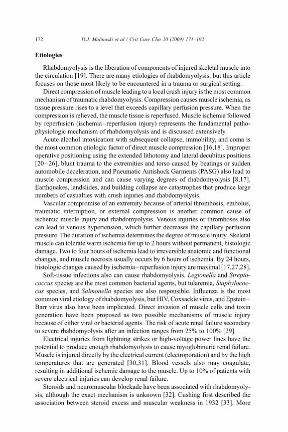
Muscle compression leads to mechanical stress, which opens stretch-activated
channels in the muscle cell membrane [37]. This results in the influx of fluid and
electrolytes, including Na+ and Ca++. Cells swell, and their intracellular Ca++
concentration rises, resulting in several pathologic processes (Fig. 1). An increase
in the activity of cytoplasmic neutral proteases leads to the degradation of myo-
fibrillar proteins [38]; Ca++ -dependent phosphorylases are activated, and cell
membranes are degraded [39]. Additionally, nucleases are activated, and mito-
chondrial ATP production is diminished because of an inhibition of cellular
respiration [40].
Muscle ischemia caused by prolonged compression or vascular injury results in
anaerobic metabolism and a further decrease in ATP production. This diminishes
the activity of the Na/K ATPase, which leads to the accumulation of intracellular
Fig. 1. The pathogenesis of rhabdomyolysis.
Page 4
D.J. Malinoski et al / Crit Care Clin 20 (2004) 171–192174
fluid and further increases the intracellular Ca++ concentration. In addition,
increased concentrations of neutrophil chemoattractants are present in postische-
mic tissues, which results in a high local concentration of activated neutrophils
once reperfusion is initiated. These neutrophils damage reperfused tissue by
releasing proteolytic enzymes, generating free radicals [41], producing large
amounts of hypochlorous acid, and by increasing microvascular resistance [42].
Reperfusion of ischemic tissue after the release of entrapped victims or the
reestablishment of vascular flow results in an ischemia–reperfusion injury. In
addition to delivering activated neutrophils to previously ischemic tissue, reperfu-
sion leads to the conversion of oxygen and hypoxanthine to xanthine by xanthine
oxidase, with the generation of superoxide anions, which are highly reactive
oxygen-derived free radicals [43].
Free radicals are potent oxidizing agents that damage intracellular and
extracellular molecules when their concentrations become excessive. Lipid
peroxidation occurs when free radicals oxidize and damage the lipid bilayer of
cell membranes. The ferrous–ferric ion pair provided by iron within the porphyrin
ring in myoglobin catalyzes this process and facilitates widespread lipid peroxi-
dation in muscle tissue [44]. Cell membrane degradation impairs the normal
permeability of the cell and results in further cellular edema, Ca++ influx, and
Na+ influx. Cell lysis follows, and the intracellular contents of the muscle cells
are released into the circulation.
Lipid peroxidation leads to cellular membrane leakiness, and, when combined
with a reduction in active ionic extrusion caused by the depletion of ATP, causes
cellular swelling and an accumulation of fluid in the interstitial space [45,46].
Three hours of ischemia followed by reperfusion appear to be the minimum
requisites for this effect [47–49]. In muscle groups confined in tight, fibrous
sheaths with low compliance, such as the calf and forearm, intracompartmental
pressure rises rapidly [50]. When this pressure exceeds the arteriolar–perfusion
pressure, muscle tamponade and myoneuronal damage occur, resulting in com-
partment syndrome. Signs and symptoms of compartment syndrome include a
tense, swollen muscle compartment, pain with passive stretch (the most sensitive
finding), paresthesias or anesthesia, weakness or paralysis of the affected extremity,
and, in late stages, diminished peripheral pulses. Palpable pulses, however, often
are found in the presence of significantly elevated compartment pressures and
should not lead one to dismiss the diagnosis of compartment syndrome.
Crush syndrome
After muscle compression is relieved or vascular interruption is corrected, the
cellular contents of the affected muscle tissue are released into the circulation.
Large volumes of intravascular fluid also can be sequestered in the involved
extremities because of increased capillary permeability. The systemic manifesta-
tions of rhabdomyolysis, caused by hypovolemia and toxin exposure, are the
components of the crush syndrome.
Page 5
D.J. Malinoski et al / Crit Care Clin 20 (2004) 171–192 175
Hypovolemia is often the first manifestation of the crush syndrome. In 1931,
Blalock was able to show that large volumes of plasma accumulated in traumatized
extremities, depleting intravascular volume and resulting in shock [51]. Bywaters
and Beall recognized shock as a component of the crush syndrome in 1941 [3]. In a
landmark animal experiment, Bywaters and Popjak demonstrated that several
hours of hind limb ischemia created by a tourniquet, followed by restoration of
blood flow, resulted in cold, firm, edematous, and paralyzed extremities. Although
the extremities were ischemic, the rabbits did not experience any significant
physiologic derangements. After reperfusion occurred, however, their blood
became hemoconcentrated and their blood urea nitrogen levels rose. When both
extremities were involved, the animals died from hypovolemic shock [52]. Clinical
observations in human studies also have demonstrated that large volumes of fluid
can leak into the interstitial space and cause hypotension [3,53]. In a series of
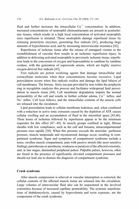
372 patients with crush syndrome, Oda [14] found that hypovolemic shock was the
most common cause of death during the first 4 days after crush injury (23/35, 66%)
(Fig. 2).
In addition to intravascular depletion, patients with the crush syndrome are
faced with a large toxin load and can develop life-threatening electrolyte abnor-
malities (Table 1). Hypocalcemia results from the influx of Ca++ into the affected
muscle tissue. This becomes dangerous when combined with hyperkalemia and
acidemia, both of which can occur upon reperfusion of ischemic tissue. Hyper-
kalemia and its associated cardiotoxicity represent the second most common cause
of early deaths following crush injury [14]. Volume replacement is the mainstay of
Fig. 2. The causes of death in 50 patients with the crush syndrome following the Hanshin–Awaji
Earthquake. Deaths from hypovolemia and hyperkalemia were the most common in the early period,
while sepsis leading to multiple organ failure was responsible for most of the late deaths. (Adapted
from Oda J, Tanaka H, Yoshioka T, et al. Analysis of 372 patients with crush syndrome caused by the
Hanshin–Awaji Earthquake. J Trauma 1997;(42):470–6; with permission.)
Page 6
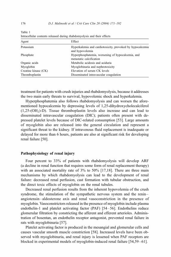
Table 1
Intracellular contents released during rhabdomyolysis and their effects
Agent Effect
Potassium Hyperkalemia and cardiotoxicity, provoked by hypocalcemia
and hypovolemia
Phosphate Hyperphosphatemia, worsening of hypocalcemia, and
metastatic calcification
Organic acids Metabolic acidosis and aciduria
Myoglobin Myoglobinuria and nephrotoxicity
Creatine kinase (CK) Elevation of serum CK levels
Thromboplastin Disseminated intravascular coagulation
D.J. Malinoski et al / Crit Care Clin 20 (2004) 171–192176
treatment for patients with crush injuries and rhabdomyolysis, because it addresses
the two main early threats to survival, hypovolemic shock and hyperkalemia.
Hyperphosphatemia also follows rhabdomyolysis and can worsen the afore-
mentioned hypocalcemia by depressing levels of 1,25-dihydroxycholecalciferol
(1,25-(OH2)-D). Tissue thromboplastin levels also increase and can lead to
disseminated intravascular coagulation (DIC); patients often present with de-
pressed platelet levels because of DIC-related consumption [53]. Large amounts
of myoglobin also are released into the general circulation and represent a
significant threat to the kidney. If intravenous fluid replacement is inadequate or
delayed for more than 6 hours, patients are also at significant risk for developing
renal failure [50].
Pathophysiology of renal injury
Four percent to 33% of patients with rhabdomyolysis will develop ARF
(a decline in renal function that requires some form of renal replacement therapy)
with an associated mortality rate of 3% to 50% [17,18]. There are three main
mechanisms by which rhabdomyolysis can lead to the development of renal
failure: decreased renal perfusion, cast formation with tubular obstruction, and
the direct toxic effects of myoglobin on the renal tubules.
Decreased renal perfusion results from the inherent hypovolemia of the crush
syndrome, the stimulation of the sympathetic nervous system and the renin–
angiotensin–aldosterone axis and renal vasoconstriction in the presence of
myoglobin. Vasoconstrictors released in the presence of myoglobin include plasma
endothelin-1 and platelet activating factor (PAF) [54–56]. Endothelins reduce
glomerular filtration by constricting the afferent and efferent arterioles. Adminis-
tration of bosentan, an endothelin receptor antagonist, prevented renal failure in
rats with myoglobinuria [57].
Platelet activating factor is produced in the mesangial and glomerular cells and
causes vascular smooth muscle constriction [58]. Increased levels have been ob-
served with myoglobinuria, and renal injury is lessened when PAF receptors are
blocked in experimental models of myoglobin-induced renal failure [54,59–61].
Page 7
D.J. Malinoski et al / Crit Care Clin 20 (2004) 171–192 177
Myoglobin is a respiratory pigment protein that comprises 1% to 3% of the wet
weight of skeletal muscle [19]. It has a molecular weight of 17,800 d and is filtered
readily by the kidney. At the center of the myoglobin molecule is a single heme
prosthetic group with an iron atom that serves as an oxygen-binding site [17]. Low
levels of circulating myoglobin are (50% to 85%) bound mostly to haptoglobin
and a2–globulin and are cleared from the circulation by the reticuloendothelial
system [62]. When circulating levels of myoglobin are elevated, as occurs after
rhabdomyolysis, the binding capacity of haptoglobin is saturated, and free plasma
levels rise. Plasma levels greater than 0.5 to 1.5 mg/dL are filtered by the kidney,
resulting in myoglobinuria. Normal urinary myoglobin levels are less than
5 ng/mL. Myoglobin is not reabsorbed in the renal tubules and increases in con-
centration as water is reabsorbed from the tubular filtrate, resulting in dark, tea-
colored urine. The presence of myoglobin in the urine can be confirmed with a
positive urine dipstick, which reacts with the heme groups of both myoglobin and
hemoglobin. The absence of red cells on microscopy confirms that the discolored
urine is caused by myoglobin. Myoglobinuria and hemoglobinuria can occur
simultaneously, however.
Myoglobin cast formation and tubular obstruction occur in patients with acidic
urine and a high concentration of myoglobin in the renal tubules. The myoglobin
reacts with the Tamm–Horsfell (THP) protein and precipitates, forming casts. This
binding is enhanced under acidic conditions, and urinary alkalinization with
sodium bicarbonate has been shown to reduce cast formation [63]. Whether this
effect is directly caused by the alkaline pH or the induced solute diuresis is not
certain. The hypothesis that myoglobin casts obstruct urine flow [63,64] and cause
a transtubular leakage of glomerular filtrate [65] has been promoted widely.
Micropuncture experiments have shown that intratubular pressures are low, how-
ever, and that perfusion of the tubule with buffer at low pressures easily removes
the casts [66].
The direct toxic effect of myoglobin is likely the main component in the
development of renal failure after rhabdomyolysis. There is an increasing body of
evidence that supports free radical-mediated renal injury, especially to the proximal
tubule. It has been shown that endogenous radical scavenging agents are depleted
during myoglobinuria-induced renal failure, while antioxidant administration can
improve renal function [67–70]. Myoglobin separates into protein and ferrihemate
moieties in an acidic environment [71]. Iron catalyzes the formation of free radicals
by ways of the Fenton reaction, which, in turn, leads to lipid peroxidation in the
renal tubules [70]. The heme group of the myoglobin molecule itself also may
promote lipid peroxidation during redox cycling between its different oxidation
states. The reactivity of ferryl myoglobin, which is responsible for inducing lipid
peroxidation, is attenuated markedly at alkaline pH [72]. This further supports the
role of an induced alkaline diuresis in the treatment of patients with rhabdomyoly-
sis. Additionally, desferrioxamine (DFO), an iron chelator, has been shown to
protect against renal dysfunction in animal models of rhabdomyolysis [68,73]. This
protects against the exposure to free iron and the redox cycling of myoglobin, and
thus lipid peroxidation [74].
Page 8
D.J. Malinoski et al / Crit Care Clin 20 (2004) 171–192178
Additional vasoconstrictors also are generated by the presence of myoglobin-
uria and contribute to the renal dysfunction following rhabdomyolysis. F2-
isoprostanes are a group of prostaglandin-like compounds that are formed during
free radical-induced lipid peroxidation [75]. They have been found to be elevated in
people with rhabdomyolysis [76] and are themselves renal vasoconstrictors. Nitric
oxide (NO) is another vasoactive mediator that is involved in maintaining renal
blood flow [77]. NO donors have been found to preserve renal function in the face
of heme protein injury, while NO synthase inhibitors aggravate injury [78]. This
may be attributable to the ability of myoglobin to scavenge NO [79] or prevent its
redox cycling [80].
Diagnosis
Early diagnosis is crucial in patients with rhabdomyolysis. Patients who have
sustained significant soft tissue injury (because of crush injuries or prolonged
immobilization) or ischemia–reperfusion injury (because of vascular interrup-
tion) are at risk of developing rhabdomyolysis, myoglobinuria, and renal failure.
Patients often present with painful, swollen extremities and should be monitored
for the development of an extremity compartment syndrome. Physical exami-
nation of patients with central nervous system injury, intoxication, or multiple
life-threatening injuries can be difficult and unreliable. Dark, tea-colored urine
that is dipstick positive for blood despite the absence of red blood cells on
microscopy is suggestive of myoglobinuria and rhabdomyolysis. The most rapid
and least expensive screening test for rhabdomyolysis is the serum CK level.
Patients who are believed to be at risk on the basis of history and physical
examination should have their urine output monitored and serial serum CK
levels drawn. When serum CK levels are elevated, it is important to rule out a
cardiac muscle injury, such as a myocardial infarction or contusion, as the source
of the enzyme elevation.
Management
Preventing the systemic and renal complications of the crush syndrome
requires early, vigorous fluid resuscitation, preferably started at the site of injury
before extrication [81]. Better et al [50,53] demonstrated the importance of early
fluid resuscitation by reporting their differing treatments of two groups of patients
(one in 1979 and one in 1982) with extensive rhabdomyolysis as a result of the
collapse of buildings. There were a total of 15 patients in the two groups who
suffered roughly the same degree of crush injury to the lower extremities after
being trapped for a mean of 12 hours. The seven men in 1979 did not receive
aggressive fluid resuscitation until at least 6 hours after extrication. Despite
receiving adequate volume replacement (average 11 L of intravenous fluids per
day until central venous pressure began to rise), all seven men developed ARF
Page 9
D.J. Malinoski et al / Crit Care Clin 20 (2004) 171–192 179
and required dialysis. In contrast, seven of the eight men treated in 1982 received
intravenous saline before the completion of extrication and were evacuated to a
hospital within 2 hours, where they received a forced mannitol–alkaline diuresis
with a target urine output of 300 mL per hour. All seven had serum CK levels
greater than 30,000 U/L, and none developed azotemia or ARF. One of the eight
men experienced a delay of 24 hours in the diagnosis and treatment of his
rhabdomyolysis and did develop ARF. Although the authors concluded that early,
aggressive volume replacement followed by a forced mannitol–alkaline diuresis
may protect against ARF in patients with traumatic rhabdomyolysis, they
recognized that the relative benefits of early fluid resuscitation versus a forced
solute diuresis are unclear and that a goal of 300 mL per hour of urine output
may be more than required.
The benefits of mannitol to induce a solute diuresis have been established in
several experimental studies. Mannitol is an osmotic diuretic, promoting an
increase in urine output and the washout of tubular myoglobin, and it is a volume
expander. Because mannitol can cause volume overload in patients with marginal
cardiac function and established ARF, it should not be used until urine output has
been established. Mannitol also has been shown to reduce intracompartmental
pressure in a canine model of compartment syndrome [82], and therefore it may be
a conservative alternative to fasciotomy. In addition, mannitol is an effective
hydroxyl-free radical scavenger that may protect the kidney from oxidant injury
[73]. Because mannitol can produce a hyperosmolar state and electrolyte derange-
ments, frequent monitoring of serum electrolytes, osmolarity, and patient volume
status is recommended.
Alkalinization of the urine with sodium bicarbonate to prevent the development
of ARF after crush injuries and rhabdomyolysis has been supported by numerous
animal studies [63,69], case reports [25,53,83–85], and retrospective clinical
studies [50,53,84]. Bicarbonate assists in the creation of alkaline urine, which
serves to decrease cast formation and lessen the direct toxic effects of myo-
globin [63]. Moore et al [72] found that alkalinization inhibits redox cycling of
myoglobin and lipid peroxidation in rhabdomyolysis. In isolated perfused kidneys,
myoglobin induced renal vasoconstriction only at an acid pH [86]. Ron and
Michaelson [53] found that large amounts of bicarbonate were required to alka-
linize the urine of patients with rhabdomyolysis. An average of 685 mEq of
bicarbonate was administered during the first 60 hours of treatment to maintain a
urine pH of greater than 6.5. To prevent alkalemia, an average of 1.5 doses of
250 mg of acetazolamide was needed, and plasma pH did not exceed 7.45 in any
patient. Other authors have argued that large volume infusion of crystalloid alone
can produce a sufficient solute diuresis to maintain alkalotic urine [8,19].
A retrospective analysis of 24 nonrandomized patients with rhabdomyolysis of
different causes attempted to determine if there was a benefit to treatment with
saline, mannitol, and bicarbonate (group 1) over that of saline alone (group 2) [87].
The authors concluded that mannitol and bicarbonate were not necessary to avoid
the development of renal failure. None of the 24 patients developed renal
dysfunction, however, and their degree of muscle injury was low (average
Page 10
D.J. Malinoski et al / Crit Care Clin 20 (2004) 171–192180
CPK = 2750 U/L). The results of this study neither support nor negate the use of a
forced alkaline diuresis. There is a need for a prospective randomized controlled
trial comparing the effectiveness of crystalloid, mannitol, and bicarbonate with that
of standard crystalloid resuscitation. Despite the fact that class 1 evidence in
support of using a forced alkaline diuresis is lacking, bicarbonate can ameliorate the
systemic acidosis and hyperkalemia that occur during the crush syndrome, and
bicarbonate may be useful in patients with the crush syndrome regardless of its
effects on urine pH.
Several experimental therapies are under investigation for treating patients
with crush injuries. Free radical-induced oxidant injury has emerged as one of the
most important mechanisms of rhabdomyolysis and renal failure. Free radical
scavengers have been shown to reduce the magnitude of muscle necrosis caused
by ischemia–reperfusion injury [88], and the administration of antioxidants such
as glutathione and vitamin E analogs improve renal function in experimental
models [67]. As mentioned previously, desferrioxamine [68,73,89], PAF receptor
blockers [61], and the endothelin receptor antagonist bosentan [57] have been
shown to reduce the direct toxic effects of myoglobin on the kidney in
experimental models of myoglobinuric renal failure. In addition, dantrolene
inhibits the release of Ca++ from the sarcoplasmic reticulum and accelerates
the return of intracellular levels of Ca++ to baseline [90]. This may help to lessen
the degree of muscle cell death in patients with crush injuries. The loop diuretic,
furosemide, was able to convert 7 of 14 patients with oliguric renal failure to
nonoliguric renal failure in a series of 200 patients with traumatic rhabdomyolysis
from severe beatings in South Africa [8]. Loop diuretics, however, have the
theoretical disadvantage of acidifying the urine, which may increase the amount
of myoglobin toxicity.
Treatment of ARF
Despite adequate resuscitation and prophylaxis against myoglobinuric renal
injury, up to one third of patients with rhabdomyolysis will develop ARF. These
patients require some form of renal replacement therapy. Daily hemodialysis or
continuous hemodialysis/hemofiltration will correct the fluid and electrolyte
abnormalities that accompany rhabdomyolysis and renal failure. Continuous
hemodialysis and hemofiltration do not cause hypotension and cardiac arrhythmias
that are associated with the rapid fluid shifts that occur with intermittent hemo-
dialysis [91]. This may make these modalities more appropriate in critically ill
patients with hemodynamic instability.
Intractable hyperkalemia and acidosis, refractory to volume expansion and
bicarbonate administration, are the main early threats to survival in patients with
rhabdomyolysis, and dialysis must be instituted promptly. The hypocalcemia that
accompanies rhabdomyolysis should not be corrected unless there is danger of a
hyperkalemic arrhythmia [92], for most of the infused calcium is deposited in
injured muscles andmay aggravate the pathogenesis of rhabdomyolysis and lead to
metastatic calcification [93].
Page 11
Treatment of compartment syndrome
When compartment syndrome is suspected based on mechanism of injury and
clinical findings, muscle compartment pressures should be measured directly.
The most commonly used compartment pressure measurement device is the
Stryker STIC Device (Stryker Corporation, Kalamazoo, Michigan). Any clinical
electronic arterial pressure monitoring device, however, can be adopted to
perform compartment pressure measurements. Normal compartment pressures
range from 0 to 15 mm Hg [94]. Compartment pressures in excess of 30 to
50 mm Hg produce clinically significant muscle ischemia [95–97]. Patients with
higher diastolic pressures are able to tolerate higher tissue pressures without
ischemic damage, and therefore a fasciotomy is recommended when the com-
partment pressure approaches 20 mm Hg below diastolic pressure [94]. Hypo-
tensive trauma patients may experience significant muscle ischemia at lower
compartment pressures.
The most common location of compartment syndrome is in the lower leg. A
lower leg fasciotomy involves incising the skin and fascia overlying its four
muscle compartments: anterior, lateral, superficial posterior, and deep posterior.
One and two-incision techniques have been described [94]. Compartment
syndrome also can be encountered in the forearm, thigh, and buttock muscle
groups. The purpose of a fasciotomy is to decrease intracompartmental pressure
and restore perfusion to ischemic muscle. To be effective in preventing irrevers-
ible ischemic muscle damage, however, a fasciotomy must be performed early.
Once irreversible ischemic damage is present, as evidenced by the onset of
painlessness or paralysis, there will be little functional recovery and a higher rate
of infection [98].
Experimental studies have found that irreversible muscle and nerve damage
occur after 6 to 8 hours of total ischemia [94]. Several studies have attempted to
define a specific cutoff period, after injury, that represents the onset of irreversible
damage and precludes the use of fasciotomy. Sheridan and Matsen [99] found that
only 8% of patients who underwent fasciotomy more than 12 hours after the onset
of symptoms of compartment syndrome had restoration of normal function. They
also reported a 46% infection rate and a 21% amputation rate. A meta-analysis by
Bradley [98] found fasciotomy in patients with paralysis to be unsatisfactory in
over 80% of cases.
In a retrospective study of five patients who underwent fasciotomy 35 to
96 hours after injury, one patient died of sepsis and multi-organ failure, and the
other four required amputation, either because of local infection and sepsis (three
patients) or the presence of an insensate, functionless limb (one patient). Finkel-
stein [100] concluded that delayed fasciotomy converts a closed injury into an open
one, and cannot correct the muscle or nerve damage that already has occurred.
Better et al [50] also condemned late fasciotomy for its potential to produce
uncontrollable infection in necrotic muscle. They went on to state that the excision
of noninfected, necrotic tissue is not essential and may delay healing. Reis and
Michaelson [85] had to perform above-knee amputations for uncontrolled sepsis in
D.J. Malinoski et al / Crit Care Clin 20 (2004) 171–192 181
Page 12
D.J. Malinoski et al / Crit Care Clin 20 (2004) 171–192182
three of six patients who previously had undergone fasciotomies more than
24 hours after injury. They concluded that the sequelae of infection in a crushed
extremity are much worse than the late muscle contracture that may result from
fibrosis of muscle.
Two years after the earthquake in Kobe, Japan, Matsuoka et al [101] performed
follow-up examinations in 42 crush syndrome patients to investigate sensory and
motor functions of the affected lower extremities. Fasciotomies were done in
17 patients, and all were performed after 12 hours. Severe disabilities related to the
lower extremity were found in 47% of patients who underwent fasciotomy and in
16% of those who did not. Although the two groups of patients were not similar,
multivariate analysis suggested that delayed rescue, delayed fasciotomy, and
radical debridement all worsen the patient’s prognosis.
It has been suggested that fasciotomy should not be performed more than
10 to 12 hours after injury in patients with compartment syndrome. Seddon
[102] found some degree of spontaneous muscle recovery up to 3 months after
injury, and suggested that an interval fasciotomy (6 to 12 months after injury)
combined with reconstructive procedures should be able to correct most
ischemic contractures. This approach may offer the best physical outcome for
patients with crush injuries and compartment syndrome that are not addressed
within 12 hours.
Amputation may be required when massive extremity necrosis progresses to an
extent that limb salvage is not possible. Because necrotic muscle is a rich source of
myoglobin, potassium, and tissue thromboplastin, retention of a nonsalvageable
limb can be life-threatening. Necrotic muscle may become infected and serve as a
source of uncontrollable sepsis. If it becomes evident that amputation will be
required, early amputation is tolerated better than late amputation both physiologi-
cally and emotionally [103].
Assessing risk of renal failure
Several studies have attempted to identify which patients will progress to
myoglobinuric renal failure based on their laboratory values. The prospective
identification of patients who are at high risk of developing acute renal dysfunction
(ARD; serum creatinine, Cr greater than 2.0 mg/dL without need for dialysis) or
ARFwould allow therapy to be delivered to those most likely to benefit. SerumCK
levels and serum and urine myoglobin levels have been proposed as screening
tools, but no consensus exists.
A series of 200 victims of severe beatings in South Africa found that base
deficit, delay in treatment, and CK levels were significant risk factors for the
development of ARD and death [8]. Similarly, in a study of 157 patients, Ward
[18] found that patients with a peak serum CK level greater than 16,000 U/L had
the highest risk of developing ARD. Notably, 90% of patients had their CK levels
peak within 24 hours of admission. A retrospective review of 93 patients with
severe rhabdomyolysis (serum CK greater than 5000 U/L) found that patients with
a peak CK level of greater than 15,000 U/L had significantly higher rates of ARD
Page 13
D.J. Malinoski et al / Crit Care Clin 20 (2004) 171–192 183
(72% versus 38%, P < 0.01), hyperkalemia (22% versus 7%, P < 0.05), and
hypocalcemia (63% versus 28%, P < 0.05). Additionally, the patients with ARD
had a higher mortality compared with those patients with normal renal function
(51% versus 17%, P < 0.01) [104].
A recent analysis of 372 patients with crush syndrome after the 1995 earthquake
in Kobe, Japan, demonstrated a significant correlation between peak serum CK
levels and the number of extremities crushed [14]. In addition, patients with a peak
CK level greater than 75,000 U/L had a higher rate of ARF andmortality than those
with a peak CK less than 75,000 U/L (84% versus 39%, P < 0.01 and 4% versus
17%, P < 0.05, respectively). Eneas et al [84] found that only patients with a peak
CK greater than 20,000 U/L failed to respond to a mannitol–bicarbonate diuresis
and went on to require dialysis. The nonresponders also had significantly higher
serum phosphate levels and hematocrit readings upon admission, indicative of
more severe muscle injury and hemoconcentration.
Several studies have attempted to predict the development of renal failure
using serum or urine myoglobin levels. A prospective study of eight patients
by Feinfeld [105] found that four of five patients with urine myoglobin levels
greater than 1000 ng/mL (normal = < 10ng/mL) developed ARD, while none
of the three patients with urine myoglobin levels less than 300 ng/mL developed
ARD. Of note, initial CK levels were not found to be predictive of renal
dysfunction in this study. Another report found that elevated urine myoglobin
levels greater than 20,000 ng/mL were associated with a significantly increased
risk of renal dysfunction [106].
The conclusions from these studies of patients with rhabdomyolysis and
myoglobinuria must be viewed with caution because of their heterogeneity. They
often used varying definitions of renal failure, ranging from an elevation in
serum Cr of greater than 2.0 mg/dL to the need for hemodialysis. There also
were large variations in study design and patient selection; the number of patients
studied ranged from 8 to 372; there was an inconsistent amount of time between
injury and treatment, and the treatment of myoglobinuria differed in each of
the studies.
Despite conflicting reports regarding the utility of CK and myoglobin levels,
several conclusions can be drawn. CK levels can be obtained in less than 1 hour;
they are readily available in most institutions, and they are less expensive than
myoglobin levels ($15 versus $97 in the authors’ laboratory). Serum and urine
myoglobin levels can take over 24 hours to obtain, depending on the distance from
one’s hospital to the regional laboratory that has the capability to perform the assay.
In addition, myoglobin has faster elimination kinetics than CK, making it a less
sensitive marker of muscle injury [107]. In a prospective study of 13 patients
with rhabdomyolysis, Lappalainen [108] found that the average times to reach
the 50% level of initial values were 12 hours for myoglobin and 42 hours for CK
(P < 0.01). In a review of laboratory investigations, Beetham [109] concluded
that serum CK levels should be used to screen patients with suspected rhab-
domyolysis and that there is no reason for laboratories to have assays for
myoglobin available until further studies are able to demonstrate its utility.
Page 14
D.J. Malinoski et al / Crit Care Clin 20 (2004) 171–192184
Page 15
Fig.3.Rhabdomyolysistreatm
entalgorithm.
D.J. Malinoski et al / Crit Care Clin 20 (2004) 171–192 185
Page 16
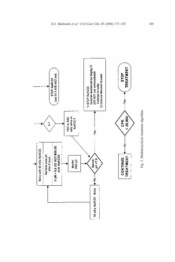
Treatment algorithm
A treatment algorithm for patients with rhabdomyolysis was established at the
authors’ institution in 1992 (Fig. 3). Based on a review of the literature, a forced
alkaline diuresis with mannitol and bicarbonate was chosen as the authors’ method
of preventing renal failure in high-risk patients. A serum CK level of 20,000 U/L is
the current threshold for identifying those patients who are at risk and require
treatment. The primary objective of the algorithm is to prevent ARF. The treatment
goals are (1) to achieve a urine output of 200 mL per hour, (2) to maintain urine
pH between 6 and 7, (3) to keep serum pH below 7.50, and (4) to achieve
hemodynamic stability and prevent volume overload. Treatment begins with a
bolus of 1 L of D5 0.22% NaCl + 100 mEq NaHCO3 over 30 minutes, followed
by an infusion at 2 to 5 mL/kg per hour. At the same time, a bolus of 0.5 gm/kg
20% mannitol is given over 15 minutes and followed by an infusion at 0.1 gm/kg
per hour. Infusions and additional boluses are titrated to maintain a urine output of
200mL per hour.When serum pH exceeds 7.45, or urine pH remains below 6.0, the
administration of acetazolamide can help to increase the excretion of bicarbonate
in the urine. The patient should be monitored for hypernatremia, hyperosmolality
(normal range in the authors’ laboratory 275 to 295 mOsm/kg water), and volume
overload, as these are possible complications of treatment.
This treatment algorithm represents a suggested method of managing patients
with rhabdomyolysis, but it has not been validated in a prospective randomized
controlled trial. Further research is needed to identify the specific components of
treatment that lead to significant improvements in outcome.
D.J. Malinoski et al / Crit Care Clin 20 (2004) 171–192186
Mass casualties
During the last 15 years, several natural disasters have generated large numbers
of patients with rhabdomyolysis and, consequently, enhanced the understanding of
the presentation of the crush syndrome and the importance of instituting early
treatment to prevent renal failure and death. Earthquakes have the unique ability to
affect the widespread collapse of buildings, trapping victims beneath their fallen
debris and inflicting crush injuries. In addition to destroying homes and businesses,
earthquakes disrupt roads, communication lines, and hospitals, making the delivery
of care to injured patients difficult at best. Another confounding factor is that
several hundred patients may require dialysis to prevent life-threatening hyper-
kalemia at roughly the same time. This often overwhelms local health care systems
and contributes to mortality [10].
On Dec. 7, 1988, an earthquake with a magnitude of 6.9 on the Richter scale
struck near Spitak, Armenia, in the former Soviet Union. Official estimates of the
number of people killed range from 26,000 to 50,000 [10]. Over 600 patients
developed renal failure and required dialysis [110]. There was a paucity of patients
with major trauma to the head, chest, or abdomen seen in hospitals after the
earthquake, suggesting that these injuries lead to asphyxiation and are generally
Page 17
D.J. Malinoski et al / Crit Care Clin 20 (2004) 171–192 187
fatal when they occur in the setting of a disaster that generates significant crush
injuries [15,17]. It became clear early on that the local medical resources were
going to be insufficient to care for the large number of injured patients, and dialysis
teams from several countries were flown to the area. Relief efforts, however, were
hindered by poor coordination, a lack of sufficient equipment, and their arrival on
the scene several days after the disaster [107]. A group of nephrologists from
London were one of the teams deployed to the disaster, and they concluded that a
permanent relief team, available to depart at a few hours notice, would be required
to achieve optimal outcomes [10]. The International Society of Nephrology created
a Disaster Relief Task Force (DRTF) in 1995 to prevent and treat crush injury-
induced ARF that occurs following traumatic rhabdomyolysis [111].
The European branch of the DRTF was dispatched to Northwest Turkey on
Aug. 17, 1999, when an earthquake with a magnitude of 7.4 on the Richter scale
struck Marmara. Personnel, equipment, and supplies were delivered to assist in
approximately 5000 dialysis sessions in 462 patients with ARF caused by traumatic
rhabdomyolysis. The observed mortality rate of 19% in these patients was a
significant improvement on previous relief efforts [11].
The Hanshin–Awaji earthquake struck the island of Japan near the city of Kobe
on Jan. 17, 1995. It measured 7.2 on the Richter scale, injured 41,000 people, and
killed approximately 5500 people. A review of the medical records of 95 hospitals
identified 372 patients with the crush syndrome; 202 patients developed ARD,
including 78 people who needed dialysis. The patients requiring dialysis were
significantly older (54 versus 45 years, P < 0.05) and experienced a longer delay
from injury to treatment (8 versus 3.6 hours, P < 0.05). As stated previously, peak
CK levels correlated with the number of injured extremities, and a peak CK of
greater than 75,000 was associated with a significantly higher prevalence of ARF
and death [14]. A retrospective analysis of eight patients who developed ARF after
the Kobe earthquake found that serum CK levels and serum myoglobin levels
significantly correlated with serum potassium concentrations (r = 0.87, P = 0.005,
and r = 0.81, P = 0.03, respectively) [13]. Another retrospective review of
14 earthquake survivors concluded that renal function after crush injury is depen-
dent on injury severity (peak CK levels) and initial fluid resuscitation [12]. Several
factors were found that contributed to the morbidity of patients who suffered crush
injuries: local physicians’ unfamiliarity with the crush syndrome, the destruction of
local medical facilities, and a delay in transport to tertiary care hospitals.
Better [50] has made several recommendations regarding relief efforts in
future disasters. He suggests that teams outfitted with continuous arteriovenous
hemofiltration equipment should be available for rapid transport to the site of a
rescue operation. This mode of renal replacement therapy is ideal in the setting
of a natural disaster, for it allows hemodialysis with only 12 L of water every
12 hours; it does not require electricity or pumps; it does not necessitate dialy-
sate delivery; its equipment is disposable, and large quantities of potassium can
be removed. Better also emphasizes the need to re-establish communication
networks immediately following future disasters, as this has limited previous
relief efforts.
Page 18
D.J. Malinoski et al / Crit Care Clin 20 (2004) 171–192188
Summary
Crush injuries resulting in traumatic rhabdomyolysis are an important cause of
acute renal failure. Ischemia–reperfusion is the main mechanism of muscle injury.
Intravascular volume depletion and renal hypoperfusion, combined with myoglo-
binuria, result in renal dysfunction. The infusion of intravenous fluids before
extrication or soon after injury may lessen the severity of the crush syndrome.
Serum CK levels can be used to screen patients with crush injuries to determine
injury severity. Once intravascular volume has been stabilized, and the presence of
urine flow has been confirmed, a forced mannitol–alkaline diuresis for prophylaxis
against hyperkalemia and acute renal failure should be instituted. If an extremity
compartment syndrome is suspected, one should have a low threshold for checking
the intracompartmental pressures. Further studies are needed to demonstrate if any
treatment regimen is truly superior to early, aggressive crystalloid infusion.
References
[1] Fleisher R. Ueber eine form von Haemoglbinuric bein Menschen. Berl Klin Wochenschr 1881;
18:691.
[2] Meyer-Betz F. Beobachtungen an einem Eigentartigen mit Muskellahmungen ver bunden fall
van Haemoglobinuric. Dtsch Arch Klin Med 1911;101:85.
[3] Bywaters E, Beall D. Crush injuries with impairment of renal function. BMJ 1941;1:427–32.
[4] Beall D, Bywaters E, Belsey R, Miles J. A case of crush injury with renal failure. BMJ 1941;1:
432–4.
[5] Bywaters E, Stead J. The production of renal failure following injection of solutions containing
myohaemoglobin. Quarterly Journal of Experimental Physiology 1944;33:53.
[6] Jones R. Crush syndrome in a Cornish tin mine. Injury 1984;15:282.
[7] Bentley G, Jeffereys T. The crush syndrome in coal miners. J Bone Joint Surg Br 1968;50:
588–94.
[8] Knottenbelt JD. Traumatic rhabdomyolysis from severe beating—experience of volume diu-
resis in 200 patients. Journal of Trauma– Injury Infection & Critical Care 1994;37(2):214–9.
[9] Noji EK, Kelen GD, Armenian HK, Oganessian A, Jones NP, Sivertson KT. The 1988 earth-
quake in Soviet Armenia: a case study. Ann Emerg Med 1990;19(8):891–7.
[10] Richards NT, Tattersall J, McCann M, Samson A, Mathias T, Johnson A. Dialysis for acute
renal failure due to crush injuries after the Armenian earthquake [erratum appears in BMJ 1989;
298(6674):655]. BMJ 1989;298(6671):443–5.
[11] Sever MS, Erek E, Vanholder R, Akoglu E, Yavuz M, Ergin H, et al. The Marmara earthquake:
epidemiological analysis of the victims with nephrological problems. Kidney Int 2001;60(3):
1114–23.
[12] Shimazu T, Yoshioka T, Nakata Y, Ishikawa K, Mizushima Y, Morimoto F, et al. Fluid resus-
citation and systemic complications in crush syndrome: 14 Hanshin–Awaji earthquake patients.
Journal of Trauma–Injury Infection & Critical Care 1997;42(4):641–6.
[13] Oda Y, Shindoh M, Yukioka H, Nishi S, Fujimori M, Asada A. Crush syndrome sustained in the
1995 Kobe, Japan, earthquake: treatment and outcome. Ann Emerg Med 1997;30(4):507–12.
[14] Oda J, Tanaka H, Yoshioka T, Iwai A, Yamamura H, Ishikawa K, et al. Analysis of 372 patients
with crush syndrome caused by the Hanshin–Awaji earthquake [discussion]. Journal of Trau-
ma–Injury Infection & Critical Care 1997;42(3):470–5.
[15] Pretto EA, Angus DC, Abrams JI, Shen B, Bissell R, Ruiz Castro VM, et al. An analysis of
prehospital mortality in an earthquake. Disaster Reanimatology Study Group. Prehospital Dis-
aster Med 1994;9(2):107–17.
Page 19
D.J. Malinoski et al / Crit Care Clin 20 (2004) 171–192 189
[16] Gabow P, Kaehny W, Kelleher S. The spectrum of rhabdomyolysis. Medicine 1982;62:141.
[17] Slater M, Mullins R. Rhabdomyolysis and myoglobinuric renal failure in trauma and surgical
patients: a review. J Am Coll Surg 1998;186(6):693–716.
[18] Ward MM. Factors predictive of acute renal failure in rhabdomyolysis. Arch Intern Med 1988;
148(7):1553–7.
[19] Knochel JP. Rhabdomyolysis and myoglobinuria. Annu Rev Med 1982;33:435–43.
[20] Ali H, Nieto JG, Rhamy RK, Chandarlapaty SK, Vaamonde CA. Acute renal failure due to
rhabdomyolysis associated with the extreme lithotomy position. Am J Kidney Dis 1993;22(6):
865–9.
[21] Bruce RG, Kim FH, McRoberts W. Rhabdomyolysis and acute renal failure following radical
perineal prostatectomy. Urology 1996;47(3):427–30.
[22] Nimmo GR, Stewart SM, English PJ. Myoglobinuric acute renal failure associated with major
urological surgery—an avoidable problem? Intensive Care Med 1988;14(3):244–5.
[23] Guzzi LM, Mills LM, Greenman P. Rhabdomyolysis, acute renal failure, and the exaggerated
lithotomy position. Anesth Analg 1993;77(3):635–7.
[24] Bildsten SA, Dmochowski RR, Spindel MR, Auman JR. The risk of rhabdomyolysis and acute
renal failure with the patient in the exaggerated lithotomy position. J Urol 1994;152:1970–2.
[25] Mathes DD, Assimos DG, Donofrio PD. Rhabdomyolysis and myonecrosis in a patient in the
lateral decubitus position. Anesthesiology 1996;84(3):727–9.
[26] Szewczyk D, Ovadia P, Abdullah F, Rabinovici R. Pressure-induced rhabdomyolysis and acute
renal failure. Journal of Trauma– Injury Infection & Critical Care 1998;44(2):384–8.
[27] Tountas CP, Bergman RA. Tourniquet ischemia: ultrastructural and histochemical observations
of ischemic human muscle and of monkey muscle and nerve. Journal of Hand Surgery—
American Volume 1977;2(1):31–7.
[28] Sapega AA, Heppenstall RB, Chance B, Park YS, Sokolow D. Optimizing tourniquet appli-
cation and release times in extremity surgery. A biochemical and ultrastructural study. J Bone
Joint Surg Am 1985;67(2):303–14.
[29] Singh U, Scheld WM. Infectious etiologies of rhabdomyolysis: three case reports and review
[comment]. Clin Infect Dis 1996;22(4):642–9.
[30] Frank D, Fisher J. Complications of electrical injury. In: Greenfield L, editor. Complications in
surgery and trauma. Philadelphia: Lippincott-Raven; 1990.
[31] Bhatt DL, Gaylor DC, Lee RC. Rhabdomyolysis due to pulsed electric fields. Plastic and
Reconstructive Surgery 1990;86(1):1–11.
[32] Hansen-Flaschen J, Cowen J, Raps EC. Neuromuscular blockade in the intensive care unit.
More than we bargained for. Am Rev Respir Dis 1993;147(1):234–6.
[33] Cushing H. The basophil adenoma of the pituitary body and their clinical manifestations. Johns
Hopkins Med J 1932;50:137.
[34] Kupfer Y, Namba T, Kaldawi E, Tessler S. Prolonged weakness after long-term infusion of
vecuronium bromide [comment]. Ann Intern Med 1992;117(6):484–6.
[35] Douglass JA, Tuxen DV, Horne M, Scheinkestel CD, Weinmann M, Czarny D, et al. Myopathy
in severe asthma. Am Rev Respir Dis 1992;146(2):517–9.
[36] Baker PF, Blaustein MP, Hodgkin AL, Steinhardt RA. The influence of calcium on sodium
efflux in squid axons. J Physiol 1969;200(2):431–58.
[37] Christensen O. Mediation of cell volume regulation by Ca2 + influx through stretch-activated
channels. Nature 1987;330(6143):66–8.
[38] Cheung JY, Bonventre JV, Malis CD, Leaf A. Calcium and ischemic injury. N Engl J Med
1986;314(26):1670–6.
[39] Chien KR, Abrams J, Serroni A, Martin JT, Farber JL. Accelerated phospholipid degradation
and associated membrane dysfunction in irreversible, ischemic liver cell injury. J Biol Chem
1978;253(13):4809–17.
[40] Wrogemann K, Pena SD. Mitochondrial calcium overload: a general mechanism for cell-
necrosis in muscle diseases. Lancet 1976;1(7961):672–4.
[41] Suzuki M, Inauen W, Kvietys PR, Grisham MB, Meininger C, Schelling ME, et al. Superoxide
Page 20
D.J. Malinoski et al / Crit Care Clin 20 (2004) 171–192190
mediates reperfusion-induced leukocyte-endothelial cell interactions. Am J Physiol 1989;257:
H1740–1745.
[42] Harris AG, Skalak TC. Effects of leukocyte activation on capillary hemodynamics in skeletal
muscle. Am J Physiol 1993;264(3 Pt 2):H909–16.
[43] Shackford S, Rich N. Peripheral Vascular Injury. In: Mattox K, Feliciano D, Moore E, editors.
Trauma. 4th edition. San Francisco: McGraw-Hill; 1996. p. 1011–44.
[44] Recknagel R, Glende E, Hruszkewycz A. Chemical mechanisms of carbon tetrachloride toxi-
city. In: Pryor W, editor. Free radicals in biology, volume 3. New York: Academic Press; 1977.
[45] Harris K, Walker PM, Mickle DA, Harding R, Gatley R, Wilson GJ, et al. Metabolic response
of skeletal muscle to ischemia. Am J Physiol 1986;250:H213–220.
[46] Sanderson RA, Foley RK, McIvor GW, Kirkaldy-Willis WH. Histological response on skeletal
muscle to ischemia. Clinical Orthop 1975;113:27–35.
[47] Diana JN, Laughlin MH. Effect of ischemia on capillary pressure and equivalent pore radius in
capillaries of the isolated dog hind limb. Circ Res 1974;35(1):77–101.
[48] Miller SH, Price G, Buck D, Neeley J, Kennedy TJ, Graham III WP, et al. Effects of tourniquet
ischemia and postischemic edema on muscle metabolism. Journal of Hand Surgery–American
Volume 1979;4(6):547–55.
[49] Strock PE, Majno G. Microvascular changes in acutely ischemic rat muscle. Surg Gynecol
Obstet 1969;129(6):1213–24.
[50] Better OS, Stein JH. Early management of shock and prophylaxis of acute renal failure in
traumatic rhabdomyolysis [comment]. N Engl J Med 1990;322(12):825–9.
[51] Blalock A. Experimental shock: the probable cause for the reduction in the blood pressure
following mild trauma to as extremity. Arch Surg 1931;22:598–609.
[52] Bywaters E, Popjak G. Experimental crushing injury: peripheral circulatory collapse and other
effects of muscle necrosis in the rabbit. Surg Gynecol Obstet 1942;75:612–27.
[53] Ron D, Taitelman U, Michaelson M, Bar-Joseph G, Bursztein S, Better OS. Prevention of acute
renal failure in traumatic rhabdomyolysis. Arch Intern Med 1984;144(2):277–80.
[54] Badr KF, DeBoer DK, Takahashi K, Harris RC, Fogo A, Jacobson HR. Glomerular responses to
platelet-activating factor in the rat: role of thromboxane A2. Am J Physiol 1989;256(1 Pt 2):
F35–43.
[55] Yanagisawa M, Kurihara H, Kimura S, Tomobe Y, Kobayashi M, Mitsui Y, et al. A novel potent
vasoconstrictor peptide produced by vascular endothelial cells [comment]. Nature 1988;
332(6163):411–5.
[56] Holt SG, Moore KP. Pathogenesis and treatment of renal dysfunction in rhabdomyolysis [com-
ment]. Intensive Care Med 2001;27(5):803–11.
[57] Karam H, Bruneval P, Clozel JP, Loffler BM, Bariety J, Clozel M. Role of endothelin in acute
renal failure due to rhabdomyolysis in rats. J Pharmacol Exp Ther 1995;274(1):481–6.
[58] Schlondorff D, Goldwasser P, Neuwirth R, Satriano JA, Clay KL. Production of platelet-
activating factor in glomeruli and cultured glomerular mesangial cells. Am J Physiol 1986;
250:F1123–1127.
[59] Hebert RL, Sirois P, Braquet P, Plante GE. Hemodynamic effects of PAF-acether on the dog
kidney. Prostaglandins Leukot Med 1987;26(3):189–202.
[60] Braquet P, Touqui L, Shen TY, Vargaftig BB. Perspectives in platelet-activating factor research.
Pharmacol Rev 1987;39(2):97–145.
[61] Lopez-Farre A, Gomez-Garre D, Bernabeu F, Ramon y Cajal S, Perez-Rodrigo P, Braquet P,
et al. Platelet-activating factor mediates glycerol-induced acute renal failure in rats. Clin Sci
(Lond) 1990;79(6):551–8.
[62] Bunn HF, Jandl JH. The renal handling of hemoglobin. II. Catabolism. J Exp Med 1969;
129(5):925–34.
[63] Zager RA. Studies of mechanisms and protective maneuvers in myoglobinuric acute renal
injury. Lab Invest 1989;60(5):619–29.
[64] Baker S, Dodds E. Obstruction of the renal tubules during the excretion of haemoglobin. Br J
Exp Pathol 1925;6:247–60.
Page 21
D.J. Malinoski et al / Crit Care Clin 20 (2004) 171–192 191
[65] Myers BD, Chui F, Hilberman M, Michaels AS. Transtubular leakage of glomerular filtrate in
human acute renal failure. Am J Physiol 1979;237(4):F319–25.
[66] Oken DE, Arce ML, Wilson DR. Glycerol-induced hemoglobinuric acute renal failure in the
rat. I. Micropuncture study of the development of oliguria. J Clin Invest 1966;45(5):724–35.
[67] Abul-Ezz SR, Walker PD, Shah SV. Role of glutathione in an animal model of myoglobinuric
acute renal failure. Proc Natl Acad Sci U S A 1991;88(21):9833–7.
[68] Shah SV, Walker PD. Evidence suggesting a role for hydroxyl radical in glycerol-induced acute
renal failure. Am J Physiol 1988;255:F438–443.
[69] Salahudeen AK, Wang C, Bigler SA, Dai Z, Tachikawa H. Synergistic renal protection by
combining alkaline-diuresis with lipid peroxidation inhibitors in rhabdomyolysis: possible
interaction between oxidant and nonoxidant mechanisms. Nephrol Dial Transplant 1996;
11(4):635–42.
[70] Zager RA. Mitochondrial free radical production induces lipid peroxidation during myohemo-
globinuria. Kidney Int 1996;49(3):741–51.
[71] Bunn HF, Jandl JH. Exchange of heme among hemoglobin molecules. Proc Natl Acad Sci
U S A 1966;56(3):974–8.
[72] Moore KP, Holt SG, Patel RP, Svistunenko DA, Zackert W, Goodier D, et al. A causative role
for redox cycling of myoglobin and its inhibition by alkalinization in the pathogenesis and
treatment of rhabdomyolysis-induced renal failure. J Biol Chem 1998;273(48):31731–7.
[73] Zager RA. Combined mannitol and deferoxamine therapy for myohemoglobinuric renal injury
and oxidant tubular stress. Mechanistic and therapeutic implications. J Clin Invest 1992;90(3):
711–9.
[74] Cooper CE, Green ES, Rice-Evans CA, Davies MJ, Wrigglesworth JM. A hydrogen-donat-
ing monohydroxamate scavenges ferryl myoglobin radicals. Free Radic Res 1994;20(4):
219–27.
[75] Morrow JD, Hill KE, Burk RF, Nammour TM, Badr KF, Roberts II LJ. A series of prosta-
glandin F2-like compounds are produced in vivo in humans by a noncyclooxygenase, free
radical-catalyzed mechanism. Proc Natl Acad Sci U S A 1990;87(23):9383–7.
[76] Holt S, Reeder B, Wilson M, Harvey S, Morrow JD, Roberts II LJ, et al. Increased lipid
peroxidation in patients with rhabdomyolysis. Lancet 1999;353(9160):1241.
[77] Vetterlein F, Hoffmann F, Pedina J, Neckel M, Schmidt G. Disturbances in renal microcircu-
lation induced by myoglobin and hemorrhagic hypotension in anesthetized rat. Am J Physiol
1995;268:F839–46.
[78] Maree A, Peer G, Schwartz D, Serban I, Blum M, Wollman Y, et al. Role of nitric oxide in
glycerol-induced acute renal failure in rats. Nephrol Dial Transplant 1994;9(Suppl 4):78–81.
[79] Gorbunov NV, Osipov AN, Day BW, Zayas-Rivera B, Kagan VE, Elsayed NM. Reduction of
ferrylmyoglobin and ferrylhemoglobin by nitric oxide: a protective mechanism against ferryl
hemoprotein-induced oxidations. Biochemistry 1995;34(20):6689–99.
[80] Dee G, Rice-Evans C, Obeyesekera S, Meraji S, Jacobs M, Bruckdorfer KR. The modulation of
ferryl myoglobin formation and its oxidative effects on low density lipoproteins by nitric oxide.
FEBS Lett 1991;294:38–42.
[81] Odeh M. The role of reperfusion-induced injury in the pathogenesis of the crush syndrome
[comment]. N Engl J Med 1991;324(20):1417–22.
[82] Better OS, Zinman C, Reis DN, Har-Shai Y, Rubinstein I, Abassi Z. Hypertonic mannitol
ameliorates intracompartmental tamponade in model compartment syndrome in the dog. Neph-
ron 1991;58(3):344–6.
[83] Ferreira TA, Pensado A, Dominguez L, Aymerich H, Molins N. Compartment syndrome with
severe rhabdomyolysis in the postoperative period following major vascular surgery. Anaes-
thesia 1996;51(7):692–4.
[84] Eneas JF, Schoenfeld PY, Humphreys MH. The effect of infusion of mannitol– sodium bicar-
bonate on the clinical course of myoglobinuria. Arch Intern Med 1979;139(7):801–5.
[85] Reis ND, Michaelson M. Crush injury to the lower limbs. Treatment of the local injury. J Bone
Joint Surg Am 1986;68(3):414–8.
Page 22
D.J. Malinoski et al / Crit Care Clin 20 (2004) 171–192192
[86] Heyman SN, Greenbaum R, Shina A, Rosen S, Brezis M. Myoglobinuric acute renal failure in
the rat: a role for acidosis? Exp Nephrol 1997;5(3):210–6.
[87] Homsi E, Barreiro MF, Orlando JM, Higa EM. Prophylaxis of acute renal failure in patients
with rhabdomyolysis. Ren Fail 1997;19(2):283–8.
[88] Walker PM, Lindsay TF, Labbe R, Mickle DA, Romaschin AD. Salvage of skeletal muscle with
free radical scavengers. J Vasc Surg 1987;5(1):68–75.
[89] Paller MS. Hemoglobin- and myoglobin-induced acute renal failure in rats: role of iron in
nephrotoxicity. Am J Physiol 1988;255:F539–44.
[90] Lopez JR, Rojas B, Gonzalez MA, Terzic A. Myoplasmic Ca2 + concentration during exer-
tional rhabdomyolysis. Lancet 1995;345(8947):424–5.
[91] Forni LG, Hilton PJ. Continuous hemofiltration in the treatment of acute renal failure [com-
ment]. N Engl J Med 1997;336(18):1303–9.
[92] Knochel JP. Serum calcium derangements in rhabdomyolysis. N Engl JMed 1981;305(3):161–3.
[93] Meroney W, Arney G, Segar W, Balch H. The acute calcification of traumatized muscle, with
particular reference to acute post-traumatic renal insufficiency. J Clin Invest 1957;36:825–32.
[94] Whitesides T, Heckman M. Acute compartment syndrome: update on diagnosis and treatment.
J Am Acad Orthop Surg 1996;4:209–18.
[95] Whitesides TE, Haney TC, Morimoto K, Harada H. Tissue pressure measurements as a deter-
minant for the need of fasciotomy. Clinical Orthop 1975;113:43–51.
[96] Matsen III FA, Krugmire Jr RB. Compartmental syndromes. Surg Gynecol Obstet 1978;147(6):
943–9.
[97] Paton DF. The anterior/tibial/syndrome. Practitioner 1981;225(1352):151–3.
[98] Bradley III EL. The anterior tibial compartment syndrome. Surg Gynecol Obstet 1973;136(2):
289–97.
[99] Sheridan GW, Matsen III FA. Fasciotomy in the treatment of the acute compartment syndrome.
J Bone Joint Surg Am 1976;58(1):112–5.
[100] Finkelstein JA, Hunter GA, Hu RW. Lower limb compartment syndrome: course after delayed
fasciotomy. Journal of Trauma Injury Infection & Critical Care 1996;40(3):342–4.
[101] Matsuoka T, Yoshioka T, Tanaka H, Ninomiya N, Oda J, Sugimoto H, et al. Long-term physical
outcome of patients who suffered crush syndrome after the 1995 Hanshin–Awaji earthquake:
prognostic indicators in retrospect. Journal of Trauma—Injury Infection & Critical Care 2002;
52(1):33–9.
[102] Seddon H. Volkmann’s contracture: treatment by excision of the infarct. J Bone Joint Surg Br
1956;38:152.
[103] Bondurant FJ, Cotler HB, Buckle R, Miller-Crotchett P, Browner BD. The medical and eco-
nomic impact of severely injured lower extremities. Journal of Trauma Injury Infection &
Critical Care 1988;28(8):1270–3.
[104] Veenstra J, Smit WM, Krediet RT, Arisz L. Relationship between elevated creatine phosphoki-
nase and the clinical spectrum of rhabdomyolysis. Nephrol Dial Transplant 1994;9(6):637–41.
[105] Feinfeld DA, Cheng JT, Beysolow TD, Briscoe AM. A prospective study of urine and serum
myoglobin levels in patients with acute rhabdomyolysis. Clin Nephrol 1992;38(4):193–5.
[106] Loun B, Astles R, Copeland KR, Sedor FA. Adaptation of a quantitative immunoassay for urine
myoglobin. Predictor in detecting renal dysfunction. Am J Clin Pathol 1996;105(4):479–86.
[107] Vanholder R, Sever MS, Erek E, Lameire N. Rhabdomyolysis. J Am Soc Nephrol 2000;11(8):
1553–61.
[108] Lappalainen H, Tiula E, Uotila L, Manttari M. Elimination kinetics of myoglobin and creatine
kinase in rhabdomyolysis: implications for follow-up. Crit Care Med 2002;30(10):2212–5.
[109] Beetham R. Biochemical investigation of suspected rhabdomyolysis. Ann Clin Biochem 2000;
37(Pt 5):581–7.
[110] Collins AJ. Kidney dialysis treatment for victims of the Armenian earthquake. N Engl J Med
1989;320(19):1291–2.
[111] Solek K, Bihari D, Collins A, Eliahou H, Federov V, Kjellstrand C, et al. International dialysis
aid in earthquakes and other disasters. Kidney Int 1993;44:479–83.