ORIGINAL RESEARCH published: 30 May 2017 doi: 10.3389/fnmol.2017.00165 Cryptochrome Is a Regulator of Synaptic Plasticity in the Visual System of Drosophila melanogaster Milena Damulewicz 1 *, Gabriella M. Mazzotta 2 , Elena Sartori 2 , Ezio Rosato 3 , Rodolfo Costa 2 and Elzbieta M. Pyza 1 1 Department of Cell Biology and Imaging, Institute of Zoology and Biomedical Research, Faculty of Biology and Earth Sciences, Jagiellonian University, Krakow, Poland, 2 Department of Biology, University of Padova, Padova, Italy, 3 Department of Genetics, University of Leicester, Leicester, United Kingdom Edited by: Teresa Duda, Salus University, United States Reviewed by: Andreas Prokop, University of Manchester, United Kingdom Thomas Dieter Riemensperger, University of Göttingen, Germany *Correspondence: Milena Damulewicz [email protected]Received: 08 February 2017 Accepted: 11 May 2017 Published: 30 May 2017 Citation: Damulewicz M, Mazzotta GM, Sartori E, Rosato E, Costa R and Pyza EM (2017) Cryptochrome Is a Regulator of Synaptic Plasticity in the Visual System of Drosophila melanogaster. Front. Mol. Neurosci. 10:165. doi: 10.3389/fnmol.2017.00165 Drosophila CRYPTOCHROME (CRY) is a blue light sensitive protein with a key role in circadian photoreception. A main feature of CRY is that light promotes an interaction with the circadian protein TIMELESS (TIM) resulting in their ubiquitination and degradation, a mechanism that contributes to the synchronization of the circadian clock to the environment. Moreover, CRY participates in non-circadian functions such as magnetoreception, modulation of neuronal firing, phototransduction and regulation of synaptic plasticity. In the present study we used co-immunoprecipitation, yeast 2 hybrid (Y2H) and in situ proximity ligation assay (PLA) to show that CRY can physically associate with the presynaptic protein BRUCHPILOT (BRP) and that CRY-BRP complexes are located mainly in the visual system. Additionally, we present evidence that light-activated CRY may decrease BRP levels in photoreceptor termini in the distal lamina, probably targeting BRP for degradation. Keywords: bruchpilot, circadian clock, tetrad synapses, active zone, photoreceptors INTRODUCTION Drosophila CRYPTOCHROME (CRY) is a blue light sensitive protein that conveys photic signals to the circadian clock (Rosato et al., 2001; Busza et al., 2004). The strong hypomorphic mutation cry b causes aberrant synchronization to light (Emery et al., 1998; Stanewsky et al., 1998), while flies overexpressing cry show increased responsiveness to photic stimuli (Ishikawa et al., 1999; Emery et al., 2000). This suggests that CRY modulates the light-dependent regulation of circadian function. The current model of the clock highlights the direct intervention of CRY on the molecular constituents of the circadian system. Evidence has shown that light generates a conformational change in CRY (Ozturk et al., 2011), enabling it to interact with the core circadian protein TIMELESS (TIM, Ceriani et al., 1999). This event triggers the intervention of kinases and E3-ubiquitin ligases. Thus, TIM is phosphorylated, ubiquitinated and degraded by the proteasome (Naidoo et al., 1999; Peschel et al., 2009), explaining its daily oscillations that are in phase with the light-dark (LD) cycle (Hunter-Ensor et al., 1996). Moreover, CRY directly modulates the firing of neurons and influences the circadian system through processes that are independent from the core components of the clock. These involve the redox- sensor function of the voltage-gated K + channel β-subunit (Kvβ) HYPERKINETIC (HK) and Frontiers in Molecular Neuroscience | www.frontiersin.org 1 May 2017 | Volume 10 | Article 165
Transcript
ORIGINAL RESEARCHpublished: 30 May 2017
doi: 10.3389/fnmol.2017.00165
Cryptochrome Is a Regulator ofSynaptic Plasticity in the VisualSystem of Drosophila melanogasterMilena Damulewicz1*, Gabriella M. Mazzotta2, Elena Sartori2, Ezio Rosato3,Rodolfo Costa2 and Elzbieta M. Pyza1
1Department of Cell Biology and Imaging, Institute of Zoology and Biomedical Research, Faculty of Biology and EarthSciences, Jagiellonian University, Krakow, Poland, 2Department of Biology, University of Padova, Padova, Italy, 3Departmentof Genetics, University of Leicester, Leicester, United Kingdom
Edited by:Teresa Duda,
Salus University, United States
Reviewed by:Andreas Prokop,
University of Manchester,United Kingdom
Thomas Dieter Riemensperger,University of Göttingen, Germany
Received: 08 February 2017Accepted: 11 May 2017Published: 30 May 2017
Citation:Damulewicz M, Mazzotta GM,
Sartori E, Rosato E, Costa R andPyza EM (2017) Cryptochrome Is a
Regulator of Synaptic Plasticity in theVisual System of Drosophila
melanogaster.Front. Mol. Neurosci. 10:165.
doi: 10.3389/fnmol.2017.00165
Drosophila CRYPTOCHROME (CRY) is a blue light sensitive protein with a keyrole in circadian photoreception. A main feature of CRY is that light promotes aninteraction with the circadian protein TIMELESS (TIM) resulting in their ubiquitinationand degradation, a mechanism that contributes to the synchronization of the circadianclock to the environment. Moreover, CRY participates in non-circadian functionssuch as magnetoreception, modulation of neuronal firing, phototransduction andregulation of synaptic plasticity. In the present study we used co-immunoprecipitation,yeast 2 hybrid (Y2H) and in situ proximity ligation assay (PLA) to show that CRYcan physically associate with the presynaptic protein BRUCHPILOT (BRP) and thatCRY-BRP complexes are located mainly in the visual system. Additionally, we presentevidence that light-activated CRY may decrease BRP levels in photoreceptor termini inthe distal lamina, probably targeting BRP for degradation.
Keywords: bruchpilot, circadian clock, tetrad synapses, active zone, photoreceptors
INTRODUCTION
Drosophila CRYPTOCHROME (CRY) is a blue light sensitive protein that conveys photicsignals to the circadian clock (Rosato et al., 2001; Busza et al., 2004). The strong hypomorphicmutation cryb causes aberrant synchronization to light (Emery et al., 1998; Stanewsky et al.,1998), while flies overexpressing cry show increased responsiveness to photic stimuli (Ishikawaet al., 1999; Emery et al., 2000). This suggests that CRY modulates the light-dependent regulationof circadian function. The current model of the clock highlights the direct intervention ofCRY on the molecular constituents of the circadian system. Evidence has shown that lightgenerates a conformational change in CRY (Ozturk et al., 2011), enabling it to interact withthe core circadian protein TIMELESS (TIM, Ceriani et al., 1999). This event triggers theintervention of kinases and E3-ubiquitin ligases. Thus, TIM is phosphorylated, ubiquitinatedand degraded by the proteasome (Naidoo et al., 1999; Peschel et al., 2009), explaining its dailyoscillations that are in phase with the light-dark (LD) cycle (Hunter-Ensor et al., 1996). Moreover,CRY directly modulates the firing of neurons and influences the circadian system throughprocesses that are independent from the core components of the clock. These involve the redox-sensor function of the voltage-gated K+ channel β-subunit (Kvβ) HYPERKINETIC (HK) and
Frontiers in Molecular Neuroscience | www.frontiersin.org 1 May 2017 | Volume 10 | Article 165
Damulewicz et al. Cryprochrome Regulates Visual Synaptic Plasticity
additional signaling mechanisms not yet described (Dissel et al.,2014; Fogle et al., 2011, 2015). In line with this findings,CRY accumulates in the projections of neurons where it isexpressed (Klarsfeld et al., 2004), it binds to components ofthe phototransduction pathway in the retinal photoreceptors(Mazzotta et al., 2013) and is involved in magnetoreception(Gegear et al., 2008; Ritz et al., 2010; Fedele et al., 2014a,b;Bazalova et al., 2016). Moreover, CRY has an essential rolein circadian plasticity in the lamina: in fact, in cry-null (cry0)mutants the cyclic expression of genes regulating circadianchanges in morphology of neurons and synapses is altered(Górska-Andrzejak et al., 2013; Damulewicz et al., 2015).
In our previous work we looked at the rhythmic plasticity ofthe synapses in the visual system by examining the expression ofBRUCHPILOT (BRP) under light/dark (LD 12:12) and constantdark (DD) conditions (Górska-Andrzejak et al., 2013). Usingmutants we revealed that the expression of BRP in the distallamina is under control of both the circadian clock and thelight-dark cycle, and that CRY possibly exerts an additionalcontrol. In fact, cry-null flies showed a reduction in BRP levelsat night that was not found in mutants affecting vision (norpA7)or the clock (per01, tim01). BRP is a prominent constituentof the T-bar, and shows homology in its N-terminus to themammalian active zone proteins ELKS/CAST/ERC (Kittel et al.,2006; Wagh et al., 2006; Fouquet et al., 2009; Hida andOhtsuka, 2010). In higher Diptera, the T-bar is an electron densespecialization of the presynaptic active zone, which is the site ofneurotransmitter release (Wichmann and Sigrist, 2010). BRP isfound as two isoforms of 170 kDa and 190 kDa, respectively.They differ in their N-terminal start but their specific functionsare not precisely described. Null mutants for brp (brp69) donot produce viable adults but some larvae escape lethality. Inthose, the model synapse at the larval neuromuscular junctionreveals defective active zone membranes, a complete loss ofpresynaptic specializations and decreased vesicle release (Kittelet al., 2006).
In this study we investigate further the involvementof CRY in the regulation of synaptic plasticity in thevisual system, in particular in the lamina, the first opticneuropil.
The results obtained in the present study reveal thatCRY forms a complex with the presynaptic scaffoldingprotein BRP and that it may be involved in themechanism of BRP degradation in the distal lamina,where the majority of synapses constitute tetrad synapses(Meinertzhagen and O’Neil, 1991).
MATERIALS AND METHODS
Flies StrainsThe following strains of Drosophila melanogaster were used:Canton S, w1118 (Bloomington Stock Centre), cry01—a nullmutant of CRY (Dolezelova et al., 2007), cry-GAL4.39 (Picotet al., 2007), yw;tim-GAL4 (Emery et al., 1998), UAS-cry∆, UAS-HAcry (Dissel et al., 2004), brp∆170, brp∆190 (Matkovic et al.,2013).
Flies were maintained on a standard cornmeal medium underLD 12:12 regime and at constant 24◦C.
Co-ImmunoprecipitationFlies were collected in liquid N2 at specific time points, usingred light when sampling the dark phase. The heads wereseparated from the bodies by vortexing and then were collectedusing a sieve while still frozen. Fifty heads were used foreach extraction. Protein were extracted mechanically (usingmotor-operated micro-pestles) and by sonication (Hielscher,60 Hz) in 50 µl of extraction buffer (20 mM Hepes,100 mM KCl, 2.5 mM EDTA, 5% glycerol, 0.5% TritonX-100, 1 mM DTT, complete protease inhibitors, Roche).The extracts were cleared by centrifugation (1 h at fullspeed in a microcentrifuge at 4◦C) and the supernatantswere moved into new 1.5 ml tubes. We precipitated BRPfrom 50 µl of supernatant using a specific antibody (nc82[α-BRP], mouse, DSHB) that were bound to Dynabeadsmagnetic beads (Invitrogen) following the manufacturer’sinstructions. Immunoprecipitation reactions were carried outat 4◦C overnight. After washing and elution the immuno-complexes were resolved by polyacrylamide gel electrophoresis(PAGE) and Western blot. HACRY was immunoprecipitated asdescribed in Mazzotta et al. (2013).
Western BlotProteins were separated by electrophoresis using commercialpolyacrylamide gradient gels 4%–12% (Life Technologies).Proteins were transferred onto a PVDF membrane (Invitrogen)and blocked with 5% powder milk in TBST. The membraneswere then incubated overnight with α-CRY (rabbit, 1:500,Dissel et al., 2014), nc82 (mouse, 1:1000, DSHB) orα-HA (mouse, 1:5000, Sigma), as required. TUBULIN(α-TUBULIN, mouse, 1:10,000, Developmental StudiesHybridoma Bank) was used as loading control. For detectionwe used HRP conjugated secondary antibodies (anti-mouseor anti-rabbit, 1:10,000, Abcam) and a commercial ECLkit (PerkinElmer, Western Lightning Plus-ECL). Proteinlevels across time points were compared by densitometry(ImageJ). Non parametric Mann-Whitney test was used fordata analysis.
Yeast Two-Hybrid AssaysTwo hybrid assays were performed with the LexA/B42 systemby Golemis and Brent (1997), using BRP as prey (B42-BRP) and CRY as bait (LexA-CRY) in the yeast strainEGY48 (MATα, ura3, trp1, his3, 3LexA-operator-LEU).The full-length brp coding sequence (isoform D) was amplifiedfrom cDNA obtained from heads of w1118 flies with primerspJG_inf_BRP_F (5′-GATGTGCCAGATTATGCCTCTCCCGAATTCGGTACCCATATGATGGGCAGTCA TACTACCGCGAC) and pJG_inf_BRP_R (ACCAAACCTCTGGCGAAGAAGTCCAAAGCTTCTCGAG GGTACCTTAGAAAAAGCTCTTCAAGAAGC) and cloned into the prey vector pJG4-5 using theIn-Fusionr HD Cloning Kit (Clontech). The construct was fullysequenced to assess the in-frame insertion of the cDNA and tocontrol for unwanted mutations. The bait construct pEG202cry
Frontiers in Molecular Neuroscience | www.frontiersin.org 2 May 2017 | Volume 10 | Article 165
Damulewicz et al. Cryprochrome Regulates Visual Synaptic Plasticity
was already available (Rosato et al., 2001). LexA-CRY waschallenged with B42-BRP under darkness and under light. As acontrol LexA-CRY was challenged with B42 only (pJG4-5 emptyvector).
Quantification of β-galactosidase activity was performedin liquid culture as in Ausbel (1998) and the experimentwas repeated three times. Unpaired t test was used fordata analysis.
ImmunohistochemistryMale flies 7 days old were decapitated at Zeitgeber Time(ZT, with ZT0 = lights ON, and ZT12 = lights OFF) 1, 4,13 and 16 under LD 12:12 conditions. Heads were fixed in 4%paraformaldehyde for 4 h, washed twice in PBS, cryoprotectedin 12.5% and 25% sucrose, frozen in liquid nitrogen, and thensectioned (20 µm thickness) on a cryostat. The sections werewashed in PBS for 30 min and then five times in phosphatebuffer with added 0.2% Triton X 100 (PBT). Afterwards, theywere incubated in a mix of 5% Normal Goat Serum (NGS)and 0.5% Bovine Serum Albumin (BSA) for 30 min. Mousenc82 primary antibodies were added to the mix (1:25) andincubated for 48 h at 4◦C. The sections were then washedsix times in PBT/BSA, blocked in 5% NGS for 45 min andincubated with Cy3 conjugated goat anti-mouse secondaryantibodies (Jackson Immuno Research, 1:500), overnight at 4◦C.After a series of washes the sections were mounted in Vectashieldmedium (Vector) and examined with a Zeiss Meta510 LaserScanning Microscope. Confocal images of the distal laminawere analyzed using ImageJ. The fluorescence intensities ofsingle cartridges were measured as mean gray values. GraphPadPrism software was used for statistics and making graphs.Data were analyzed using one way ANOVA Tukey’s multiplecomparisons test.
Proximity Ligation Assay (PLA)Canton S and cry01 flies were collected at ZT0. Heads werefixed in 4% paraformaldehyde for 4 h. They were cryoprotected,frozen in liquid nitrogen and then sectioned on a cryostat as20 µm thick sections. The sections were treated accordingto the protocol used for immunohistochemistry until theaddition of the primary antibodies, α-CRY (1:100) and nc82(1:25). On the following day the sections were washed andthen incubated with the secondary antibodies conjugatedto proximity ligation assay (PLA) probes (Duolink). Theseare short DNA sequences that hybridize to connectoroligoes when less than 40 nm apart. The circular structureobtained is then stabilized by ligation and it is amplifiedby the addition of a rolling circle DNA polymerase.After amplification the newly synthetized DNA is heavilydecorated with fluorescent detection probes, making eachcomplex visible under a confocal microscope as a singlefluorescent dot.
Walking Optomotor ResponseThe walking optomotor response was tested essentially asdescribed by Burnet et al. (1968). Flies were entrained toLD 12:12 conditions. At selected time points (ZT1, ZT4, ZT8,
ZT13, ZT16, and ZT20) 7 days old males were placed separatelyin a T-shaped tube. The longer arm of the T was opaque andlocated in the center of an arena inside a rotating drum. Theinternal walls of the drum were painted with alternating blackand white vertical stripes, and the apparatus was illuminatedfrom above with a white light (2000 lx). The drum was constantlyrotated at 30 rpm. The fly walked out toward the light reachinga choice point where it could turn into the transparent rightor left arm of the T-shaped tube. Normal flies are expected tofollow the direction of rotation. The test was repeated 10 timesfor each fly: five times with clockwise and five times withcounterclockwise rotation. Each fly was then scored for thenumber of correct turns taken in the 10 trials. For each timepoint we analyzed 100 flies for Canton S and cry > CRY∆
and 35 flies for the parental (UAS-cry and cry-Gal4) genotypes.GraphPad Prism software was used for statistics and makinggraphs. Data were analyzed using one way ANOVA Tukey’smultiple comparisons test.
RESULTS
CRY Interacts with the Presynaptic ProteinBRP Especially Under LightBRP has a central function in the assembly and maturationof the presynaptic active zone where it interacts with manyproteins. Immunoprecipitation of BRP followed by PAGEand silver staining showed co-precipitation of dozens ofproteins (Supplementary Figure S1). Some of those werearound 65 kDa in size, which is the molecular weightpredicted for CRY. Prompted by this observation and byprevious work showing a genetic interaction between cry andbrp (Górska-Andrzejak et al., 2013) we explored whether aphysical interaction occurs between the two proteins. Weprecipitated BRP with the nc82 antibody, which targets theC-terminus of the protein hence both the 170 kDa andthe 190 kDa isoforms (Matkovic et al., 2013). After PAGE,probing a Western blot with α-CRY antibodies revealeda band in wild type (Canton S) flies but not in cry01mutants, suggesting that CRY co-immunoprecipitates withBRP (Figure 1A). Furthermore, we used the mutants brp∆190
and brp∆170 expressing only one BRP isoform, the 170 kDaand the 190 kDa type, respectively (Matkovic et al., 2013).Immunoprecipitation with nc82 antibodies followed by Westernblot to identify co-precipitating CRY indicated that bothisoforms are likely able to form a complex with CRY,although the results were not conclusive due to very weekco-immunoprecipitation bands (Supplementary Figure S1). Toconfirm these results we then increased the expression ofCRY using tim-GAL4 > HACRY flies, namely overexpressinghemagglutinin (HA) tagged CRY in all clock cells. We collectedsamples at ZT24 (dark) and ZT24 + 15 min light. Weprecipitated HACRY with α-HA antibodies and probed for BRPon a Western blot. We identified two immuno-positive bandscompatible with the 170 kDa and 190 kDa isoforms knownfor BRP. The interaction was stronger under light conditions(Figure 1B).
Frontiers in Molecular Neuroscience | www.frontiersin.org 3 May 2017 | Volume 10 | Article 165
Damulewicz et al. Cryprochrome Regulates Visual Synaptic Plasticity
FIGURE 1 | CRYPTOCHROME (CRY) interacts with the presynaptic protein BRUCHPILOT (BRP). (A) BRP was precipitated from whole head protein extracts ofCanton S and cry01 flies with nc82 antibodies bound onto magnetic beads (Dynabeads). Co-immunoprecipitating (coIP) proteins were resolved by polyacrylamide gelelectrophoresis (PAGE), transferred onto membrane by Western blot, and probed with α-CRY primary antibodies. This resulted in a band of ca. 60 kDa in the CantonS but not in the cry01 lane, suggesting a specific CRY-BRP interaction. (B) Samples were collected at ZT24 (darkness, D) and at ZT24 + 15 min of light (L) fromtim > HACRY and yw;tim-GAL4 flies, the latter used as a negative control. Whole head protein samples were precipitated with a-HA antibodies and coIP BRP wasrevealed with nc82 antibodies specifically in flies overexpressing HACRY. BRP is visible as a double band suggesting that both the 170 and the 190 kDa isoformscoIP with CRY. Stronger bands in the L sample suggest that CRY and BRP form a complex more readily under light. (C) Full-length CRY (bait) was challenged withfull-length BRP (prey) in a yeast 2 hybrid (Y2H) assay with β-galactosidase activity been a measure of interaction. As negative control, full-length CRY was challengedwith empty prey vector, and the measured activity was considered as background. The graph reports relative β-galactosidase activity (Miller units) as mean ± SEM ofseven independent clones analyzed in triplicate and corrected for background. The asterisk marks a statistically significant difference (t test, p < 0.0001) between theexperiments conducted under darkness and under light.
Finally, we used a yeast 2 hybrid (Y2H) system to examinewhether the physical interaction between BRP and CRY isdirect. Full-length CRY was challenged as bait with full-lengthBRP as prey. We observed a specific increase of β-galactosidaseactivity and consequent activation of the reporter under lightin seven independent clones. Overall these results suggest thatlight promotes the direct binding between the two proteins(Figure 1C).
CRY-BRP Complexes Are Formed In VivoWe used in situ PLA to confirm whether BRP and CRY canform complexes in vivo. PLA produces a ‘‘dotted’’ fluorescentsignal in regions where two antigens targeted by specificantibodies exist in close physical proximity. Cryostat sectionsof the optic lobes of Canton S and cry01 flies where challengedwith α-CRY and nc82 antibodies. Confocal analyses revealedfluorescent signals in the retina, in the lamina and in the
medulla of Canton S (Figure 2A), but not of cry01 flies(Figure 2B).
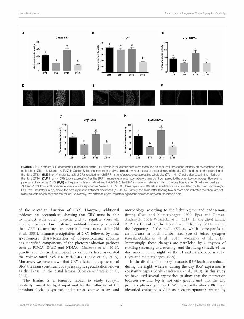
CRY Affects BRP Degradation in theLaminaIn the lamina the photoreceptor terminals from the retinaare arranged in cylindrical modules called cartridges. Besidethe photoreceptor terminals R1–R6, they consist of the laminainterneurons and processes of cells projecting from opticneuropils and from the central brain. Within cartridges manysynaptic contacts are formed between cells, including tetradsynapses between the photoreceptor terminals R1–R6 andfour postsynaptic partners among the following cell types:L1, L2, β-processes of amacrine cells, glial cells or L3(Prokop and Meinertzhagen, 2006). In the distal lamina BRPexpression is rhythmic, showing a light-dependent peak at thebeginning of the day (ZT1) and a clock dependent peak at
Frontiers in Molecular Neuroscience | www.frontiersin.org 4 May 2017 | Volume 10 | Article 165
Damulewicz et al. Cryprochrome Regulates Visual Synaptic Plasticity
FIGURE 2 | CRY-BRP complexes are formed in vivo. CRY-BRP complexes were visualized in vivo on 20 µm cryosections of the optic lobe with proximity ligationassay (PLA). (A) Canton S and (B) cry01 Drosophila brains. Complexes between CRY and BRP are seen as fluorescent dots in the retina (R), the lamina (L) and themedulla (M; arrows in A) of Canton S but not cry01 flies.
the beginning of the night (ZT13, Górska-Andrzejak et al.,2013 and Figures 3A,D,G,H). The cry01 mutation changesthis pattern as BRP levels are high across the whole dayand only decrease in the middle of the night (ZT16, Górska-Andrzejak et al., 2013 and Figures 3B,E). This suggests thatCRY might be involved in the light-dependent degradationof BRP, possibly providing a functional explanation for thebinding between the two proteins in analogy to what it isknown for the CRY-TIM interaction. To test this hypothesiswe used immunofluorescence to measure BRP levels in thelamina of cry > CRY∆ flies. The latter overexpress CRY∆, aC-terminal deletion that results in a constitutively active formof CRY, in all cry-positive cells (Rosato et al., 2001; Disselet al., 2004). cry > CRY∆ flies showed maximal immune-signal for BRP at ZT13, but showed no difference betweenthe beginning and the middle of the day (ZT1 and ZT4, seeFigures 3C,F). Thus, the pattern of expression is reminiscent ofwild type flies under DD (see Figure 2B in Górska-Andrzejaket al., 2013). However, there was an important difference, whichis that at each time point the BRP signal was dramaticallyreduced compared to control (Canton S, cry-Gal4, UAS-CRY∆)and cry01 flies (Figures 3A–H). We also tested by Westernblot whether the same pattern of expression might be foundfor BRP in the whole head. Whole head protein extractsobtained at ZT1 from Canton S, cry01 and cry > CRY∆, wereimmune-stained with nc82 antibodies. Both cry01 and CRY∆
flies revealed an overall reduction in the levels of the two BRPisoforms (Figures 4A,B). However, they also showed muchgreater variability in the expression of BRP than Canton S, asindicated by the large standard deviations reported in Figure 4B.
This suggests that CRY affects the regulation of BRP in a morecomplex and diverse fashion compared to the distal laminaalone.
CRY∆ Affects the Optomotor ResponseSuch a reduced level of BRP in the lamina observed incry > CRY∆ flies should result in measurable effects onvision, which we assessed using the optomotor walkingresponse. Control flies (Canton S, cry-Gal4, UAS-CRY∆)showed daily modulation of the optomotor response withabout 60% correct choices during the day (ZT1, 4, 8) andabout 70%–80% correct choices at night (ZT13, 16, 20). Theoptomotor response of cry > CRY∆ flies was almost flatwith significant differences only between ZT8, with about 40%correct choices, and ZT20, scoring about 50% correct choices(Figure 5).
DISCUSSION
The discovery of CRY was triggered by its ability to modulatethe stability of other proteins. The first mutant cryb wasidentified through the loss of rhythmic expression of PERand TIM in peripheral tissues and a dampening of thoserhythms in the circadian neurons in the brain (Stanewskyet al., 1998). Subsequently, it emerged that CRY binds to TIMunder light (Ceriani et al., 1999) and that such interactiontriggers the light-dependent degradation of both proteins(Peschel et al., 2009). This cell-autonomous model of light-induced degradation has dominated the understanding
Frontiers in Molecular Neuroscience | www.frontiersin.org 5 May 2017 | Volume 10 | Article 165
Damulewicz et al. Cryprochrome Regulates Visual Synaptic Plasticity
FIGURE 3 | CRY affects BRP degradation in the distal lamina. BRP levels in the distal lamina were measured as immunofluorescence intensity on cryosections of theoptic lobe at ZTs 1, 4, 13 and 16. (A,D) In Canton S flies the immune-signal was bimodal with one peak at the beginning of the day (ZT1) and one at the beginning ofthe night (ZT13). (B,E) In cry01 mutants, lack of CRY resulted in high BRP immunofluorescence across the whole day (ZTs 1, 4, 13) but a decrease in the middle ofthe night (ZT16). (C,F) In cry > CRY∆ overexpressing flies the BRP immune-signal was lower at every time point compared to the other two genotypes. However, apeak was observed at ZT13. (G,H) In the parental lines cry-Gal4 and UAS-CRY∆ the BRP immune-signal was similar to the one from Canton S, with two peaks atZT1 and ZT13. Immunofluorescence intensities are reported as Mean ± SD. N = 30, three repetitions. Statistical significance was calculated by ANOVA using Tukey’sHSD test. The letters (a,b,c) above the bars represent statistical differences (p < 0.05). Namely, the same letter labeling two or more bars indicates that there are notstatistical differences between the values. Conversely, two different letters indicate a significant difference between the labeled bars.
of the circadian function of CRY. However, additionalevidence has accumulated showing that CRY must be ableto interact with other proteins and to regulate cross-talkamong neurons. For instance, antibody staining revealedthat CRY accumulates in neuronal projections (Klarsfeldet al., 2004), immune-precipitation of CRY followed by massspectrometry characterization of co-precipitating proteinshas identified components of the phototransduction pathwaysuch as RDGA, INAD and NINAC (Mazzotta et al., 2013),genetic and electrophysiological experiments have associatedthe voltage-gated Kvβ HK with CRY (Fogle et al., 2015).Moreover, we have shown that CRY affects the expression ofBRP, the main constituent of a presynaptic specialization knownas the T-bar, in the distal lamina (Górska-Andrzejak et al.,2013).
The lamina is a fantastic model to study synapticplasticity caused by light input and by the influence of thecircadian clock, as synapses and neurons change in size and
morphology according to the light regime and endogenoustiming (Pyza and Meinertzhagen, 1999; Pyza and Górska-Andrzejak, 2004; Woznicka et al., 2015). In the distal laminaBRP levels peak at the beginning of the day (ZT1) and atthe beginning of the night (ZT13), which corresponds toan increase in both number and size of tetrad synapses(Górska-Andrzejak et al., 2013; Woznicka et al., 2015).Interestingly, these changes are paralleled by a rhythm ofswelling (morning and evening) and shrinking (middle of theday, middle of the night) of the L1 and L2 monopolar cells(Pyza and Meinertzhagen, 1999).
In the distal lamina of cry0 mutants BRP levels are reducedduring the night, whereas during the day BRP expression isconstantly high (Górska-Andrzejak et al., 2013). In this studywe have used several approaches to show that the interactionbetween cry and brp is not only genetic and that the twoproteins physically interact. We have pulled-down BRP andidentified endogenous CRY as a co-precipitating protein by
Frontiers in Molecular Neuroscience | www.frontiersin.org 6 May 2017 | Volume 10 | Article 165
Damulewicz et al. Cryprochrome Regulates Visual Synaptic Plasticity
FIGURE 4 | CRY has a complex effect on BRP degradation in whole headprotein extracts. BRP levels in whole head protein extracts were determinedby Western blot analyses in Canton S, cry01 and cry > CRY∆ flies collected atZT1. (A) Representative immunoblot stained with nc82 antibodies showing the170 kDa and 190 kDa BRP isoforms. Alfa Tubulin (αTub) was used as loadingcontrol. (B) Densitometry of BRP isoforms normalized to αTub levels.BRP/αTub ratios are reported as Mean ± SD, N = 50, three repetitions.A non-parametric Kruskal-Wallis test showed no statistical difference amongthe strains.
Western blot (Figure 1A). Moreover, when we pulled-downoverexpressed HA-CRY and probed for the endogenous BRP,we observed that both the 170 kDa and the 190 kDaBRP isoforms co-precipitated with HA-CRY (Figure 1B). Weinterpret this finding as an indirect confirmation of theweak co-immunoprecipitation results obtained with brp∆190
and brp∆170 mutants that express only the 170 kDa andthe 190 kDa BRP isoform, respectively (Matkovic et al.,2013). In a Y2H assay we verified that CRY and BRPdirectly bind to each other and that light promotes thisinteraction (Figure 1C). This result is in agreement with theco-immunoprecipitation experiment presented in Figure 1B,showing that more BRP co-precipitated together with HA-CRYunder light conditions than in darkness. We note thatalthough the Y2H experiment suggests a direct interactionbetween CRY and BRP, additional proteins might be involvedin vivo to stabilize the complex and/or to initiate a signalingcascade.
Using PLA with anti-BRP and anti-CRY antibodies wediscovered PLA-positive signal in the retina, in the laminaand in the medulla of Canton S but not of cry01 flies,showing that the interaction between CRY and BRP occursin vivo and in situ (Figure 2). The pattern of fluorescenceis in agreement with the known distribution of CRY (BRPis found in all photoreceptors and neurons) but it wassurprisingly sparse. We interpret this result as a consequence
FIGURE 5 | CRY∆ affects the optomotor response. Optomotor walkingresponse of Canton S, cry-Gal4, UAS-CRY∆ and cry > CRY∆ flies during theday (ZT1, ZT4, ZT8) and during the night (ZT13, ZT16, ZT20). Optomotorresponses are reported as Mean ± SE; N = 100 for genotypes Canton S andcry > CRY∆. N = 35 for genotypes cry-Gal4 and UAS-CRY∆. Statisticalsignificance was calculated by ANOVA using Tukey’s HSD test. Significantdifferences. Canton S: ZT1 vs. ZT13 (p ≤ 0.0001), ZT1 vs. ZT16 (p ≤ 0.001),ZT1 vs. ZT20 (p ≤ 0.01), ZT4 vs. ZT13, ZT16, ZT20 (p ≤ 0.0001), ZT8 vs.ZT13 (p ≤ 0.0001), ZT8 vs. ZT16 (p ≤ 0.001), ZT8 vs. ZT20 (p ≤ 0.01).cry-Gal4: ZT1 vs. ZT13, ZT16, ZT20 (p ≤ 0.0001), ZT4 vs. ZT13, ZT16(p ≤ 0.001), ZT4 vs. ZT20 (p ≤ 0.0001), ZT8 vs. ZT13, ZT16 (p ≤ 0.0001),ZT8 vs. ZT20 (p ≤ 0.001). UAS-CRY∆: ZT1 vs. ZT13, ZT16, ZT20(p ≤ 0.0001), ZT4 vs. ZT13, ZT16, ZT20 (p ≤ 0.0001), ZT8 vs. ZT13, ZT16,ZT20 (p ≤ 0.0001). cry > CRY∆: ZT8 vs. ZT20 (p ≤ 0.05).
of the complexity of the technique. One possibility is thatthe PLA-positive signal is limited to those areas where CRYand BRP are expressed at the highest level. It is unlikelythat within tissue sections the primary and the secondaryantibodies will always bind with the right steric arrangementto allow the best interaction between the antibodies-boundPLA probes and the connectors. Another possibility is that20 µm sections are quite difficult to penetrate by enzymessuch as ligase and rolling circle DNA polymerase. Hence signalmay be prevalent in areas that are more exposed and/orhave a ‘‘looser’’ structure. Although we did not investigatethese possibilities, it is likely that both aspects played a rolein determining the tissue distribution of the PLA-signals werevealed.
To test whether BRP may be targeted for degradationfollowing its interaction with CRY, we turned to CRY∆, aC-terminal deletion of CRY. We have previously shown ina Y2H assay that CRY∆ binds to TIM independently fromlight, and that the overexpression of CRY∆ in flies resultsin phenotypes suggesting that this form of the protein doesnot require activation by light (Rosato et al., 2001; Disselet al., 2004). For instance, in CRY∆ flies TIM is expressedat lower levels than in wild type, which agrees with the ideaof a constitutive interaction with an active CRY. Moreover,CRY∆ does not accumulate or marginally so; this is alsoexpected, considering that the interaction between TIM andCRY drives the degradation of both proteins (Peschel et al.,2009). Thus we hypothesized that cry > CRY∆ flies wouldshow reduced immunostaining for BRP at each time point.Indeed, that is what we observed in the distal lamina incomparison to both Canton S and cry01 flies (Figure 3).This suggests that BRP, like TIM, is targeted for degradationfollowing the formation of a complex with CRY. Although
Frontiers in Molecular Neuroscience | www.frontiersin.org 7 May 2017 | Volume 10 | Article 165
Damulewicz et al. Cryprochrome Regulates Visual Synaptic Plasticity
the overall BRP immune-signal was reduced, we could stilldetect a significant oscillation in BRP levels. At its peak atZT13, the BRP signal was about twice the size than for theother time points (Figure 3C). Interestingly, this expressionprofile mimics the temporal distribution of BRP immune-signal in wild type flies maintained under constant darkness(compare the expression profiles of BRP in Figure 3C toFigure 2B of Górska-Andrzejak et al., 2013). This is in spiteof the fact that constitutive active CRY, which would arguablysimulate constant light, might be expected to produce anon-rhythmic phenotype. When we measured BRP levels inwhole heads we observed the reduction of BRP in both cry01and cry > CRY∆ in comparing with Canton S; however,these differences were not statistically significant due to aremarkable variability among experiments (Figure 4B). Ourinterpretation is that although CRY does have an effect on theexpression of BRP in general, the mechanisms are complex andpossibly tissue/brain area specific. We also note that CRY isnot uniformly expressed across the brain or the head. Thus, wemight have detected a mixture of direct and indirect effects,and arguably the latter might amplify noise. Thus, we concludethat CRY affects BRP expression in the distal lamina, likelyregulating its stability. In addition, we propose that the laminais a particularly attractive model to investigate the mode ofaction of CRY.
Finally we reasoned that such a reduction in BRP expressionin the distal lamina of cry > CRY∆ flies (Figure 3), should giverise to behavioral phenotypes. The optomotor response measuresthe ability to detect and respond to amoving environment.Whenthe environment moves it generates an apparent self-motion towhich a spectator responds with movement to stabilize theirapparent course. The optomotor response depends to a largeextent on the time of day, with best performances observedbetween the end of the day and the middle of the night andit reflects the presence of a functional circadian clock in thephotoreceptor system (Barth et al., 2010; Mazzotta et al., 2013;Mazzotta and Costa, 2016). In order to study the motionvision of flies, we analyzed their optomotor walking response.As previously reported (Mazzotta et al., 2013) Canton S fliesperformed better at night (ZT13, 16, 20) than during the day(ZT1, 4, 8) with 70% and 60% correct choices, respectively(Figure 5).
Light adaptation in the retina depends on horizontalmigration of screening pigment granules towards therhabdomeres (Nilsson and Ro, 1994). In Musca domesticascreening pigment granules migrate also vertically inphotoreceptors, with maximal accumulation in the proximalpart of the lamina at the end of day and higher pigmentnumber in the distal lamina at the end of night. This patternis clock-dependent (Pyza and Meinertzhagen, 1997). Becausethe absence of screening pigment causes loss of visual acuity(Burnet et al., 1968), the daily changes in optomotor walkingresponse may be correlated with the pattern of pigment granulesmigration. However, BRP levels peak at ZT1 and ZT13 inthe distal lamina of Canton S (Figure 3A), and we wouldhave expected a similar behavioral outcome at these two timepoints. Thus, the optomotor response did not reflect the daily
differences in number and size of tetrad synapses of whichBRP levels in the distal lamina are a proxy. Nevertheless, wecould see significant differences between the performances ofcontrol and of cry > CRY∆ flies (Figure 5). The latter genotypeshowed 40%–50% of correct choices during the whole day,which is the value expected by chance alone. Thus, these flieseither could not detect the movement of the stripes or theywere unable to process the information, or may be both. Againwe did not see a correlation between the expression profile ofBRP and the optomotor response. In an earlier study on thehousefly, we found that motor stimulation is more effectivethan visual stimulation in eliciting morphological changes inthe lamina. Thus the lack of correlation between BRP levels andoptomotor response is not surprising (Kula and Pyza, 2007).This behavioral assay tests the functioning of the visual systemfrom photoreceptors to higher order motion vision processingneurons in the lobula plate, a region in the optic lobes thatis the final destination of visual information (Heisenberg andWolf, 1984). Our results are in agreement with the previousobservation where flies in which CRY lacked its C-terminus tail(cryM, Busza et al., 2004) showed a reduced performance in theoptomotor response (Mazzotta et al., 2013). In addition, flies withbrp expression silenced in the visual system (gmr > brpRNAi)showed changes in the electroretinogram (ERG; Wagh et al.,2006), which measures extracellular activity of photoreceptorsand interneurons in response to light (Heisenberg, 1971). Lackof BRP in the photoreceptor terminals causes severe defectsin synaptic transmission which is visualized in ERG, sincelight-induced depolarization of photoreceptors is normal butON/OFF transients originating from the interneurons areabsent (Wagh et al., 2006). We speculate that the presence ofa constitutively active form of CRY (due to the absence of itsregulatory C-terminus) likely impacts on the organization ofthe visual system beyond the photoreceptors and the laminaand may have similar effect on the retina functionality as brpsilencing. This consideration calls for additional studies todissect the role of CRY in the visual system of Drosophila.
In conclusion, we have identified a physical interactionbetween CRY and BRP, the main constituent of the presynapticactive zone T bar. We have confirmed this interaction usingdifferent techniques such as immune-precipitation, Y2H andPLA. Our data suggest that CRY and BRP can interactdirectly especially under light, but they do not preclude thatCRY and BRP might be part of a larger complex in vivo.We have evidence that CRY regulates the stability of BRPin the distal lamina, possibly mirroring what is known forthe CRY-TIM interaction. However, the regulation of BRPin the head is more complex, since not all synapses peakin number at the same time during day and night. Finally,we have presented data showing that the functionality of thevisual system is compromised in cry > CRY∆ flies, probablyaffecting higher order processing neurons. This suggests thatthe role exerted by CRY on the development and physiologyof the visual system is greater than currently appreciated,with important consequences for the interpretation of theeffects of CRY on the light entrainment of the circadianclock.
Frontiers in Molecular Neuroscience | www.frontiersin.org 8 May 2017 | Volume 10 | Article 165
Damulewicz et al. Cryprochrome Regulates Visual Synaptic Plasticity
AUTHOR CONTRIBUTIONS
MD, GMM and ES carried out experiments. MD preparedfigures. MD, GMM and ER, RC and EMP discussed results andreviewed themanuscript. MD and ERwrote themainmanuscripttext.
FUNDING
The study was supported by the Polish National ScienceCentre (Narodowe Centrum Nauki, NCN) grant no. UMO-2014/15/D/NZ3/05207 to MD. ER was supported by theUK Biotechnology and Biological Sciences Research Council(BBSRC, grant nos. BB/F008988/1 and BB/H018093/1). RC was
supported by a grant from National Research Council of Italy(EPIGEN Progetto Bandiera Epigenomica—Subproject 4).
ACKNOWLEDGMENTS
We would like to thank F. Rouyer, D. Dolezel and S. Sigrist forflies.
SUPPLEMENTARY MATERIAL
The Supplementary Material for this article can be foundonline at: http://journal.frontiersin.org/article/10.3389/fnmol.2017.00165/full#supplementary-material
REFERENCES
Ausbel, F. M. (1998). Current Protocols in Molecular Biology. New York, NY:John Wiley & Sons.
Bazalova, O., Kvicalova, M., Valkova, T., Slaby, P., Bartos, P., Netusil, R.,et al. (2016). Cryptochrome 2 mediates directional magnetoreception incockroaches. Proc. Natl. Acad. Sci. U S A 113, 1660–1665. doi: 10.1073/pnas.1518622113
Barth, M., Schultze, M., Schuster, C.M., and Strauss, R. (2010). Circadianplasticity in photoreceptor cells controls visual coding efficiency in Drosophilamelanogaster. PLoS One 5:e9217. doi: 10.1371/journal.pone.0009217
Burnet, B., Connolly, K., and Beck, J. (1968). Phenogenetic studies on visual acuityin Drosophila melanogaster. J. Insect Physiol. 14, 855–860. doi: 10.1016/0022-1910(68)90196-0
Busza, A., Emery-Le, M., Rosbash, M., and Emery, P. (2004). Roles ofthe two Drosophila CRYPTOCHROME structural domains in circadianphotoreception. Science 304, 1503–1506. doi: 10.1126/science.1096973
Ceriani, M. F., Darlington, T. K., Staknis, D., Más, P., Petti, A. A., Weitz, C. J., et al.(1999). Light-dependent sequestration of TIMELESS by CRYPTOCHROME.Science 285, 553–556. doi: 10.1126/science.285.5427.553
Damulewicz, M., Loboda, A., Bukowska-Strakova, K., Jozkowicz, A., Dulak, J.,and Pyza, E. (2015). Clock and clock-controlled genes are differently expressedin the retina, lamina and in selected cells of the visual system of Drosophilamelanogaster. Front. Cell. Neurosci. 9:353. doi: 10.3389/fncel.2015.00353
Dissel, S., Codd, V., Fedic, R., Garner, K. J., Costa, R., Kyriacou, C. P., et al.(2004). A constitutively active cryptochrome in Drosophila melanogaster. Nat.Neurosci. 7, 834–840. doi: 10.1038/nn1285
Dissel, S., Hansen, C. N., Özkaya, Ö, Hemsley, M., Kyriacou, C. P., and Rosato, E.(2014). The logic of circadian organization in Drosophila. Curr. Biol. 24,2257–2266. doi: 10.1016/j.cub.2014.08.023
Dolezelova, E., Dolezel, D., and Hall, J. C. (2007). Rhythm defects caused by newlyengineered null mutations in Drosophila’s cryptochrome gene. Genetics 177,329–345. doi: 10.1534/genetics.107.076513
Emery, P., So, W. V., Kaneko, M., Hall, J. C., and Rosbash, M. (1998).CRY, a Drosophila clock and light-regulated cryptochrome, is a majorcontributor to circadian rhythm resetting and photosensitivity. Cell 95,669–679. doi: 10.1016/s0092-8674(00)81637-2
Emery, P., Stanewsky, R., Hall, J. C., and Rosbash, M. (2000). A unique circadian-rhythm photoreceptor. Nature 404, 456–457. doi: 10.1038/35006558
Fedele, G., Edwards, M. D., Bhutani, S., Hares, J. M., Murbach, M., Green, E. W.,et al. (2014a). Genetic analysis of circadian responses to low frequencyelectromagnetic fields in Drosophila melanogaster. PLoS Genet. 10:e1004804.doi: 10.1371/journal.pgen.1004804
Fedele, G., Green, E. W., Rosato, E., and Kyriacou, C. P. (2014b). Anelectromagnetic field disrupts negative geotaxis in Drosophila via aCRY-dependent pathway. Nat. Commun. 5:4391. doi: 10.1038/ncomms5391
Fogle, K. J., Baik, L. S., Houl, J. H., Tran, T. T., Roberts, L., Dahm, N. A., et al.(2015). CRYPTOCHROME-mediated phototransduction bymodulation of the
potassium ion channel β-subunit redox sensor. Proc. Natl. Acad. Sci. U S A 112,2245–2250. doi: 10.1073/pnas.1416586112
Fogle, K. J., Parson, K. G., Dahm, N. A., and Holmes, T. C. (2011).CRYPTOCHROME is a blue-light sensor that regulates neuronal firing rate.Science 331, 1409–1413. doi: 10.1126/science.1199702
Fouquet, W., Owald, D., Wichmann, C., Mertel, S., Depner, H., Dyba, M., et al.(2009). Maturation of active zone assembly by Drosophila Bruchpilot. J. CellBiol. 186, 129–145. doi: 10.1083/jcb.200812150
Gegear, R. J., Casselman, A.,Waddell, S., and Reppert, S. M. (2008). Cryptochromemediates light-dependent magnetosensitivity in Drosophila. Nature 454,1014–1018. doi: 10.1038/nature07183
Golemis, E. A., and Brent, R. (1997). ‘‘Searching for interacting proteins with thetwo-hybrid system, III,’’ in The Yeast Two-Hybrid System, eds P. L. Bartel andS. Fields (New York, NY: Oxford University Press), 43–72.
Górska-Andrzejak, J., Makuch, R., Stefan, J., Görlich, A., Semik, D., andPyza, E. (2013). Circadian expression of the presynaptic active zone proteinbruchpilot in the lamina ofDrosophila melanogaster.Dev. Neurobiol. 73, 14–26.doi: 10.1002/dneu.22032
Heisenberg, M. (1971). Separation of receptor and lamina potentials inthe electroretinogram of normal and mutant Drosophila. J. Exp. Biol.55, 85–100. Available online at: www.ncbi.nlm.nih.gov/pubmed/5001616.
Heisenberg, M., and Wolf, R. (1984). Vision in Drosophila: Genetics ofMicrobehavior. Berlin, New York, NY: Springer-Verlag.
Hida, Y., and Ohtsuka, T. (2010). CAST and ELKS proteins: Structural andfunctional determinants of the presynaptic active zone. J. Biochem. 148,131–137. doi: 10.1093/jb/mvq065
Hunter-Ensor, M., Ousley, A., and Sehgal, A. (1996). Regulation of the Drosophilaprotein timeless suggests a mechanism for resetting the circadian clock by light.Cell 84, 677–685. doi: 10.1016/s0092-8674(00)81046-6
Ishikawa, T., Matsumoto, A., Kato, T., Togashi, S., Ryo, H., Ikenaga, M., et al.(1999). DCRY is a Drosophila photoreceptor protein implicated in lightentrainment of circadian rhythm. Genes Cells 4, 57–65. doi: 10.1046/j.1365-2443.1999.00237.x
Kittel, R. J., Hallermann, S., Thomsen, S., Wichmann, C., Sigrist, S. J., andHeckmann, M. (2006). Active zone assembly and synaptic release. Biochem.Soc. Trans. 34, 939–941. doi: 10.1042/BST0340939
Klarsfeld, A., Malpel, S., Michard-Vanhée, C., Picot, M., Chélot, E., andRouyer, F. (2004). Novel features of cryptochrome-mediated photoreceptionin the brain circadian clock of Drosophila. J. Neurosci. 24, 1468–1477.doi: 10.1523/JNEUROSCI.3661-03.2004
Kula, E., and Pyza, E. (2007). Effects of locomotor stimulation and proteinsynthesis inhibition on circadian rhythms in size changes of L1 andL2 interneurons in the fly’s visual system. Dev. Neurobiol. 67, 1433–1442.doi: 10.1002/dneu.20518
Matkovic, T., Siebert, M., Knoche, E., Depner, H., Mertel, S., Owald, D., et al.(2013). The bruchpilot cytomatrix determines the size of the readily releasablepool of synaptic vesicles. J. Cell Biol. 202, 667–683. doi: 10.1083/jcb.201301072
Frontiers in Molecular Neuroscience | www.frontiersin.org 9 May 2017 | Volume 10 | Article 165
Damulewicz et al. Cryprochrome Regulates Visual Synaptic Plasticity
Mazzotta, G., Rossi, A., Leonardi, E., Mason, M., Bertolucci, C., Caccin, L., et al.(2013). Fly cryptochrome and the visual system. Proc. Natl. Acad. Sci. U S A110, 6163–6168. doi: 10.1073/pnas.1212317110
Mazzotta, G. M., and Costa, R. (2016). Circadian control of visual plasticityin arthropods. Ethol. Ecol. Evol. 28, 1–19. doi: 10.1080/03949370.2015.1064037
Meinertzhagen, I. A., and O’Neil, S. D. (1991). Synaptic organization of columnarelements in the lamina of the wild type in Drosophila melanogaster. J. Comp.Neurol. 305, 232–263. doi: 10.1002/cne.903050206
Naidoo, N., Song, W., Hunter-Ensor, M., and Sehgal, A. (1999). A role for theproteasome in the light response of the timeless clock protein. Science 285,1737–1741. doi: 10.1126/science.285.5434.1737
Nilsson, D. E., and Ro, A. I. (1994). Did neural pooling for night vision lead tothe evolution of neural superposition eyes? J. Comp. Physiol. A 175, 289–302.doi: 10.1007/BF00192988
Ozturk, N., Selby, C. P., Annayev, Y., Zhong, D., and Sancar, A. (2011). Reactionmechanism of Drosophila cryptochrome. Proc. Natl. Acad. Sci. U S A 108,516–521. doi: 10.1073/pnas.1017093108
Peschel, N., Chen, K. F., Szabo, G., and Stanewsky, R. (2009). Light-dependent interactions between the Drosophila circadian clock factorscryptochrome, jetlag and timeless. Curr. Biol. 19, 241–247. doi: 10.1016/j.cub.2008.12.042
Picot, M., Cusumano, P., Klarsfeld, A., Ueda, R., and Rouyer, F. (2007).Light activates output from evening neurons and inhibits output frommorning neurons in the Drosophila circadian clock. PLoS Biol. 5:e315.doi: 10.1371/journal.pbio.0050315
Prokop, A., and Meinertzhagen, I. A. (2006). Development and structure ofsynaptic contacts inDrosophila. Semin. Cell Dev. Biol. 17, 20–30. doi: 10.1016/j.semcdb.2005.11.010
Pyza, E., and Górska-Andrzejak, J. (2004). Involvement of glial cells in rhythmicsize changes in neurons of the housefly’s visual system. J. Neurobiol. 59,205–215. doi: 10.1002/neu.10307
Pyza, E., andMeinertzhagen, I. A. (1997). Circadian rhythms in screening pigmentand invaginating organelles in photoreceptor terminals of the housefly’sfirst optic neuropile. J. Neurobiol. 32, 517–529. doi: 10.1002/(sici)1097-4695(199705)32:5<517::aid-neu6>3.3.co;2-4
Pyza, E., and Meinertzhagen, I. A. (1999). Daily rhythmic changes of cell size andshape in the first optic neuropil in Drosophila melanogaster. J. Neurobiol. 40,77–88. doi: 10.1002/(sici)1097-4695(199907)40:1<77::aid-neu7>3.0.co;2-0
Ritz, T., Yoshii, T., Helfrich-Foerster, C., and Ahmad, M. (2010). Cryptochrome: aphotoreceptor with the properties of a magnetoreceptor?Commun. Integr. Biol.3, 24–27.
Rosato, E., Codd, V., Mazzotta, G., Piccin, A., Zordan, M., Costa, R., et al. (2001).Light-dependent interaction between Drosophila CRY and the clock proteinPER mediated by the carboxy terminus of CRY. Curr. Biol. 11, 909–917.doi: 10.1016/s0960-9822(01)00259-7
Stanewsky, R., Kaneko, M., Emery, P., Beretta, B., Wager-Smith, K., Kay, S. A.,et al. (1998). The cryb mutation identifies cryptochrome as a circadianphotoreceptor in Drosophila. Cell 95, 681–692. doi: 10.1016/s0092-8674(00)81638-4
Wagh, D. A., Rasse, T. M., Asan, E., Hofbauer, A., Schwenkert, I., Dürrbeck, H.,et al. (2006). Bruchpilot, a protein with homology to ELKS/CAST, is requiredfor structural integrity and function of synaptic active zones in Drosophila.Neuron 49, 833–844. doi: 10.1016/j.neuron.2006.02.008
Wichmann, C., and Sigrist, S. J. (2010). The active zone T-bar—a plasticitymodule? J. Neurogenet. 24, 133–145. doi: 10.3109/01677063.2010.489626
Woznicka, O., Görlich, A., Sigrist, S., and Pyza, E. (2015). BRP-170 andBRP190 isoforms of bruchpilot protein differentially contribute to thefrequency of synapses and synaptic circadian plasticity in the visual system ofDrosophila. Front. Cell. Neurosci. 9:238. doi: 10.3389/fncel.2015.00238
Conflict of Interest Statement: The authors declare that the research wasconducted in the absence of any commercial or financial relationships that couldbe construed as a potential conflict of interest.