6/30/2016
1
Ayesha Iqbal, M.D.
Crystal Arthropathies Crystal Arthropathies Gout – Monosodium urate
CPPD – Calcium pyrophosphate
dihydrate crystal deposition
Basic Calcium Phosphate Dz – Hydroxyapaptite
Brief review of etiology and pathophysiology
Recognize predisposing factors
Review diagnostic criteria and evaluation
Select appropriate treatment
Objectives Gout
Gout is an inflammatory arthritis
resulting from deposition of
monosodium urate crystals in joints and
other connective tissue structures
Most common inflammatory arthritis in men
Prevalence in US: 3.9% (~8 million)
↑ incidence and prevalence worldwide
Male to female – 4:1
Rare before puberty and in premenopausal
women
Epidemiology Hyperuricemia and Gout Humans have inactivated the Uricase gene
which degrades uric acid to water soluble
Allantoin.
Hyperuricemia is defined as levels >2
standard deviations above nL
6.8 mg/dl in men.
6.0 mg/dl in women.
Solubility of MSU is 6.8 mg/dl
6/30/2016
2
Hyperuricemia and Gout Hyperuricemia is a risk factor for gout
Prevalence: 2.3 - 41.3%
Less than 20% get gout
↑ incidence of gout with ↑ serum uric acid (SUA) levels.
The annual incidence rates for gout 0.1% at SUA levels less than 7 mg/dl
0.5% at SUA levels between 7- 8.9 mg/dl
4.9% at SUA levels above 9 mg/dl
Decreased excretion of Uric acid: 80-90%
Overproduction of Uric acid: 10%
Lesch- Nyhan Syndrome: Hypoxanthine -guanine
phosphoribosyl transferase deficiency(HGPRT)
Combined Mechanism
Lesch-Nyhan Synd: Young boy with arthritis(gouty), kidney stones,
abnormal involuntary movements, self injury/mutilation
Hyperuricemia - Mechanism
Ethanol consumption
Diet : High purine diet, high fructose
beverages
Obesity
Metabolic syndrome
Acute illness, Post - Op
Renal insufficiency
Hypertension
Chronic lead toxicity- Saturnine gout
Predisposing factors
Drugs: Diuretics, low dose salicylates, b-
Blockers, Pyrazinamide, Ethambutol,
Cyclosporine, tacrolimus, Insulin
Strong disease association – CKD, Metabolic
syndrome, HTN, CAD
Predisposing factors
Pathophysiology Not completely understood
Better idea about the inflammatory response
• MSU crystals undergo phagocytosis by
macrophages which stimulates a cyropyrin
inflammasome (NLRP3), this induces release
of pro- inflammatory cytokines (IL- 1beta, IL-
6,8 and TNF)
• These cytokines lead to recruitment and
activation of leukocytes resulting in signs of
inflammation in acute gouty arthritis
Classic presentations in the natural course of
urate deposition disease:
Asymptomatic Hyperuricemia
Acute gouty arthritis
Intercritical gout or interval gout
Chronic gout:
Tophaceous
Clinical manifestations
6/30/2016
3
Sudden intense inflammation, severe pain,
redness, swelling, warmth and disability
First attack: 4th-6th decade in men
Monoarticular in 80-90% cases
Polyarticular in 3-14%
1st MTP commonly involved- 50%
Fever, leukocytosis and ↑ inflammatory markers
Self-limited
Acute gouty arthritis Acute gouty arthritis
Asymptomatic periods in between acute
attacks
Approx 70% will have a second attack in 2
years
10% never have a recurrence
Over time, attacks are less explosive,
polyarticular, and take longer to resolve
Intercritical Gout Approx takes12 years between initial attack
and development of chronic arthritis in
untreated patients..
Pain free intercritical periods are rare
Tophi: Collections of MSU crystal core in
connective tissues enclosed by granulation
tissue
Joint destruction
Confused with other inflammatory arthritides
Chronic Gout
Chronic Tophaceous Gout Chronic Tophaceous Gout
6/30/2016
4
Cyclosporine induced tophaceous gout Diagnosis
Mostly on the clinical basis of: An acute monoarthritis
Hyperuricemia
Dramatic improvement of articular symptoms in response to Colchicine
Accuracy ↑ with typical presentation Definitive diagnosis: demonstration of MSU
crystals in synovial fluid or from tophi
Aspiration! Aspiration! Aspiration!
Assess predisposing factors Labs SUA: May be normal in acute attacks SUA level needed to monitor therapy
Renal uric acid excretion: Age of onset before 25 FH of early onset gout Nephrolithiasis
Inflammatory markers: non specific Imaging
Non specific in early and acute gout Typical features may be seen in chronic gout
Synovial fluid analysis
Diagnosis Microscopy:
Cell counts: 5,000 – 100,000 (Inflammatory) Neutrophilic predominance Light microscopy
Needle shaped MSU crystals Compensated polarizing light microscopy:
Intracellular MSU crystals Needle shaped Strong negative birefringence Acute attack: Sensitivity 85% Inter-critical period: Sensitivity 70%
(Extracellular Crystals)
Septic arthritis should always be ruled out by a gram stain and culture in these situations
Synovial Fluid Analysis
Plain microscopy Compensated polarizing
Yellow when parallel to Z’ axis
Tophus- multiple MSU crystals
Synovial Fluid Analysis
6/30/2016
5
Imaging Plain Radiography
Acute Gout:
Non specific
Soft tissue swelling
Chronic Tophaceous Gout:
Tophi
Punched out erosions with sclerotic borders
and overhanging edge
High resolution USG, dual energy CT can diagnose earlier disease.
Imaging
"Punched out" erosion of tophaceous gout
USG- Crystal deposits Double Contour
USG- 1st MTP
Diagnosis
Differential diagnosis: Infection:
Septic arthritis Cellulitis Septic bursitis
Other crystal induced arthropathies Other inflammatory arthritides
Rheumatoid arthritis Spondyloarthropathy
Trauma
Inflammatory monoarthritis and elevated uric acid not always gout
6/30/2016
6
Treatment Therapeutic aims:
To terminate the acute attack promptly
To prevent recurrences of acute gouty arthritis
To prevent or reverse complications of the
disease
Treatment options vary
Acute gouty arthritis
Prophylaxis
Chronic gout
2012 ACR Gout Guidelines
Assess benefits and risks of treatments
Cost-effectiveness not asssessed
Grades of evidence supporting recommendations
Level A: supported by multiple(more than 1) randomized
clinical trials or meta-analyses (20%)
Level B: supported from a single randomized trial, or non-
randomized studies (30%)
Level C: consensus opinion of experts, case studies, or
standard of care (50%)
Acute gouty arthritis
Pharmacologic: NSAIDS
Colchicine
Steroids
? IL-1 inhibitors
Step up or combination therapy can be used (C)
Non Pharmacologic: Co morbid risk reduction
Assess and manage drug interferences
Education
NSAIDS: (A) Traditionally Indomethacin has been used
Various NSAIDS have showed similar efficacy and tolerability within the class
ADR: GI irritability, HTN, fluid retention, headaches, rash, hypersensitivity, renal failure
Acute gouty arthritis
Colchicine: (A)
Use within 24 hours of acute attack (C)
FDA approved:
1.2mg orally once followed by 0.6mg EULAR recommendation:
0.6mg orally two or three times per day Dose adjustments for renal and severe hepatic
impairment New FDA recommendations: caution concurrent use
CYP3A4 inhibitors and P-glycoprotein
ADR: GI discomfort- diarrhea, BM suppression, Rhabdomyolysis, Dermatoses, Peripheral neuritis
Acute gouty arthritis
Steroids: (A) Unable to tolerate NSAIDs / colchicine
NSAIDs contraindicated
Suboptimal response
Rapidly effective
Caution in diabetic patients
ADR: Hyperglycemia, Infection, Fluid
retention, Osteoporosis, Cataracts, Psychosis
Acute gouty arthritis
6/30/2016
7
Steroids: Intra-articular:
Used for monoarticular and oligoarticular arthritis
Infection must be ruled out
Orally:
30-40 mg once a day, tapered over 2 weeks
Parenteral:
Unable to tolerate PO
Low dose colchicine (0.6-1.2mg/day) prevents rebound flares (B)
Do not initiate or change Urate lowering therapy during an acute attack
Acute gouty arthritis Treatment Chronic Gout
Pharmacologic:
Urate lowering therapy:
Xanthine oxidase inhibitors
Uricosuric agents
Uricase
Others
Prophylactic agents:
Colchicine
NSAIDs
Indications 2 or more major attacks per year(A)
Tophaceous gout(A)
Nephrolithiasis(C)
Renal uric acid excretion more than 1100 mg/dl(C)
CKD stage 2 and greater(C)
SUA level less than 6.0 mg/dl needed for prevention of acute attacks
SUA around 5.0 mg/dl required for resorption of tophi
Urate Lowering Therapy Urate Lowering Therapy Xanthine oxidase inhibitors:
Purine - Allopurinol
Non-purine -Febuxostat
Uricosuric agents:
Probenacid
Sulfinpyrazone
Uricase - Pegloticase
Others:
Losartan
Fenofibrate
Vitamin C
Allopurinol (A)
Blocks the conversion of hypoxanthine to
xanthine and of xanthine to uric acid
Mean effective daily dose is 300 mg
Initiated at 100 mg/day, titrated up every 2- 4
weeks to maintain the desired
antihyperuricemic effect
SUA levels fall within 2 days of treatment and
reach stable levels in 1-2 weeks
Dosage to be adjusted in renal insufficiency
Urate Lowering Therapy
Adverse reactions: Precipitate acute gouty arthritis
↓ w/ colchicine prophylaxis
Rash: 3 - 5%; 0.1% can progress to AHS (severe exfoliative dermatitis, ARF) Leukopenia Thrombocytopenia Drug fever Vasculitis Interstitial nephritis Drug interactions especially 6-mercaptopurine and
Azathioprine
HLA –B*5801 screening in high risk population(Han Chinese, Koreans with CKD 3 and Thai descent)
Urate Lowering Therapy
6/30/2016
8
Febuxostat (A)
Non-purine, selective inhibitor of Xanthine oxidase
No dose adjustments in mild - mod renal and
hepatic impairment
80-120mg per day
ADR- Acute flare, ↑ LFTs
Use contraindicated with 6-MP, azathioprine and
theophylline
Urate Lowering Therapy
Uricosuric agents: Promote renal excretion of urate
Contraindications Tophi
CRI (GFR less than 35ml/min)
H/O urolithiasis
Intolerance
Rapid cell turnover states
25% failure rate – mild CRI
Interact with ASA, NSAIDs, PCN, captopril
Watch for rash, GI, HA, dyscrasias, nephrosis
Urate Lowering Therapy
Uricosuric agents:
Probenecid (B)
Start 250 mg BID, titrated every few weeks to
a maintenance dose of 500-1000 mg 2-
3x/daily
Losartan (B)
An angiotensin II-receptor antagonist has been shown to have a modest uricosuric effect in a study of hypertensive patients by Wurzner et al.
Urate Lowering Therapy
Fenofibrate (B)
Vitamin C
High dose Salicylate
Combination ULT recommended if SUA not at target
Initiate concomitant anti-inflammatory gout flare prophylaxis (A)
Urate Lowering Therapy
Uricase - converts uric acid to allantoin Pegloticase
FDA approved for tx of refractory gout
Pegylated recombinant uricase
Dose: 8mg IV infusion every 2 weeks
Premedicate with antihistamines or corticosteroids
Prophylaxis recommended
Contra-indication: G6PD deficiency
ADR: Severe gout flares, Infusion reactions(boxed warning)
New Treatments URAT-1( Urate-anion exchange transporter)
inhibitor – Lesinurad
Increases uric acid excretion
Approved in combination with Xanthine
Oxidase Inhibitors for treatment of difficult to
treat hyperurecemia in Gout
Nephrotoxicity noted when used alone.
Contraindication:
CrCl less than 30ml/min, ESRD, Dialysis, Kidney
transplant, Tumor Lysis synd and Lysch-Nyhan synd
6/30/2016
9
IL-1 Beta Inhibitors NLRP3 inflammasome implicated in inflammatory response
to gout crystals Role – Acute and Chronic active gouty arthritis
Anakinra: IL-1receptor antagonist Rilonacept: IL-1alpa and Beta soluble receptor
antagonist Canakinumab: fully human monoclonal
antibody SC administered Role in treatment of acute flare and possibly
prophylaxis Rejected by FDA, approved by EU for acute
treatment
New Treatments CPPD
Precipitation of calcium pyrophosphate
dihydrate crystals in connective tissue
Mostly presents in 6th decade of life
Slight predominance in women
Mostly asymptomatic
Etiology and disease associations: Strong
Idiopathic- aging
Complication of primary osteoarthritis
Mechanical joint trauma or knee meniscectomy
Moderate Familial
Systemic metabolic syndromes Hemochromatosis
Hyperparathyroidism
Hypomagnesemia
Dialysis –dependent RF
Low X-linked hypophosphatemic rickets
Familial hypocalciuric hypercalcemia
Ochronosis
Gout
Wilson’s disease
Hypothyroidism
Amyloidosis
Etiology and disease associations:
CPPD
Clinical Syndromes Asymptomatic with radiological findings-
Chondrocalcinosis
Pseudogout
Pseudo-rheumatoid arthritis
Pseudo-osteoarthritis
Pseudo-neuropathic arthritis
Chondrocalcinosis
Radiographic calcification in hyaline and or fibrocartilage
Radiographic surveys demonstrate an age related increase in prevalence
65 -74 yrs: 15%
> 84 yrs: 50%
Most are asymptomatic
> 50% of these patients have evidence of DJD
25% of these will get pseudogout
CPPD
6/30/2016
10
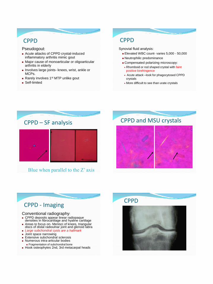
Pseudogout: Acute attacks of CPPD crystal-induced
inflammatory arthritis mimic gout
Major cause of monoarticular or oligoarticular arthritis in elderly
Involves large joints- knees, wrist, ankle or MCPs.
Rarely involves 1st MTP unlike gout
Self-limited
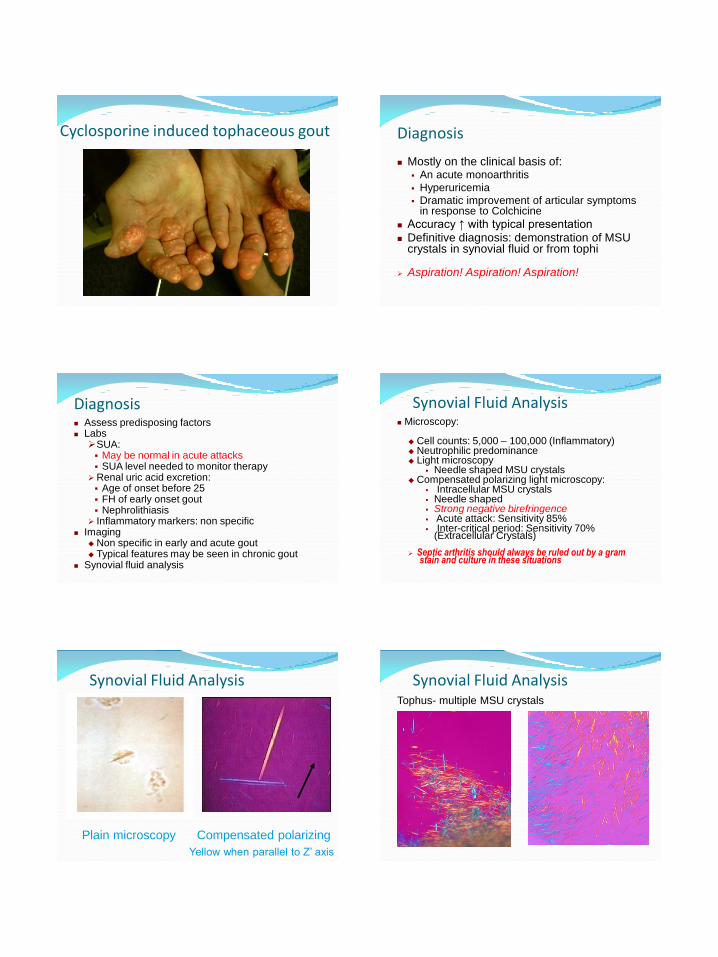
CPPD Synovial fluid analysis:
Elevated WBC count- varies 5,000 - 50,000
Neutrophilic predominance
Compensated polarizing microscopy:
Rhomboid or rod shaped crystal with faint
positive birefringence
Acute attack –look for phagocytosed CPPD
crystals
More difficult to see than urate crystals
CPPD
Blue when parallel to the Z’ axis
CPPD – SF analysis CPPD and MSU crystals
Conventional radiography: CPPD deposits appear linear radiopaque
densities in fibrocartilage and hyaline cartilage Areas to focus on- Menisci of knees, triangular
discs of distal radioulnar joint and glenoid labra Large subchondral cysts are a hallmark Joint space narrowing Extensive subchondral sclerosis Numerous intra-articular bodies
Fragmentation of subchondral bone Hook osteophytes 2nd, 3rd metacarpal heads
CPPD - Imaging CPPD
6/30/2016
11
CPPD CPPD
Ultrasound findings Diagnosis Aspiration! Aspiration! Aspiration!
Definitive diagnosis- demonstration of
intracellular CPPD crystals
+/- typical cartilage or joint calcification on
imaging
Screening for associated diseases
Serum calcium, phosphorus, magnesium,
alkaline phosphatase, ferritin, iron, TSH
2nd and 3rd MCP joint osteoarthritis with beak
osteophytes and chondrocalcinosis think of
hemachromatosis
Acute pseudogout NSAIDS
Colchicine 0.6 mg orally two or three times per day
Steroids
Joint aspiration
Prophylaxis Colchicine 0.6 mg orally twice a day
Treatment Common inflammatory arthritis
Easily misdiagnosed
Aspirate! Aspirate! Aspirate!
Summary
6/30/2016
12
Basic Calcium Phosphate Dz Hydroxyapatite
Dz association – OA (70% SF)
Difficult to detect on SF analysis- non birefringent
crystals
Alizarin red S stain
Milwaukee Shoulder / Knee Syndrome Intra-articular and periarticular hydroxyapatite
crystals
large effusion, minimally inflammatory
+ joint and surrounding structure destruction
+ pain