Crystal structure of Hfq from Bacillus subtilis in complex with SELEX-derived RNA aptamer: insight into RNA-binding properties of bacterial Hfq Tatsuhiko Someya 1, *, Seiki Baba 2 , Mai Fujimoto 1 , Gota Kawai 3,4 , Takashi Kumasaka 2 and Kouji Nakamura 1 1 Graduate School of Life and Environmental Sciences, University of Tsukuba, 1-1-1 Tennodai, Tsukuba-shi, Ibaraki 305-8572, 2 Japan Synchrotron Radiation Research Institute, 1-1-1 Kouto, Sayo-cho, Sayo-gun, Hyogo 679-5198, 3 Department of Life and Environmental Sciences, Faculty of Engineering, Chiba Institute of Technology, 2-17-1 Tsudanuma, Narashino-shi, Chiba 275-0016 and 4 RIKEN SPring-8 Center, 1-1-1 Kouto, Mikazuki-cho, Sayo-gun, Hyogo 679-5148, Japan Received June 29, 2011; Revised October 2, 2011; Accepted October 3, 2011 ABSTRACT Bacterial Hfq is a protein that plays an important role in the regulation of genes in cooperation with sRNAs. Escherichia coli Hfq (EcHfq) has two or more sites that bind RNA(s) including U-rich and/or the poly(A) tail of mRNA. However, functional and struc- tural information about Bacillus subtilis Hfq (BsHfq) including the RNA sequences that specifically bind to it remain unknown. Here, we describe RNA aptamers including fragment (AG) 3 A that are recognized by BsHfq and crystal structures of the BsHfq–(AG) 3 A complex at 2.2 A ˚ resolution. Mutational and structural studies revealed that the RNA fragment binds to the distal site, one of the two binding sites on Hfq, and identified amino acid residues that are critical for sequence-specific interactions between BsHfq and (AG) 3 A. In particu- lar, R32 appears to interact with G bases in (AG) 3 A. Poly(A) also binds to the distal site of EcHfq, but the overall RNA structure and protein–RNA interaction patterns engaged in the R32 residues of BsHfq– (AG) 3 A differ from those of EcHfq–poly(A). These findings provide novel insight into how the Hfq homologue recognizes RNA. INTRODUCTION Gene expression is controlled in bacteria through re- sponses to environmental changes that affect growth. Hfq is an important component in the regulation of gene expression in cooperation with sRNAs. Hfq (also known as HF-I) was originally identified as an Escherichia coli host factor required for the RNA replication of phage Qb (1) and further studies revealed that Hfq in Gram-negative bacteria functions as a post-transcriptional regulator by interacting with various RNAs. Escherichia coli Hfq (EcHfq), which is the most studied among this family of proteins, promotes inter- actions between small untranslated RNA regulatory mol- ecules (such as OxyS, DsrA, Spot42, RyhB and SgrS sRNAs) and their target mRNAs (2–6). Escherichia coli Hfq interacts with many target mRNAs as an RNA chap- erone (7,8) and stimulates polyadenylation that is catalyzed by poly(A) polymerase I, by binding to the poly(A) tail (9–11). Furthermore, EcHfq is involved in RNA stability control by protecting RNA from degrad- ation by the 3 0 –5 0 exoribonucleases, RNase II and PNPase (12,13). About half of all sequenced Gram-negative (such as E. coli spp., Salmonella enterica, Pseudomonas aeruginosa, Neisseria meningitidis and Vibrio cholerae) and Gram-positive (e.g. Staphylococcus aureus, Listeria monocytogenes and Bacillus subtilis) bacteria express Hfq [reviewed in Ref. (14)]. Growth rates of Hfq deletion strains of E. coli K-12 and P. aeruginosa O1 are reduced (15,16). In addition, growth of the Hfq deletion strain of N. meningitidis is impaired in nutrient-rich media and proteomics analysis has revealed that the expression of 28 genes is affected in this strain (17). Moreover, Hfq is required for intestinal colonization by V. cholerae (18). On the other hand, little is known about the function of Hfq in Gram-positive bacteria. The growth of S. aureus and L. monocytogenes Hfq deletion strains is not affected (19,20). Although Hfq in S. aureus (SaHfq) does not play important role in the stress response, RNA sta- bility or exoprotein expression (19), L. monocytogenes Hfq controls the expression of numerous stress- and virulence-associated genes and binds to sRNAs (20,21). *To whom correspondence should be addressed. Tel: +81 29 853 6419; Fax: +81 29 853 7723; Email: [email protected]1856–1867 Nucleic Acids Research, 2012, Vol. 40, No. 4 Published online 3 November 2011 doi:10.1093/nar/gkr892 ß The Author(s) 2011. Published by Oxford University Press. This is an Open Access article distributed under the terms of the Creative Commons Attribution Non-Commercial License (http://creativecommons.org/licenses/ by-nc/3.0), which permits unrestricted non-commercial use, distribution, and reproduction in any medium, provided the original work is properly cited. Downloaded from https://academic.oup.com/nar/article-abstract/40/4/1856/2411658 by guest on 09 April 2018
Transcript
Crystal structure of Hfq from Bacillus subtilis incomplex with SELEX-derived RNA aptamer: insightinto RNA-binding properties of bacterial HfqTatsuhiko Someya1,*, Seiki Baba2, Mai Fujimoto1, Gota Kawai3,4, Takashi Kumasaka2
and Kouji Nakamura1
1Graduate School of Life and Environmental Sciences, University of Tsukuba, 1-1-1 Tennodai, Tsukuba-shi,Ibaraki 305-8572, 2Japan Synchrotron Radiation Research Institute, 1-1-1 Kouto, Sayo-cho, Sayo-gun,Hyogo 679-5198, 3Department of Life and Environmental Sciences, Faculty of Engineering, Chiba Institute ofTechnology, 2-17-1 Tsudanuma, Narashino-shi, Chiba 275-0016 and 4RIKEN SPring-8 Center, 1-1-1 Kouto,Mikazuki-cho, Sayo-gun, Hyogo 679-5148, Japan
Received June 29, 2011; Revised October 2, 2011; Accepted October 3, 2011
ABSTRACT
Bacterial Hfq is a protein that plays an importantrole in the regulation of genes in cooperation withsRNAs. Escherichia coli Hfq (EcHfq) has two or moresites that bind RNA(s) including U-rich and/or thepoly(A) tail of mRNA. However, functional and struc-tural information about Bacillus subtilis Hfq (BsHfq)including the RNA sequences that specificallybind to it remain unknown. Here, we describeRNA aptamers including fragment (AG)3A that arerecognized by BsHfq and crystal structures ofthe BsHfq–(AG)3A complex at 2.2 A resolution.Mutational and structural studies revealed that theRNA fragment binds to the distal site, one of the twobinding sites on Hfq, and identified amino acidresidues that are critical for sequence-specificinteractions between BsHfq and (AG)3A. In particu-lar, R32 appears to interact with G bases in (AG)3A.Poly(A) also binds to the distal site of EcHfq, but theoverall RNA structure and protein–RNA interactionpatterns engaged in the R32 residues of BsHfq–(AG)3A differ from those of EcHfq–poly(A). Thesefindings provide novel insight into how the Hfqhomologue recognizes RNA.
INTRODUCTION
Gene expression is controlled in bacteria through re-sponses to environmental changes that affect growth.Hfq is an important component in the regulation ofgene expression in cooperation with sRNAs. Hfq (alsoknown as HF-I) was originally identified as anEscherichia coli host factor required for the RNA
replication of phage Qb (1) and further studies revealedthat Hfq in Gram-negative bacteria functions as apost-transcriptional regulator by interacting with variousRNAs. Escherichia coli Hfq (EcHfq), which is the moststudied among this family of proteins, promotes inter-actions between small untranslated RNA regulatory mol-ecules (such as OxyS, DsrA, Spot42, RyhB and SgrSsRNAs) and their target mRNAs (2–6). Escherichia coliHfq interacts with many target mRNAs as an RNA chap-erone (7,8) and stimulates polyadenylation that iscatalyzed by poly(A) polymerase I, by binding to thepoly(A) tail (9–11). Furthermore, EcHfq is involved inRNA stability control by protecting RNA from degrad-ation by the 30–50 exoribonucleases, RNase II andPNPase (12,13).
About half of all sequenced Gram-negative (such asE. coli spp., Salmonella enterica, Pseudomonas aeruginosa,Neisseria meningitidis and Vibrio cholerae) andGram-positive (e.g. Staphylococcus aureus, Listeriamonocytogenes and Bacillus subtilis) bacteria express Hfq[reviewed in Ref. (14)]. Growth rates of Hfq deletionstrains of E. coli K-12 and P. aeruginosa O1 are reduced(15,16). In addition, growth of the Hfq deletion strain ofN. meningitidis is impaired in nutrient-rich media andproteomics analysis has revealed that the expression of28 genes is affected in this strain (17). Moreover, Hfq isrequired for intestinal colonization by V. cholerae (18). Onthe other hand, little is known about the function of Hfqin Gram-positive bacteria. The growth of S. aureus andL. monocytogenes Hfq deletion strains is not affected(19,20). Although Hfq in S. aureus (SaHfq) doesnot play important role in the stress response, RNA sta-bility or exoprotein expression (19), L. monocytogenesHfq controls the expression of numerous stress- andvirulence-associated genes and binds to sRNAs (20,21).
*To whom correspondence should be addressed. Tel: +81 29 853 6419; Fax: +81 29 853 7723; Email: [email protected]
1856–1867 Nucleic Acids Research, 2012, Vol. 40, No. 4 Published online 3 November 2011doi:10.1093/nar/gkr892
� The Author(s) 2011. Published by Oxford University Press.This is an Open Access article distributed under the terms of the Creative Commons Attribution Non-Commercial License (http://creativecommons.org/licenses/by-nc/3.0), which permits unrestricted non-commercial use, distribution, and reproduction in any medium, provided the original work is properly cited.
Downloaded from https://academic.oup.com/nar/article-abstract/40/4/1856/2411658by gueston 09 April 2018
The deletion of B. subtilis Hfq (BsHfq), which isencoded by the ymaH gene, does not affect either thegrowth or sporulation (22). BsHfq binds to SR1 sRNAand to ahrC mRNA and the interaction of SR1 sRNAwith its primary target ahrC mRNA causes the inhibitionof ahrC mRNA translation. However, BsHfq neither sta-bilizes SR1 sRNA, nor promotes complex formationbetween SR1 sRNA and ahrC mRNA (23,24).Alternatively, BsHfq is required to activate ahrC mRNAtranslation (24).
Crystal structures for several bacterial Hfqs andarchaeal Hfq-like proteins show that Hfq forms ahomohexameric ring consisting of Sm-like folds and twoRNA-binding sites at proximal/distal locations in bothsides of the ring (25–30). A co-crystal structure of SaHfqin complex with AU5G has revealed that the RNA isrecognized by residues at its proximal site (two loopsbetween b2–b3 and b4–b5), although evidence that RNAbinds to the distal site of SaHfq has not been uncovered(25). Site-directed mutagenesis of EcHfq has revealed thatthe two binding sites recognize their respective RNAs,namely poly(A) and U-rich RNA; the poly(A) tail of thetarget mRNA interacts with residues (Y25 and I30) onthe distal site of EcHfq and short U-rich RNA (AU5G)recognizes residues (Y55, K56 and H57) on the proximalsite (31). Moreover, a co-crystal structure of EcHfq incomplex with poly(A) RNA has recently been described.The structure shows that the poly(A) RNA binds to thedistal site (on loop between b1–b2, b4 and b5) and thepoly(A)-binding site comprises an adenosine specificitysite, a purine nucleotide selectivity site, and a sequence-non-discriminating RNA entrance/exit site (30).
In this article, we present the RNA sequence motif thatis recognized by BsHfq and the crystal structure ofBsHfq–RNA complex. We first performed a systematicevolution of ligands by exponential enrichment (SELEX)experiment and identified a single stranded AG repeatsequence in specific RNA sequences that are recognizedby BsHfq. Structures of BsHfq in complex with the RNAfragment (AG)3A were determined at a resolution of 2.2 Aby X-ray crystallography using molecular replacement.The overall structure of BsHfq is a hexameric ringcomprising a Sm-like motif, and the RNA fragment(AG)3A is bound to the distal site of BsHfq. Thisbinding mode was confirmed by gel electrophoresismobility shift assays (EMSA) with site-directed mutagen-esis. The present results provide novel insight into the rec-ognition pattern of RNA with AG repeats located at thedistal site of Hfq.
MATERIALS AND METHODS
Plasmid construction
We constructed pQE60Hfq for C-terminal 6� His fusionBsHfq (BsHfq–His) overexpression by inserting aPCR-amplified ymaH gene into the NcoI–BglII digestedpQE60 expression vector (Qiagen). The PCR product wasobtained using ymaH-NcoI and ymaH-BglII primers(Supplementary Table S1). Site-directed mutagenesis wasperformed using a QuikChange site-directed mutagenesis
kit (Stratagene). Single specific mutations were introducedinto BsHfq–His protein using pQE60Hfq as a templateDNA together with mutagenic primers (see primer list inSupplementary Table S1). All constructs were verified byDNA sequencing.
Protein overexpression and purification
The expression of BsHfq–His in E. coli M15/pREP4 wasinduced for 5 h in the presence of 2mM IPTG. The cellswere harvested by centrifugation at 4000 g at 4�C for15min. Wet cells were suspended in lysis buffer (10mMTris–HCl, 100mM NaH2PO4, 8M Urea, pH 8.0), lysedduring incubation at room temperature for 30min andcentrifuged at 15 000 g at room temperature for 15min.The supernatant was incubated with His-selectTM NickelAffinity gel (Sigma) at room temperature for 1 h, andeluted with elution buffer A (10mM Tris–HCl, 100mMNaH2PO4, 8M Urea, 200mM Imidazole, pH 6.3). Thepurity of the BsHfq–His protein was analyzed by 15%SDS–PAGE. Eluted samples were dialyzed against20mM Tris–HCl buffer (pH 7.5) containing 500mMNaCl and 50% glycerol. The present study assesseshexameric conformations of purified single specificBsHfq mutations by gel filtration chromatography onSuperdexTM 200 10/300 GL columns (GE Healthcare)equilibrated with binding buffer A (10mM Tris–HCl,pH 7.5, 50mM NaCl, 50mM KCl, 1mM MgCl2).Mutant BsHfqs (48 mg) with lysozyme (50 mg) wereeluted with the same buffer. We purified GST-taggedBsHfq and removed the GST-tag using PreScissionprotease for crystal structure analysis and EMSA experi-ments with short RNAs. The purified BsHfq contained anextra GPLGS sequence at the N-terminus as describedpreviously (32).
SELEX experiment
RNA selection experiments proceeded as described (33)using the template 50-TAATACGACTCACTATAGGGACACAATGGACG – N30 – TAACGGCCGACATGAGAG-30 where N30 represents 30 random nucleotide pos-itions (T7 RNA polymerase promoter is underlined) andthe primer sequence, 50-CTCTCATGTCGGCCGTTA-30.We enzymatically synthesized RNA pools usingAmpliScribeTM T7 High Yield Transcription kit(Epicentre Biotechnologies). After the transcribed RNAswere incubated with DNase I at 37�C for 15min, RNAswere separated by 6% PAGE under denaturing conditionwith 8M urea and then purified from the gel.Aptamers that bind to BsHfq–His were obtained after
nine rounds of selection. The first six rounds comprisedfiltering the Hfq–RNA aptamer complex through 0.22mmnitrocellulose filter (Millipore). The samples were mixed inbinding buffer A. The filters were washed with the bindingbuffer A and then bound RNA aptamers were eluted fromthe filter using elution buffer B (0.3M NaOAc, 0.1%SDS). The RNA aptamers were reverse transcribed intocDNA, amplified by PCR and then RNA was synthesizedfrom the synthesized cDNA at the next round of selection.The next two rounds of selection comprised purificationusing the MagneHis Protein Purification System
Nucleic Acids Research, 2012, Vol. 40, No. 4 1857
Downloaded from https://academic.oup.com/nar/article-abstract/40/4/1856/2411658by gueston 09 April 2018
(Promega). The final round of selection consisted of filtra-tion through nitrocellulose filter. The yielded ligand DNAwas cloned into the pGEM-T vector (Promega) and indi-vidual clones were sequenced using the ABI PRISM 310genetic Analyzer with the Big-Dye Terminator v3.1 cyclesequencing kit (Applied Biosystems).
Preparation of RNA samples
Samples of short RNAs (�18 nt) were chemicallysynthesized using a DNA/RNA synthesizer (Expedite8909, Perseptive). Long RNA samples (�45 nt) wereenzymatically synthesized by in vitro transcription usingthe AmpliScribeTM T7 High Yield Transcription kit.Template DNA plasmids for transcription were con-structed by inserting the DNA fragment into thepGEM-3zf(+) vector (Promega) digested with EcoRI–HindIII and cleaved by HindIII digestion to enablerun-off transcription with T7 RNA polymerase. TheDNA fragments were prepared by annealing primerpairs (see the primer list in Supplementary Table S1).RNA samples were purified by resolution on PAGEunder denaturing conditions with 8M urea andconcentrated by ethanol precipitation.
EMSA experiments
Long and short RNAs were mixed in binding buffer Acontaining 1mM DTT, 0.3% ribonucleoside vanadylcomplex and 5% glycerol and in binding buffer B(10mM Tris–HCl, pH 7.5 and 10mM NaCl), respectively,before EMSA with PAGE. The mixtures were reacted for30min at 37 and 4�C for the long and short RNAs, re-spectively. Incubation temperatures and buffer conditionswere optimized to establish appropriate binding condi-tions and reproducibility between long or short RNAsand BsHfqs. Protein-bound long and short RNAs wereseparated from free RNA by 6% or 8% PAGE usingacrylamide:bisacrylamide (40:1) in 1� and 0.5�TEB(Tris–borate–EDTA) buffer, respectively. Non-denaturinggel electrophoresis proceeded for 60–70min at 100V/cmon 100� 100mm plates at an ambient temperature of 4�C.Bands containing RNA on the gels were visualized bystaining with ethidium bromide or Toluidinblau O(Chroma Gesellschaft Schmidt & Co.).
Footprinting assay
We prepared 50- or 30- 32P-labeled RNAs (0.07 or0.3 pmol) in RNA structure buffer (Ambion) containing1 mg of yeast tRNA in the absence or presence of0.58 pmol BsHfq–His. The samples were incubated at37�C for 30min before digestion with RNases in areaction volume of 10 ml. Each sample was then adjustedwith either 2 ml of 0.003 U/ml RNase T1 (Ambion), 2 ml of0.0005 U/ml RNase V1 (Ambion) or 2 ml of 5 U/ml RNaseS1 (Ambion) at 37�C for 5min. The reaction samples werequenched by adding 20 ml of inactivation/precipitationbuffer (Ambion) at �20�C for 1 h, separated by centrifu-gation at 20 000 g at 4�C for 20min, and then pellets werewashed with 70% ethanol. Dried pellets were dissolved ingel loading buffer II (Ambion) and then the digests wereseparated by 10% PAGE under denaturing conditions
with 8M Urea, and detected by autoradiography(Fujifilm).
UV cross-link
Samples of 32P-RNA and BsHfq–His were incubated at37�C for 30min in binding buffer A containing 1mMDTT and 5% glycerol. Concentrations of the samples inthe binding buffer A were adjusted to 50 000 cpm/ml forthe labeled RNA and 0.8 mM for BsHfq–His. The mixtureswere exposed for 15min to five 8W germicidal lamps(254 nm wavelength) using UV Stratalinker 1800(Stratagene) and then incubated with RNase A at 37�Cfor 10min. Cross-linked samples were analyzed by 15%SDS–PAGE and visualized using the Bio-imagingAnalyzer System (Fujifilm).
X-ray crystallography
GST-tagged protein was expressed and purified for crystalstructure analysis and BsHfq–RNA complexes werecrystallized as described previously (32). The crystal struc-tures of BsHfq–RNA were resolved at BsHfq:RNA molarratios of 1:1 and 1:2 by molecular replacement with amodel based on the structure of S. aureus Hfq (1KQ2)using the program MOLREP (34) in the CCP4 package(35) and refined with the program REFMAC in the CCP4package and CNS 1.2 (36). Atomic models were fitted intoelectron density maps using the graphics programXtalView/Xfit (37) and COOT (38). The quality of bothcrystal structures, assessed using PROCHECK (39), wereas predicted or better for a structure at this resolution.
RESULTS
In vitro selection of RNA bound to BsHfq
To define a specific RNA sequence that BsHfq recognizes,RNA aptamers were obtained by nine SELEX cycles usingnitrocellulose filter and magnetic beads. Table 1 summar-izes the concentrations of BsHfq and RNAs for each cycle.Sequencing the 47 clones isolated from the RNA poolafter nine selection cycles showed that the 22 RNAaptamers possessed AG repeats (AG)n where n� 2. Theother 25 RNA aptamers did not contain AG repeats.
To elucidate how BsHfq recognizes AG repeats in RNAaptamers, we prepared 25 RNA aptamers with or withoutAG repeats by in vitro transcription using T7 RNA poly-merase and investigated their ability to bind BsHfq usingan EMSA (Supplementary Figure S1). Bands correspond-ing to RNA–protein complexes of aptamers containingAG repeats shifted, except for aptamer m36f that con-tained a (AG)2A sequence but did not bind BsHfq. Incontrast, bands for aptamers without AG repeats didnot shift (m4f, m18f and m33f in SupplementaryFigure S1). These findings indicated that the AG repeatplays a key role in BsHfq binding. Figure 1 shows thesequences of RNA aptamers with high binding affinityfor BsHfq in EMSA. Analysis of the aligned sequencesindicated that all A residues in the sequence of the AGrepeat were strictly conserved, whereas G residues couldbe replaced with A or U residues. To reconfirm the
1858 Nucleic Acids Research, 2012, Vol. 40, No. 4
Downloaded from https://academic.oup.com/nar/article-abstract/40/4/1856/2411658by gueston 09 April 2018
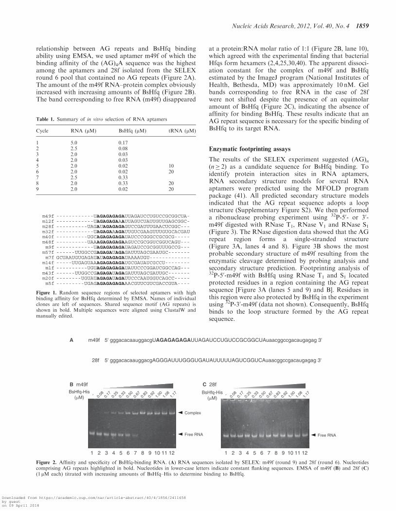
relationship between AG repeats and BsHfq bindingability using EMSA, we used aptamer m49f of which thebinding affinity of the (AG)4A sequence was the highestamong the aptamers and 28f isolated from the SELEXround 6 pool that contained no AG repeats (Figure 2A).The amount of the m49f RNA–protein complex obviouslyincreased with increasing amounts of BsHfq (Figure 2B).The band corresponding to free RNA (m49f) disappeared
at a protein:RNA molar ratio of 1:1 (Figure 2B, lane 10),which agreed with the experimental finding that bacterialHfqs form hexamers (2,4,25,30,40). The apparent dissoci-ation constant for the complex of m49f and BsHfqestimated by the ImageJ program (National Institutes ofHealth, Bethesda, MD) was approximately 10 nM. Gelbands corresponding to free RNA in the case of 28fwere not shifted despite the presence of an equimolaramount of BsHfq (Figure 2C), indicating the absence ofaffinity for binding BsHfq. These results indicate that anAG repeat sequence is necessary for the specific binding ofBsHfq to its target RNA.
Enzymatic footprinting assays
The results of the SELEX experiment suggested (AG)n(n� 2) as a candidate sequence for BsHfq binding. Toidentify protein interaction sites in RNA aptamers,RNA secondary structure models for several RNAaptamers were predicted using the MFOLD programpackage (41). All predicted secondary structure modelsindicated that the AG repeat sequence adopts a loopstructure (Supplementary Figure S2). We then performeda ribonuclease probing experiment using 32P-50- or 30-m49f digested with RNase T1, RNase V1 and RNase S1(Figure 3). The RNase digestion data showed that the AGrepeat region forms a single-stranded structure(Figure 3A, lanes 4 and 8). Figure 3B shows the mostprobable secondary structure of m49f resulting from theenzymatic cleavage determined by probing analysis andsecondary structure prediction. Footprinting analysis of32P-50-m49f with BsHfq using RNase T1 and S1 locatedprotected residues in a region containing the AG repeatsequence [Figure 3A (lanes 5 and 9) and B]. Residues inthis region were also protected by BsHfq in the experimentusing 32P-30-m49f (data not shown). Consequently, BsHfqbinds to the loop structure formed by the AG repeatsequence.
BsHfq-His(µM)
A 5' gggacacaauggacgUAGAGAGAGAUUAGAUCCUGUCCGCGGCUAuaacggccgacaugagag 3'
Figure 2. Affinity and specificity of BsHfq-binding RNA. (A) RNA sequences isolated by SELEX: m49f (round 9) and 28f (round 6). Nucleotidescomprising AG repeats highlighted in bold. Nucleotides in lower-case letters indicate constant flanking sequences. EMSA of m49f (B) and 28f (C)(1 mM each) titrated with increasing amounts of BsHfq–His to determine binding to BsHfq.
Figure 1. Random sequence regions of selected aptamers with highbinding affinity for BsHfq determined by EMSA. Names of individualclones are left of sequences. Shared sequence motif (AG repeats) isshown in bold. Multiple sequences were aligned using ClustalW andmanually edited.
Table 1. Summary of in vitro selection of RNA aptamers
We constructed mutant RNAs based on the sequence ofaptamer m49f (hereafter referred to as 49R, Figure 4A) todetermine the importance of residues in AG repeat se-quences for BsHfq binding. We initially examined theBsHfq-binding affinities of three mutants in which GAresidues were replaced with CC residues (named49R-mt1, 49R-mt2 and 49R-mt3; Figure 4A). MutantRNAs were labeled with 32P, exposed to short-wavelengthUV light in the presence of BsHfq and then analyzed bySDS–PAGE (Figure 4B). The intensity of bands corres-ponding to three 32P-labeled mutants (49R-mt1, 49R-mt2and 49R-mt3) was apparently reduced, comparedwith that of 49R that contains an (AG)4A sequence(Figure 4B, lanes 1–4). The EMSA results showed thatthe mutants cannot form stable complexes with BsHfqeven in the case of 49R-mt1 (data not shown). Thesefindings indicated that reducing the number of AGresidue repeats decreased their protein binding affinity.The sequence of aptamers obtained from the SELEX
experiment also indicated that G residues in the AGrepeat sequence can be replaced by either A or U
residues (Figure 1). The G residue at the sixth positionof the AG repeat sequence is in fact, frequently substitutedin this manner. To determine the nucleotide sequence spe-cificity for the binding of these aptamers by BsHfq, wegenerated four more mutants based on the 49Rsequence (bottom of Figure 4A); the G residue at thesixth position of the AG repeat sequence of 49R wasreplaced with U, A, C or GG residues and the mutantswere named 49R-U, 49R-A, 49R-C and 49R-GG, respect-ively. The results of UV cross-linking indicated thatmutant 49R-A and 49R formed similarly stable complexes(Figure 4B, lanes 5 and 7). However, the 32P signal inten-sity of 49R-U, 49R-C and 49R-GG was reduced,indicating that these mutants do not easily formcomplexes (Figure 4B, lanes 6, 8 and 9). The results with49R and mutant proteins in EMSA were the same (datanot shown). Thus, dinucleotide repeat motifs (AG) play animportant role in the efficiency of RNA aptamer bindingto BsHfq.
We constructed a series of RNA fragments by deletingresidues from the 30-end of the 9-mer 50-(AG)4A-30 andexamined interactions between the fragments and BsHfq
G
A
AG A
GAG
AUU
AG
G
GG
AC
ACA
AU G
GA
CG
U
AU
CC
UG
UC
C
GC
GG
CU
AU
A
A
CGG
CC
G
A
C
AU
GAGA
G
5’
3’
10
20
30
4050
60
C AH G- + - + - +
T1 V1 S1
(BsHfq-His)
60
50
40
30
20
10
A B
1 2 3 4 5 6 7 8 9
Res
idue
Num
ber
(nt)
T1
V1
S1
StrongWeakStrongWeakStrongWeak
Figure 3. Identification of BsHfq-binding site on RNA aptamer m49f. (A) Enzymatic footprinting of BsHfq bound to m49f. Cleavage products of32P-50- m49f modified by RNase T1, V1 and S1 with or without BsHfq-His were analyzed by denaturing gel electrophoresis. Lanes C, AH and Grepresent untreated RNA, alkaline hydrolysis ladder and G ladder, respectively. The vertical line on the right of the gel indicates protected residuesthat are resistant to RNase digestion in the presence of BsHfq-His. (B) Summarized results of enzymatic probing analysis of RNA aptamer m49f.Secondary structure was obtained by combining experimental results with MFOLD predictions (41). Major and minor cuts are indicated as symbols(see box). Residues corresponding to vertical line in (A) are highlighted in bold.
1860 Nucleic Acids Research, 2012, Vol. 40, No. 4
Downloaded from https://academic.oup.com/nar/article-abstract/40/4/1856/2411658by gueston 09 April 2018
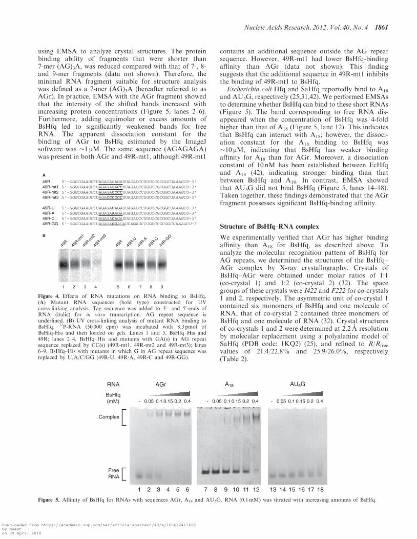
using EMSA to analyze crystal structures. The proteinbinding ability of fragments that were shorter than7-mer (AG)3A, was reduced compared with that of 7-, 8-and 9-mer fragments (data not shown). Therefore, theminimal RNA fragment suitable for structure analysiswas defined as a 7-mer (AG)3A (hereafter referred to asAGr). In practice, EMSA with the AGr fragment showedthat the intensity of the shifted bands increased withincreasing protein concentrations (Figure 5, lanes 2–6).Furthermore, adding equimolar or excess amounts ofBsHfq led to significantly weakened bands for freeRNA. The apparent dissociation constant for thebinding of AGr to BsHfq estimated by the ImageJsoftware was �1 mM. The same sequence (AGAGAGA)was present in both AGr and 49R-mt1, although 49R-mt1
contains an additional sequence outside the AG repeatsequence. However, 49R-mt1 had lower BsHfq-bindingaffinity than AGr (data not shown). This findingsuggests that the additional sequence in 49R-mt1 inhibitsthe binding of 49R-mt1 to BsHfq.Escherichia coli Hfq and SaHfq reportedly bind to A18
and AU5G, respectively (25,31,42). We performed EMSAsto determine whether BsHfq can bind to these short RNAs(Figure 5). The band corresponding to free RNA dis-appeared when the concentration of BsHfq was 4-foldhigher than that of A18 (Figure 5, lane 12). This indicatesthat BsHfq can interact with A18; however, the dissoci-ation constant for the A18 binding to BsHfq was�10 mM, indicating that BsHfq has weaker bindingaffinity for A18 than for AGr. Moreover, a dissociationconstant of 10 nM has been established between EcHfqand A18 (42), indicating stronger binding than thatbetween BsHfq and A18. In contrast, EMSA showedthat AU5G did not bind BsHfq (Figure 5, lanes 14–18).Taken together, these findings demonstrated that the AGrfragment possesses significant BsHfq-binding affinity.
Structure of BsHfq–RNA complex
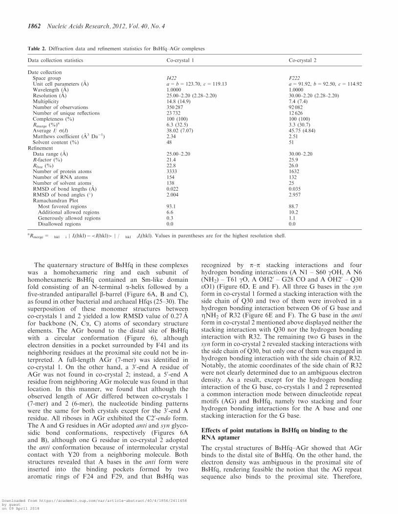
We experimentally verified that AGr has higher bindingaffinity than A18 for BsHfq, as described above. Toanalyze the molecular recognition pattern of BsHfq forAG repeats, we determined the structures of the BsHfq–AGr complex by X-ray crystallography. Crystals ofBsHfq–AGr were obtained under molar ratios of 1:1(co-crystal 1) and 1:2 (co-crystal 2) (32). The spacegroups of these crystals were I422 and F222 for co-crystals1 and 2, respectively. The asymmetric unit of co-crystal 1contained six monomers of BsHfq and one molecule ofRNA, that of co-crystal 2 contained three monomers ofBsHfq and one molecule of RNA (32). Crystal structuresof co-crystals 1 and 2 were determined at 2.2 A resolutionby molecular replacement using a polyalanine model ofSaHfq (PDB code: 1KQ2) (25), and refined to R/Rfree
values of 21.4/22.8% and 25.9/26.0%, respectively(Table 2).
Figure 4. Effects of RNA mutations on RNA binding to BsHfq.(A) Mutant RNA sequences (bold type) constructed for UVcross-linking analysis. Tag sequence was added to 30- and 50-ends ofRNA (italic) for in vitro transcription. AG repeat sequence isunderlined. (B) UV cross-linking analysis of mutant RNA binding toBsHfq. 32P-RNA (50 000 cpm) was incubated with 8.3 pmol ofBsHfq-His and then loaded on gels. Lanes 1 and 5, BsHfq–His and49R; lanes 2–4, BsHfq–His and mutants with GA(s) in AG repeatsequence replaced by CC(s) (49R-mt1, 49R-mt2 and 49R-mt3); lanes6–9, BsHfq–His with mutants in which G in AG repeat sequence wasreplaced by U/A/C/GG (49R-U, 49R-A, 49R-C and 49R-GG).
Nucleic Acids Research, 2012, Vol. 40, No. 4 1861
Downloaded from https://academic.oup.com/nar/article-abstract/40/4/1856/2411658by gueston 09 April 2018
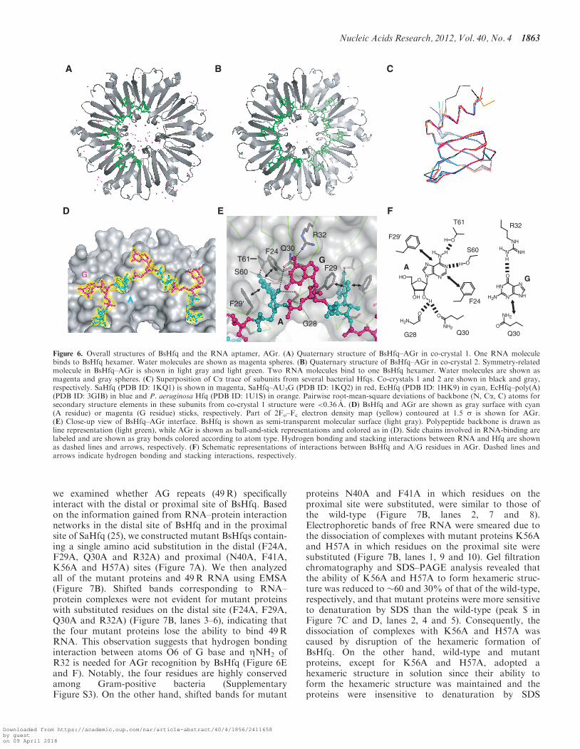
The quaternary structure of BsHfq in these complexeswas a homohexameric ring and each subunit ofhomohexameric BsHfq contained an Sm-like domainfold consisting of an N-terminal a-helix followed by afive-stranded antiparallel b-barrel (Figure 6A, B and C),as found in other bacterial and archaeal Hfqs (25–30). Thesuperposition of these monomer structures betweenco-crystals 1 and 2 yielded a low RMSD value of 0.27 Afor backbone (N, Ca, C) atoms of secondary structureelements. The AGr bound to the distal site of BsHfqwith a circular conformation (Figure 6), althoughelectron densities in a pocket surrounded by F41 and itsneighboring residues at the proximal site could not be in-terpreted. A full-length AGr (7-mer) was identified inco-crystal 1. On the other hand, a 30-end A residue ofAGr was not found in co-crystal 2; instead, a 50-end Aresidue from neighboring AGr molecule was found in thatlocation. In this manner, we found that although theobserved length of AGr differed between co-crystals 1(7-mer) and 2 (6-mer), the nucleotide binding patternswere the same for both crystals except for the 30-end Aresidue. All riboses in AGr exhibited the C20-endo form.The A and G residues in AGr adopted anti and syn glyco-sidic bond conformations, respectively (Figures 6Aand B), although one G residue in co-crystal 2 adoptedthe anti conformation because of intermolecular crystalcontact with Y20 from a neighboring molecule. Bothstructures revealed that A bases in the anti form wereinserted into the binding pockets formed by twoaromatic rings of F24 and F29, and that BsHfq was
recognized by p–p stacking interactions and fourhydrogen bonding interactions (A N1 – S60 gOH, A N6(NH2) – T61 gO, A OH20 – G28 CO and A OH20 – Q30eO1) (Figure 6D, E and F). All three G bases in the synform in co-crystal 1 formed a stacking interaction with theside chain of Q30 and two of them were involved in ahydrogen bonding interaction between O6 of G base andZNH2 of R32 (Figure 6E and F). The G base in the antiform in co-crystal 2 mentioned above displayed neither thestacking interaction with Q30 nor the hydrogen bondinginteraction with R32. The remaining two G bases in thesyn form in co-crystal 2 revealed stacking interactions withthe side chain of Q30, but only one of them was engaged inhydrogen bonding interaction with the side chain of R32.Notably, the atomic coordinates of the side chain of R32were not clearly determined due to an ambiguous electrondensity. As a result, except for the hydrogen bondinginteraction of the G base, co-crystals 1 and 2 representeda common interaction mode between dinucleotide repeatmotifs (AG) and BsHfq, namely two stacking and fourhydrogen bonding interactions for the A base and onestacking interaction for the G base.
Effects of point mutations in BsHfq on binding to theRNA aptamer
The crystal structures of BsHfq–AGr showed that AGrbinds to the distal site of BsHfq. On the other hand, theelectron density was ambiguous in the proximal site ofBsHfq, rendering feasible the notion that the AG repeatsequence also binds to the proximal site. Therefore,
Table 2. Diffraction data and refinement statistics for BsHfq–AGr complexes
Data collection statistics Co-crystal 1 Co-crystal 2
RefinementData range (A) 25.00–2.20 30.00–2.20R-factor (%) 21.4 25.9Rfree (%) 22.8 26.0Number of protein atoms 3333 1632Number of RNA atoms 154 132Number of solvent atoms 138 25RMSD of bond lengths (A) 0.022 0.035RMSD of bond angles (�) 2.004 2.957Ramachandran Plot
Most favored regions 93.1 88.7Additional allowed regions 6.6 10.2Generously allowed regions 0.3 1.1Disallowed regions 0.0 0.0
aRmerge=�hkl �i j Ii(hkl)�<I(hkl)> j / �hkl �iIi(hkl). Values in parentheses are for the highest resolution shell.
1862 Nucleic Acids Research, 2012, Vol. 40, No. 4
Downloaded from https://academic.oup.com/nar/article-abstract/40/4/1856/2411658by gueston 09 April 2018
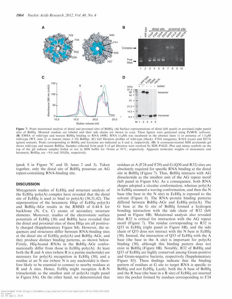
we examined whether AG repeats (49R) specificallyinteract with the distal or proximal site of BsHfq. Basedon the information gained from RNA–protein interactionnetworks in the distal site of BsHfq and in the proximalsite of SaHfq (25), we constructed mutant BsHfqs contain-ing a single amino acid substitution in the distal (F24A,F29A, Q30A and R32A) and proximal (N40A, F41A,K56A and H57A) sites (Figure 7A). We then analyzedall of the mutant proteins and 49R RNA using EMSA(Figure 7B). Shifted bands corresponding to RNA–protein complexes were not evident for mutant proteinswith substituted residues on the distal site (F24A, F29A,Q30A and R32A) (Figure 7B, lanes 3–6), indicating thatthe four mutant proteins lose the ability to bind 49RRNA. This observation suggests that hydrogen bondinginteraction between atoms O6 of G base and ZNH2 ofR32 is needed for AGr recognition by BsHfq (Figure 6Eand F). Notably, the four residues are highly conservedamong Gram-positive bacteria (SupplementaryFigure S3). On the other hand, shifted bands for mutant
proteins N40A and F41A in which residues on theproximal site were substituted, were similar to those ofthe wild-type (Figure 7B, lanes 2, 7 and 8).Electrophoretic bands of free RNA were smeared due tothe dissociation of complexes with mutant proteins K56Aand H57A in which residues on the proximal site weresubstituted (Figure 7B, lanes 1, 9 and 10). Gel filtrationchromatography and SDS–PAGE analysis revealed thatthe ability of K56A and H57A to form hexameric struc-ture was reduced to �60 and 30% of that of the wild-type,respectively, and that mutant proteins were more sensitiveto denaturation by SDS than the wild-type (peak $ inFigure 7C and D, lanes 2, 4 and 5). Consequently, thedissociation of complexes with K56A and H57A wascaused by disruption of the hexameric formation ofBsHfq. On the other hand, wild-type and mutantproteins, except for K56A and H57A, adopted ahexameric structure in solution since their ability toform the hexameric structure was maintained and theproteins were insensitive to denaturation by SDS
AA
G
A B C
D FT61
S60
F24
Q30G28
F29'
AN
OOH
OH
N
NN
N
O
HH
H
OH
OH
ONH2
O
NH2
G
Q30
R32
N
NH
NH
N
NH2
O
O
NH2
NH
NH
NH
H
R32
G28
F29'
F29
F24 Q30T61
S60
A
G
E
Figure 6. Overall structures of BsHfq and the RNA aptamer, AGr. (A) Quaternary structure of BsHfq–AGr in co-crystal 1. One RNA moleculebinds to BsHfq hexamer. Water molecules are shown as magenta spheres. (B) Quaternary structure of BsHfq–AGr in co-crystal 2. Symmetry-relatedmolecule in BsHfq–AGr is shown in light gray and light green. Two RNA molecules bind to one BsHfq hexamer. Water molecules are shown asmagenta and gray spheres. (C) Superposition of Ca trace of subunits from several bacterial Hfqs. Co-crystals 1 and 2 are shown in black and gray,respectively. SaHfq (PDB ID: 1KQ1) is shown in magenta, SaHfq–AU5G (PDB ID: 1KQ2) in red, EcHfq (PDB ID: 1HK9) in cyan, EcHfq–poly(A)(PDB ID: 3GIB) in blue and P. aeruginosa Hfq (PDB ID: 1U1S) in orange. Pairwise root-mean-square deviations of backbone (N, Ca, C) atoms forsecondary structure elements in these subunits from co-crystal 1 structure were <0.36 A. (D) BsHfq and AGr are shown as gray surface with cyan(A residue) or magenta (G residue) sticks, respectively. Part of 2Fo–Fc electron density map (yellow) contoured at 1.5 s is shown for AGr.(E) Close-up view of BsHfq–AGr interface. BsHfq is shown as semi-transparent molecular surface (light gray). Polypeptide backbone is drawn asline representation (light green), while AGr is shown as ball-and-stick representations and colored as in (D). Side chains involved in RNA-binding arelabeled and are shown as gray bonds colored according to atom type. Hydrogen bonding and stacking interactions between RNA and Hfq are shownas dashed lines and arrows, respectively. (F) Schematic representations of interactions between BsHfq and A/G residues in AGr. Dashed lines andarrows indicate hydrogen bonding and stacking interactions, respectively.
Nucleic Acids Research, 2012, Vol. 40, No. 4 1863
Downloaded from https://academic.oup.com/nar/article-abstract/40/4/1856/2411658by gueston 09 April 2018
(peak $ in Figure 7C and D, lanes 2 and 3). Takentogether, only the distal site of BsHfq possesses an AGrepeat-containing RNA-binding site.
DISCUSSION
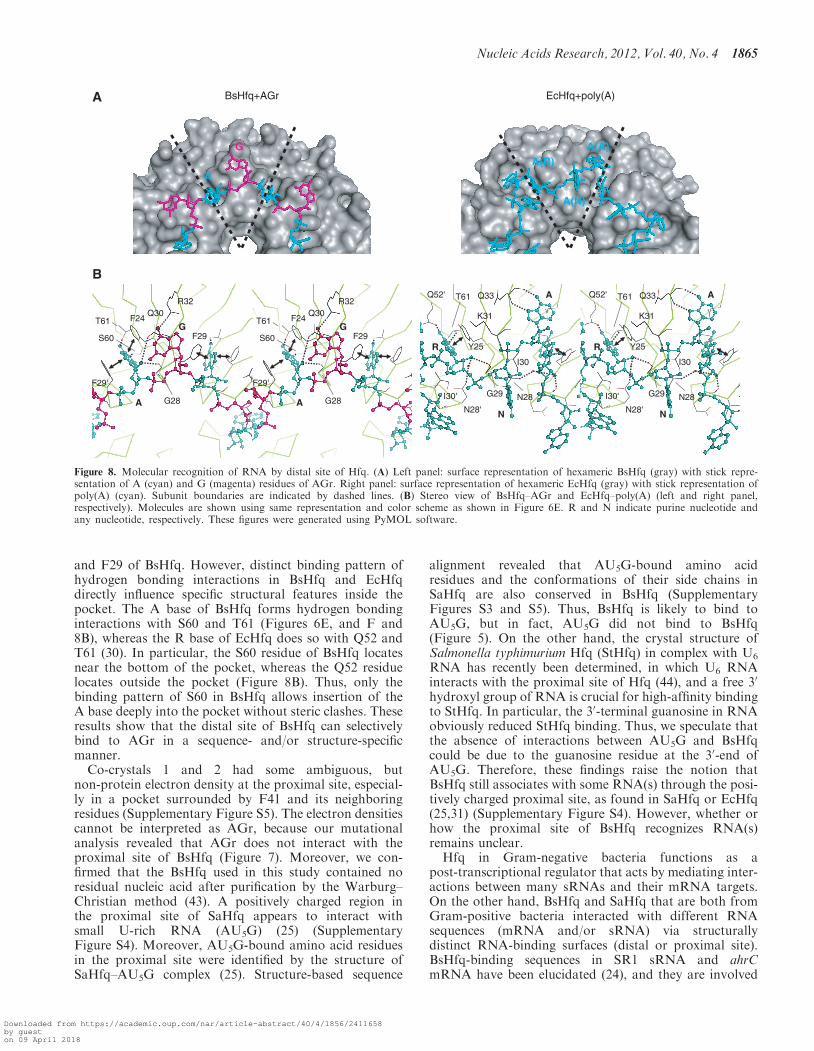
Mutagenesis studies of EcHfq and structure analysis ofthe EcHfq–poly(A) complex have revealed that the distalsite of EcHfq is used to bind to poly(A) (30,31,42). Thesuperposition of the hexameric Hfqs of EcHfq–poly(A)and BsHfq–AGr results in the RMSD of 0.44 A forbackbone (N, Ca, C) atoms of secondary structureelements. Moreover, studies of the electrostatic surfacepotentials of EcHfq (30) and BsHfq have revealed thatthe distal and proximal sites of these Hfqs are all positive-ly charged (Supplementary Figure S4). However, the se-quences and structures differ between RNA-binding sitesat the distal site of EcHfq–poly(A) and BsHfq–AGr, andthey produce distinct binding patterns, as shown below.Firstly, Hfq-bound RNAs in the BsHfq–AGr confor-mationally differ from those in EcHfq–poly(A). At leastboth the R and A sites (where R is a purine nucleotide) arenecessary for poly(A) recognition in EcHfq (30), and aresidue at an N site (where N is any nucleotide) is there-fore likely to be essential to connect to nearby residues atR and A sites. Hence, EcHfq might recognize A-R-Ntrinucleotide as the smallest unit of poly(A) (right panelin Figure 8A). On the other hand, we demonstrated that
residues at A (F24 and F29) and G (Q30 and R32) sites areabsolutely required for specific RNA binding at the distalsite in BsHfq (Figure 7). Thus, BsHfq interacts with AGdinucleotide as the smallest unit of the AG repeat motif(left panel in Figure 8A). As a consequence, both RNAshapes adopted a circular conformation, whereas poly(A)in EcHfq assumed a waving conformation, and then the Nbase (the base in the N site) in EcHfq is exposed to thesolvent (Figure 8). The RNA–protein binding patternsdiffered between BsHfq–AGr and EcHfq–poly(A). TheG base at the G site of BsHfq formed a hydrogenbonding interaction with the side chain of R32 (leftpanel in Figure 8B). Mutational analysis also revealedthat R32 is critical for interaction with the AG repeatmotif (Figure 7). The residue corresponding to R32 isQ33 in EcHfq (right panel in Figure 8B), and the sidechain of Q33 does not interact with the N base in EcHfq(30). Instead, the interaction of Q33 of EcHfq with the Abase (the base in the A site) is important for poly(A)binding (30), although this binding pattern does notexist in BsHfq (Figure 8B). Notably, R32 of BsHfq andQ33 of EcHfq are highly conserved among Gram-positiveand Gram-negative bacteria, respectively (SupplementaryFigure S3). These findings indicate that the bindingpattern of residues at G site to target RNA is specific toBsHfq and not EcHfq. Lastly, both the A base of BsHfqand the R base (the base in a R site) of EcHfq are insertedinto the pocket formed by residues corresponding to F24
F24
F29
Q30
R32
N40
F41
H57
K56
A B
C D
F24A
F29A
Q30
AR32
AN40
AF4
1A
H57A
- K56A
FreeRNA
Complex
1 2 3 4 5 6 7 8 9 10
distal site proximal site
$ #
wild
type
0
50
100
150
10.0 15.0 20.0 25.0
Elution volume (ml)
Abs
orba
nce
280
nm (
mA
U)
boiled-+ - - -wild
type
K56A
F29A
H57A
hexamer
monomer
31
21.5
14.5
6.5
4566
97.4
M 1 2 3 4 5
(kDa)
Figure 7. Point mutational analysis of distal and proximal sites of BsHfq. (A) Surface representations of distal (left panel) or proximal (right panel)sites of BsHfq. Mutated residues are labeled and their side chains are shown in cyan. These figures were generated using PyMOL software.(B) EMSA of wild-type and mutant BsHfq binding to RNA (49R). RNA (1 mM) was incubated in the absence (lane 1) or presence of 1.5 mMwild-type (WT, lane 2) or mutant (lanes 3–10) BsHfqs. (C) Gel filtration profiles of wild-type (black), F29A (magenta), K56A (cyan) and H57A(green) mutants. Peaks corresponding to BsHfq and lysozyme are indicated as $ and #, respectively. (D) A coomassie-stained SDS acrylamide gelshows wild-type and mutant BsHfqs. Samples collected from peak $ of gel filtration were resolved by SDS–PAGE. Plus and minus symbols on thetop of the gel indicate samples boiled or not in SDS buffer for 10min at 95�C, respectively. Apparent molecular weights of monomeric andhexameric BsHfqs are �9.6 and 58 kDa, respectively.
1864 Nucleic Acids Research, 2012, Vol. 40, No. 4
Downloaded from https://academic.oup.com/nar/article-abstract/40/4/1856/2411658by gueston 09 April 2018
and F29 of BsHfq. However, distinct binding pattern ofhydrogen bonding interactions in BsHfq and EcHfqdirectly influence specific structural features inside thepocket. The A base of BsHfq forms hydrogen bondinginteractions with S60 and T61 (Figures 6E, and F and8B), whereas the R base of EcHfq does so with Q52 andT61 (30). In particular, the S60 residue of BsHfq locatesnear the bottom of the pocket, whereas the Q52 residuelocates outside the pocket (Figure 8B). Thus, only thebinding pattern of S60 in BsHfq allows insertion of theA base deeply into the pocket without steric clashes. Theseresults show that the distal site of BsHfq can selectivelybind to AGr in a sequence- and/or structure-specificmanner.
Co-crystals 1 and 2 had some ambiguous, butnon-protein electron density at the proximal site, especial-ly in a pocket surrounded by F41 and its neighboringresidues (Supplementary Figure S5). The electron densitiescannot be interpreted as AGr, because our mutationalanalysis revealed that AGr does not interact with theproximal site of BsHfq (Figure 7). Moreover, we con-firmed that the BsHfq used in this study contained noresidual nucleic acid after purification by the Warburg–Christian method (43). A positively charged region inthe proximal site of SaHfq appears to interact withsmall U-rich RNA (AU5G) (25) (SupplementaryFigure S4). Moreover, AU5G-bound amino acid residuesin the proximal site were identified by the structure ofSaHfq–AU5G complex (25). Structure-based sequence
alignment revealed that AU5G-bound amino acidresidues and the conformations of their side chains inSaHfq are also conserved in BsHfq (SupplementaryFigures S3 and S5). Thus, BsHfq is likely to bind toAU5G, but in fact, AU5G did not bind to BsHfq(Figure 5). On the other hand, the crystal structure ofSalmonella typhimurium Hfq (StHfq) in complex with U6
RNA has recently been determined, in which U6 RNAinteracts with the proximal site of Hfq (44), and a free 30
hydroxyl group of RNA is crucial for high-affinity bindingto StHfq. In particular, the 30-terminal guanosine in RNAobviously reduced StHfq binding. Thus, we speculate thatthe absence of interactions between AU5G and BsHfqcould be due to the guanosine residue at the 30-end ofAU5G. Therefore, these findings raise the notion thatBsHfq still associates with some RNA(s) through the posi-tively charged proximal site, as found in SaHfq or EcHfq(25,31) (Supplementary Figure S4). However, whether orhow the proximal site of BsHfq recognizes RNA(s)remains unclear.Hfq in Gram-negative bacteria functions as a
post-transcriptional regulator that acts by mediating inter-actions between many sRNAs and their mRNA targets.On the other hand, BsHfq and SaHfq that are both fromGram-positive bacteria interacted with different RNAsequences (mRNA and/or sRNA) via structurallydistinct RNA-binding surfaces (distal or proximal site).BsHfq-binding sequences in SR1 sRNA and ahrCmRNA have been elucidated (24), and they are involved
A(R)
A(N)
A(A)
A
G
BsHfq+AGr EcHfq+poly(A)A
B
Q33
K31
Q52' T61
Y25
N28'
I30
G29I30'
A
N
R
N28
Q33
K31
Q52' T61
Y25
N28'
I30
G29I30'
A
N
R
N28
R32
G28
F29'
F29
F24Q30
T61
S60
A
G
R32
G28
F29'
F29
F24Q30
T61
S60
A
G
Figure 8. Molecular recognition of RNA by distal site of Hfq. (A) Left panel: surface representation of hexameric BsHfq (gray) with stick repre-sentation of A (cyan) and G (magenta) residues of AGr. Right panel: surface representation of hexameric EcHfq (gray) with stick representation ofpoly(A) (cyan). Subunit boundaries are indicated by dashed lines. (B) Stereo view of BsHfq–AGr and EcHfq–poly(A) (left and right panel,respectively). Molecules are shown using same representation and color scheme as shown in Figure 6E. R and N indicate purine nucleotide andany nucleotide, respectively. These figures were generated using PyMOL software.
Nucleic Acids Research, 2012, Vol. 40, No. 4 1865
Downloaded from https://academic.oup.com/nar/article-abstract/40/4/1856/2411658by gueston 09 April 2018
in short AG repeats GAAUAAGAGA (SR1 sRNA) or AAAUAGAG (ahrCmRNA). Therefore, these results whencombined with the findings of the SELEX experimentsindicated that the target sequence for BsHfq certainlycomprises AG repeats. Taken together with these data,the results presented herein suggest that SR1 and ahrCmRNA recognize the same distal site of BsHfq. Wefurther examined whether the intergenic regions ofB. subtilis 168 genome include the AGr sequence usingthe SubtiList database (http://genolist.pasteur.fr/SubtiList/). We identified the sequence in 110 intergenicregions and in 215 protein coding regions, indicating thatthe AGr sequence does in fact exists in the B. subtilisgenome.Amino acid residues on the surface of the distal
site, including those that bind to AGr in BsHfq, arehighly conserved in Gram-positive bacteria such asS. aureus and L. monocytogenes (SupplementaryFigure S3). Moreover, lmo0850 mRNA, which binds toL. monocytogenes Hfq, also possesses AG repeats (45).Therefore, we speculate that several other Hfqs mightfunction through interaction with the AG repeat motifamong Gram-positive bacteria. To date, various RNAtarget sequences have been identified and they can dis-criminate the RNA-binding surface(s) of hexameric Hfqat the atomic level. Furthermore, multiple functional rolesof Hfq have been identified in several bacterial species,probably as a consequence of this. Hence, bacterial Hfqsmight have species-specific functions among bacterialphyla. To understand Hfq activities, recognition patternsbetween RNA(s) and Hfq across various biological speciesshould be further elucidated in future studies from bothstructural and functional perspectives.
ACCESSION NUMBERS
Coordinates and structure factors for the co-crystal 1 andco-crystal 2 of BsHfq–AGr have been deposited with theProtein Data Back under accession codes 3HSB and3AHU.
SUPPLEMENTARY DATA
Supplementary Data are available at NAR online:Supplementary Table 1, Supplementary Figures 1–5.
ACKNOWLEDGEMENTS
We thank Ms S. Tamura, S. Suzuki, and A. Shino forexcellent technical assistance and the staff of SPring-8BL38B1 (2008B1506 and 2011A2043) for help with col-lecting the X-ray data. We also thank Ms N. Foster forcritical reading of the manuscript.
FUNDING
Funding for open access charge: University of Tsukuba.
Conflict of interest statement. None declared.
REFERENCES
1. Franze de Fernandez,M.T., Hayward,W.S. and August,J.T. (1972)Bacterial proteins required for replication of phage Qb ribonucleicacid. Purification and properties of host factor I, a ribonucleicacid-binding protein. J. Biol. Chem., 247, 824–831.
2. Zhang,A., Wassarman,K.M., Ortega,J., Steven,A.C. and Storz,G.(2002) The Sm-like Hfq protein increases OxyS RNA interactionwith target mRNAs. Mol. Cell, 9, 11–22.
3. Sledjeski,D.D., Whitman,C. and Zhang,A. (2001) Hfq is necessaryfor regulation by the untranslated RNA DsrA. J. Bacteriol., 148,1997–2005.
4. Møller,T., Franch,T., Højrup,P., Keene,D.R., Bachinger,H.P.,Brennan,R.G. and Valentin-Hansen,P. (2002) Hfq: a bacterialSm-like protein that mediates RNA-RNA interaction. Mol. Cell,9, 23–30.
5. Geissmann,T.A. and Touati,D. (2004) Hfq, a new chaperoningrole: binding to messenger RNA determines access for smallRNA regulator. EMBO J., 23, 396–405.
6. Morita,T., Maki,K. and Aiba,H. (2005) RNase E-basedribonucleoprotein complexes: mechanical basis of mRNAdestabilization mediated by bacterial noncoding RNAs.Genes Dev., 19, 2176–2186.
7. Moll,I., Leitsch,D., Steinhauser,T. and Blasi,U. (2003) RNAchaperone activity of the Sm-like Hfq protein. EMBO Rep., 4,284–289.
8. Vecerek,B., Moll,I. and Blasi,U. (2005) Translational autocontrolof the Escherichia coli hfq RNA chaperone gene. RNA, 11,976–984.
9. Hajnsdorf,E. and Regnier,P. (2000) Host factor Hfq ofEscherichia coli stimulates elongation of poly(A) tails by poly(A)polymerase I. Proc. Natl Acad. Sci. USA, 97, 1501–1505.
10. Mohanty,B.K., Maples,V.F. and Kushner,S.R. (2004) The Sm-likeprotein Hfq regulates polyadenylation dependent mRNA decay inEscherichia coli. Mol. Microbiol., 54, 905–920.
11. Folichon,M., Allemand,F., Regnier,P. and Hajnsdorf,E. (2005)Stimulation of poly(A) synthesis by Escherichia coli poly(A)polymerase I is correlated with Hfq binding to poly(A) tails.FEBS J., 272, 454–463.
12. Folichon,M., Arluison,V., Pellegrini,O., Huntzinger,E., Regnier,P.and Hajnsdorf,E. (2003) The poly(A) binding protein Hfqprotects RNA from RNase E and exoribonucleolytic degradation.Nucleic Acids Res., 31, 7302–7310.
13. Afonyushkin,T., Vecerek,B., Moll,I., Blasi,U. and Kaberdin,V.R.(2005) Both RNase E and RNase III control the stability of sodBmRNA upon translational inhibition by the small regulatoryRNA RyhB. Nucleic Acids Res., 33, 1678–1689.
14. Chao,Y. and Vogel,J. (2010) The role of Hfq in bacterialpathogens. Curr. Opin. Microbiol., 13, 24–33.
15. Tsui,H.-C.T., Leung,H.C.E. and Winkler,M.E. (1994)Characterization of broadly pleiotropic phenotypes caused by anhfq insertion mutation in Escherichia coli K-12. Mol. Microbiol.,13, 35–49.
16. Sonnleitner,E., Hagens,S., Rosenau,F., Wilhelm,S., Habel,A.,Jager,K.E. and Blasi,U. (2003) Reduced virulence of a hfqmutant of Pseudomonas aeruginosa O1. Microb. Pathog., 35,217–228.
17. Pannekoek,Y., Huis in’t Veld,R., Hopman,C.Th P.,Langerak,A.A.J., Speijer,D. and van der Ende,A. (2009)Molecular characterization and identification of proteins regulatedby Hfq in Neisseria meningitidis. FEMS Microbiol. Lett., 294,216–224.
18. Ding,Y., Davis,B.M. and Waldor,M.K. (2004) Hfq is essential forVibrio cholerae virulence and downregulates sigma expression.Mol. Microbiol., 53, 345–354.
19. Bohn,C., Rigoulay,C. and Bouloc,P. (2007) No detectable effectof RNA-binding protein Hfq absence in Staphylococcus aureus.BMC Microbiol., 7, 10.
20. Christiansen,J.K., Larsen,M.H., Ingmer,H., Søgaard-Andersen,L.and Kallipolitis,B.H. (2004) The RNA-binding protein Hfq ofListeria monocytogenes: Role in stress tolerance and virulence.J. Bacteriol., 186, 3355–3362.
21. Christiansen,J.K., Nielsen,J.S., Ebersbach,T., Valentin-Hansen,P.,Søgaard-Andersen,L. and Kallipolitis,B.H. (2006) Identification of
1866 Nucleic Acids Research, 2012, Vol. 40, No. 4
Downloaded from https://academic.oup.com/nar/article-abstract/40/4/1856/2411658by gueston 09 April 2018
small Hfq-binding RNAs in Listeria monocytogenes. RNA, 12,1383–1396.
22. Silvaggi,J.M., Perkins,J.B. and Losick,R. (2005) SmallUntranslated RNA antitoxin in Bacillus subtilis. J. Bacteriol., 187,6641–6650.
23. Heidrich,N., Chinali,A., Gerth,U. and Brantl,S. (2006) The smalluntranslated RNA SR1 from the Bacillus subtilis genome isinvolved in the regulation of arginine catabolism. Mol. Microbiol.,62, 520–536.
24. Heidrich,N., Moll,I. and Brantl,S. (2007) In vitro analysis of theinteraction between the small RNA SR1 and its primary targetahrC mRNA. Nucleic Acids Res., 35, 4331–4346.
25. Schumacher,M.A., Pearson,R.F., Møller,T., Valentin-Hansen,P.and Brennan,R.G. (2002) Structures of the pleiotropictranslational regulator Hfq and an Hfq-RNA complex: a bacterialSm-like protein. EMBO J., 21, 3546–3556.
26. Sauter,C., Basquin,J. and Suck,D. (2003) Sm-like proteins inEubacteria: the crystal structure of the Hfq protein fromEscherichia coli. Nucleic Acids Res., 31, 4091–4098.
27. Nikulin,A., Stolboushkina,E., Perederina,A., Vassilieva,I.,Blaesi,U., Moll,I., Kachalova,G., Yokoyama,S., Vassylyev,D.,Garber,M. et al. (2005) Structure of Pseudomonas aeruginosa Hfqprotein. Acta. Crystallogr. D Biol. Crystallogr., 61, 141–146.
28. Nielsen,J.S., Bøggild,A., Andersen,C.B., Nielsen,G., Boysen,A.,Brodersen,D.E. and Valentin-Hansen,P. (2007) An Hfq-likeprotein in archaea: crystal structure and functionalcharacterization of the Sm protein from Methanococcus jannaschii.RNA, 13, 2213–2223.
29. Boggild,A., Overgaard,M., Valentin-Hansen,P. andBrodersen,D.E. (2009) Cyanobacteria contain a structuralhomologue of the Hfq protein with altered RNA-bindingproperties. FEBS J., 276, 3904–3915.
30. Link,T.M., Valentin-Hansen,P. and Brennan,R.G. (2009)Structure of Escherichia coli Hfq bound to polyriboadenylateRNA. Proc. Natl Acad. Sci. USA, 106, 19292–19297.
31. Mikulecky,P.J., Kaw,M.K., Brescia,C.C., Takach,J.C.,Sledjeski,D.D. and Feig,A.L. (2004) Escherichia coli Hfq hasdistinct interaction surfaces for DsrA, rpoS and poly(A) RNAs.Nat. Struct. Mol. Biol., 11, 1206–1214.
32. Baba,S., Someya,T., Kawai,G., Nakamura,K. and Kumasaka,T.(2010) Expression, crystallization and preliminary crystallographicanalysis of RNA-binding protein Hfq (YmaH) from Bacillussubtilis in complex with an RNA aptamer. Acta. Crystallogr. Sect.F Struct. Biol. Cryst. Commun., 66, 563–566.
33. Conrad,R.C., Giver,L., Tian,Y. and Ellington,A.D. (1996) In vitroselection of Nucleic Acid Aptamer that bind proteins. MethodsEnzymol., 267, 336–367.
34. Vagin,A. and Teplyakov,A. (1997) MOLREP: an automatedprogram for molecular replacement. J. Appl. Cryst., 30,1022–1025.
35. Collaborative Computational Project, Number 4. (1994) TheCCP4 suite: programs for protein crystallography. Acta.Crystallogr. D Biol. Crystallogr., 50, 760–763.
36. Brunger,A.T., Adams,P.D., Clore,G.M., DeLano,W.L., Gros,P.,Grosse-Kunstleve,R.W., Jiang,J.S., Kuszewski,J., Nilges,M.,Pannu,N.S. et al. (1998) Crystallography & NMR system: A newsoftware suite for macromolecular structure determination. Acta.Crystallogr. D Biol. Crystallogr., 54, 905–921.
37. McRee,D.E. (1999) XtalView/Xfit—A versatile program formanipulating atomic coordinates and electron density. J. Struct.Biol., 125, 156–165.
38. Emsley,P. and Cowtan,K. (2004) Coot: Model-building tools formolecular graphics. Acta. Crystallogr. D Biol. Crystallogr., 60,2126–2132.
39. Laskowski,R.A., Macarthur,M.W., Moss,D.S. and Thornton,J.M.(1993) Procheck – a program to check the stereochemical qualityof protein structures. J. Appl. Crystallogr., 26, 283–291.
40. Fender,A., Elf,J., Hampel,K., Zimmermann,B. and Wagner,E.G.(2010) RNAs actively cycle on the Sm-like protein Hfq. GenesDev., 24, 2621–2626.
41. Zuker,M. (2003) Mfold web server for nucleic acid folding andhybridization prediction. Nucleic Acids Res., 31, 3406–3415.
42. Sun,X. and Wartell,P.M. (2006) Escherichia coli Hfq binds A18
and DsrA domain II with similar 2:1 Hfq6/RNA stoichiometryusing different surface site. Biochemistry, 45, 4875–4887.
43. Stoscheck,C.M. (1990) Quantitation of protein. MethodsEnzymol., 182, 50–68.
44. Sauer,E. and Weichenrieder,O. (2011) Structural basis for RNA30-end recognition by Hfq. Proc. Natl Acad. Sci. USA, 108,13065–13070.
45. Nielsen,J.S., Lei,L.K., Ebersbach,T., Olsen,A.S., Klitgaard,J.K.,Valentin-Hansen,P. and Kallipolitis,B.H. (2010) Defining a rolefor Hfq in Gram-positive bacteria: evidence for Hfq-dependentantisense regulation in Listeria monocytogenes. Nucleic Acids Res.,38, 907–919.
Nucleic Acids Research, 2012, Vol. 40, No. 4 1867
Downloaded from https://academic.oup.com/nar/article-abstract/40/4/1856/2411658by gueston 09 April 2018