Mark Cushman,⊥ Markus Fischer,| and Rudolf Ladenstein§
Karolinska Institutet, NOVUM, Centre for Structural Biochemistry, S-14157 Huddinge, Sweden, Lehrstuhl fu¨r OrganischeChemie und Biochemie, Technische UniVersitat Munchen, Lichtenbergstrasse 4, D-85747 Garching, Germany, andDepartment of Medicinal Chemistry and Molecular Pharmacology, School of Pharmacy and Pharmacal Sciences,
Purdue UniVersity, West Lafayette, Indiana 47907
ReceiVed October 6, 2004; ReVised Manuscript ReceiVed NoVember 30, 2004
ABSTRACT: The enzymes involved in the biosynthesis of riboflavin represent attractive targets for thedevelopment of drugs against bacterial pathogens, because the inhibitors of these enzymes are not likelyto interfere with enzymes of the mammalian metabolism. Lumazine synthase catalyzes the penultimatestep in the riboflavin biosynthesis pathway. A number of substituted purinetrione compounds represent anew class of highly specific inhibitors of lumazine synthase fromMycobacterium tuberculosis.To developpotent antibiotics for the treatment of tuberculosis, we have determined the structure of lumazine synthasefrom M. tuberculosisin complex with two purinetrione inhibitors and have studied binding via isothermaltitration calorimetry. The structures were determined by molecular replacement using lumazine synthasefrom Saccharomyces cereVisiae as a search model and refined at 2 and 2.3 Å resolution. TheR-factorswere 14.7 and 17.4%, respectively, and theRfree values were 19.3 and 26.3%, respectively. The enzymewas found to be a pentamer consisting of five subunits related by 5-fold local symmetry. The comparisonof the active site architecture with the active site of previously determined lumazine synthase structuresreveals a largely conserved topology with the exception of residues Gln141 and Glu136, which participatein different charge-charge interactions in the core space of the active site. The impact of structural changesin the active site on the altered binding and catalytic properties of the enzyme is discussed. Isothermaltitration calorimetry measurements indicate highly specific binding of the purinetrione inhibitors to theM. tuberculosisenzyme with dissociation constants in micromolar range.
The prevalence of multiple-drug resistance in pathogenicmicroorganisms is at a steadily increasing rate becoming amajor threat to human health around the world. An alarmingexample is the worldwide increasing multidrug resistanceof Mycobacterium tuberculosis, a human pathogen, whichis responsible for the death of millions of people every year.This disease has become such a public health threat that theWorld Health Organization has declared a public healthemergency (1). The increasing incidence of antibioticresistance has brought a new sense of urgency to thediscovery and development of antibacterial drugs. Theantibiotics, however, that are currently available attack onlya handful of targets. As a consequence, the development ofdrugs that act on new targets is urgently needed.
Among the known strategies for designing new antibiotics,cofactor/coenzyme biosynthesis offers several importantadvantages for drug development. Most of the knownmicroorganisms are strictly dependent on endogenous bio-synthesis of cofactors, because they lack specific transportproteins and are therefore devoid of efficient uptake systems(2). The enzymes involved in cofactor biosynthesis pathwaysrepresent very specific targets for antibacterial growthinhibition, because these enzymes are usually not present inthe human or animal host.
Recent genomic studies have suggested that the riboflavinbiosynthesis pathway is essential for bothM. tuberculosisandMycobacterium leprae(3). The development of inhibitorsagainst enzymes involved in riboflavin (vitamin B2) biosyn-thesis opens a novel approach for the chemotherapy ofmycobacterial infections. Notably, flavoenzymes, which usederivatives of riboflavin as their coenzyme, are indispensablefor electron transport reactions in all cellular organisms. Theenzyme targets of our interest are in the first line lumazinesynthase and riboflavin synthase because those enzymes arewell characterized in terms of structure and mechanism. Itshould be noted that compounds interfering with the bio-synthesis of folic acid, a vitamin characterized by many
† This work was supported by the Swedish Research Council(Vetenskapsrådet) (Project 621-2001-3195).
‡ The atomic coordinates and structure factors of MbtLS-TS-44 andMbtLS-TS-70 complexes have been deposited in the Protein Data Bank(entries 1W19 and 1W29, respectively).
* To whom correspondence should be addressed. E-mail:[email protected]. Telephone:+46-8-6089290. Fax:
structural and biosynthetic similarities with riboflavin, havea long and highly successful history as chemotherapeuticagents.
Lumazine synthase (LS)1 and riboflavin synthase (RS) aretwo enzymes in the riboflavin biosynthesis pathway, cata-lyzing the formation of 6,7-dimethyl-8-(D-ribityl)lumazineand riboflavin, respectively. LS catalyzes the penultimate stepof riboflavin biosynthesis, namely, the formation of 6,7-dimethyl-8-(D-ribityl)lumazine (3) from 3,4-dihydroxy-2-butanone 4-phosphate (2) and 5-amino-6-ribitylamino-2,4-(1H,3H)-pyrimidinedione (1) (Figure 1). Sequences oflumazine synthases from different organisms are 30-48%similar, and these enzymes exist in at least two oligomer-ization states (4, 5). The enzymes fromBacillus subtilis,Aquifex aeolicus, Escherichia coli, and Spinacia oleraceaform icosahedral capsids that consist of 60 identical subunitswhich can alternatively be described as dodecamers ofpentamers.B. subtilisalso forms a 1 MDa enzyme complexconsisting of the LS capsid and three RS subunits enclosedin the central core. Several structures of empty capsidsobtained by recombinant DNA technology have been deter-mined in native form as well as in complex with inhibitorsor substrate analogues (6-10). LS from SaccharomycescereVisiae, Schizosaccharomyces pombe, Brucella abortus,Magnaporthe grisea, and M. tuberculosisassemble intostable homopentamers with a topology very similar to thatof the pentameric modules in the icosahedral enzymes (7,11-13). In all LS structures, the topologically equivalent
active sites are located at the interfaces between adjacentsubunits in the pentamer. By comparing inhibitor bindingdata and considering structural constraints implied by thegeometry of the active site, Zhang et al. (9) have suggesteda catalytic mechanism which represents an extended andrefined version of the mechanism proposed previously byKis et al. (14). This mechanism consisted of the followingsteps: substrate binding, nucleophilic attack, formation of aSchiff base intermediate, phosphate elimination, and ringclosure. The involvement of conserved key residues, i.e.,Phe22, Arg127, and His88, and the Lys135/Glu138 coupleof LS from A. aeolicushas been discussed on the basis ofstructural proximity and activity measurements on mutantsof the highly homologous LS fromB. subtilis(9).
Our work on the structure-function relationships ofriboflavin biosynthetic enzymes as well as the potential ofriboflavin biosynthesis as a target for antibacterial therapyhas resulted in cooperative efforts concerning the design andsynthesis of a number of inhibitors of LS and RS (15-18).At present, the kinetic inhibition constants and experimentallydetermined structures of complexes with several substrateanalogue inhibitors, such as 5-nitroso-6-ribitylamino-2,4-(1H,3H)-pyrimidinedione (7, 9), 5-nitro-6-ribitylamino-2,4-(1H,3H)-pyrimidinedione (8, 12), and 5-[6-D-ribitylamino-2,4(1H,3H)-pyrimidinedione-5-yl]-1-pentylphosphonic acid(9, 13), and also of the product analogues 6,7-dioxo-5H-8-ribitylaminolumazine and 3,7-hydroxy-8-ribityllumazine (9)are available. To obtain inhibitors of potential value asantibiotics, a series of ribitylpurinetriones bearing an alkylphosphate group were recently designed and synthesized(19). These substances mimic a reaction intermediate of LSthat binds to the active site. The compounds were found tobe rather effective inhibitors for bothB. subtilisLS andE.coli RS. Surprisingly, those purinetrione compounds showedthe highest affinity for LS fromM. tuberculosis(MbtLS)with kinetic inhibition constants in the nanomolar range (19).It is obvious that the inhibition effect of the ribitylpurinetri-one phosphates is strongly dependent on the length of thealiphatic linker between the purinetrione ring system and thephosphate moiety.
To study in detail the binding of the potential MbtLSinhibitors with the future aim of developing antibacterialdrugs directed againstMycobacterium, we have investigatedtwo complexes of MbtLS with 3-(1,3,7,9-tetrahydro-9-D-ribityl-2,6,8-trioxopurin-7-yl)-1-propane 1-phosphate (TS-44) and 3-(1,3,7,9-tetrahydro-9-D-ribityl-2,6,8-trioxopurin-7-yl)-1-butane 1-phosphate (TS-70) by X-ray crystallography.In this paper, we present the results of the structuredetermination, the comparison of MbtLS with the previouslyknown structures of lumazine synthases from differentspecies, and the results of calorimetric binding studies.
MATERIALS AND METHODS
Molecular Biology and Enzymatic Methods.5-Amino-6-ribitylamino-2,4(1H,3H)-pyrimidinedone and 6,7-dimethyl-8-ribityllumazine were synthesized by published procedures(20, 21). Recombinant 3,4-dihydroxy-2-butanone 4-phos-phate synthase ofE. coli (22) was used for preparation of3,4-dihydroxy-2-butanone 4-phosphate (23). Restriction en-zymes were from New England Biolabs (Schwalbach,Germany). T4 DNA ligase was from Gibco BRL (Eggen-
FIGURE 1: Biosynthesis of riboflavin: (1) 5-amino-6-ribitylamino-2,4(1H,3H)-pyrimidinedione, (2) 3,4-dihydroxy-2-butanone 4-phos-phate, (3) 6,7-dimethyl-8-ribityllumazine, and (4) riboflavin.
B Morgunova et al. Biochemistry
stein, Germany). Taq Polymerase was from Finnzyme(Epsoo, Finland). DNA fragments were purified with theQIAquick PCR purification kit from Qiagen (Hilden, Ger-many). Oligonucleotides were custom-synthesized by MWGBiotech (Ebersberg, Germany).
Strains and Plasmids. Bacterial strains and plasmids usedin this study are summarized in Table 1.
Restriction Enzyme Digestion of DNA. DNA was digestedat 37°C with restriction enzymes in reaction buffers specifiedby the supplier. The digested DNA was analyzed byhorizontal electrophoresis in 0.8 to 3% agarose gels.
Enzyme Assay. The assay for lumazine synthase wasperformed as described previously (24).
Estimation of Protein Concentrations. Protein concentra-tions were estimated by the modified Bradford procedurereported by Read and Northcote (25).
Gene Synthesis. The overlapping oligonucleotides MT-LS-1 and MT-LS-2 were annealed, and a double-strandedDNA segment of 108 bp was obtained by DNA polymerasetreatment. In a sequence of five PCR amplifications usingthat oligonucleotide as a template, the oligonucleotides listedin Table 2 were used pairwise (starting with MT-LS-3 andMT-LS-4) for the elongation of each prior amplificate. Thefinal 510 bp amplificate was digested withEcoRI andBamHIand ligated into the pNCO113 plasmid, which had beentreated with the same restriction enzymes. The resultingplasmid designated pNCO-MT-LS was transformed intoE.coli XL1-Blue cells by published procedures (26). Trans-formants were selected on LB agar plates supplemented withampicillin (170 mg/L). The plasmid was re-isolated andtransformed intoE. coli M15[pREP4] cells (27) carrying thepREP4 repressor plasmid for the overexpression of thelacrepressor protein. Kanamycin (15 mg/L) and ampicillin (170mg/L) were added to secure the presence of both plasmidsin the host strain.
SDS-Polyacrylamide Gel Electrophoresis. Sodium dode-cyl sulfate-polyacrylamide gel electrophoresis was per-
formed as described by La¨mmli (28). Molecular weightstandards were supplied by Sigma (Munich, Germany).
Analytical Ultracentrifugation. Experiments were per-formed with an Optima XL-A analytical ultracentrifuge fromBeckman Instruments (Palo Alto, CA) equipped with ab-sorbance optics. Aluminum double-sector cells equipped withquartz windows were used throughout. The partial specificvolume was estimated from the amino acid composition (29).The protein concentration was monitored photometrically at280 nm. For boundary sedimentation experiments, a solutioncontaining 100 mM potassium phosphate (pH 7.0) and 2.3mg/mL protein was centrifuged at 59 000 rpm and 20°C.Sedimentation equilibrium experiments were performed witha solution containing 0.76 mg of protein per milliliter of 100mM potassium phosphate (pH 7.0).
Purification. All purification steps were performed at 4°C. Frozen cell mass (20 g) was thawed in 100 mL of 50mM potassium phosphate (pH 7.0) containing 0.5 mM EDTAand 1.0 mM DTT (buffer A). The suspension was subjectedto treatment with a French press and was then centrifuged.The supernatant was passed through a column of Q-Sepharose Fast Flow (2 cm× 18 cm) (Amersham PharmaciaBiotech, Freiburg, Germany) pre-equilibrated with buffer A(flow rate of 5 mL/min). The column was washed with 100mL of buffer A and developed with a linear gradient of 0 to1.0 M potassium chloride in buffer A (total volume of 700mL). Lumazine synthase was eluted from 310 to 370 mL.The enzyme fraction was brought to a concentration of 1 Mammonium sulfate by slow addition of an equal volume of2 M ammonium sulfate in buffer A. The solution was passedthrough a column of octyl-Sepharose 4 FF (1.5 cm× 12cm) (Amersham Pharmacia Biotech) pre-equilibrated with1 M ammonium sulfate in buffer A (flow rate of 5 mL/min).The column was washed with 40 mL of 1 M ammoniumsulfate in buffer A and developed with a decreasing lineargradient of 1.0 to 0 M ammonium sulfate in buffer A (totalvolume of 300 mL). Fractions were combined, concentratedby ultrafiltration, and brought to a concentration of 1 M
Biochemistry Crystal Structure of Lumazine SynthaseC
ammonium sulfate in buffer A. The solution was passedthrough a column of phenyl-Sepharose FF 16/10 (AmershamPharmacia Biotech) pre-equilibrated with 1 M ammoniumsulfate in buffer A (flow rate of 3 mL/min). The columnwas washed with 100 mL of 1 M ammonium sulfate in bufferA and developed with a linear gradient of 1.0 to 0 Mammonium sulfate in buffer A (total volume of 100 mL).The enzyme was eluted from 75 to 82 mL. Fractions werecombined, concentrated by ultrafiltration, and dialyzedagainst 100 mM potassium phosphate (pH 7.0) containing0.5 mM EDTA and 1 mM DTT (buffer B). The solutionwas passed through a column of Superdex 200 HiPrep 26/10 (Amersham Pharmacia Biotech) which had been equili-brated with buffer B (flow rate of 3 mL/min). The columnwas developed with 360 mL of buffer B. The enzyme waseluted from 210 to 240 mL. Fractions were combined,concentrated by ultrafiltration, and transferred to 100 mMpotassium phosphate (pH 7.1) containing 1 mM DTT (bufferC). According to SDS-PAGE, the protein sample containedless than 3% impurities.
Crystallization.MbtLS was crystallized in the presenceof two inhibitor compounds 3-(1,3,7-trihydro-9-D-ribityl-2,6,8-purinetrione-7-yl)propane 1-phosphate (TS-44) and3-(1,3,7-trihydro-9-D-ribityl-2,6,8-purinetrion-7-yl)butane1-phosphate (TS-70). The crystals were obtained in sittingdrops by the vapor diffusion technique with the followingmacroseeding procedure: 1µL of protein solution (8 mg/mL) containing 50 mM potassium phosphate buffer (pH 7.0)and 1 mM DTT was mixed with 1µL of a 4 mM inhibitorsolution and 2µL of the reservoir solution containing 100mM ADA [ N-(2-acetamido)-2-iminodiacetic acid] buffer atpH 6.4, 3.5 M potassium acetate, and 50 mM DTT. Smallcrystals appeared after 3-4 days and were used as seeds formacroseeding. Small single crystals were transferred to drops,containing 1µL of protein solution and 1µL of inhibitorsolution, under the same conditions as in the original dropswith 2 µL of a solution containing 100 mM ADA (pH 6.4),50 mM DTT, 2 M potassium acetate, and 8-10% MPD.After equilibration of the drops for 2-3 weeks against thereservoir solution, the seeds grew to a size of 0.4 mm× 0.3mm × 0.2 mm. Additional crystals appeared around theinitial seed. All crystals were of sufficient quality for X-raydata collection.
Data Collection. Two X-ray intensity data sets forcomplexes of MbtLS with TS-44 and of MbtLS with TS-70were collected on a MAR Research 345 Image plate detectorsystem (DESY synchrotron beamline BW7B at the EMBLOutstation, Hamburg, Germany) at 100 K each from a singlecrystal using the reservoir solution as a cryoprotectant. Eachdata set was obtained at a wavelength of 0.85 Å and anoscillation range 1°. Space group and cell parameters weredetermined using the auto-indexing routine in DENZO (30)and have been checked with pseudoprecession imagesgenerated with Pattern (31). The X-ray data were evaluatedand scaled with DENZO and SCALEPACK (30). Statisticsof the data collection are given in Table 3.
Structure Determination.To determine the local symmetryin the asymmetric unit, self-rotation functions were calculatedfor both data sets using MOLREP (32) at 4 Å resolutionwith an integration radius of 30 Å. The search was carriedout for ø angles of 180°, 120°, 90°, and 72° to check for thepresence of 2-, 3-, 4-, and 5-fold axes, respectively. Ten
peaks with a signal-to-noise ratio higher than 5σ were foundfor the ø angle of 180°, and only one peak with the sameratio was found atø of 72°. No other peaks with a signal-to-noise ratio higher than 1.5σ were found in the list of self-rotation correlation maxima. Thus, theø angles of the localsymmetry axes supported the pentameric organization of theprotein subunits.
To determine the position and orientation of the pentamericMbtLS molecule, the structure of a pentamer ofS. cereVisiaeLS (13) was used as a search model with all nonconservedresidues replaced with alanine [Protein Data Bank (PDB)entry 1EJB].
The cross-rotation and translation search was conductedwith AMORE as implemented in the CCP4 program package(32). A cross-rotation function was calculated in the resolu-tion range of 15-4 Å with an integration sphere radius of30 Å. From the list of correlation peaks with a possibleorientation of the pentamer, the 10 highest peaks were usedfor the translation search, giving a correlation coefficient of31.8%. The next highest peak had a correlation coefficientof 23.2%. After rigid body refinement, only one pentamerorientation was found with anR-factor of 53.4% and acorrelation coefficient of 44.4%. Examination of the crystalpacking with O (33) demonstrated reasonable crystal-lographic contacts of two pentamers around the crystal-lographic 2-fold axis. The asymmetric unit was found tocontain only one pentamer with a corresponding Matthewscoefficient of 2.4 Å3/Da and 48% solvent content (34). Therefined structure of the MbtLS-TS-44 complex was suc-cessfully used as a search model for the molecular replace-ment search of MbtLS in the data set of the MbtLS-TS-70complex.
Table 3: Data Collection and Refinement Statistics
MbtLS-TS-44 MbtLS-TS-70
data collectionresolution limit (Å) 2.0 2.3no. of observed reflections 108126 71430no. of unique reflections 52169 33902highest-resolution shell (Å) 2.02-2.00 2.33-2.3overall completeness (%) 99.4 98.0completeness of the highest-resolution
a Rsym ) ∑i|Ii - ⟨Ii⟩|/∑i|⟨Ii⟩|, whereIi is the scaled intensity of theith observation and⟨Ii⟩ is the mean intensity for that reflection.b Rcryst
) ∑hkl||Fobs| - |Fcalc||/∑hkl|Fobs|. c Rfree is the cross-validationR-factorcomputed for the test set of 5% of the unique reflections.
D Morgunova et al. Biochemistry
Refinement.The initial pentameric model was subjectedto rigid body refinement conducted with CNS (35). Each ofthe five subunits was considered a rigid group. Two cyclesof refinement, 250 steps each, with applied restrained non-crystallographic symmetry using data from 20 to 3 Å reducedthe R-factors (Rfree) from 51.8 (53.8%) to 51.3% (52.5%).The resulting coordinates were used to calculate an initial2|Fo| - |Fc| electron density map. Solvent flattening,histogram matching, and 5-fold noncrystallographic averag-ing were applied to the initial electron density with DM asimplemented in the CCP4 package (32) The mask coveringone subunit was calculated with NCSMASK (32), and thenoncrystallographic symmetry operators were improved afterevery cycle of averaging. This procedure dramaticallyimproved the quality of the electron density map and allowedthe building of almost all residues that had been replacedwith alanine in the original model. The electron density forthe inhibitor was clearly visible in the averaged map.
Further refinement using the TLS option and modelbuilding were carried out with REFMAC5 (32) and O (33),respectively. The progress of refinement was monitored bytheRfree with 5% of the data set aside from the calculations.Five protein subunits and later five inhibitor molecules wereassigned as separate TLS groups. The molecular model forthe inhibitor was generated with O and the Monomer LibrarySketcher (32). The dictionaries and libraries needed for therebuilding and refinement were prepared by HIC-Up (36).The optimization of the geometric parameters was performedwith CNS. After inclusion of the bound inhibitor andsubsequent refinement of the protein models, theR-factorwas reduced to 22.3% (Rfree ) 26.4%). At this stage, solventmolecules were added with the help of ARP/WARP asimplemented in the CCP4 package. In addition to 5 MbtLSsubunits and 5 TS-44 molecules, a total of 631 watermolecules, 13 K+ ions, 6 DTT molecules, and 5 acetate ionswere built into the|Fo| - |Fc| map during several cycles ofARP/WARP, REFMAC5 refinement and manual rebuildingwith O. The values of the finalRcryst andRfree are 14.7 and19.3%, respectively. Details of the refinement statistics arepresented in Table 3.
The structure of MbtLS in complex with inhibitor TS-70was determined by molecular replacement with the MbtLSpentamer as a search model. Rigid body refinement, solventflattening, averaging, and refinement were performed usinga protocol similar to the one used for the MbtLS-TS-44structure. The final model consists of 5 protein subunits, 5inhibitor molecules, 469 water molecules, 13 K+ ions, 4acetate ions, and 1 DTT molecule. The finalRcryst value is17.4%, and theRfree value is 26.3% at 2.3 Å resolution. Therefinement statistics are presented in Table 3.
Purinetrione Inhibitors.3-(1,3,7-Trihydro-9-D-ribityl-2,6,8-purinetrion-7-yl)propane 1-phosphate (TS-44) and 3-(1,3,7-trihydro-9-D-ribityl-2,6,8-purinetrion-7-yl)butane 1-phosphate(TS-70) were prepared as described elsewhere (19).
Isothermal Titration Calorimetry.Calorimetric measure-ments were carried out using a VP-ITC MicroCalorimeter(MicroCal, Inc., Northampton, MA). The reference cell wasfilled with water, and the instrument was calibrated usingstandard electrical pulses. All solutions were carefullydegassed by stirring under vacuum before use. The bindingisotherms of both ligands were measured by direct titration.A solution of MbtLS (0.002-0.06 mM) in 50 mM potassium
phosphate buffer (pH 7.08) was titrated at 30°C with 25identical 3µL injections at 2 min intervals. The syringe wasfilled with 1.6-2.2 mM inhibitor dissolved in the samebuffer. The heat evolved after each injection was obtainedfrom the integral of the calorimetric signal. The measuredheat was obtained as a difference between the heat of bindingand the corresponding heat of dilution. All data wereevaluated with Microcal ORIGIN50 (Microcal Software).The association constants (Ka), binding enthalpy (∆H), andstoichiometry (n) were obtained by fitting the data to standardequations for the binding using a model for sequentialbinding sites as implemented in Microcal ORIGIN50. Thebinding entropy (∆S) and free energy (∆G) of the bindingwere calculated from the basic thermodynamic equations(∆G ) -RT ln K) and the Gibbs-Helmholtz equation (∆G) ∆H - T∆S).
RESULTS AND DISCUSSION
Cloning, Expression, and Purification.The hypotheticalopen reading frame (GenBank accession number E70902)of M. tuberculosis, which predicts a protein of 154 aminoacid residues that are 46% identical to those of lumazinesynthase ofA. aeolicus, was amplified by PCR and wascloned into the pNCO113 expression vector. A recombinantE. coli strain carrying the resulting plasmid failed to producesignificant amounts of the cognate protein. We thereforeconstructed a synthetic gene that was optimized for expres-sion of theM. tuberculosisgene inE. coli by stepwise PCRelongation using six oligonucleotide pairs as primers. Thesynthetic DNA segment was cloned into the pNCO113plasmid, and its sequence was verified by dideoxy termina-tion sequencing. The sequence has been deposited in theGenBank (accession number AY730463).
A plasmid harboring the synthetic gene under the controlof a T5 promoter and alac operator directed the abundantsynthesis of a protein with an approximate mass of 15.9 kDain a recombinantE. coli strain. The recombinant protein waspurified and appeared to be homogeneous as judged bysodium dodecyl sulfate-polyacrylamide gel electrophoresis.
The enzyme sediments at an apparent velocity of 4.8 S at20 °C. Orthologous lumazine synthases with pentamericstructure from the yeastsSc. pombeandS. cereVisiae havebeen reported to sediment at similar rates of 5.0 and 5.5 S,respectively (37, 38). Sedimentation equilibrium experimentsindicated a molecular mass of 79 kDa using a model of anideal monodisperse solution for calculation. The calculatedsubunit molecular mass of 15 901 Da implicates a pentamermass of 79.5 kDa, in good agreement with the experimentaldata.
Crystallization, Data Collection, and Structure Determi-nation. The first crystallization trials were performed withprotein dissolved in Tris hydrochloride at pH 7.0 withoutany inhibitors. These attempts only yielded badly shapedcrystals, which however could be used later as macro seedsfor the cocrystallization of MbtLS with inhibitors. Using themacroseeding procedure described in Materials and Methods,well-shaped crystals with maximum dimensions of 0.4 mm× 0.3 mm× 0.2 mm were obtained 2-3 weeks after seedshad been added to the pre-equilibrated drop. The space groupwas determined to be monoclinicC2 for both complexes withslightly different cell dimensions:a ) 131.3 Å,b ) 80.7
Biochemistry Crystal Structure of Lumazine SynthaseE
Å, c ) 86.2 Å, R ) γ ) 90°, and â ) 120.3° for theMbtLS-TS-44 complex anda ) 131.4 Å,b ) 80.8 Å,c )86.0 Å, R ) γ ) 90°, andâ ) 120.2° for the MbtLS-TS-70 complex. The asymmetric unit contained one pentamer.
Both structures were determined by molecular replacementat 2.0 and 2.3 Å resolution, respectively. The self-rotationfunction calculated with MOLREP revealed clear evidencefor 5-fold particle symmetry. Theø ) 72° section indicateda 5-fold axis lying in thea-c plane at 58° inclination withrespect to thea axis. Theø ) 180° section indicated twosets of 2-fold, generated by the interaction of crystallographicand noncrystallographic symmetry operations. They areperpendicular to the 5-fold, one set of five peaks with aheight of 37.8% and an angle of 36° between them as wellas a second set of five peaks with a height of 17.9% and, asin the first set, with a 36° angle between the peaks. An angleof 18° was found between the neighboring peaks from bothsets (Figure 2). This pattern of correlation peaks was wellin line with the assumption of one pentamer in the asym-metric unit and showed that the 5-fold axes of the fourpentamers in the unit cell are parallel. The correlationbetween the peaks in self- and cross-rotation functionsconfirmed the correct orientation of one pentamer in theasymmetric unit. Assuming one pentamer per asymmetricunit, the calculated Matthews coefficient appears to be 2.4Å3/Da with a respective solvent content of 48%.
MbtLS-TS-44 Complex.The cross-rotation and translationsearch performed in the MbtLS-TS-44 data set with thepolyalanine model yielded a single dominant solution withgood packing in the crystal lattice. The packing is character-ized by face-to-face contact of two pentamers sharing acommon 5-fold symmetry axis. This kind of contact isreminiscent of a similar packing interaction that was observedbetween pentamers in crystals ofS. cereVisiaeLS (13). TheR-factor after rigid body refinement with noncrystallographicrestraints and one monomer defined as a rigid body was51.3% (Rfree ) 52.5%). The initial electron density mapcalculated at that stage was well-defined for a major part ofthe main polypeptide chains of five subunits. Ten cycles ofsolvent flattening followed by 5-fold noncrystallographicaveraging with DM improved the map dramatically andallowed the building of all side chains of the MbtLS sequencewhich had earlier been replaced with alanine residues. Theelectron density of the first 14 N-terminal residues was
invisible for all five subunits and remained undefinedthroughout the refinement. The model was rebuilt and refinedwith isotropic B-factors during several cycles until theR-factor reached a value of 22.3% (Rfree ) 26.4%). Theelectron density of the inhibitor was very well defined, andthe inhibitor molecule was built after all side chains, forwhich there was interpretable density, had been introducedand refined. At the final stage of refinement, water molecules,potassium ions, acetate ions, and DTT molecules wereincorporated into the model and refined. The finalR-factorwas 14.7%, and the finalRfree was 19.3%.
The analysis of the final model with PROCHECK showed92% of the residues in the most favored regions and 7.8%in the additional allowed regions of the Ramachandran plot.The final pentamer model corresponding to one asymmetricunit contained 5294 protein atoms, 5 inhibitor molecules,631 water molecules, 13 potassium ions, 5 acetate ions, and6 DTT molecules. Fourteen N-terminal residues remaineduntraceable in all five subunits. The electron density ofresidues Asp49, Asp50, Glu122, and Glu123 was ratherdiffuse with B-factors of up to 30 Å3, indicating multipleconformations.
MbtLS-TS-70 Complex.The structure determination ofthe MbtLS-TS-70 complex was performed using a similarprotocol with the MbtLS-TS-44 structure as a search model.The inhibitor molecule as well as potassium and acetate ionswas removed from the model. The molecular replacementsearch yielded an unambiguous solution. After rigid bodyrefinement, solvent flattening, and noncrystallographic 5-foldaveraging, the electron density was of very good quality.Most side chains with the exception of the 15 N-terminalresidues were clearly visible. The side chains for Asp49,Asp50, Glu122, and Glu123 again appeared to be ratherdiffused. The TS-70 molecules were clearly visible in eachactive site as well as 13 potassium ions at the same placesas in the MbtLS-TS-44 complex. Because of a lowerresolution, only 4 acetate ions, 1 DTT molecule, and 469water molecules could be incorporated into the final electrondensity map. The refinement with individual isotropicB-factors reduced theR-factor to 17.3% andRfree to 26.8%.The conformations of 92% of the polypeptide bonds werefound in the most favored region and 8% within theadditionally allowed regions.
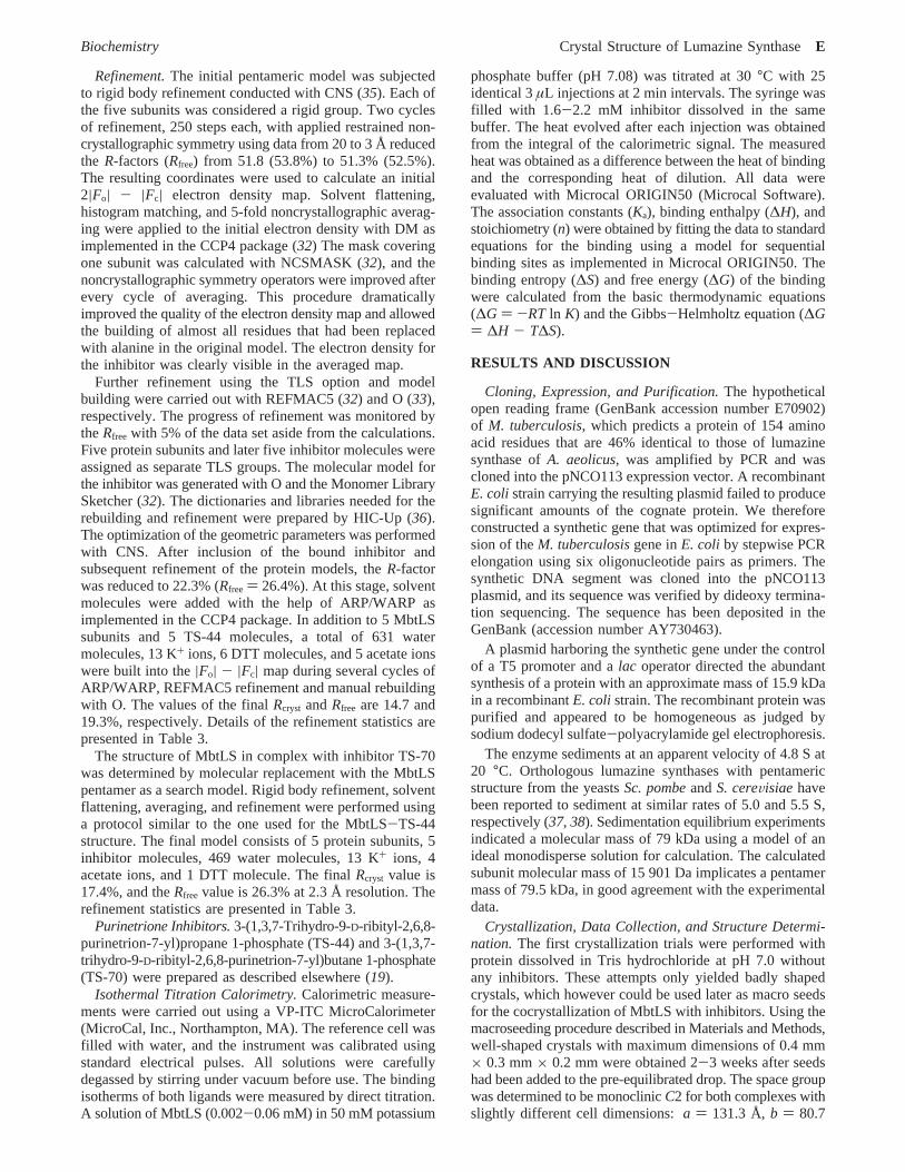
Structural Comparison.MbtLS as well as all other knownLS orthologues belong to the family ofR/â proteins withR/â/R sandwich topology (Figure 3). The overall topologyof this fold isâ2R1â3R2â4R3â5R4R5, with R2/R3 andR1/R4/R5 forming the flanking helices andâ2â3â4â5 the centralâ-sheet. In icosahedral LSs (LSs fromB. subtilis, A. aeolicus,and Sp. oleracea), the first eight N-terminal residues areknown to build an extra fifthâ-strand of the centralâ-sheetof the adjacent subunit, but they have a flexible conformationin the pentameric enzymes. In a LS sequence comparison,the presence of at least one proline among the first 10residues in the pentameric enzymes and an absence of prolinein this sequence region in the icosahedral oligomers werenoted (7). It was proposed that the geometry of the Proresidue does not allow a proper alignment of the fifthâ strandto the adjacent subunit and thus prevents LS containing aPro in this area from forming icosahedral assemblies (Figure4). The loop connectingR1 with â3 is exposed to the bulksolvent. It is the shortest among all known structures and
FIGURE 2: Stereogram plots of the self-rotation functionø ) 180°(a) andø ) 72° (b) sections. The radius of integration was 30 Å,and the resolution limits were 20-4.0 Å. Polar angles are definedas implemented in the CCP4 package (32). Contour lines are drawnfrom 10.0σ in steps of 6σ. The plot was generated with MOLREPas implemented in the CCP4 program package.
F Morgunova et al. Biochemistry
possesses almost no sequence similarity with other LSsequences. It is stabilized by formation of two hydrogenbonds between main chain carbonyl oxygens and amidenitrogens in the Ala43-Leu48 and Ala44-Gly47 pairs. Thefirst H-bond is well conserved in some of the knownstructures (PDB entries 1RVV, 1KYV, 1C41, and 1HQK),but the second one is hitherto only observed in MbtLS.
The loop betweenR2 andâ4 is also shortest in MbtLS incomparison to the other known LS structures. The sequencedoes not show any homology in this area, but insertions ofdifferent lengths are observed in all known LSs, the longestone consisting of 36 residues inM. grisea (7). In MbtLS,this loop participates in the face-to-face contact with theadjacent pentamer by forming the Arg71-Glu68′ salt bridge.
Theâ-hairpin loop connectingR4 andR5 is known as anessential region for the formation of the icosahedral capsid.In all LS structures which assemble to icosahedral particles,this loop comprises the rather conserved G(T/G)K(A/H)GNmotif. In the pentameric enzymes, however, the loop showsdifferent lengths with an insertion of up to four residues andadopts different conformations. In this loop region, theMbtLS sequence differs from sequences forming icosahedralLSs by four residues, whereas the structural alignment shows
that this loop differs from the corresponding loop in theicosahedral ones by only two residues. InB. abortusLS,the insertion forms a part of a continuous helix with nomultiple contacts with the neighboring pentamer, but it stillprevents the isocahedral assembly (11). In this respect,B.abortusLS deviates from all other known pentameric LSstructures. The conformation of the loop in MbtLS isstabilized by five hydrogen bonds formed between main andside chain atoms inside the loop. Its orientation is strictlyfixed by three hydrogen bonds between main and side chainatoms with residue Asp127 from helixR4. The alignmentof three MbtLS subunits with a trimer of the icosahedralB.subtilisLS revealed that even the short two-residue insertionis enough to produce a clash with the loop connecting strandâ2 and helixR1 in the adjacent subunit. Interestingly, thefour-residue addition in this loop inA. aeolicusLS resultedin the formation of largeT ) 3 icosahedral particles but notin the disassembly ofT ) 1 icosahedrons to pentamers (X.Zhang et al., unpublished results).
Thirteen peaks of strong spherical electron density over5σ were found in the|Fo| - |Fc| electron density map andwere interpreted as potassium ions because of the highconcentration of potassium acetate (3.5 M) used as aprecipitating agent during the crystallization. Each subunitcoordinates two K+ ions. Every pair of subunits, connectedby a local 2-fold axis, generated by the interaction ofcrystallographic and noncrystallographic symmetry opera-tions, shares one K+ ion, and two subunits connected by acrystallographic 2-fold axis share one K+ ion, too (Figure3). Both K+ ions found inside the monomer exhibit a slightlydistorted octahedral coordination provided by three or fourmain chain and side chain oxygen atoms and two or threewater molecules, respectively, with an average distance of2.85 Å. One potassium ion is found to make two contactswith the main chain oxygen atoms of Ala70 and His73 inthe loop linking helixR2 and strandâ4 and one contact withthe side chain OG1 atom from Thr110 in the loop betweenhelix R3 and strandâ5. The other K+ ion stabilizes theconformation of the loop that connects helicesR4 andR5.It makes two contacts with main chain oxygen atoms ofAla129 and Gly130 and one contact with the carboxyloxygen of Asp137. The fourth contact of this K+ ion isformed with the main chain oxygen of Ala34 from helixR1.The potassium ion located on the 2-fold axis is ligandedsymmetrically by two main chain oxygens of each subunitwith a distance of 3 Å from the metal ion and two watermolecules in addition to the octahedral coordination.
Pentamer Assembly.MbtLS belongs to the class ofpentameric LSs in contrast to the other icosahedral LSs. Thepentameric nature of MbtLS was shown by analyticalultracentrifugation and confirmed by the structural investiga-tion presented in this paper (Figure 5). The central left-handed twisted superhelix channel, with a length of 30 Åand a smallest diameter of 8.6 Å, is formed by theR3 helicesarranged around the 5-fold axis. Hydrophilic residuesAsp107, Arg103, and Asp95 and polar residues Tyr95 andGln99 expose their side chains to the channel and participatein hydrogen bond network formation with water molecules.A salt bridge between Arg103 and Asp107 is stabilizing oneof the entrances, while the opposite one is stabilized byhydrophobic residues Pro88 and Phe90 and polar residueTyr92. In the crystal structure, the channel is filled with water
FIGURE 3: Secondary structure alignment of one subunit oflumazine synthase fromM. tuberculosis (MbtLS) (red) withlumazine synthases fromS. pombe(pink), S. cereVisiae (yellow),B. abortus(brown), M. grisea (orange),B. subtilis (violet), A.aeolicus(cyan), andSp. oleracea(green). The secondary structureelements are assigned with respect to the MbtLS structure andlabeled according toB. subtilis lumazine synthase (6, 8). Bluespheres represent K+ ions. Black arrowheads mark the loops, whichshow the structural differences. This figure was generated withPYMOL (41).
Biochemistry Crystal Structure of Lumazine SynthaseG
molecules. The structural superposition of theR3 helicesfrom eight known LS structures reveals a highly conservedarrangement with an rms deviation of 0.628 Å inM. griseaLS and 1.293 Å withB. abortusLS. However, sequencehomology shows that none of the residues ofR3 helices isconserved in all LSs. The alignment of the whole pentamerswith the exception of 15 N-terminal residues indicates atendency toward increasing structural differences betweenpentamers extending from the central channel to the periph-eral parts. The rmsd between the remaining parts of theMbtLS pentamer and LSs from different organisms variesfrom 0.999 in M. grisea LS up to 1.402 in LS fromB.abortus.
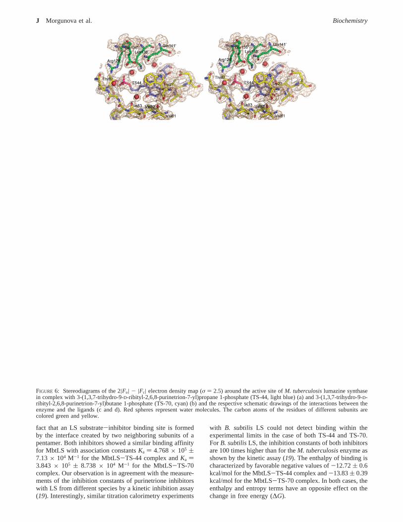
Inhibitor Binding Mode.The active site of all LSs knownat present is located at the interface between two neighboringsubunits in the pentameric assembly. The overall conforma-tion of the MbtLS active site is very similar to those of theactive sites of other LSs. All five substrate-product bindingpockets within a pentamer have the same overall topology.
It is formed by the residues from threeâ-loops connectingstrandsâ2-â4 to helicesR1-R3 (residues 26-28, 58-61,and 81-87, respectively) from one subunit as well asresidues 128-141 from helicesR4 and R5 and residueAsn114 from strandâ5 of the neighboring subunit (Figure6). There are in total 20 hydrogen bonds and 2 ionic contactsthrough which the TS-44 molecule is bound to the twosubunits forming the active site: 7 contacts are made withone subunit, 4 made with the second one, and 11 contactsmediated by water molecules (Figure 6a,c). The heteroaro-matic ring systems of both inhibitors are effectively packedin the hydrophobic part of the active site formed by Trp27,Ile60, Val81, Val82, Ile83, Phe90, and Val93 of one subunit.The purinetrione rings are located in a stacking position withthe aromatic ring system of Trp27 3.5 and 4.5 Å from theside chains of Val83 and Val93, respectively, from the othersides of the cavity. The hydrophobic environment is highlyconserved in all known LS active sites with respect to theequivalent substitutions of the hydrophobic residues; in
FIGURE 4: Sequence alignment of eight lumazine synthases with known structure. The residues involved in formation of the active site arecolored red; putative residues, which may prevent icosahedral assembly, are colored magenta, and residues thought to be responsible forthe specificity of lumazine synthase fromM. tuberculosis(MbtLS) are colored cyan. Secondary structure elements are shown above thesequences as they are found in MbtLS. Sequence alignment was performed with CLUSTAL W (42).
H Morgunova et al. Biochemistry
particular, Trp27 is often replaced with Phe and Val93 canbe replaced with Ile. It is important to note that this cavityin MbtLS is lacking the offset stacking aromatic interactionobserved in almost all known LSs between Trp57/Phe/Tyrand Phe113/Leu (A. aeolicusLS numbering) as a result ofsubstitution by Ile60′ and Asn114′, respectively. The hydro-phobic interaction of the purinetrione rings is supported byhydrogen bonds formed between the main chain O atom ofVal81 and N3 from purinetrione rings, the main chain Natom of Ala59 and O2 of purinetrione, the main chain Natom of Val81 and the main chain O atom of Trp27 mediatedby Wat25 with O2, and the NE2 atom of His28 and the mainchain O atom of Ile83 with O4 mediated by Wat202. Aschematic diagram of protein-inhibitor interactions is pre-sented in Figure 6a,c. The carbonyl O6 of the purinetrionering is fixed by a unique H-bond with the amide NZ atomof Lys138′ with a distance of 3.03 Å. This Lys is conservedin many LSs; however, in some LSs, it is replaced with His.Usually, this residue adopts a different conformation and issupposed to play some role in the phosphate transferringprocess during the catalytic reaction. The purinetrionecompounds are the first of a series of rationally designedinhibitors, which make contact with the Lys138 side chain(18, 19). The binding of the ribityl moiety of the purinetrioneinhibitors is very similar to the binding of the ribityl chainsof all previously known inhibitors. The ribityl chain isembedded in a surface depression formed by residues 56-62 from one subunit and one end ofâ5 (residues 113 and114) of the adjacent subunit with several H-bonds to bothsubunits. The main chain N and O atoms of Asn114′ arehydrogen-bonded to O5′ and O4′, respectively, and thecarboxyl oxygen atoms of Glu61 form hydrogen bonds withO3′ and O5′, respectively; O3′ forms a hydrogen bond tothe OE1 atom of Glu61, and O2′ is H-bonded to the mainchain N atom of Ile60. Those contacts are strictly conservedin all structurally investigated LSs.
The phosphate moiety occupies the putative position of3,4-hydroxy-2-butanone 4-phosphate (2) (Figure 1), thesecond substrate of LS. Several contacts formed by thephosphate moiety of the novel inhibitors are strictly con-served among all known structures of LSs. O32 is H-bondedto the main chain N atom of Gln86 through Wat470, andO33 forms several contacts with the main chain N atom andthe side chain OG1 atom of Thr87 and with the N atom ofGly85 and the O atom of Thr87, mediated by Wat19. O31forms a H-bond with the NE atom of Arg128. Severalcontacts are mediated by water molecules: to OG1 of Thr87through Wat386 and to OE1 and OE2 of Glu136′ and to NZof Lys138′ through Wat259. Wat259 is also mediating thecontact of O27 with Glu136′ and Lys138′.
The structural alignment of the two bound inhibitorsreveals a slight difference in their binding modes that couldbe explained by the fact that the aliphatic chain of the TS-70 molecule bearing the phosphate group is one C-C bondlonger than in TS-44. The conformation of this chain isslightly different in both inhibitors (Figure 6). The positionof the phosphorus atom in TS-70 is shifted toward NH2 ofArg128′ by 1.1 Å on average in all five subunits, and CZ ofArg128′ is repositioned by 0.7 Å. The purinetrione ringsoccupy the same position as in the MbtLS-TS-44 complex,and the ribityl chain participates in the same contacts as inthe MbtLS-TS-44 complex. The longer aliphatic linker ofthe phosphate moiety results in additional contacts, namely,O32 with the N atom of Gln86 and the NH atom of Arg128′from the phosphate group part of the inhibitor and additionalcontacts of O2 and O4 with the OG atom of Ser25 and theNE2 atom of His28 from the purinetrione ring system (Figure6b,d).
Isothermal Titration Calorimetry.Isothermal titrationcalorimetry (ITC) is the most direct method of measuringthe heat that is generated or consumed upon formation of aligand-macromolecule complex at a constant temperature.By least-squares fitting of the binding isotherms, we canderive the association constant (Ka), the binding enthalpy(∆H), and the stoichiometry (n) of the process. Moreover,the entropy of the binding reaction (∆S) and the free energychange (∆G) are easily obtained from the relation∆G )-RT ln Ka ) ∆H - T∆S.
To test purinetrione-based compounds for their inhibitionpotential, calorimetric measurements of the binding affinitywere performed. Figure 7 shows representative calorimetrictitration curves of MbtLS in 50 mM potassium phosphatebuffer (pH 7.0) at 30°C obtained with TS-44 and TS-70.The heat changes upon binding for each individual injectionwere plotted as a function of the molar inhibitor-to-proteinratio. The binding of both inhibitors is exothermic withnegative changes in the binding enthalpy. To derive thethermodynamic parameters, the binding isotherms wereinitially fitted with a binding model assuming identical andindependent binding sites. The fitting with this model,however, gave rather unsatisfactory results. Good fits of theisotherms could only be obtained by applying a model withfive sequential binding sites. The analysis of the data revealeda molar binding stoichiometry (n) of 5 bound ligandmolecules per pentamer which is in agreement with the X-raystructure presented in this paper. It appeared, however, thatthe five binding sites are not independent. They showed aweak cooperative behavior, which seems to be related to the
FIGURE 5: Pentameric assembly of lumazine synthase fromM.tuberculosisviewed along the 5-fold noncrystallographic axis. Theactive sites located between subunits are occupied by 3-(1,3,7-trihydro-9-D-ribityl-2,6,8-purinetrion-7-yl)propane 1-phosphate (TS-44). Blue spheres represent potassium ions.
Biochemistry Crystal Structure of Lumazine SynthaseI
fact that an LS substrate-inhibitor binding site is formedby the interface created by two neighboring subunits of apentamer. Both inhibitors showed a similar binding affinityfor MbtLS with association constantsKa ) 4.768× 10⁵ (7.13× 10⁴ M-1 for the MbtLS-TS-44 complex andKa )3.843 × 10⁵ ( 8.738 × 10⁴ M-1 for the MbtLS-TS-70complex. Our observation is in agreement with the measure-ments of the inhibition constants of purinetrione inhibitorswith LS from different species by a kinetic inhibition assay(19). Interestingly, similar titration calorimetry experiments
with B. subtilis LS could not detect binding within theexperimental limits in the case of both TS-44 and TS-70.ForB. subtilisLS, the inhibition constants of both inhibitorsare 100 times higher than for theM. tuberculosisenzyme asshown by the kinetic assay (19). The enthalpy of binding ischaracterized by favorable negative values of-12.72( 0.6kcal/mol for the MbtLS-TS-44 complex and-13.83( 0.39kcal/mol for the MbtLS-TS-70 complex. In both cases, theenthalpy and entropy terms have an opposite effect on thechange in free energy (∆G).
FIGURE 6: Stereodiagrams of the 2|Fo| - |Fc| electron density map (σ ) 2.5) around the active site ofM. tuberculosislumazine synthasein complex with 3-(1,3,7-trihydro-9-D-ribityl-2,6,8-purinetrion-7-yl)propane 1-phosphate (TS-44, light blue) (a) and 3-(1,3,7-trihydro-9-D-ribityl-2,6,8-purinetrion-7-yl)butane 1-phosphate (TS-70, cyan) (b) and the respective schematic drawings of the interactions between theenzyme and the ligands (c and d). Red spheres represent water molecules. The carbon atoms of the residues of different subunits arecolored green and yellow.
J Morgunova et al. Biochemistry
The most favorable enthalpy change and most unfavorableentropy change were observed in titration studies with TS-44 and may result from a weakened ability of TS-44 to fillthe active site properly due to, compared to TS-70, a shorteraliphatic linker. For both ligands, the enthalpic terms arelarger than the entropic terms at 30°C, indicating that theinteractions between MbtLS and both compounds are en-thalpically driven processes. However, because the interac-tion between MbtLS and TS-44 or TS-70 involves theremoval of a phosphate ion which occupies the binding siteof substrate2 as well as several protonation and deproto-nation events in the binding interface, a two-state (freefbound) model of the binding process is likely to be anoversimplification. Attempts to resolve the observed coop-erativity of the binding process and the competitive removalof the phosphate ion by the inhibitor are in progress.
Implications for the Mechanism of Lumazine Formation.A landmark for the characterization of the enzymaticmechanism of LS was the identification of the secondsubstrate, a four-carbon precursor of the pyrazine ring of6,7-dimethyl-8-ribityllumazine (3) (Figure 1), which wasidentified by Volk and Bacher (23) as (3S)-3,4-dihydroxy-2-butanone 4-phosphate. A mechanism describing lumazineformation was suggested by Kis et al. (14) (Figure 8) andextended by Schramek et al. (39) and Haase et al. (40). Thedetails of the catalytic mechanism have been investigatedand discussed by Zhang et al. (9) on the basis of the structureof LS from A. aeolicusin free form as well as in complexwith substrate-product analogues. According to the mech-anism described by Zhang et al. (9) for A. aeolicusLS,residues Arg127′, Lys135′, and Glu138′ (A. aeolicusLSnumbering, Figure 9b) are involved in positioning of thephosphate moiety of substrate 2. The side chain of Glu138′can adopt different conformations in a cooperative manner
together with Lys135′. A salt bridge between these residuesis therefore conserved even after the change in theirrespective conformation. Since Lys135′ is in one of twoobserved conformations close to the phosphate moiety ofbound substrate 2, it can build a salt bridge to the phosphategroup. After conformational reordering of Lys135′ withretention of this salt bridge, the phosphate moiety could bereoriented and thus be removed from the original bindingsite. Enzyme kinetic studies of MbtLS performed by Cush-man et al. (19) suggest tighter binding of the phosphate ionas well as a considerably decreased speed of the enzymaticreaction compared to the reaction speed ofB. subtilisLS.The structure of MbtLS reveals differences in the active site,which could result in a slightly different kinetic behavior ofthe enzyme (Figure 9). Lys135′ and Glu138′ are in MbtLS
FIGURE 7: Typical isothermal titration calorimetry curves for lumazine synthase fromM. tuberculosistitrated with 3-(1,3,7-trihydro-9-D-ribityl-2,6,8-purinetrion-7-yl)propane 1-phosphate (TS-44) (a) and 3-(1,3,7-trihydro-9-D-ribityl-2,6,8-purinetrion-7-yl)butane 1-phosphate(TS-70) (b). The top plots show titration data for the inhibitor binding. The bottom plots represent binding isotherms. The experiments werecarried out as described in Materials and Methods.
FIGURE 8: Hypothetical mechanism for the biosynthesis of lumazinesuggested by Kis et al. (14).
Biochemistry Crystal Structure of Lumazine SynthaseK
replaced with Lys138′ and Gln141′, respectively. A Glnresidue in position 141 is unique for MbtLS among all LSswith known structure. A salt bridge between Lys138′ andGln141′ is not possible anymore; however, a new negativelycharged binding partner, Glu136′, is introduced into the activesite as a replacement for the uncharged Gly133′. Glu136′can probably form a salt bridge to Lys138′ and can thereforeact as a negatively charged counterpart for Lys138′. Thevariation of its charge environment might hamper Lys138′in its conformational variability and thus decrease the speedof the enzymatic reaction by reducing its ability to mediatea proper reorientation of the phosphate-bearing aliphaticchain that is needed to make the subsequent ring closurepossible. The position and conformation of all other residuesin the active site that were thought to take part in catalysisare conserved.
Conclusion.The structure of lumazine synthase fromM.tuberculosisreveals new specific features of the active sitethat can be used as a structural basis for the development ofspecific inhibitors with an aim of designing new tuberculo-static agents. We have also demonstrated that compoundsderived from purinetrione may serve as potential and specificinhibitors for MbtLS. Our future plans will include in vivotests of the inhibitors described in this paper.
ACKNOWLEDGMENT
We thank Xiaofeng Zhang for technical assistance in thepreparation of the manuscript. We gratefully acknowledgethe access to synchrotron radiation facilities at the EMBLOutstation and thank the staff for help at the beamline.
REFERENCES
1. Nakajima, H. (1993) Tuberculosis: a global emergency,WorldHealth 46, 3.
2. Dahl, S. G., Ingebrigt, S., and Westrheim, R. A. (2004) Structureand Models of Transporter Proteins,J. Pharmacol. Exp. Ther.309, 853-860.
3. Cole, T. S., Eiglmeier, K., Parkhill, J., James, K. D., Thomson,N. R., Wheeler, P. R., Honore, N., Garnier, T., Churcher, C.,Harris, D., Mungall, K., Basham, D., Brown, D., Chillingworth,T., Connor, R., Davies, R. M., Devlin, K., Duthoy, S., Feltwell,T., Fraser, A., Hamlin, N., Holroyd, S., Jagels, K., Lacroix, C.,Maclean, J., Moule, S., Murphy, L., Oliver, K., Quail, M. A.,Rajandream, M.-A., Rutherford, K. M., Rutter, S., Seeger, K.,Simmonds, M., Skelton, J., Squares, R., Squares, S., Stevens, K.,Taylor, K., Whitehead, S., Woodward, J. R., and Barrel, B.G.(2001) Massive gene decay in the leprosy bacillus,Nature 409,1007-1011.
4. Bacher, A., Ludwig, H. C., Schnepple, H., and Ben-Shaul, Y.(1986) Heavy riboflavin synthase fromBacillus subtilis. Quater-nary structure and reaggregation,J. Mol. Biol. 187, 75-86.
5. Zylberman, V., Craig, P. O., Klinke, S., Braden, B. C., Cauerhff,A., and Goldbaum, F. A. (2004) High order quaternary arrange-ment confers increased structural stability toBrucella spp.lumazine synthase,J. Biol. Chem. 279, 8093-8101.
6. Ladenstein, R., Schneider, M., Huber, R., Bartunik, H. D., Wilson,K., Schott, K., and Bacher, A. (1988) Heavy riboflavin synthasefrom Bacillus subtilis. Crystal structure analysis of the icosahedralâ60 capsid at 3.3 Å resolution,J. Mol. Biol. 203, 1045-1070.
7. Persson, K., Schneider, G., Jordan, D. B., Viitanen, P. V., andSandalova, T. (1999) Crystal structure analysis of a pentamericfungal and an icosahedral plant lumazine synthase reveals thestructural basis for differences in assembly,Protein Sci. 8, 2355-2365.
8. Ritsert, K., Huber, R., Turk, D., Ladenstein, R., Schmidt-Ba¨se,K., and Bacher, A. (1995) Studies on the lumazine synthase/riboflavin synthase complex ofBacillus subtilis: Crystal structure
FIGURE 9: Stereoview of the surface representation of the active sites of lumazine synthase fromM. tuberculosisin complex with theinhibitor 3-(1,3,7-trihydro-9-D-ribityl-2,6,8-purinetrion-7-yl)propane 1-phosphate (TS-44) (a) andA. aeolicuslumazine synthase in complexwith 3-(7-hydroxy-8-ribityllumazin-6-yl)propionic acid (RPL) (PDB entry 1NQX) (b). The accessible surfaces were calculated with a waterprobe radius of 1.4 Å. Two adjacent subunits, constituting the active site, are shown in different colors (pink and wheat); the inhibitormolecules are colored red, and water molecules are colored light blue.
L Morgunova et al. Biochemistry
analysis of reconstituted, icosahedralâ-subunit capsids with boundsubstrate analogue inhibitor at 2.4 Å resolution,J. Mol. Biol. 253,151-167.
9. Zhang, X., Meining, W., Cushman, M., Haase, I., Fischer, M.,Bacher, A., and Ladenstein, R. (2003) A structure-based modelof the reaction catalyzed by lumazine synthase fromAquifexaeolicus, J. Mol. Biol. 328, 167-182.
10. Zhang, X., Meining, W., Fischer, M., Bacher, A., and Ladenstein,R. (2001) X-ray structure analysis and crystallographic refinementof lumazine synthase from the hyperthermophileAquifex aeolicusat 1.6 Å resolution: Determinants of thermostability revealed fromstructural comparisons,J. Mol. Biol. 306, 1099-1114.
11. Braden, B. C., Velikovsky, C. A., Cauerhff, A. A., Polikarpov, I.,and Goldbaum, F. A. (2000) Divergence in macromolecularassembly: X-ray crystallographic structure analysis of lumazinesynthase fromBrucella abortus, J. Mol. Biol. 297, 1031-1036.
12. Gerhardt, S., Haase, I., Steinbacher, S., Kaiser, J. T., Cushman,M., Bacher, A., Huber, R., and Fischer, M. (2002) The StructuralBasis of Riboflavin Binding toSchizosaccharomyces pombe6,7-Dimethyl-8-ribityllumazine Synthase,J. Mol. Biol. 318, 1317-1329.
13. Meining, W., Mortl, S., Fischer, M., Cushman, M., Bacher, A.,and Ladenstein, R. (2000) The atomic structure of pentamericlumazine synthase fromSaccharomyces cereVisiae at 1.85 Åresolution reveals the binding mode of a phosphonate intermediateanalogue,J. Mol. Biol. 299, 181-197.
14. Kis, K., Volk, R., and Bacher, A. (1995) Biosynthesis of riboflavin.Studies on the reaction mechanism of 6,7-dimethyl-8-ribityllu-mazine synthase,Biochemistry 34, 2883-2892.
15. Cushman, M., Mihalic, J. T., Kis, K., and Bacher, A. (1999) Designand synthesis of 6-(6-D-ribitylamino-2,4-dihydroxypyrimidin-5-yl)-1-hexyl phosphonic acid, a potent inhibitor of lumazinesynthase,Bioorg. Med. Chem. Lett. 9, 39-42.
16. Cushman, M., Mihalic, J. T., Kis, K., and Bacher, A. (1999)Design, synthesis, and biological evaluation of homologousphosphonic acids and sulfonic acids as inhibitors of lumazinesynthase,J. Org. Chem. 64, 3838-3845.
17. Cushman, M., Yang, D., Gerhardt, S., Huber, R., Fischer, M., Kis,K., and Bacher, A. (2002) Design, synthesis, and evaluation of6-carboxyalkyl and 6-phosphonoxyalkyl derivatives of 7-oxo-8-ribitylaminolumazines as inhibitors of riboflavin synthase andlumazine synthase,J. Org. Chem. 67, 5807-5816.
18. Cushman, M., Yang, D., Kis, K., and Bacher, A. (2001) Design,synthesis, and evaluation of 9-D-ribityl-1,3,7-trihydro-2,6,8-puri-netrione, a potent inhibitor of riboflavin synthase and lumazinesynthase,J. Org. Chem. 66, 8320-8327.
19. Cushman, M., Sambaiah, T., Jin, G., Illarionov, B., Fischer, M.,and Bacher, A. (2004) Design, synthesis, and evaluation of 9-D-ribitylamino-1,3,7,9-tetrahydro-2,6,8-purinetriones bearing alkylphosphate andR,R-difluorophosphonate substituents as inhibitorsof tiboflavin synthase and lumazine synthase,J. Org. Chem. 69,601-612.
20. Sedlmaier, H., Mu¨ller, F., Keller, P. J., and Bacher, A. (1987)Enzymatic synthesis of riboflavin and FMN specifically labeledwith 13C in the xylene ring,Z. Naturforsch. C42, 425-429.
21. Vervoort, J., Muller, F., Mayhew, S. G., Berg, W. A. v. d.,Moonen, C. T., and Bacher, A. (1986) A comparative carbon-13,nitrogen-15, and phosphorus-31 nuclear magnetic resonance studyon the flavodoxins from Clostridium MP, Megasphaera elsdenii,andAzotobacterVinelandii, Biochemistry 25, 6789-6799.
22. Richter, G., Volk, R., Krieger, C., Lahm, H. W., Ro¨thlisberger,U., and Bacher, A. (1992) Biosynthesis of riboflavin: Cloning,sequencing, and expression of the gene coding for 3,4-dihydroxy-2-butanone 4-phosphate synthase ofEscherichia coli, J. Bacteriol.174, 4050-4056.
23. Volk, R., and Bacher, A. (1990) Studies on the 4-carbon precursorin the biosynthesis of riboflavin. Purification and properties ofL-3,4-dihydroxy-2-butanone-4-phosphate synthase,J. Biol. Chem.265, 19479-19485.
24. Kis, K., and Bacher, A. (1995) Substrate channeling in thelumazine synthase/riboflavin synthase complex ofBacillus subtilis,J. Biol. Chem. 270, 16788-16795.
25. Read, S. M., and Northcote, D. H. (1981) Minimization of variationin the response to different proteins of the Coomassie blue G dye-binding assay for protein,Anal. Biochem. 116, 53-64.
26. Bullock, W. O., Fernandez, J. M., and Shout, J. M. (1987) XL1-blue: A high efficiency plasmid transforming recAEscherichiacoli strain withâ-galactosidase selection,BioTechniques 5, 376-380.
27. Stuber, D., Matile, H., and Garotta, G. (1990) inImmunologicalMethods (Lefkovits, I., and Pernis, B., Eds.) pp 121-152,Academic Press, New York.
28. Lammli, U. K. (1970) Cleavage of structural proteins during theassembly of the head of bacteriophage T4,Nature 227, 680-685.
29. Laue, T. M., Shah, B. D., Ridgeway, T. M., and Pelletier, S. L.(1992)Computer-aided interpretation of analytical sedimentationdata for proteins, Royal Society of Chemistry, Cambridge, U.K.
30. Otwinowski, Z., and Minor, W. (1997) Processing of X-rayDiffraction Data Collected in Oscillation Mode,Methods Enzymol.276, 307-326.
31. Lu, G. (1999) PATTERN: A precession simulating program fordisplaying reflection data of reciprocal space,J. Appl. Crystallogr.32, 375-376.
32. Collaborative Computational Project No. 4 (1994) The CCP4Suite: Programs for Protein Crystallography,Acta Crystallogr.D50, 760-763.
33. Jones, A. T., Zou, J. Y., Cowtan, J. Y., and Kjeldgaard, M. (1991)Improved methods for building protein models in electron densitymaps and the location of errors in the model,Acta Crystallogr.A47, 110-119.
34. Matthews, B. W. (1968) Solvent content of protein crystals,J.Mol. Biol. 33, 491-497.
35. Brunger, A. T., Adams, P. D., Clore, G. M., DeLano, W. L., Gros,P., Grosse-Kunstleve, R. W., Jiang, J.-S., Kuszewski, J., Nilges,M., Pannu, N. S., Read, R. J., Rice, L. M., Simonson, T., andWarren, G. L. (1998) Crystallography & NMR System: A NewSoftware Suite for Macromolecular Structure Determination,ActaCrystallogr. D54, 905-921.
36. Kleywegt, G. J., and Jones, T. A. (1998) Databases in proteincrystallography,Acta Crystallogr. D54, 1119-1131.
37. Bacher, A., Fischer, M., Kis, K., Kugelbrey, K., Mo¨rtl, S.,Scheuring, J., Weinkauf, S., Eberhardt, S., Schmidt-Ba¨se, K.,Huber, R., Ritsert, K., Cushman, M., and Ladenstein, R. (1996)Biosynthesis of riboflavin: Structure and mechanism of lumazinesynthase,Biochem. Soc. Trans. 24, 89-94.
38. Braun, N., Meining, W., Hars, U., Fischer, M., Ladenstein, R.,Huber, R., Bacher, A., Weinkauf, S., and Bachmann, L. (2002)Formation of metal nanoclusters on specific surface sites of proteinmolecules,J. Mol. Biol. 321, 341-353.
39. Schramek, N., Haase, I., Fischer, M., and Bacher, A. (2003)Biosynthesis of riboflavin. Single turnover kinetic analysis of 6,7-dimethyl-8-ribityllumazine synthase,J. Am. Chem. Soc. 125,4460-4466.
40. Haase, I., Fischer, M., Bacher, A., and Schramek, N. (2003)Temperature-dependent presteady-state kinetics of lumazine syn-thase from the hyperthermophilic eubacterium Aquifex aeolicus,J. Biol. Chem. 278, 37909-37915.
41. DeLano, W. L. (2002)PYMOL, DeLano Scientific, San Carlos,CA.
42. Thompson, J. D., Higgins, D. G., and Gibson, T. J. (1994)CLUSTAL W: Improving the sensitivity of progressive multiplesequence alignment through sequence weighting, position-specificgap penalties and weight matrix choice,Nucleic Acids Res. 22,4673-4680.
BI047848A
Biochemistry PAGE EST: 12.9 Crystal Structure of Lumazine SynthaseM