26
CSF ANALYSIS CSF ANALYSIS
| Date post: | 02-Jan-2016 |
| Category: |
Documents |
| Upload: | dora-johnston |
| View: | 240 times |
| Download: | 4 times |
CSF ANALYSISCSF ANALYSIS
CSF Formation CSF Formation
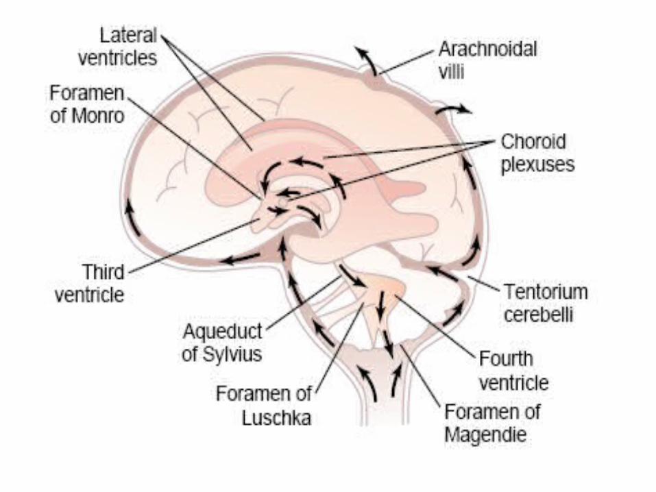
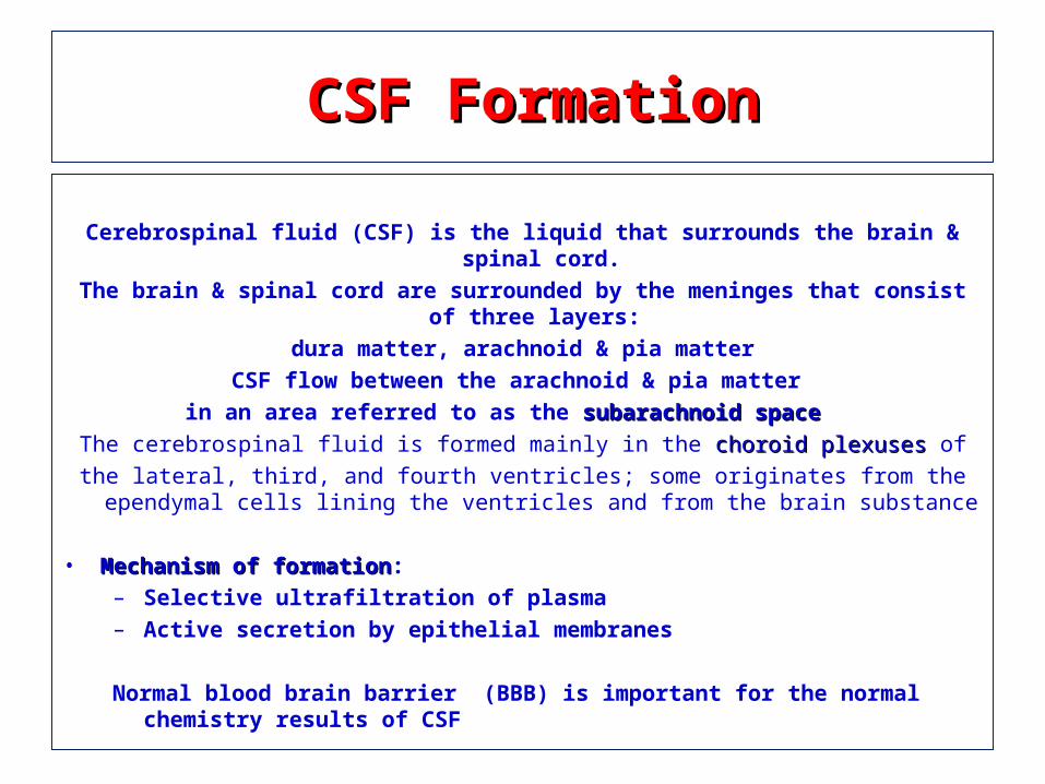
Cerebrospinal fluid (CSF) is the liquid that surrounds the brain & spinal cord.The brain & spinal cord are surrounded by the meninges that consist of three layers:
dura matter, arachnoid & pia matterCSF flow between the arachnoid & pia matter
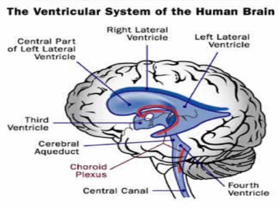
in an area referred to as the subarachnoid space subarachnoid space The cerebrospinal fluid is formed mainly in the choroid plexuses choroid plexuses of
the lateral, third, and fourth ventricles; some originates from the ependymal cells lining the ventricles and from the brain substance
• Mechanism of formationMechanism of formation:– Selective ultrafiltration of plasma– Active secretion by epithelial membranes
Normal blood brain barrier (BBB) is important for the normal chemistry results of CSF
Functions of CSFFunctions of CSF
Clinical Indications for CSF AnalysisClinical Indications for CSF Analysis
CSF is performed in cases of CSF is performed in cases of suspectedsuspected::
1- CNS 1- CNS infectionsinfections (infectious meningitis & encephalitis) (infectious meningitis & encephalitis)
2- CNS 2- CNS malignancymalignancy (as malignant infiltrates as in leukemia ..etc) (as malignant infiltrates as in leukemia ..etc)
3- CNS 3- CNS hemorrhageshemorrhages (as subarachnoid hemorrhage) (as subarachnoid hemorrhage)
4- CNS 4- CNS demylineatingdemylineating diseases (as multiple sclerosis) diseases (as multiple sclerosis)
Routine Laboratory CSF AnalysisRoutine Laboratory CSF Analysis
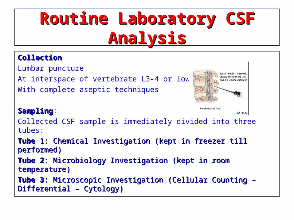
CollectionCollectionLumbar puncture
At interspace of vertebrate L3-4 or lower
With complete aseptic techniques
SamplingSampling:
Collected CSF sample is immediately divided into three tubes:
Tube Tube 1: Chemical Investigation (kept in freezer till performed)1: Chemical Investigation (kept in freezer till performed)
Tube 2Tube 2: Microbiology Investigation (kept in room temperature): Microbiology Investigation (kept in room temperature)
Tube 3Tube 3: Microscopic Investigation (Cellular Counting – Differential – Cytology): Microscopic Investigation (Cellular Counting – Differential – Cytology)
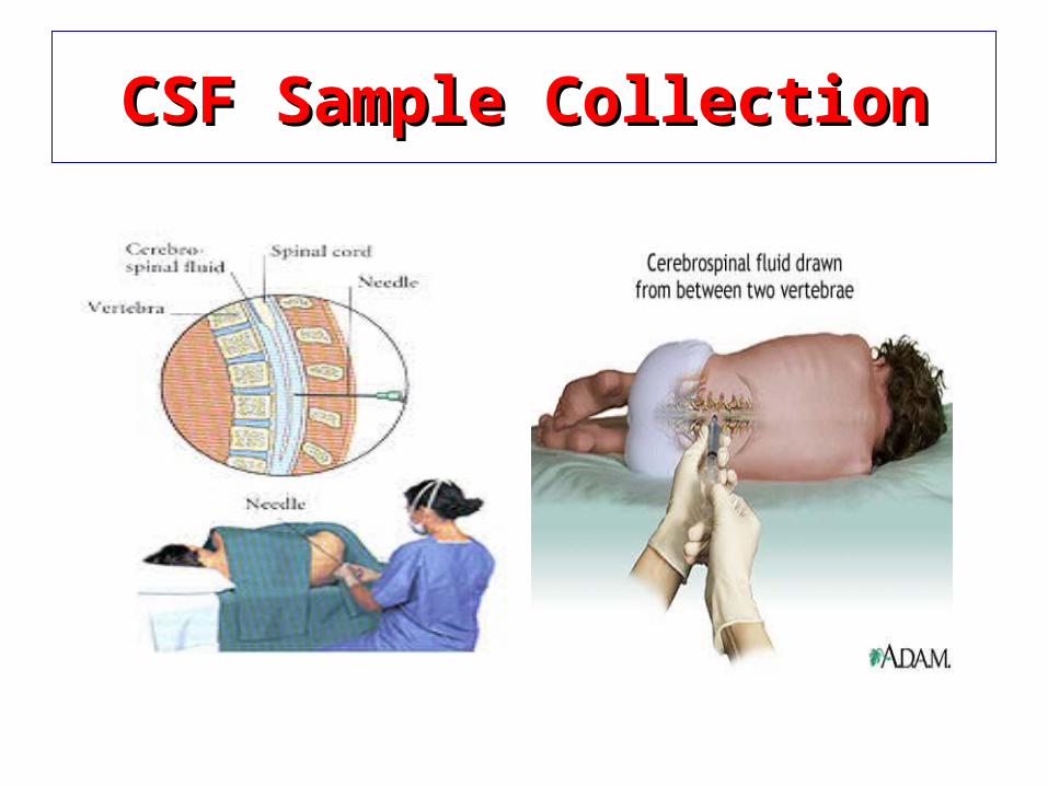
CSF Sample CollectionCSF Sample Collection
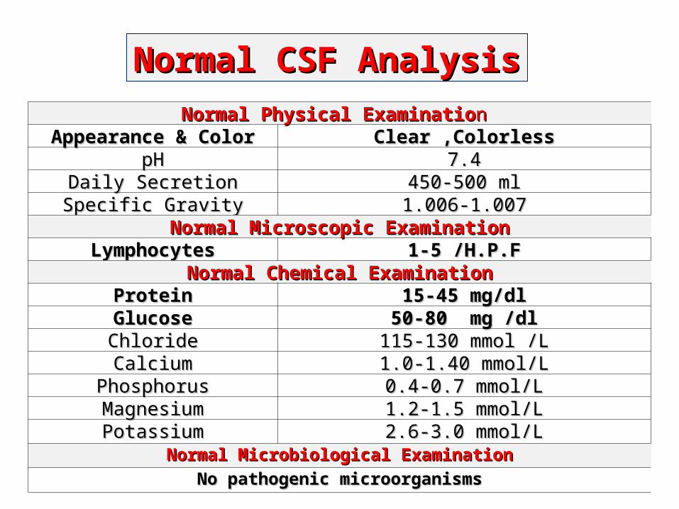
Normal Physical ExaminatioNormal Physical Examination n Appearance & ColorAppearance & Color Clear ,ColorlessClear ,Colorless
pHpH 7.47.4Daily SecretionDaily Secretion 450-500 ml450-500 mlSpecific GravitySpecific Gravity 1.006-1.0071.006-1.007
Normal Microscopic ExaminationNormal Microscopic ExaminationLymphocytesLymphocytes 1-5 /H.P.F1-5 /H.P.F
Normal Chemical ExaminationNormal Chemical ExaminationProteinProtein 15-45 mg/dl15-45 mg/dlGlucoseGlucose 50-80 mg /dl50-80 mg /dlChlorideChloride 115-130 mmol /L115-130 mmol /LCalciumCalcium 1.0-1.40 mmol/L1.0-1.40 mmol/L
PhosphorusPhosphorus 0.4-0.7 mmol/L0.4-0.7 mmol/LMagnesiumMagnesium 1.2-1.5 mmol/L1.2-1.5 mmol/LPotassiumPotassium 2.6-3.0 mmol/L2.6-3.0 mmol/L
Normal Microbiological ExaminationNormal Microbiological ExaminationNo pathogenic microorganismsNo pathogenic microorganisms
Normal CSF AnalysisNormal CSF Analysis
Physical examination of CSFPhysical examination of CSF
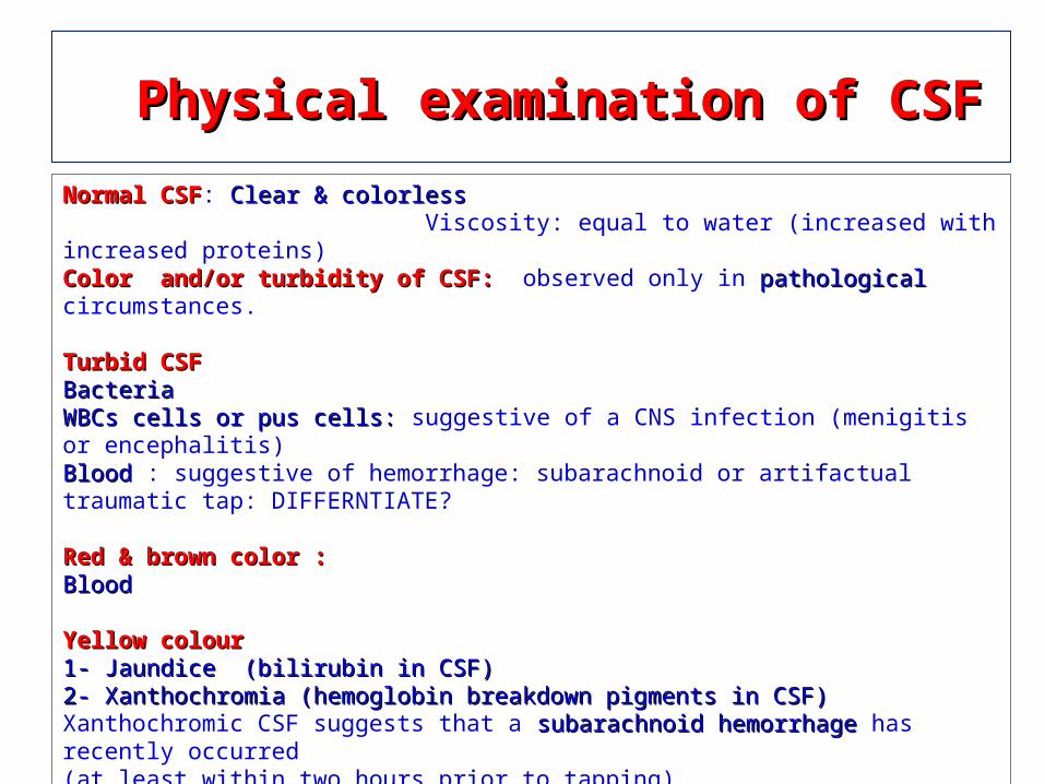
Normal CSFNormal CSF: Clear & colorless Clear & colorless Viscosity: equal to water (increased with increased proteins)Color and/or turbidity of CSF: Color and/or turbidity of CSF: observed only in pathologicalpathological circumstances.
Turbid CSFTurbid CSF BacteriaBacteriaWBCs cells or pus cells: WBCs cells or pus cells: suggestive of a CNS infection (menigitis or encephalitis)BloodBlood : suggestive of hemorrhage: subarachnoid or artifactual traumatic tap: DIFFERNTIATE?
Red & brown color :Red & brown color :Blood Blood
Yellow colour Yellow colour 1- Jaundice (bilirubin in CSF)1- Jaundice (bilirubin in CSF)2- Xanthochromia (hemoglobin breakdown pigments in CSF)2- Xanthochromia (hemoglobin breakdown pigments in CSF)Xanthochromic CSF suggests that a subarachnoid hemorrhage subarachnoid hemorrhage has recently occurred (at least within two hours prior to tapping). The yellow color is due to bilirubin generated in the CNS by the breakdown of hemoglobin released from RBC's. (so jaundice should be excluded).
Microscopic examination of CSFMicroscopic examination of CSF
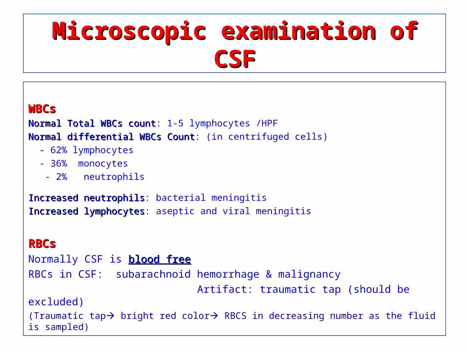
WBCsWBCsNormal Total WBCs countNormal Total WBCs count: 1-5 lymphocytes /HPF
Normal differential WBCs CountNormal differential WBCs Count: (in centrifuged cells)
- 62% lymphocytes
- 36% monocytes
- 2% neutrophils
Increased neutrophilsIncreased neutrophils: bacterial meningitis
Increased lymphocytesIncreased lymphocytes: aseptic and viral meningitis
RBCsRBCsNormally CSF is blood freeblood free
RBCs in CSF: subarachnoid hemorrhage & malignancy
Artifact: traumatic tap (should be excluded)(Traumatic tap bright red color RBCS in decreasing number as the fluid is sampled)
Chemical examination of CSFChemical examination of CSF
In addition to the major ions, CSF contains oxygen, sugars (e.g. glucose, fructose), lactate, proteins (e.g. albumin,
globulins), amino acids, urea, ammonia, glutamine, creatinine, lipids, hormones (e.g. insulin) and vitamins.
CSF Glucose CSF Glucose
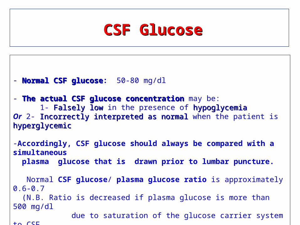
- Normal CSF glucoseNormal CSF glucose: 50-80 mg/dl
- The actual CSF glucose concentration The actual CSF glucose concentration may be: 1- Falsely low Falsely low in the presence of hypoglycemia hypoglycemia Or 2- Incorrectly interpreted as normal Incorrectly interpreted as normal when the patient is hyperglycemichyperglycemic
-Accordingly, CSF glucose should always be compared with a simultaneous plasma glucose that is drawn prior to lumbar puncture. Normal CSF glucose/ plasma glucose ratio is approximately 0.6-0.7 (N.B. Ratio is decreased if plasma glucose is more than 500 mg/dl due to saturation of the glucose carrier system to CSF
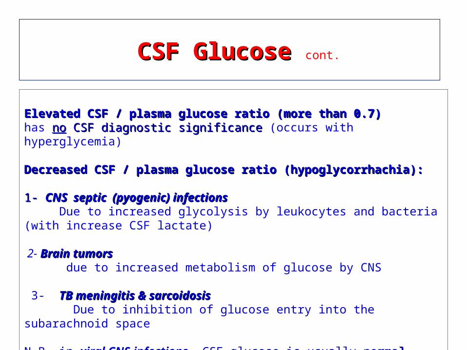
Elevated CSF / plasma glucose ratio (more than 0.7)Elevated CSF / plasma glucose ratio (more than 0.7)has nono CSF diagnostic significance CSF diagnostic significance (occurs with hyperglycemia)
Decreased CSF / plasma glucose ratio (hypoglycorrhachia):Decreased CSF / plasma glucose ratio (hypoglycorrhachia):
1- 1- CNS septic (pyogenic) infectionsCNS septic (pyogenic) infections Due to increased glycolysis by leukocytes and bacteria (with increase CSF lactate)
2- Brain tumors Brain tumors due to increased metabolism of glucose by CNS
3- TB meningitis & sarcoidosisTB meningitis & sarcoidosis Due to inhibition of glucose entry into the subarachnoid space N.B. in viral CNS infections, CSF glucose is usually normalnormal
CSF Glucose CSF Glucose cont.
CSF Protein CSF Protein cont.
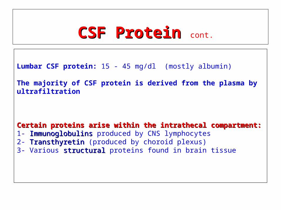
Lumbar CSF protein: 15 - 45 mg/dl (mostly albumin)
The majority of CSF protein is derived from the plasma by ultrafiltration
Certain proteins arise within the intrathecal compartment:Certain proteins arise within the intrathecal compartment:1- Immunoglobulins Immunoglobulins produced by CNS lymphocytes2- Transthyretin Transthyretin (produced by choroid plexus)3- Various structuralstructural proteins found in brain tissue
CSF Protein CSF Protein
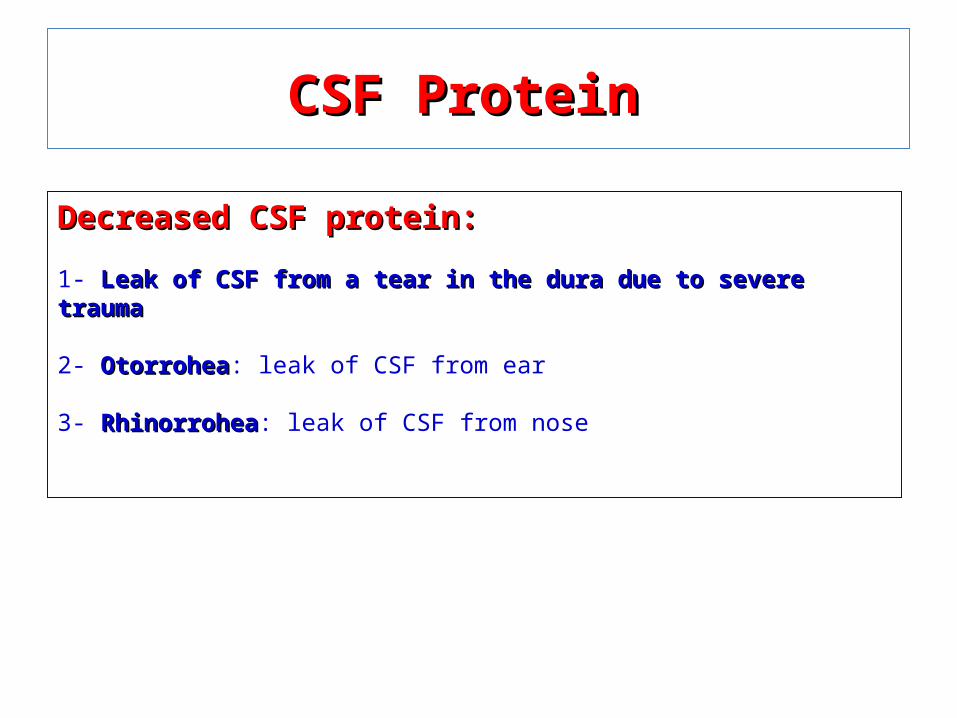
Decreased CSF protein:Decreased CSF protein:
1- Leak of CSF from a tear in the dura due to severe traumaLeak of CSF from a tear in the dura due to severe trauma 2- OtorroheaOtorrohea: leak of CSF from ear
3- RhinorroheaRhinorrohea: leak of CSF from nose
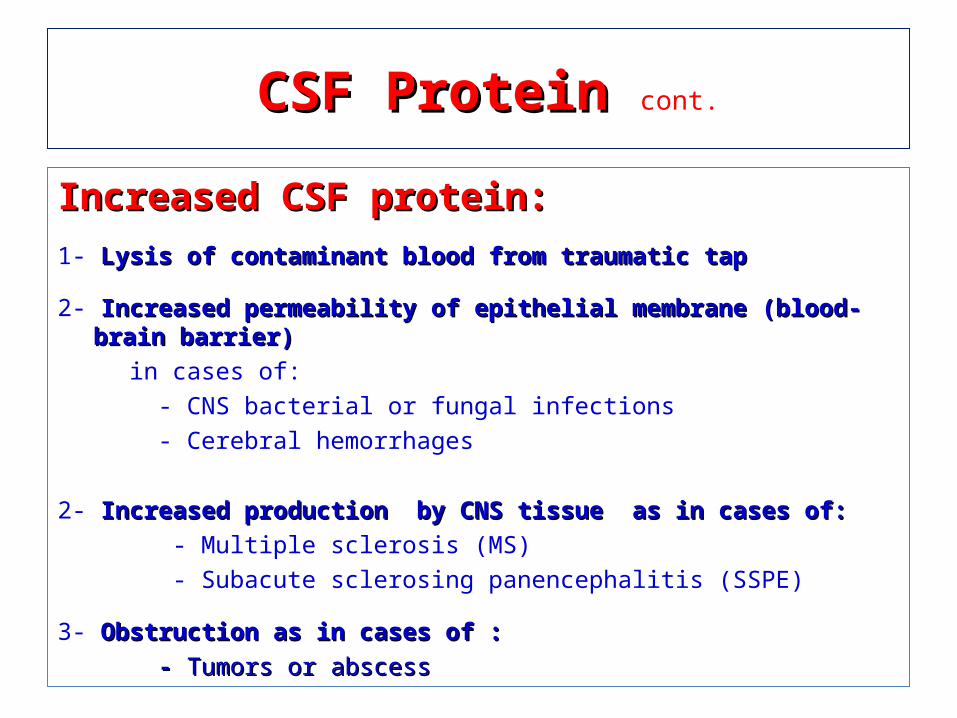
Increased CSF protein:Increased CSF protein:
1- Lysis of contaminant blood from traumatic tapLysis of contaminant blood from traumatic tap
2- Increased permeability of epithelial membrane (blood-brain barrier) Increased permeability of epithelial membrane (blood-brain barrier)
in cases of:
- CNS bacterial or fungal infections
- Cerebral hemorrhages
2- Increased production by CNS tissue as in cases of:Increased production by CNS tissue as in cases of: - Multiple sclerosis (MS)
- Subacute sclerosing panencephalitis (SSPE)
3- Obstruction as in cases of :Obstruction as in cases of :
- - Tumors or abscessTumors or abscess
CSF Protein CSF Protein cont.
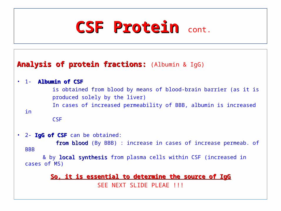
Analysis of protein fractions: Analysis of protein fractions: (Albumin & IgG)
• 1- Albumin of CSFAlbumin of CSF
is obtained from blood by means of blood-brain barrier (as it is
produced solely by the liver)
In cases of increased permeability of BBB, albumin is increased in
CSF
• 2- IgG of CSF IgG of CSF can be obtained:
from blood from blood (By BBB) : increase in cases of increase permeab. of BBB
& by local synthesis local synthesis from plasma cells within CSF (increased in cases of MS)
So, it is essential to determine the source of IgGSo, it is essential to determine the source of IgGSEE NEXT SLIDE PLEAE !!!
CSF Protein CSF Protein cont.
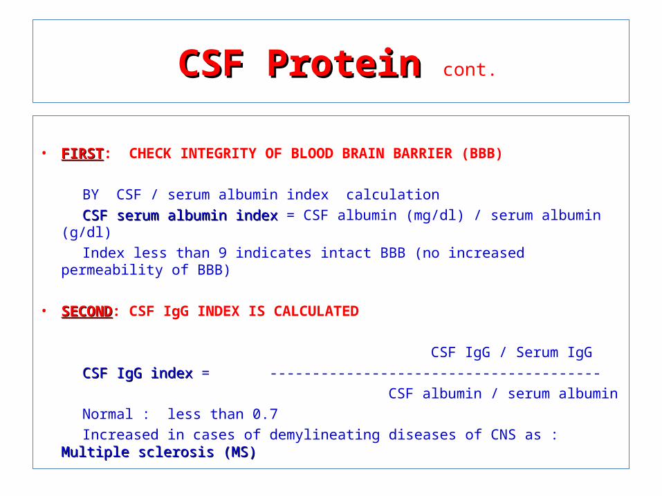
• FIRSTFIRST: CHECK INTEGRITY OF BLOOD BRAIN BARRIER (BBB) BY CSF / serum albumin index calculation CSF serum albumin index CSF serum albumin index = CSF albumin (mg/dl) / serum albumin (g/dl) Index less than 9 indicates intact BBB (no increased permeability of BBB) • SECONDSECOND: CSF IgG INDEX IS CALCULATED CSF IgG / Serum IgG CSF IgG index CSF IgG index = --------------------------------------- CSF albumin / serum albumin Normal : less than 0.7 Increased in cases of demylineating diseases of CNS as : Multiple sclerosis (MS)Multiple sclerosis (MS)
CSF Protein CSF Protein cont.
CSF ImmunoglobulinCSF Immunoglobulin
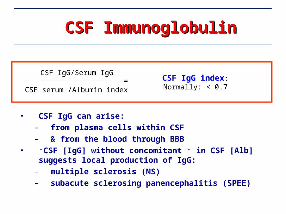
• CSF IgG can arise:– from plasma cells within CSF– & from the blood through BBB
• ↑CSF [IgG] without concomitant ↑ in CSF [Alb] suggests local production of IgG:
– multiple sclerosis (MS)– subacute sclerosing panencephalitis (SPEE)
CSF IgG/Serum IgG
CSF serum /Albumin index
CSF IgG index:Normally: < 0.7
=
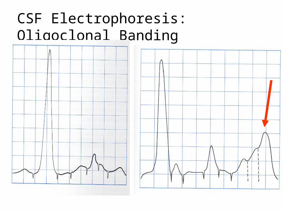
CSF Electrophoresis: Oligoclonal Banding
CSF lactateCSF lactate
CSF lactate is increased in cases of bacterial meningitis (due to increased
glycolysis by bacteria & inflammatory cells)
The level of CSF glutamine CSF glutamine reflects level of ammonia in that is normally removed in the CNS by formation of glutamine (amino acid glutamate + ammonia).
Glutamine synthesis Glutamine synthesis helps to protect the CNS from the toxic effects of increased ammonia.
Ammonia production is increase dramatically in patients with liver failure.
Accordingly, CSF glutamine production is increased in cases of hepatic hepatic encephalopathyencephalopathy
CSF glutamine CSF glutamine
Enzymes in the CSFEnzymes in the CSF
CSF lactate dehydrogenase (LDH) may be elevated in bacterial bacterial meningitismeningitis.
CSF adenosine deaminase (ADA) elevations can occur in tuberculous tuberculous meningitismeningitis.
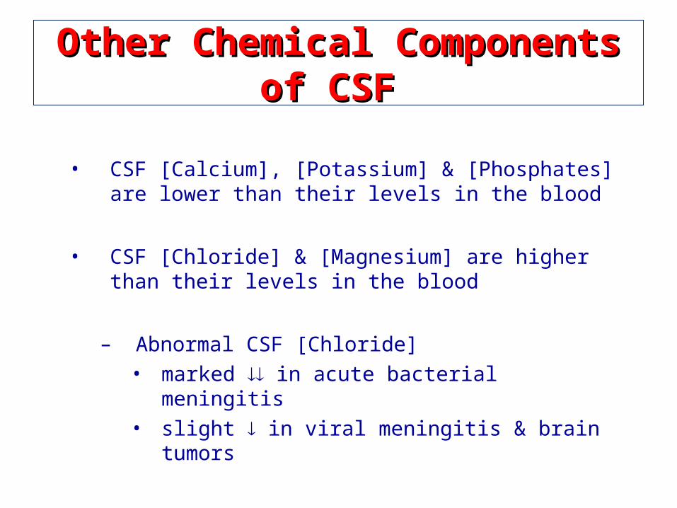
Other Chemical Components of CSF Other Chemical Components of CSF
• CSF [Calcium], [Potassium] & [Phosphates] are lower than their levels in the blood
• CSF [Chloride] & [Magnesium] are higher than their levels in the blood
– Abnormal CSF [Chloride]• marked in acute bacterial meningitis• slight in viral meningitis & brain tumors
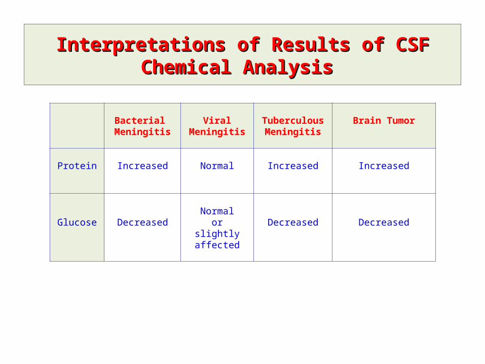
Interpretations of Results of CSF Chemical Analysis Interpretations of Results of CSF Chemical Analysis
Bacterial Meningitis
Viral Meningitis Tuberculous Meningitis
Brain Tumor
Protein Increased Normal Increased Increased
Glucose DecreasedNormal
orslightly affected
Decreased Decreased


