arlos Henrique Marques dos Santosa,∗, Jovino Nogueira da Silva Menezesb,hiago Franchi Nunesc, Letícia de Assis Martinsc
Service of Coloproctology, Hospital Universitário Maria Aparecida Pedrossian, Campo Grande, MS, BrazilService of General Surgery, Hospital Universitário Maria Aparecida Pedrossian, Campo Grande, MS, BrazilService of Radiology, Hospital Universitário Maria Aparecida Pedrossian, Campo Grande, MS, Brazil
r t i c l e i n f o
rticle history:
eceived 28 March 2015
ccepted 8 June 2015
vailable online 2 October 2015
eywords:
rohn’s disease
nterography
iagnosis
a b s t r a c t
Proposition: Crohn’s disease (CD) is a chronic inflammatory process that affects various parts
of the gastrointestinal tract, from the mouth to the anus with unknown etiology and variable
clinical presentation. CD diagnosis is based on clinical and complementary tests. Among
the complementary tests, enterography with CT enterography has shown good results in
the evaluation of this disease.
Methods: The patients evaluated were submitted to a questionnaire on the clinical manifes-
tations of the disease and an CT enterography was obtained. The studies were reviewed by
an experienced radiologist looking for radiological signs of CD.
Results: The mean age was 40 years, with a predominance of women. The main clinical
manifestations are diarrhea in 24 (70%), hematochezia in 19 (55%), abdominal pain in 29
(85%) and weight loss in 22 (64%) patients. The main findings on CT enterography were an
intestinal wall enhancement signal in 23 patients (67%), vascular engorgement (vasa recta)
in 20 (58%), parenteral fat densification in 14 (41%), intestinal wall thickening in 22 (64%),
and lymph node enlargement in 17 (50%) of patients.
Conclusion: This study showed that CT enterography presents a good assessment of intesti-
Crohn’s disease (CD) is a chronic inflammatory process thataffects various parts of the gastrointestinal tract, from themouth to the anus.1,2 CD has shown an increase in its preva-lence since the second half of the twentieth century and,despite major advances in understanding the basic mecha-nisms of inflammation and pathogenesis, its cause remainsunknown.1
With an extremely variable clinical presentation, CDexhibits symptoms and prevalent injuries that differ accordingto their location, extent, systemic manifestations and poten-tial complications. In general, CD exhibits as early symptoms:abdominal pain, associated with persistent diarrhea, weightloss, mild fever and extra-intestinal manifestations.2
The diagnosis of CD is based on the analysis of clini-cal data, as history and a complete physical and proctologicexamination, besides endoscopic, radiological, laboratory andhistological tests.3 Undoubtedly, colonoscopy has proven tobe the test of choice for the diagnosis of this disease, sinceit allows a complete evaluation of the large bowel, ileocecalvalve and terminal ileum, areas commonly affected by the dis-ease. However, in its most part, the small intestine cannot beevaluated by this method.
For several years, barium studies were considered as thegold standard in the investigation of diseases of the smallintestine, for example, conventional enteroclysis and intesti-nal transit,3 with great impact on the diagnosis, evaluationof their anatomical distribution, the presence of complica-tions such as stencils, fistulae, abscesses, and signs of acuteexacerbation.4
With the development of imaging studies, the enterogra-phy, either by computed tomography or magnetic resonanceimaging, is replacing the intestinal transit and enteroclysisprocedures in the imaging evaluation of the small intestine.The advantage of CT enterography is to allow a visualizationof the entire small intestine, without overlapping loops, thus
allowing the evaluation of the intestinal wall, detection ofextra-luminal pathological conditions, and potential associ-ated changes.
Many of these findings are not seen in traditional endo-scopic studies, which favors the progressive replacement ofold methods by the enterography as the main method of diag-nosis of inflammatory bowel disease (IBD).5–8
The early studies with CT enterography showed a highdegree of sensitivity, above 85%, for the diagnosis of activeCD, when compared with barium enteroclysis. Recent studieshave demonstrated a sensitivity rate of up to 100% and speci-ficity of 53.9% for the identification of CD in its active phase.7
Other papers have shown that CT enterography is equivalentto MR enterography for the assessment of CD activity.
Despite studies showing good results with CT enterogra-phy, there are few publications on this subject in Brazil; thus,it is essential to carry out this study in our midst.
Goal
The aim of this study was to analyze the radiological findingsof CT enterography, relating them to the clinical manifesta-tions in patients with CD.
Method
The study was approved by the Ethics Committee of theFederal University of Mato Grosso do Sul. After readingand signing an informed consent form, patients diagnosedwith Crohn’s disease referred from Coloproctology OutpatientClinics of Hospital Universitário Maria Aparecida Pedrossianand Hospital Regional de Mato Grosso do Sul were stud-ied.
Patients diagnosed with Crohn’s disease, aged over 18years, and already evaluated by colonoscopy were included.
Patients with a known gastrointestinal tract neoplasia,patients with gastrointestinal symptoms such as nausea andvomiting, pregnant women, and patients with allergy toiodinated contrast or with creatinine above 2.0 mg/dl wereexcluded from this study.
All participants responded to a questionnaire (Fig. 1) onCrohn’s disease about symptoms and signs at presentationand current medication.
j coloproctol (rio j). 2 0 1 5
Fig. 1 – Enterography by computed tomography ofabdomen (sagittal section) showing luminal dilation, wallthickening and contrast hyper-uptake.
fimsb(
(wd(patevptoaupa
ewenlEl3fia
patients. Abscesses were also seen in four (11%) patients, withlocation on the left ischioanal fossa, at the colostomy site, right
Caucasian
Race or Color
Black
Brown
Yellow
Regarding the medication used, these drugs were classi-ed as immunosuppressants (azathioprine), salicylates (oralesalazine), topical salicylates (mesalazine enemas and
uppositories), corticosteroids (prednisone and budesonide),iologicals (infliximab and adalimumab) and antibiotics
metronidazole and ciprofloxacin).An oral neutral contrast, polyethylene glycol solution
Muvinlax®), commercially available as sachets with 13.125 g,as used as per protocol in the procedures. This product wasiluted as follows: five sachets (65.625 g) in 1500 mL of water
43.75 g/L), administered during 40 min. Then, the partici-ants underwent computed tomography (CT) of the abdomennd pelvis. An Aquilon-64 tomograph with 64 rows of detec-ors (Toshiba Medical Systems, Tokyo, Japan) was used. Thexaminations were performed with multislice technique andolumetric acquisition, extending from the diaphragm to theubic symphysis, 65 s after starting the intravenous injec-ion of contrast medium. The contrast was injected at a ratef approximately 3 mL/s in a proportion of 1.5 mL/kg, with
maximum volume of 150 mL. The technical parameterssed in CT scans were as follows: collimation 64 × 0.625 mm,itch = 0.891, 3-mm thick, 120 kVp, and mAs = 1 mm. Coronalnd sagittal reconstructions were obtained.
The scans were interpreted by a single radiologist withxtensive experience in CT enterography; this professionalas blinded to clinical information at his workstation. The
valuator conducted an analysis as to the degree of intesti-al distension, divided into four areas of bowel segments:
eft hypocondrium, mesogastrium, pelvis and terminal ileum.ach region was rated according to a scale from 1 to 3, as fol-ows: 1 – loops <1 cm diameter; 2 – loops with 1–2 cm diameter;
– loops >2 cm diameter. Furthermore, the radiologist classi-ed the loop walls as visible or non-visible, also in the fourreas considered.
;3 5(4):217–222 219
The following findings indicative of disease activity wereobserved with CT enterography: wall thickening, increasedintestinal wall enhancement, parietal stratification, par-enteral fat densification, vascular engorgement (vasa recta),lymphadenomegaly, fistulae or abscesses.
Data were analyzed using the Excel Windows® program2007. The findings of CT enterography were then correlatedwith the clinical findings obtained through the questionnaireon the disease.
All information on the identity of the research subjectsand on questionnaires complied with the ethical principlesof research set out in the National Health Council Resolution466/12.
Results
A good tolerance of all patients with oral contrast intake wasnoted, and all tests have been completed, totaling 34 studies.
Ages ranged from 18 to 67 years (mean, 40 years). Of our 34patients, 20 (58%) were female and 14 (42%) were male. As forrace or color, 18 (52%) reported Caucasian ascend, four (11%)Black, 11 (32%) Brown (2%) and 1 Yellow patient (Fig. 2).
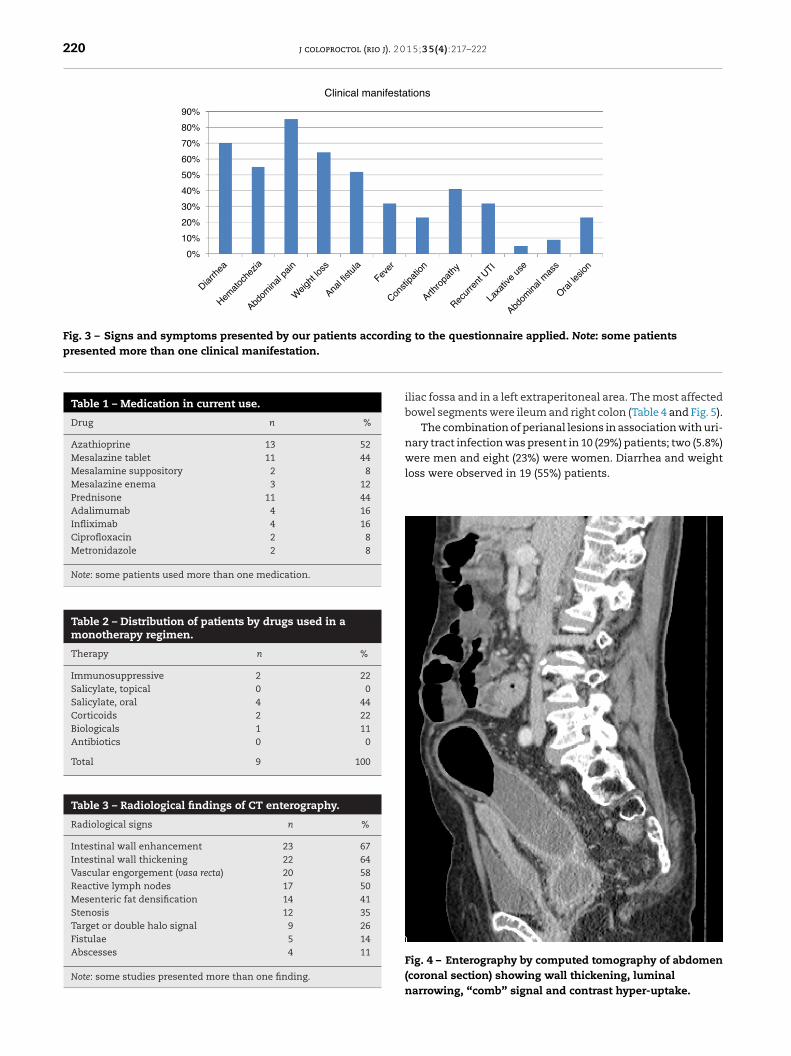
The main clinical manifestations were diarrhea in 24 (70%),hematochezia in 19 (55%), abdominal pain in 29 (85%), andweight loss in 22 (64%) patients (Fig. 3).
Twenty-five patients (73.5%) were under pharmacologicaltreatment. The major drugs used were azathioprine (52%), oralmesalamine (44%) and prednisone (44%), some of these usedin combination (Table 1). Nine patients (36%) were using onlyone drug, especially salicylate (44%), while 13 (38%) were usingmore than one drug (Table 2).
The main findings of CT enterography were intestinal wallsignal enhancement in 23 patients (67%), vascular engorge-ment (vasa recta) in 20 (58%), perienteral fat densification in14 (41%), and bowel wall thickening in 22 (64%) (Table 3 andFigs. 1 and 4). Lymphadenomegaly was observed in 17 (50%)
Fig. 2 – Distribution of patients according to race orethnicity.
220 j coloproctol (rio j). 2 0 1 5;3 5(4):217–222
Clinical manifestations
90%
80%
70%
60%
50%
40%
30%
20%
10%
0%
Diarrh
ea
Hemat
oche
zia
Abdom
inal p
ain
Weig
ht lo
ss
Anal fi
stula
Fever
Consti
patio
n
Arthro
path
y
Recur
rent
UTI
Laxa
tive
use
Abdom
inal m
ass
Oral le
sion
Fig. 3 – Signs and symptoms presented by our patients according to the questionnaire applied. Note: some patientspresented more than one clinical manifestation.
Note: some studies presented more than one finding.
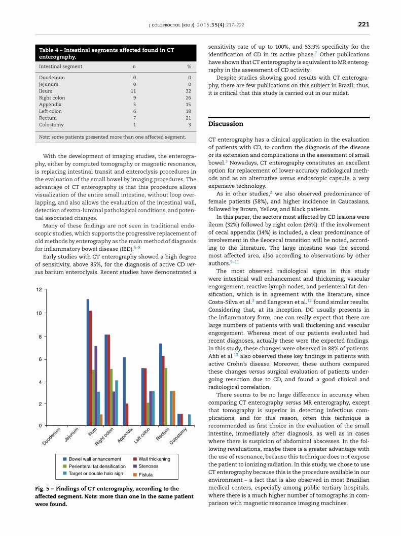
iliac fossa and in a left extraperitoneal area. The most affectedbowel segments were ileum and right colon (Table 4 and Fig. 5).
The combination of perianal lesions in association with uri-nary tract infection was present in 10 (29%) patients; two (5.8%)
were men and eight (23%) were women. Diarrhea and weightloss were observed in 19 (55%) patients.
Fig. 4 – Enterography by computed tomography of abdomen(coronal section) showing wall thickening, luminalnarrowing, “comb” signal and contrast hyper-uptake.
j coloproctol (rio j). 2 0 1 5
Table 4 – Intestinal segments affected found in CTenterography.
Note: some patients presented more than one affected segment.
With the development of imaging studies, the enterogra-hy, either by computed tomography or magnetic resonance,
s replacing intestinal transit and enteroclysis procedures inhe evaluation of the small bowel by imaging procedures. Thedvantage of CT enterography is that this procedure allowsisualization of the entire small intestine, without loop over-apping, and also allows the evaluation of the intestinal wall,etection of extra-luminal pathological conditions, and poten-ial associated changes.
Many of these findings are not seen in traditional endo-copic studies, which supports the progressive replacement ofld methods by enterography as the main method of diagnosisor inflammatory bowel disease (IBD).5–8
Early studies with CT enterography showed a high degreef sensitivity, above 85%, for the diagnosis of active CD ver-us barium enteroclysis. Recent studies have demonstrated a
Bowel wall enhancement
12
10
8
6
4
2
0
Wall thickening
Perienteral fat densification Stenoses
Duode
num
Jejun
um Ilium
Right c
olon
Appen
dix
Left
colon
Rectu
m
Colosto
my
Target or double halo sign Fistula
ig. 5 – Findings of CT enterography, according to theffected segment. Note: more than one in the same patientere found.
;3 5(4):217–222 221
sensitivity rate of up to 100%, and 53.9% specificity for theidentification of CD in its active phase.7 Other publicationshave shown that CT enterography is equivalent to MR enterog-raphy in the assessment of CD activity.
Despite studies showing good results with CT enterogra-phy, there are few publications on this subject in Brazil; thus,it is critical that this study is carried out in our midst.
Discussion
CT enterography has a clinical application in the evaluationof patients with CD, to confirm the diagnosis of the diseaseor its extension and complications in the assessment of smallbowel.3 Nowadays, CT enterography constitutes an excellentoption for replacement of lower-accuracy radiological meth-ods and as an alternative versus endoscopic capsule, a veryexpensive technology.
As in other studies,2 we also observed predominance offemale patients (58%), and higher incidence in Caucasians,followed by Brown, Yellow, and Black patients.
In this paper, the sectors most affected by CD lesions wereileum (32%) followed by right colon (26%). If the involvementof cecal appendix (14%) is included, a clear predominance ofinvolvement in the ileocecal transition will be noted, accord-ing to the literature. The large intestine was the secondmost affected area, also according to observations by otherauthors.9–11
The most observed radiological signs in this studywere intestinal wall enhancement and thickening, vascularengorgement, reactive lymph nodes, and perienteral fat den-sification, which is in agreement with the literature, sinceCosta-Silva et al.3 and Ilangovan et al.12 found similar results.Considering that, at its inception, DC usually presents inthe inflammatory form, one can really expect that there arelarge numbers of patients with wall thickening and vascularengorgement. Whereas most of our patients evaluated hadrecent diagnoses, actually these were the expected findings.In this study, these changes were observed in 88% of patients.Afifi et al.13 also observed these key findings in patients withactive Crohn’s disease. Moreover, these authors comparedthese changes versus surgical evaluation of patients under-going resection due to CD, and found a good clinical andradiological correlation.
There seems to be no large difference in accuracy whencomparing CT enterography versus MR enterography, exceptthat tomography is superior in detecting infectious com-plications; and for this reason, often this technique isrecommended as first choice in the evaluation of the smallintestine, immediately after diagnosis, as well as in caseswhere there is suspicion of abdominal abscesses. In the fol-lowing revaluations, maybe there is a greater advantage withthe use of resonance, because this technique does not exposethe patient to ionizing radiation. In this study, we chose to useCT enterography because this is the procedure available in our
environment – a fact that is also observed in most Brazilianmedical centers, especially among public tertiary hospitals,where there is a much higher number of tomographs in com-parison with magnetic resonance imaging machines.
Perianal lesions associated with urinary tract infectionwere present in 10 (29%) patients, two (5.8%) men and eight(23%) women. Diarrhea and weight loss were observed in 19(55%) patients. Such an association may be related to the mal-absorption syndrome observed in CD patients.2
Importantly, cross-sectional studies reflect certainmoments of the sample; thus, changes may occur in severalaspects analyzed over time, and with the inclusion of newpatients.
Conclusion
The study allows one to observe that the main radiologicalfindings of CT enterography were intestinal wall enhancementand thickening and vascular engorgement, mainly affectingthe ileum and right colon. The main clinical manifestationsin our patients were diarrhea and abdominal pain.
Conflicts of interest
The authors declare no conflicts of interest.
e f e r e n c e s
1. Habr-Gama A, Cerski CTS, Moreira JPT, Caserta NMG, OliveiraJúnior O, Araújo SEA. Diretrizes da Associacão MédicaBrasileira. Doenca de Crohn intestinal: manejo. Rev AssocMed Bras. 2011;57:10–3.
2. Santos SMR [Tese Mestrado] Doenca de Crohn: Etiopatogenia,aspetos clínicos, diagnóstico e tratamento. Porto, Portugal.:Universidade Fernando Pessoa; 2013. Available from:http://hdl.handle.net/10284/4100
4. Glick SN. Critérios de adequacão de exames de imagem eradioterapia. São Paulo: Colégio Brasileiro de Radiologia; 2005.p. 309.
5. D’Ippolito G, Braga FA, Resende MC, Bretas EAS, Nunes TF,Rosas GQ, et al. Enterografia por tomografiacomputadorizada: uma avaliacão de diferentes contrastesorais neutros. Radiol Bras. 2012;45:139–43.
6. Hanauer SB, Sandborn W. Practice Parameters Committee ofthe American College of Gastroenterology. Management ofCrohn’s disease in adults. Am J Gastroenterol. 2001;96:635–43.
7. Cakmakci E, Erturk SM, Cakmarkci S. Comparison of theresults of computerized tomographic and diffusion-weightedmagnetic resonance imaging techniques in inflammatorybowel diseases. Quant Imaging Med Surg. 2013;3:327–33.
8. Amitai MM, Ben-Horin S, Eliakim R, Kopylov U. Magneticresonance enterography in Crohn’s disease: a guide tocommon imaging manifestations for the IBD physician. JCrohn’s Colitis. 2013;7:603–15.
9. Faria LC, Ferrari MLA, Cunha AS. Aspectos clínicos da doencade Crohn em um centro de referência para doencasintestinais. GED Gastroenterol Endosc Dig. 2004;23:151–64.
0. Hardt MR, Kotze PG, Teixeira FV, Ludvig JC, Malluta EF,Kleinubing H Jr, et al. Epidemiological profile of 175 patientswith Crohn’s disease submitted to biological therapy. JColoproct. 2012;32:395–401.
1. Kleinubing-Junior H, Pinho MSL, Ferreira LC, Bachtold GA,Merki A. Outpatients profile with inflammatory boweldisease. ABCD Arq Bras Cir Dig. 2011;24:200–3.
2. Ilangovan R, Burling D, George A, Gupta A, Marshall M, TaylorSA. CT enterography: review of technique and practical tips.
and treatment. Evaluation using MDCT enterography andendoscopy. Egypt J Radiol Nucl Med. 2012;43:507–17.