1
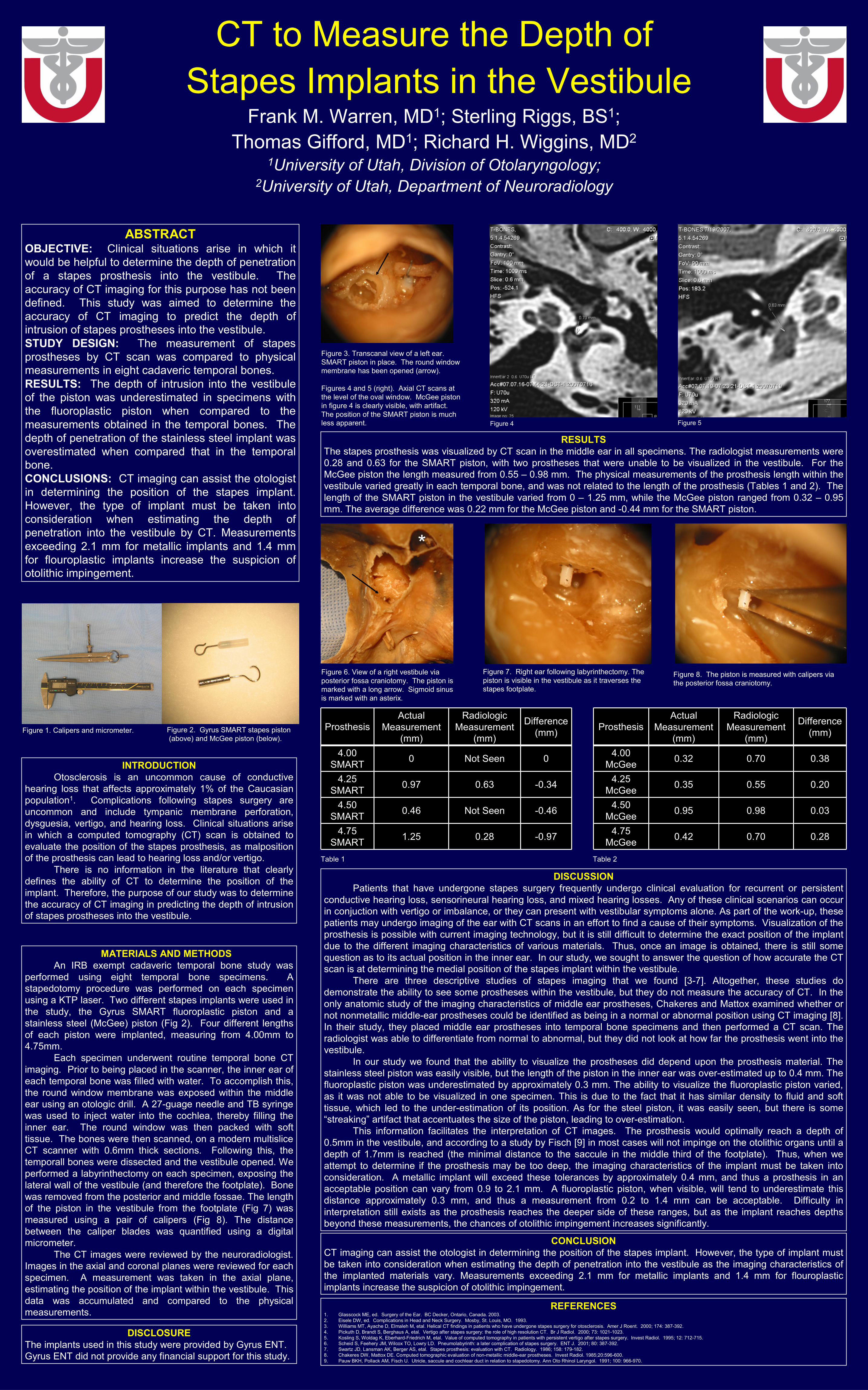
CT to Measure the Depth of Stapes Implants in the Vestibule Frank M. Warren, MD 1 ; Sterling Riggs, BS 1 ; Thomas Gifford, MD 1 ; Richard H. Wiggins, MD 2 1 University of Utah, Division of Otolaryngology; 2 University of Utah, Department of Neuroradiology ABSTRACT OBJECTIVE: Clinical situations arise in which it would be helpful to determine the depth of penetration of a stapes prosthesis into the vestibule. The accuracy of CT imaging for this purpose has not been defined. This study was aimed to determine the accuracy of CT imaging to predict the depth of intrusion of stapes prostheses into the vestibule. STUDY DESIGN: The measurement of stapes prostheses by CT scan was compared to physical measurements in eight cadaveric temporal bones. RESULTS: The depth of intrusion into the vestibule of the piston was underestimated in specimens with the fluoroplastic piston when compared to the measurements obtained in the temporal bones. The depth of penetration of the stainless steel implant was overestimated when compared that in the temporal bone. CONCLUSIONS: CT imaging can assist the otologist in determining the position of the stapes implant. However, the type of implant must be taken into consideration when estimating the depth of penetration into the vestibule by CT. Measurements exceeding 2.1 mm for metallic implants and 1.4 mm for flouroplastic implants increase the suspicion of otolithic impingement. INTRODUCTION Otosclerosis is an uncommon cause of conductive hearing loss that affects approximately 1% of the Caucasian population 1 . Complications following stapes surgery are uncommon and include tympanic membrane perforation, dysguesia, vertigo, and hearing loss. Clinical situations arise in which a computed tomography (CT) scan is obtained to evaluate the position of the stapes prosthesis, as malposition of the prosthesis can lead to hearing loss and/or vertigo. There is no information in the literature that clearly defines the ability of CT to determine the position of the implant. Therefore, the purpose of our study was to determine the accuracy of CT imaging in predicting the depth of intrusion of stapes prostheses into the vestibule. MATERIALS AND METHODS An IRB exempt cadaveric temporal bone study was performed using eight temporal bone specimens. A stapedotomy procedure was performed on each specimen using a KTP laser. Two different stapes implants were used in the study, the Gyrus SMART fluoroplastic piston and a stainless steel (McGee) piston (Fig 2). Four different lengths of each piston were implanted, measuring from 4.00mm to 4.75mm. Each specimen underwent routine temporal bone CT imaging. Prior to being placed in the scanner, the inner ear of each temporal bone was filled with water. To accomplish this, the round window membrane was exposed within the middle ear using an otologic drill. A 27-guage needle and TB syringe was used to inject water into the cochlea, thereby filling the inner ear. The round window was then packed with soft tissue. The bones were then scanned, on a modern multislice CT scanner with 0.6mm thick sections. Following this, the temporall bones were dissected and the vestibule opened. We performed a labyrinthectomy on each specimen, exposing the lateral wall of the vestibule (and therefore the footplate). Bone was removed from the posterior and middle fossae. The length of the piston in the vestibule from the footplate (Fig 7) was measured using a pair of calipers (Fig 8). The distance between the caliper blades was quantified using a digital micrometer. The CT images were reviewed by the neuroradiologist. Images in the axial and coronal planes were reviewed for each specimen. A measurement was taken in the axial plane, estimating the position of the implant within the vestibule. This data was accumulated and compared to the physical measurements. RESULTS The stapes prosthesis was visualized by CT scan in the middle ear in all specimens. The radiologist measurements were 0.28 and 0.63 for the SMART piston, with two prostheses that were unable to be visualized in the vestibule. For the McGee piston the length measured from 0.55 – 0.98 mm. The physical measurements of the prosthesis length within the vestibule varied greatly in each temporal bone, and was not related to the length of the prosthesis (Tables 1 and 2). The length of the SMART piston in the vestibule varied from 0 – 1.25 mm, while the McGee piston ranged from 0.32 – 0.95 mm. The average difference was 0.22 mm for the McGee piston and -0.44 mm for the SMART piston. DISCUSSION Patients that have undergone stapes surgery frequently undergo clinical evaluation for recurrent or persistent conductive hearing loss, sensorineural hearing loss, and mixed hearing losses. Any of these clinical scenarios can occur in conjuction with vertigo or imbalance, or they can present with vestibular symptoms alone. As part of the work-up, these patients may undergo imaging of the ear with CT scans in an effort to find a cause of their symptoms.. Visualization of the prosthesis is possible with current imaging technology, but it is still difficult to determine the exact position of the implant due to the different imaging characteristics of various materials. Thus, once an image is obtained, there is still some question as to its actual position in the inner ear. In our study, we sought to answer the question of how accurate the CT scan is at determining the medial position of the stapes implant within the vestibule. There are three descriptive studies of stapes imaging that we found [3-7]. Altogether, these studies do demonstrate the ability to see some prostheses within the vestibule, but they do not measure the accuracy of CT. In the only anatomic study of the imaging characteristics of middle ear prostheses, Chakeres and Mattox examined whether or not nonmetallic middle-ear prostheses could be identified as being in a normal or abnormal position using CT imaging [8]. In their study, they placed middle ear prostheses into temporal bone specimens and then performed a CT scan. The radiologist was able to differentiate from normal to abnormal, but they did not look at how far the prosthesis went into the vestibule. In our study we found that the ability to visualize the prostheses did depend upon the prosthesis material. The stainless steel piston was easily visible, but the length of the piston in the inner ear was over-estimated up to 0.4 mm. The fluoroplastic piston was underestimated by approximately 0.3 mm. The ability to visualize the fluoroplastic piston varied, as it was not able to be visualized in one specimen. This is due to the fact that it has similar density to fluid and soft tissue, which led to the under-estimation of its position. As for the steel piston, it was easily seen, but there is some “streaking” artifact that accentuates the size of the piston, leading to over-estimation. This information facilitates the interpretation of CT images. The prosthesis would optimally reach a depth of 0.5mm in the vestibule, and according to a study by Fisch [9] in most cases will not impinge on the otolithic organs until a depth of 1.7mm is reached (the minimal distance to the saccule in the middle third of the footplate). Thus, when we attempt to determine if the prosthesis may be too deep, the imaging characteristics of the implant must be taken into consideration. A metallic implant will exceed these tolerances by approximately 0.4 mm, and thus a prosthesis in an acceptable position can vary from 0.9 to 2.1 mm. A fluoroplastic piston, when visible, will tend to underestimate this distance approximately 0.3 mm, and thus a measurement from 0.2 to 1.4 mm can be acceptable. Difficulty in interpretation still exists as the prosthesis reaches the deeper side of these ranges, but as the implant reaches depths beyond these measurements, the chances of otolithic impingement increases significantly. CONCLUSION CT imaging can assist the otologist in determining the position of the stapes implant. However, the type of implant must be taken into consideration when estimating the depth of penetration into the vestibule as the imaging characteristics of the implanted materials vary. Measurements exceeding 2.1 mm for metallic implants and 1.4 mm for flouroplastic implants increase the suspicion of otolithic impingement. REFERENCES 1. Glasscock ME, ed. Surgery of the Ear. BC Decker, Ontario, Canada. 2003. 2. Eisele DW, ed. Complications in Head and Neck Surgery. Mosby, St. Louis, MO. 1993. 3. Williams MT, Ayache D, Elmaleh M, etal. Helical CT findings in patients who have undergone stapes surgery for otosclerosis. Amer J Roent. 2000; 174: 387-392. 4. Pickuth D, Brandt S, Berghaus A, etal. Vertigo after stapes surgery: the role of high resolution CT. Br J Radiol. 2000; 73: 1021-1023. 5. Kosling S, Woldag K, Eberhard-Friedrich M, etal. Value of computed tomography in patients with persistent vertigo after stapes surgery. Invest Radiol. 1995; 12: 712-715. 6. Scheid S, Feehery JM, Wilcox TO, Lowry LD. Pneumolabyrinth: a later complication of stapes surgery. ENT J. 2001; 80: 387-392. 7. Swartz JD, Lansman AK, Berger AS, etal. Stapes prosthesis: evaluation with CT. Radiology. 1986; 158: 179-182. 8. Chakeres DW, Mattox DE. Computed tomographic evaluation of non-metallic middle-ear prostheses. Invest Radiol. 1985;20:596-600. 9. Pauw BKH, Pollack AM, Fisch U. Utricle, saccule and cochlear duct in relation to stapedotomy. Ann Oto Rhinol Laryngol. 1991; 100: 966-970. -0.34 0.63 0.97 4.25 SMART -0.97 0.28 1.25 4.75 SMART -0.46 Not Seen 0.46 4.50 SMART 0 Not Seen 0 4.00 SMART Difference (mm) Radiologic Measurement (mm) Actual Measurement (mm) Prosthesis 0.20 0.55 0.35 4.25 McGee 0.28 0.70 0.42 4.75 McGee 0.03 0.98 0.95 4.50 McGee 0.38 0.70 0.32 4.00 McGee Difference (mm) Radiologic Measurement (mm) Actual Measurement (mm) Prosthesis Figure 1. Calipers and micrometer. Figure 2. Gyrus SMART stapes piston (above) and McGee piston (below). Figure 3. Transcanal view of a left ear. SMART piston in place. The round window membrane has been opened (arrow). Figures 4 and 5 (right). Axial CT scans at the level of the oval window. McGee piston in figure 4 is clearly visible, with artifact. The position of the SMART piston is much less apparent. Figure 4 Figure 5 Figure 6. View of a right vestibule via posterior fossa craniotomy. The piston is marked with a long arrow. Sigmoid sinus is marked with an asterix. Figure 7. Right ear following labyrinthectomy. The piston is visible in the vestibule as it traverses the stapes footplate. Figure 8. The piston is measured with calipers via the posterior fossa craniotomy. Table 1 Table 2 DISCLOSURE The implants used in this study were provided by Gyrus ENT. Gyrus ENT did not provide any financial support for this study. *


![Case Report A Rare Stapes Abnormalitydownloads.hindawi.com/journals/criot/2015/387642.pdfCase Report A Rare Stapes Abnormality ... (adapted from Bhatti and Bluestone [ ], surgical](https://static.documents.pub/doc/80x56/5e7b048b765f92114f47b80a/case-report-a-rare-stapes-case-report-a-rare-stapes-abnormality-adapted-from.jpg)


