THE QUALITY OF VISION IN THE CTENID SPIDERCUPIENNIUS SALEI
BY M. F. LAND
Neuroscience Research Centre, School of Biological Sciences,University of Sussex, Brighton BN1 9QG, UK
AND F. G. BARTH
Institut fur Zoologie der Universitat Wien, Althanstrasse 14,A-1090 Wien, Austria
Accepted 20 November 1991
Summary
Much is known about the mechanosensory behaviour of the spider Cupienniussalei Keyserling, but much less about its visual capabilities. In this study the qualityof the optical image, the retinal resolution and the fields of view were assessed foreach of the four pairs of eyes. The image is of good quality in all eyes. Theprincipal (antero-median) eyes lack a tapetum and have an inter-receptor angle of2.9°. The three secondary eyes (antero-lateral, postero-median and postero-lateral) all have 'gridiron' tapeta with receptors arranged in rows. The angularseparations (along rows x between rows) are 3.6° x 9.3°, 0.9° x 2.3° and 1.0° x3.0°, respectively. Although the disposition of eyes on the head is similar to that ofpisaurid spiders, all other features of the eyes, including the sizes and shapes of thefields of view, resemble those of lycosid spiders. The peripheral visual system ofCupiennius can thus, in principle, support a similar range of visual behaviour tothat of lycosids, which includes prey capture, predator avoidance and courtship.
Introduction
Much of the behaviour of the ctenid spider Cupiennius salei is mediated by itsmechanical senses, with both courtship and prey capture involving the detection ofsubstratum-borne vibrations (Barth, 1985). This species is largely nocturnal andshows clear peaks of locomotor activity at night. The extent of visual involvementin the various behaviours is not known, but it has been claimed that prey capture isnot impaired if the eyes are covered. In spite of this, Cupiennius and other ctenidsappear to have eyes that are similar to those of other hunting spiders, and in theirexternal appearance they do not seem to be either reduced or degenerate.Furthermore, the eyes connect to extensive neuropiles that again are typical ofother hunting spiders (N. J. Strausfeld, P. Weltzien and F. G. Barth, inpreparation). In view of the paradoxical absence of visual behaviour, we decided
to examine the eyes of Cupiennius in detail and to assess their optical perform-ance.
These spiders were originally classed as a separate family, the Ctenidae, butwere reclassified by Homann (1961, 1971) as the Cteninae, a sub-family of theLycosidae and sister group to the Lycosinae, or wolf spiders. This redesignationwas based largely on eye morphology. However, Lachmuth et al. (1985) retain thefamily name Ctenidae in a recent revision of the genus Cupiennius. Whatever theirexact taxonomic status, it is clear that the ctenids and lycosids are closely related.Some physiological information is available for wolf spider eyes (Homann, 1931;Land, 1985) and the new information in the present paper provides anotheropportunity to assess the extent of similarity between the two groups and also tomake comparisons with other groups of hunting spiders such as the salticids. Manywolf spiders are known to be visual hunters, responding to moving objects witheither attack or evasion. Thus, a strong resemblance between the eyes of theCteninae and the Lycosinae would suggest that it would be worthwhile to lookharder for visual behaviour in Cupiennius and other ctenids.
Materials and methods
Two typical adult female Cupiennius salei were used in this study. They camefrom a colony kept in Vienna, and were almost identical, with a cephalothoraxlength of 9.5 mm. Four kinds of measurement were made on one animal or both:anatomical measurements of the eyes and components of the retina, focal lengthmeasurements, ophthalmoscopic measurements of the retinal structures and fieldof view measurements.
Measurements of the external dimensions of the eyes were made fromphotographs of living spiders, narcotised with CO2. The dimensions of the retinalstructures were measured on 8/im sections from standard wax-embedded prep-arations fixed in 5 % formaldehyde in spider saline and stained with haematoxylinand eosin. Roughly 15% shrinkage is expected from this method, and allmeasurements on the sections have been increased by this amount.
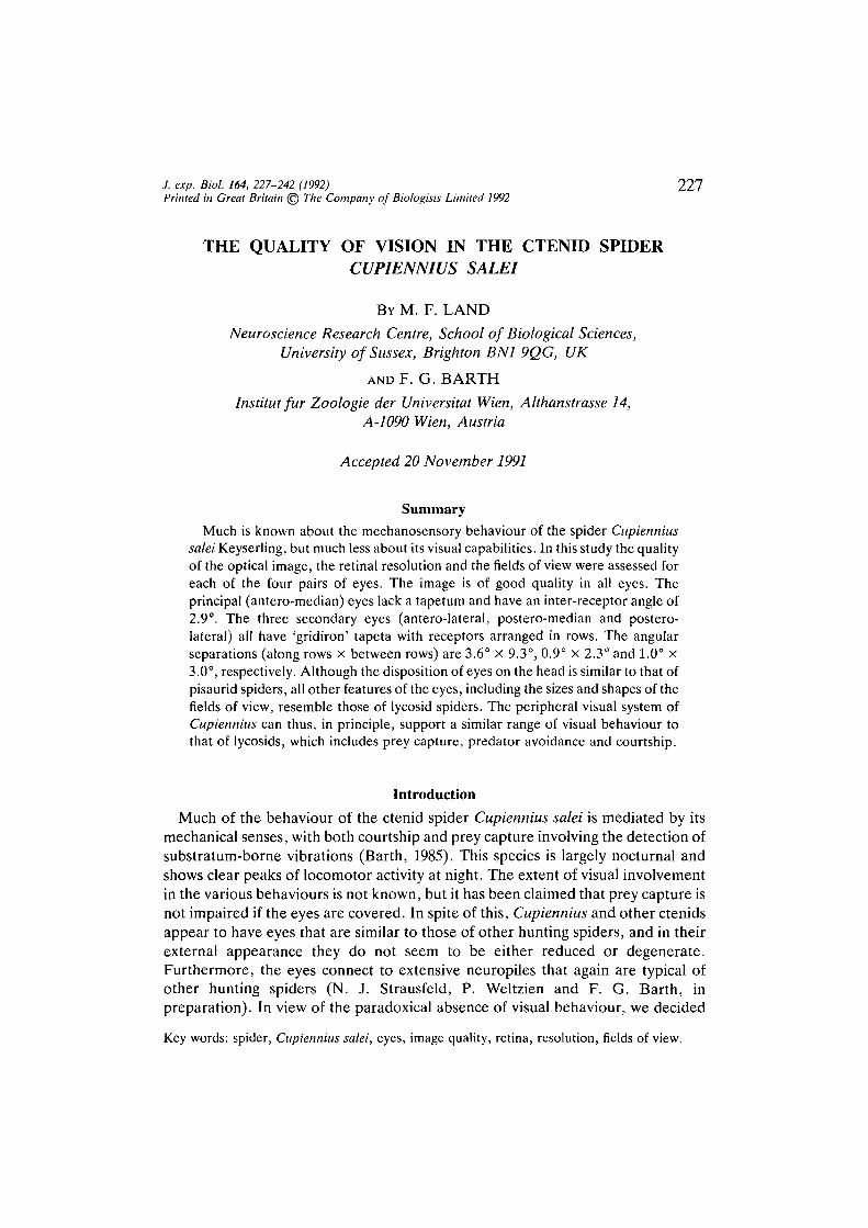
Focal lengths of the lenses were measured using the 'hanging drop' method ofHomann (1928; see also Land, 1985). The lenses are dissected from the head withsome surrounding carapace and placed in the meniscus of a hanging drop of salineso that the outer surface is in air and the inner surface in saline, as in life (see Figs1A, 4B). The image plane is then examined with a microscope while the spider'slens views a target, such as a pair of lines, at a known distance (M). The object andimage sizes {O and /) are measured, and from these the lens-image distance (v) isdetermined from: v/u=l/O. The focal length (or posterior nodal distance, / ) isthen found from the lens formula: (l/v) —(l/w) = l// .
The retina of the antero-lateral (AL), postero-medial (PM) and postero-lateral(PL) eyes, all of which have reflecting tapeta, can be studied directly in the livinganimal using an ophthalmoscope. Some detail can be seen with an ordinarymedical ophthalmoscope, but in this study a special instrument was used, more
Eyes of ctenid spiders
B
R c
Lamp A A
229
Beamsplitter Eye
Observer
Latitude
Longitude
Fig. 1. Methods used in this study. (A) Hanging drop method for measuring focallength (Homann, 1928). Symbols are explained in the text. (B) Ophthalmoscope forobserving living retina (see Fig. 4C) (Land, 1969). R, plane conjugate with infinity forobjects to be imaged on retina; C, plane conjugate with cornea for maskingilluminating beam; L, removable lens allowing observation of either retina or eyesurface. (C) Cardan-arm goniometer for measuring fields of view (see Fig. 5) (Land,1985). T, telescope; S, light source; M, mirror.
suitable for small eyes (Fig. IB). Details of its construction are given in Land(1969, 1984). The instrument has two optical paths, for viewing and illumination,separated behind the objective lens by a beam splitter. The viewing beam can beused either as a microscope to examine the external features of the eye or, byinserting lens L, it can be used to view the spider's retina, which is conjugate with
230 M. F. LAND AND F. G. BARTH
infinity (Fig. 4C). When used in this way, the field of view in the eyepiece can becalibrated directly in terms of the angle subtended by structures in the spider's eye,and so can provide a direct in vivo measure of resolution. In the illuminatingbeam, objects placed in the plane R are imaged on the retina by the spider's ownlens, and this plane can thus be used to position test objects of known angulardimensions onto the retina. The principal (AM) eyes, which lack a tapetum, donot reflect enough light for the ophthalmoscopic method to be usable.
Fields of view are measured with a small telescope mounted on a Cardan-armgoniometer (Fig. 1C). The telescope T has a narrow-angle light source (5)attached, which illuminates the viewing direction via a half-silvered mirror (M).As the telescope is rotated around the animal, each of the secondary eyes 'lightsup' in turn, as green light is reflected back from the tapetum (see Land, 1985,Fig. 3c). The angular field over which a particular eye reflects light corresponds tothe angle subtended by the tapetum, which we know from histological study to beco-extensive with the retina. This method can also be used to study the principal(AM) eyes, because although these lack a tapetum there is a noticeable changefrom a pinkish-brown reflex to a darker brown at the boundary of the retina. Thefields of view in Fig. 5 are plotted on Aitoff's equal area projection, which allowsthe whole 360° field to be shown and preserves the relationships of the field sizes.
Results
Anatomy of the eyes
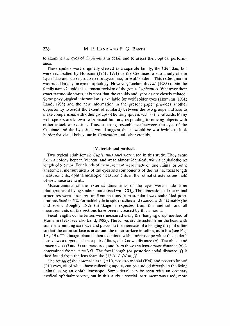
Like most other spiders, Cupiennius has eight eyes of two kinds. They are allsuperficially similar, but the antero-median, or 'principal', eyes lack a reflectingtapetum and have receptors with a quite different structure from those of the otherthree 'secondary' eyes (antero-lateral, postero-median and postero-lateral). Thearrangement of the eyes is shown in Fig. 2. They are in two strongly curved rows,the AM and AL eyes in front of the PM and PLs. The AMs lie directly in front ofthe PMs, and similarly with the ALs and PLs. This disposition is very similar tothat in the lycosid relative Pisaura, and not like that of typical lycosids, where thePL eyes are usually set much further back and the PM eyes further out, so that theylie behind the ALs rather than the AMs. The relative sizes of the eyes, however,follow the typical lycosid pattern (Table 1). The PM eyes are the largest, the PLsare slightly smaller, followed by the AMs and finally the ALs. In Pisaura, the ALsare larger than the AMs, and not much smaller than the other secondary eyes. Insalticids the order is completely different, with the AMs much the largest,followed by the ALs, PLs and PMs.
The eyes themselves all have a similar shape, the retina forming a roughlyhemispherical cup behind the lens, which is biconvex. The space between the lensand retina is filled by a cellular 'vitreous'. Both front and rear lens surfaces arenear hemispheres, and they share the same centre of curvature; however, the radiiare different, the smaller rear surface having a radius of curvature about 0.8 timesthat of the front. The focal lengths of the lenses, measured by the hanging drop
Eyes of ctenid spiders 231
AM
Salticus
Fig. 2. Layout of the eyes of Cupiennius, left, and three other hunting spiders, right(not to the same scale). AM, antero-median eyes; AL, antero-lateral; PM, postero-median; PL, postero-lateral.
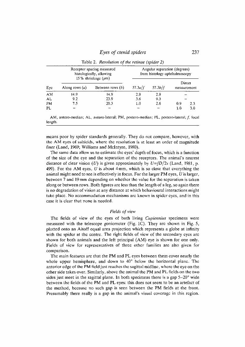
Table 1. Dimensions of the eyes (fim) (spider 1)
Eye
AMALPMPL
AM,
(D)
396256629625
antero-median; AL,
Radius of curvature
Anterior
204147320320
antero-lateral:
Posterior
157—
236-
Focal length(/)
293148448432
if ID)0.740.580.710.69
; PM, postero-median; PL, postero-lateral.
technique, are also given in Table 1. As expected, these are proportional to theeye diameters, and the F-numbers of the lenses (f/D, where/is focal length and Dis diameter) are all in the range 0.58-0.74. By the standards of camera lenses,these are astonishingly low figures, implying very bright images, but they are notatypical of hunting spiders generally (Land, 1985).
The structure of the AM retina is different from that of the other three eyes. Thereceptors cells are about 90 pum. long and 14 /im wide, with their short rhabdomericregions adjacent to the vitreous (Fig. 3A). In cross section the cells in this regionform a rather irregular lattice, with each receptor having rhabdomeres on three or
232 M. F. LAND AND F. G. BARTH
hi M
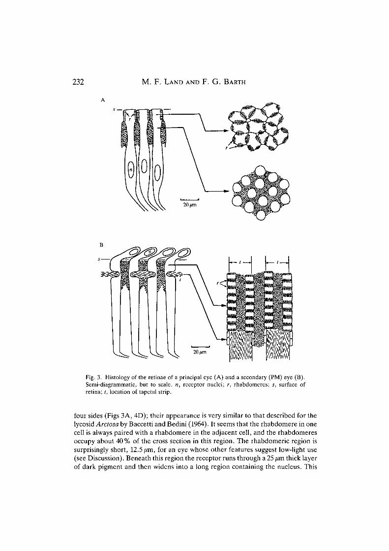
Fig. 3. Histology of the retinae of a principal eye (A) and a secondary (PM) eye (B).Semi-diagrammatic, but to scale, n, receptor nuclei; r, rhabdomeres; s, surface ofretina; t, location of tapetal strip.
four sides (Figs 3A, 4D); their appearance is very similar to that described for thelycosid Arctosa by Baccetti and Bedini (1964). It seems that the rhabdomere in onecell is always paired with a rhabdomere in the adjacent cell, and the rhabdomeresoccupy about 40% of the cross section in this region. The rhabdomeric region issurprisingly short, 12.5/im, for an eye whose other features suggest low-light use(see Discussion). Beneath this region the receptor runs through a 25 fim thick layerof dark pigment and then widens into a long region containing the nucleus. This
Eyes of ctenid spiders 233
then tapers to form an axon of the optic nerve which runs to the first opticneuropile. There are no other neurones in the eye. The retina occupies most of theback of the eyecup and appears to be uniform. There is no suggestion that thisretina has any of the characteristics of salticid retinae, namely multiple receptortypes arranged in tiers, with large differences in packing density in differentregions (Land, 1969). Blest and O'Carroll (1989) have found evidence of two-layertiering in the AM eyes of the wolf spider Geolycosa godeffroyi, although not in thetype genus Lycosa. Cupiennius does not show tiering and appears to have an AMretina typical of those described previously for lycosids (Homann, 1931; Baccettiand Bedini, 1964).
The secondary eyes all have a similar structure, except that the AL eyes aremuch smaller than the PMs and PLs and are less precisely ordered. They are allbuilt around a 'gridiron' tapetum, which consists of a series of parallel strips ofreflecting material forming a double ladder-like array (Fig. 4A). In the PM and PLeyes the strips run roughly horizontally (parallel to the animal's longitudinalplane). Each tapetal strip supports two rows of receptors as indicated in Fig. 3B.These receptors differ from those of the AM eyes in a number of ways. First, thenucleus is in the part of the cell nearest the lens, and the nuclei form a thin layerbetween the vitreous and the retina proper. Second, the rhabdomeres are on twosides of the receptors only, the cells being rectangular in cross section in the regionabove the tapetum and organised into linear arrays (Fig. 4E). As in the AM eyes,the rhabdomere of one receptor abuts the corresponding one from the nextreceptor, to form what appears to be a single rhabdom. Again, about 40% of thecross section in this region is occupied by the rhabdomeres. Third, the rhabdo-meric part of the cell 'sits' on the tapetal strip, with a narrow isthmus passingaround it before expanding again. The receptor then extends inwards withoutrhabdomeres for about 85 [xm before tapering to form an optic nerve fibre. Thisstructure means that nearly all of the rhabdomeric region is backed by tapetum,and so receives light twice, once before and once after reflection; this doubles theeffective length of the rhabdom for the purposes of light absorption. The gridironstructure of the secondary eyes results in retinae which sample the image in a verydifferent way from the AM retina. Not only are the receptors arranged strictly inrows, but the spacing of their centres differs considerably, depending on thedirection. The receptor spacing between the receptor rows is greater than thatalong the rows by more than a factor of two (see Table 2), and this must mean thatthe horizontal resolution of the eyes is much better than the vertical resolution.
This description of the secondary eye structure is once again very similar to thatgiven by Baccetti and Bedini (1964) for Arctosa. One small difference is that inArctosa there is dark pigment between receptors within a row, rather than justbetween the rows themselves as in Cupiennius. Amongst lycosids, Pardosa issimilar to Arctosa, but Geolycosa and Trochosa are like Cupiennius. Presumablythese differences in the optical isolation of one receptor from another, as well asthe asymmetries in sampling, must have functional meanings; but our ignorance ofthe behavioural role of these eyes is too profound to provide any clues.
234 M. F. LAND AND F. G. BARTH
Eyes of ctenid spiders 235
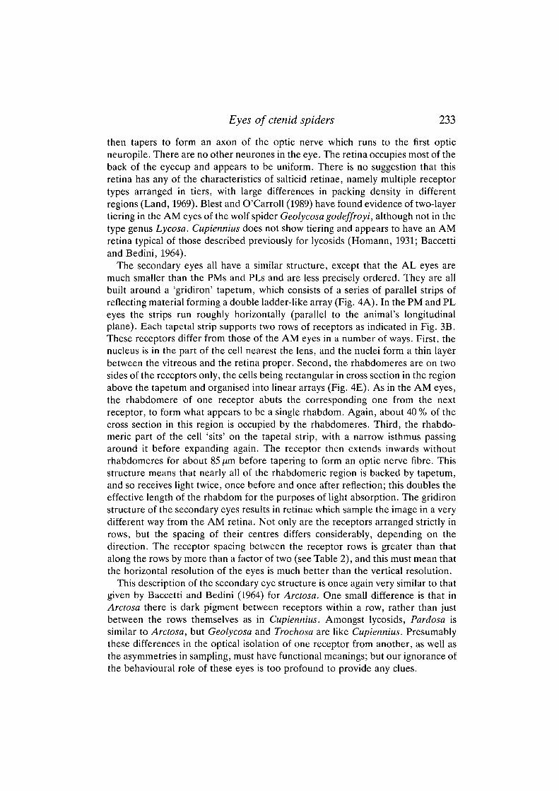
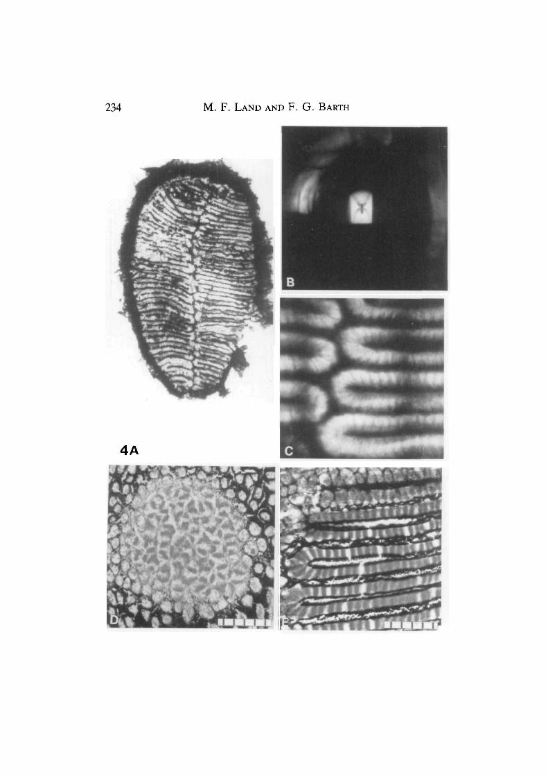
Fig. 4. (A) Whole PM retina, showing the gridiron pattern of tapetal strips. (B) Theimage of a lycosid spider photographed at the focus of the lens of a PM eye, using thehanging-drop method (Fig. 1A). The image size is approximately that of anotherCupiennius at 25cm. The magnifications in A and B are approximately the same, sothat the optical and retinal resolution can be compared directly. (C) Ophthalmoscopicimage of part of the PM retina, near the midline, photographed through the eye's ownlens using the instrument pictured in Fig. IB. The boundaries between the receptorsare clearly visible (compare Figs 3 and 4E). The two blurred vertical lines are theimages of lines 10° apart in the field of view and act as a direct angular calibration.(D) Tangential histological section of the centre of the AM retina (cf. Fig. 3A),showing the irregular arrangement of the rhabdoms. (E) Similar section of the PMretina (cf. Figs 3B and 4C), showing the linear arrangement of the rhabdoms. Scalebars, 5x 10,i/m.
Resolution
The capacity of an eye to resolve detail in the environment is limited by twofactors: the ability of the eye to produce a sharp image and the fineness of theretinal mosaic (see Land, 1981). In the human eye both factors are approximatelymatched, with the cone mosaic just able to resolve the finest pattern the optics canprovide - a grating with a period of 1 min of arc. In some spiders, such as thesalticid Portia, the resolution of the AM eyes is perhaps only a factor of 5 lowerthan that in humans (Williams and Mclntyre, 1980), but in Cupiennius the eyes'performance is not comparable with this. The relatively coarse receptor mosaic inboth the principal and secondary eyes means that the finest resolvable grating willhave a spatial period (the angular subtense of a line pair at the eye) of 2° or more.
It is clear that the potential resolution of the eyes' optics is much better than this.The ultimate limit to resolution, which cannot be improved upon, is set by theamount of blur that results from diffraction at the aperture. Expressed as theangular spatial period of the finest grating that can be resolved, this limit is givenby w/Drad, or 57.3w/D degrees, where D is the eye diameter and w thewavelength of light. Notice that the bigger D is, the finer the resolution. Eyediameters range from 256 fim (AL) up to 629 fim (PM) and, taking w as 0.5 [Am, thisgives grating periods between 0.11 and 0.05°. These values are smaller by morethan an order of magnitude than the angular receptor spacing, so diffractioncertainly does not limit resolution in any of these eyes. It is also possible that thequality of the image is affected by defects other than diffraction, the mostimportant being spherical aberration, which is potentially very serious in a lenswith an F-number as low as 0.6. However, there are good reasons for thinking thatspider lenses are at least partially corrected for this by having an opticallyinhomogeneous structure (Blest and Land, 1977).
Two kinds of direct evidence suggest that the image is as good as or better thanthe sampling ability of the retina. (The finest grating the retina can resolve willhave a period equal to twice the receptor spacing, since one receptor is requiredfor each dark and each light stripe; see Land, 1981.) Direct inspection of the imageproduced by an excised lens (Fig. 4B) shows that it is indeed of good quality and
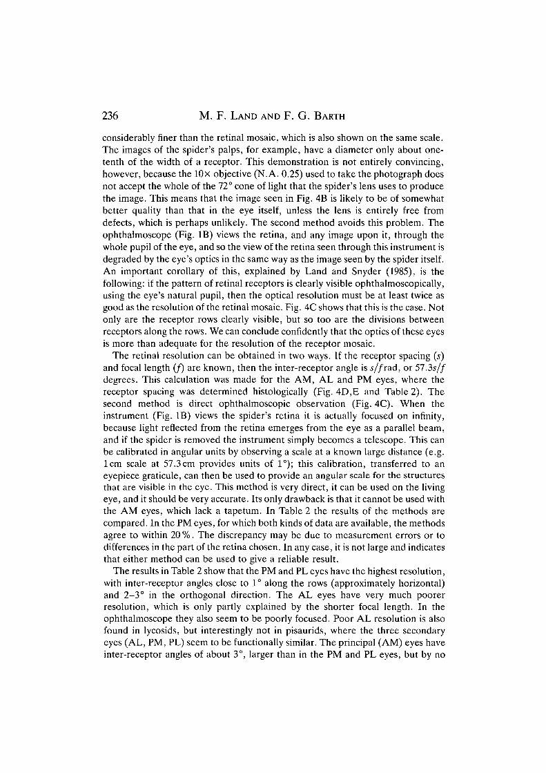
236 M. F. LAND AND F. G. BARTH
considerably finer than the retinal mosaic, which is also shown on the same scale.The images of the spider's palps, for example, have a diameter only about one-tenth of the width of a receptor. This demonstration is not entirely convincing,however, because the lOx objective (N.A. 0.25) used to take the photograph doesnot accept the whole of the 72° cone of light that the spider's lens uses to producethe image. This means that the image seen in Fig. 4B is likely to be of somewhatbetter quality than that in the eye itself, unless the lens is entirely free fromdefects, which is perhaps unlikely. The second method avoids this problem. Theophthalmoscope (Fig. IB) views the retina, and any image upon it, through thewhole pupil of the eye, and so the view of the retina seen through this instrument isdegraded by the eye's optics in the same way as the image seen by the spider itself.An important corollary of this, explained by Land and Snyder (1985), is thefollowing: if the pattern of retinal receptors is clearly visible ophthalmoscopically,using the eye's natural pupil, then the optical resolution must be at least twice asgood as the resolution of the retinal mosaic. Fig. 4C shows that this is the case. Notonly are the receptor rows clearly visible, but so too are the divisions betweenreceptors along the rows. We can conclude confidently that the optics of these eyesis more than adequate for the resolution of the receptor mosaic.
The retinal resolution can be obtained in two ways. If the receptor spacing (s)and focal length (f) are known, then the inter-receptor angle is s/frad, or 57.35//degrees. This calculation was made for the AM, AL and PM eyes, where thereceptor spacing was determined histologically (Fig. 4D,E and Table 2). Thesecond method is direct ophthalmoscopic observation (Fig. 4C). When theinstrument (Fig. IB) views the spider's retina it is actually focused on infinity,because light reflected from the retina emerges from the eye as a parallel beam,and if the spider is removed the instrument simply becomes a telescope. This canbe calibrated in angular units by observing a scale at a known large distance (e.g.lcm scale at 57.3cm provides units of 1°); this calibration, transferred to aneyepiece graticule, can then be used to provide an angular scale for the structuresthat are visible in the eye. This method is very direct, it can be used on the livingeye, and it should be very accurate. Its only drawback is that it cannot be used withthe AM eyes, which lack a tapetum. In Table 2 the results of the methods arecompared. In the PM eyes, for which both kinds of data are available, the methodsagree to within 20 %. The discrepancy may be due to measurement errors or todifferences in the part of the retina chosen. In any case, it is not large and indicatesthat either method can be used to give a reliable result.
The results in Table 2 show that the PM and PL eyes have the highest resolution,with inter-receptor angles close to 1° along the rows (approximately horizontal)and 2-3° in the orthogonal direction. The AL eyes have very much poorerresolution, which is only partly explained by the shorter focal length. In theophthalmoscope they also seem to be poorly focused. Poor AL resolution is alsofound in lycosids, but interestingly not in pisaurids, where the three secondaryeyes (AL, PM, PL) seem to be functionally similar. The principal (AM) eyes haveinter-receptor angles of about 3°, larger than in the PM and PL eyes, but by no
means poor by spider standards generally. They do not compare, however, withthe AM eyes of salticids, where the resolution is at least an order of magnitudefiner (Land, 1969; Williams and Mclntyre, 1980).
The same data allow us to estimate the eyes' depth of focus, which is a functionof the size of the eye and the separation of the receptors. The animal's nearestdistance of clear vision (£/) is given approximately by U=fD/2s (Land, 1981, p.499). For the AM eyes, U is about 4mm, which is so close that everything theanimal might need to see is effectively in focus. For the larger PM eyes, [/is larger,between 7 and 19 mm depending on whether the value for the separation is takenalong or between rows. Both figures are less than the length of a leg, so again thereis no degradation of vision at any distance at which behavioural interactions mighttake place. No accommodation mechanisms are known in spider eyes, and in thiscase it is clear that none is needed.
Fields of view
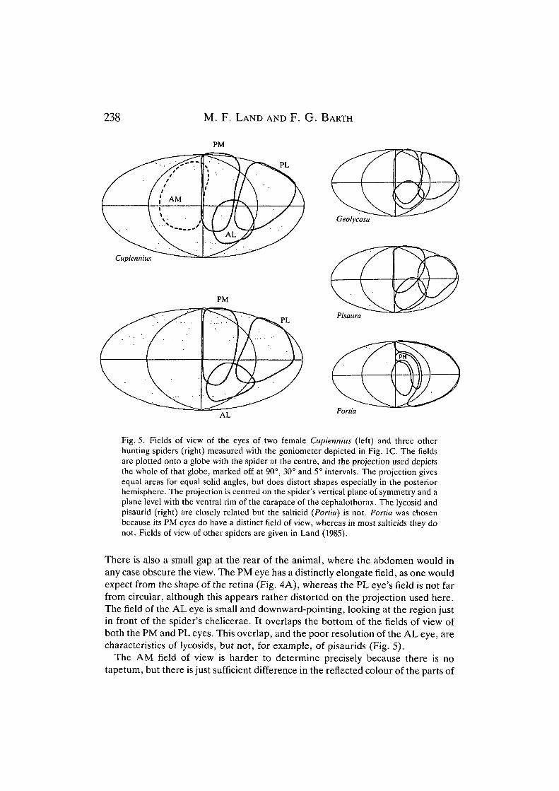
The fields of view of the eyes of both living Cupiennius specimens weremeasured with the telescope goniometer (Fig. 1C). They are shown in Fig. 5,plotted onto an Aitoff equal area projection which represents a globe at infinitywith the spider at the centre. The right fields of view of the secondary eyes areshown for both animals and the left principal (AM) eye is shown for one only.Fields of view for representatives of three other families are also given forcomparison.
The main features are that the PM and PL eyes between them cover nearly thewhole upper hemisphere, and down to 40° below the horizontal plane. Theanterior edge of the PM field just reaches the sagittal midline, where the eye on theother side takes over. Similarly, above the animal the PM and PL fields on the twosides just meet in the sagittal plane. In both specimens there is a gap 5-20° widebetween the fields of the PM and PL eyes: this does not seem to be an artefact ofthe method, because no such gap is seen between the PM fields at the front.Presumably there really is a gap in the animal's visual coverage in this region.
238 M. F. LAND AND F. G. BARTH
PM
PL
Geolycosa
Cupiennius
PLPisaura
Portia
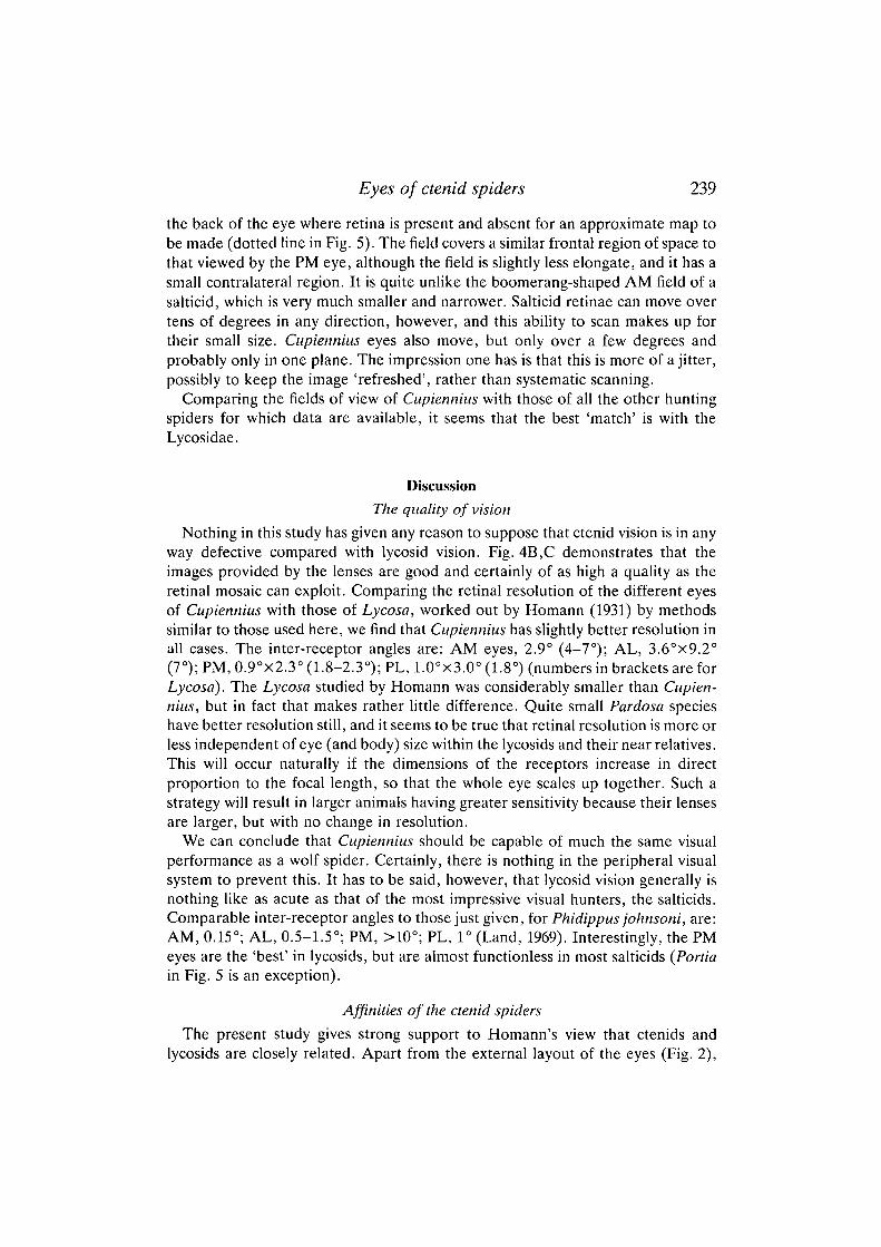
Fig. 5. Fields of view of the eyes of two female Cupiennius (left) and three otherhunting spiders (right) measured with the goniometer depicted in Fig. 1C. The fieldsare plotted onto a globe with the spider at the centre, and the projection used depictsthe whole of that globe, marked off at 90°, 30° and 5° intervals. The projection givesequal areas for equal solid angles, but does distort shapes especially in the posteriorhemisphere. The projection is centred on the spider's vertical plane of symmetry and aplane level with the ventral rim of the carapace of the cephalothorax. The lycosid andpisaurid (right) are closely related but the salticid (Portia) is not. Portia was chosenbecause its PM eyes do have a distinct field of view, whereas in most salticids they donot. Fields of view of other spiders are given in Land (1985).
There is also a small gap at the rear of the animal, where the abdomen would inany case obscure the view. The PM eye has a distinctly elongate field, as one wouldexpect from the shape of the retina (Fig. 4A), whereas the PL eye's field is not farfrom circular, although this appears rather distorted on the projection used here.The field of the AL eye is small and downward-pointing, looking at the region justin front of the spider's chelicerae. It overlaps the bottom of the fields of view ofboth the PM and PL eyes. This overlap, and the poor resolution of the AL eye, arecharacteristics of lycosids, but not, for example, of pisaurids (Fig. 5).
The AM field of view is harder to determine precisely because there is notapetum, but there is just sufficient difference in the reflected colour of the parts of
Eyes of ctenid spiders 239
the back of the eye where retina is present and absent for an approximate map tobe made (dotted line in Fig. 5). The field covers a similar frontal region of space tothat viewed by the PM eye, although the field is slightly less elongate, and it has asmall contralateral region. It is quite unlike the boomerang-shaped AM field of asalticid, which is very much smaller and narrower. Salticid retinae can move overtens of degrees in any direction, however, and this ability to scan makes up fortheir small size. Cupiennius eyes also move, but only over a few degrees andprobably only in one plane. The impression one has is that this is more of a jitter,possibly to keep the image 'refreshed', rather than systematic scanning.
Comparing the fields of view of Cupiennius with those of all the other huntingspiders for which data are available, it seems that the best 'match' is with theLycosidae.
Discussion
The quality of vision
Nothing in this study has given any reason to suppose that ctenid vision is in anyway defective compared with lycosid vision. Fig. 4B,C demonstrates that theimages provided by the lenses are good and certainly of as high a quality as theretinal mosaic can exploit. Comparing the retinal resolution of the different eyesof Cupiennius with those of Lycosa, worked out by Homann (1931) by methodssimilar to those used here, we find that Cupiennius has slightly better resolution inall cases. The inter-receptor angles are: AM eyes, 2.9° (4-7°); AL, 3.6°x9.2°(7°); PM, 0.9°x2.3° (1.8-2.3°); PL, 1.0°x3.0° (1.8°) (numbers in brackets are forLycosa). The Lycosa studied by Homann was considerably smaller than Cupien-nius, but in fact that makes rather little difference. Quite small Pardosa specieshave better resolution still, and it seems to be true that retinal resolution is more orless independent of eye (and body) size within the lycosids and their near relatives.This will occur naturally if the dimensions of the receptors increase in directproportion to the focal length, so that the whole eye scales up together. Such astrategy will result in larger animals having greater sensitivity because their lensesare larger, but with no change in resolution.
We can conclude that Cupiennius should be capable of much the same visualperformance as a wolf spider. Certainly, there is nothing in the peripheral visualsystem to prevent this. It has to be said, however, that lycosid vision generally isnothing like as acute as that of the most impressive visual hunters, the salticids.Comparable inter-receptor angles to those just given, for Phidippus johnsoni, are:AM, 0.15°; AL, 0.5-1.5°; PM, >10°; PL, 1° (Land, 1969). Interestingly, the PMeyes are the 'best' in lycosids, but are almost functionless in most salticids {Portiain Fig. 5 is an exception).
Affinities of the ctenid spiders
The present study gives strong support to Homann's view that ctenids andlycosids are closely related. Apart from the external layout of the eyes (Fig. 2),
240 M. F. LAND AND F. G. BARTH
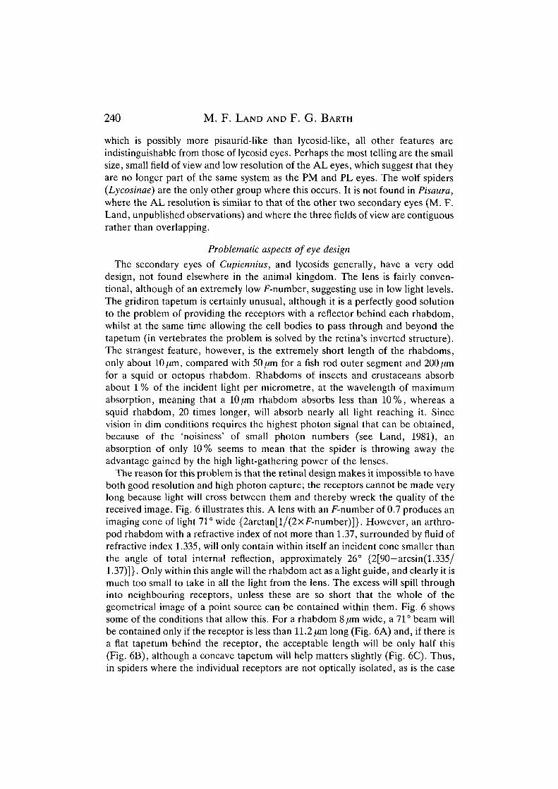
which is possibly more pisaurid-like than lycosid-like, all other features areindistinguishable from those of lycosid eyes. Perhaps the most telling are the smallsize, small field of view and low resolution of the AL eyes, which suggest that theyare no longer part of the same system as the PM and PL eyes. The wolf spiders(Lycosinae) are the only other group where this occurs. It is not found in Pisaura,where the AL resolution is similar to that of the other two secondary eyes (M. F.Land, unpublished observations) and where the three fields of view are contiguousrather than overlapping.
Problematic aspects of eye design
The secondary eyes of Cupiennius, and lycosids generally, have a very odddesign, not found elsewhere in the animal kingdom. The lens is fairly conven-tional, although of an extremely low F-number, suggesting use in low light levels.The gridiron tapetum is certainly unusual, although it is a perfectly good solutionto the problem of providing the receptors with a reflector behind each rhabdom,whilst at the same time allowing the cell bodies to pass through and beyond thetapetum (in vertebrates the problem is solved by the retina's inverted structure).The strangest feature, however, is the extremely short length of the rhabdoms,only about lO^m, compared with 50 ^m for a fish rod outer segment and 200 (imfor a squid or octopus rhabdom. Rhabdoms of insects and crustaceans absorbabout 1 % of the incident light per micrometre, at the wavelength of maximumabsorption, meaning that a lO/xm rhabdom absorbs less than 10%, whereas asquid rhabdom, 20 times longer, will absorb nearly all light reaching it. Sincevision in dim conditions requires the highest photon signal that can be obtained,because of the 'noisiness' of small photon numbers (see Land, 1981), anabsorption of only 10% seems to mean that the spider is throwing away theadvantage gained by the high light-gathering power of the lenses.
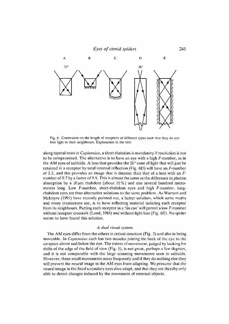
The reason for this problem is that the retinal design makes it impossible to haveboth good resolution and high photon capture; the receptors cannot be made verylong because light will cross between them and thereby wreck the quality of thereceived image. Fig. 6 illustrates this. A lens with an F-number of 0.7 produces animaging cone of light 71° wide {2arctan[l/(2xF-number)]}. However, an arthro-pod rhabdom with a refractive index of not more than 1.37, surrounded by fluid ofrefractive index 1.335, will only contain within itself an incident cone smaller thanthe angle of total internal reflection, approximately 26° {2[90—arcsin(1.335/1.37)]}. Only within this angle will the rhabdom act as a light guide, and clearly it ismuch too small to take in all the light from the lens. The excess will spill throughinto neighbouring receptors, unless these are so short that the whole of thegeometrical image of a point source can be contained within them. Fig. 6 showssome of the conditions that allow this. For a rhabdom 8/im wide, a 71° beam willbe contained only if the receptor is less than 11.2 /im long (Fig. 6A) and, if there isa flat tapetum behind the receptor, the acceptable length will be only half this(Fig. 6B), although a concave tapetum will help matters slightly (Fig. 6C). Thus,in spiders where the individual receptors are not optically isolated, as is the case
Eyes of ctenid spiders
A B c D
71° 26°
241
/v/\M
Fig. 6. Constraints on the length of receptors of different types such that they do notlose light to their neighbours. Explanation in the text.
along tapetal rows in Cupiennius, a short rhabdom is mandatory if resolution is notto be compromised. The alternative is to have an eye with a high F-number, as inthe AM eyes of salticids. A lens that provides the 26° cone of light that will just beretained in a receptor by total internal reflection (Fig. 6D) will have an F-numberof 2.2, and this provides an image that is dimmer than that of a lens with an F-number of 0.7 by a factor of 9.9. This is almost the same as the difference in photonabsorption by a lOjitrn rhabdom (about 10%) and one several hundred micro-metres long. Low F-number, short-rhabdom eyes and high F-number, long-rhabdom eyes are thus alternative solutions to the same problem. As Warrant andMclntyre (1991) have recently pointed out, a better solution, which some mothsand many crustaceans use, is to have reflecting material isolating each receptorfrom its neighbours. Putting each receptor in a 'tin can' will permit a low F-numberwithout receptor crosstalk (Land, 1984) and without light loss (Fig. 6E). No spiderseems to have found this solution.
A dual visual system
The AM eyes differ from the others in retinal structure (Fig. 3) and also in beingmoveable. In Cupiennius each has two muscles joining the back of the eye to thecarapace above and below the eye. The extent of movement, judged by looking forshifts of the edge of the field of view (Fig. 5), is not great, perhaps a few degrees,and it is not comparable with the large scanning movements seen in salticids.However, these small movements occur frequently and if they do nothing else theywill prevent the neural image in the AM eyes from adapting. We presume that theneural image in the fixed secondary eyes does adapt, and that they are thereby onlyable to detect changes induced by the movement of external objects.
242 M. F. L A N D AND F. G. BARTH
We anticipate that, as in salticids (see Land, 1972), the function of the secondaryeyes is to detect motion and that of the principal eyes is to analyse stationaryobjects. The observation that the AM and PM eyes have essentially the same fieldof view (Fig. 5) argues strongly in favour of them having different and complemen-tary roles. As we have seen, Cupiennius has potentially impressive visualcapabilities. It is time to seek some visual behaviour to go with them.
ReferencesBACCETTI, B. AND BEDINI, C. (1964). Research on the structure and physiology of the eyes of a
lycosid spider. I. Microscopic and ultramicroscopic structure. Archs ital. Biol. 102, 97-122.BARTH, F. G. (1985). Slit sensilla and the measurement of cuticular strains. In Neurobiology of
Arachnids (ed. F. G. Barth), pp. 162-188. Berlin: Springer.BLEST, A. D. AND LAND, M. F. (1977). The physiological optics of Dinopis subrufus L. Koch: a
fish lens in a spider. Proc. R. Soc. Lond. B 196, 198-222.BLEST, A. D. AND O'CARROLL, D. (1989). The evolution of the tiered principal retinae of
jumping spiders. In Neurobiology of Sensory Systems (ed. R. N. Singh and N. J. Strausfeld),pp. 155-170. New York: Plenum.
HOMANN, H. (1928). Beitrage zur Physiologie der Spinnenaugen. I. Untersuchungsmethoden.II. Das Sehvermogen der Salticiden. Z. vergl. Physiol. 7, 201-269.
HOMANN, H. (1931). Beitrage zur Physiologie der Spinnenaugen. III. Das Sehvermogen derLycosiden. Z. vergl. Physiol. 14, 40-67.
HOMANN, H. (1961). Die Stellung der Ctenidae, Textricinae und Rhoicininae im System derAraneae. Senck. Biol. 42, 397-408.
HOMANN, H. (1971). Die Augen der Araneae. Anatomie, Ontogenie und Bedeutung fur dieSystematik (Chelicerata, Arachnida). Z. Morph. Tiere. 69, 201-272.
LACHMUTH, U., GRASSHOFF, M. AND BARTH, F. G. (1985). Taxonomische Revision der GattungCupiennius Simon 1891. Senkenbergiana biol. 65, 329-372.
LAND, M. F. (1969). Structure of the principal eyes of jumping spiders (Salticidae:Dendryphantinae) in relation to visual optics. J. exp. Biol. 51, 443-470.
LAND, M. F. (1972). Mechanisms of orientation and pattern recognition in jumping spiders(Salticidae). In Information Processing in the Visual Systems of Arthropods (ed. R. Wehner),pp. 231-247. Berlin: Springer.
LAND, M. F. (1981). Optics and vision in invertebrates. In Handbook of Sensory Physiology,vol. VIII/6B (ed. H. Autrum), pp. 471-592. Berlin: Springer.
LAND, M. F. (1984). The resolving power of diurnal superposition eyes measured with anophthalmoscope. J. comp. Physiol. 154, 515-533.
LAND, M. F. (1985). Morphology and optics of spider eyes. In Neurobiology of Arachnids (ed.F. G. Barth), pp. 53-78. Berlin: Springer.
LAND, M. F. AND SNYDER, A. (1985). Cone mosaic observed directly through natural pupil oflive vertebrate. Vision Res. 25, 1519-1523.
WARRANT, E. J. AND MCINTYRE, P. D. (1991). Strategies for retinal design in arthropod eyes oflow F-number. J. comp. Physiol. A 168, 499-512.
WILLIAMS, D. S. AND MCINTYRE, P. (1980). The principal eyes of a jumping spider have atelephoto component. Nature 288, 578-580.